Computer signal transmission in optical coherence tomography on the basis of Wigner transformation...
36
Computer signal transmission in Computer signal transmission in optical coherence tomography on optical coherence tomography on the basis of Wigner transformation the basis of Wigner transformation JASS-2004 JASS-2004 , , St.- St.- Petersburg Petersburg
-
date post
19-Dec-2015 -
Category
Documents
-
view
219 -
download
2
Transcript of Computer signal transmission in optical coherence tomography on the basis of Wigner transformation...

Computer signal transmission in Computer signal transmission in optical coherence tomography on optical coherence tomography on the basis of Wigner transformationthe basis of Wigner transformation
JASS-2004JASS-2004 ,,
St.-PetersburgSt.-Petersburg

Why Use OCT?Why Use OCT?
For more than three centuries, the microscope For more than three centuries, the microscope has been considered the ultimate tool to has been considered the ultimate tool to understand the origins of disease by examining understand the origins of disease by examining tissue and microstructures in exquisite detail. tissue and microstructures in exquisite detail.

Why use OCT?Why use OCT?
While the technology contained within the While the technology contained within the microscope has evolved over time, enabling microscope has evolved over time, enabling better resolution of smaller structures, one better resolution of smaller structures, one aspect has remained constant - material to be aspect has remained constant - material to be examined must be excised from the body and examined must be excised from the body and brought to the microscope. brought to the microscope.

2 main reasons2 main reasons New thinking concerning the origins of the New thinking concerning the origins of the
world's two largest killers – world's two largest killers – heart disease and cancerheart disease and cancer – – expose the limitations of the current paradigm. expose the limitations of the current paradigm.
Both diseases are thought to originate at the Both diseases are thought to originate at the cellular levelcellular level in the in the thin (20 - 200 micron)thin (20 - 200 micron) cellular cellular layer covering the inner and outer surfaces of layer covering the inner and outer surfaces of the body. Understanding these diseases on the the body. Understanding these diseases on the microscopic level on an in vivo basis could lead microscopic level on an in vivo basis could lead to better diagnosis, earlier more precise to better diagnosis, earlier more precise treatments and development of novel therapies treatments and development of novel therapies to help eradicate these diseases to help eradicate these diseases

microscopic images from within the bodymicroscopic images from within the body
For years, scientists, physicians and For years, scientists, physicians and technology developers have struggled to technology developers have struggled to achieve microscopic images from within the achieve microscopic images from within the body. These attempts have met with limited body. These attempts have met with limited success due to two primary factors: success due to two primary factors:
1.1. the size of the apparatus being deployed and the size of the apparatus being deployed and
2.2. the resolution of the images being obtained. the resolution of the images being obtained.

Attempts to obtain microscopic images using Attempts to obtain microscopic images using external methodologies:external methodologies:
magnetic resonance, magnetic resonance, X-ray, ultrasound, and X-ray, ultrasound, and nuclear imaging,nuclear imaging,
have met with similar fates. have met with similar fates.

A New Weapon for Heart Disease and A New Weapon for Heart Disease and
CancerCancer While OCT has the potential to be used for While OCT has the potential to be used for
a variety of medical a variety of medical applicationsapplications, cancer , cancer and heart disease represent two of the and heart disease represent two of the most pressing, and promising application most pressing, and promising application areas. areas.
Cardiovascular ImagingCardiovascular Imaging
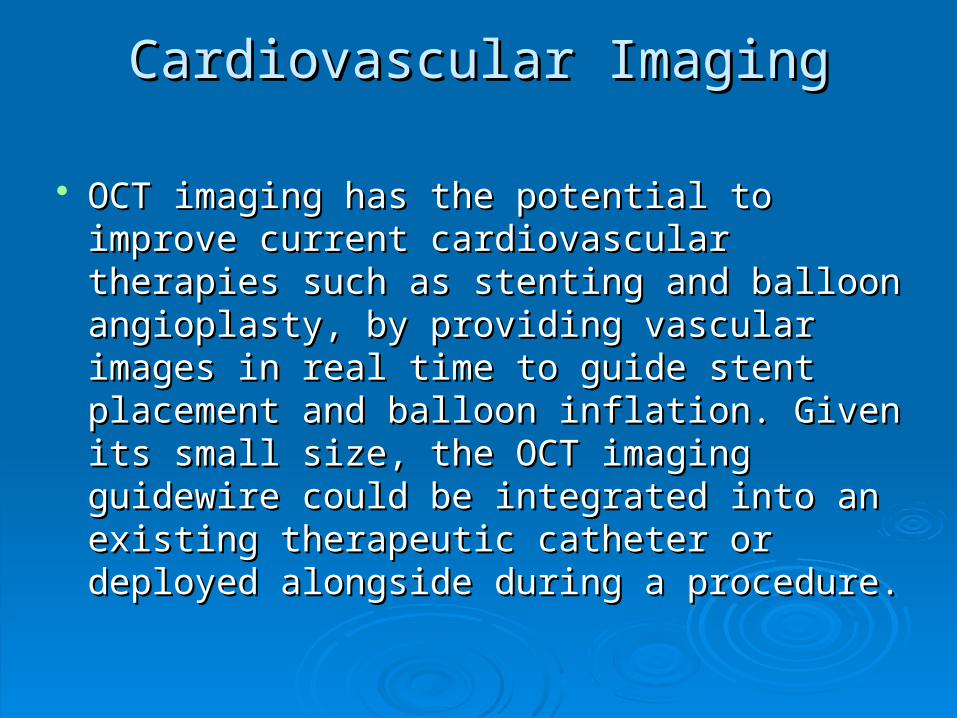
OCT imaging has the potential to improve OCT imaging has the potential to improve current cardiovascular therapies such as current cardiovascular therapies such as stenting and balloon angioplasty, by stenting and balloon angioplasty, by providing vascular images in real time to providing vascular images in real time to guide stent placement and balloon guide stent placement and balloon inflation. Given its small size, the OCT inflation. Given its small size, the OCT imaging guidewire could be integrated into imaging guidewire could be integrated into an existing therapeutic catheter or an existing therapeutic catheter or deployed alongside during a procedure. deployed alongside during a procedure.

New cardiac research indicates that unstable New cardiac research indicates that unstable plaques - arterial lesions that do not constrict the plaques - arterial lesions that do not constrict the blood vessel but rather burst releasing a bolus of blood vessel but rather burst releasing a bolus of lipids into the blood stream - may be responsible for lipids into the blood stream - may be responsible for up to 70 percent of all heart attacks. OCT has the up to 70 percent of all heart attacks. OCT has the potential to clearly identify plaques and help potential to clearly identify plaques and help differentiate unstable plaques from stable plaques. differentiate unstable plaques from stable plaques.
Cardiovascular System Atherosclerotic Disease Cardiovascular System Atherosclerotic Disease Brezinski, M.E. Circ. 93; 1206:1996 Brezinski, M.E. Circ. 93; 1206:1996

In addition to providing exquisite In addition to providing exquisite morphological detail, OCT is meeting other morphological detail, OCT is meeting other capabilities such as capabilities such as spectroscopic spectroscopic imaging,imaging, polarization imaging and Dopplerpolarization imaging and Doppler to provide further information regarding to provide further information regarding tissue composition and flow. tissue composition and flow.

Cancer Detection Cancer Detection
It is estimated that more than 85 percent of all It is estimated that more than 85 percent of all cancers originate in the epithelium, the thin (20-cancers originate in the epithelium, the thin (20-200 micron) cellular layer covering the inner and 200 micron) cellular layer covering the inner and outer surfaces of the body. Excisional biopsy, outer surfaces of the body. Excisional biopsy, removing tissue from the body and examining it removing tissue from the body and examining it under a microscope, is the gold standard for under a microscope, is the gold standard for cancer diagnosis. However, many biopsies are cancer diagnosis. However, many biopsies are done on a hit or miss basis, small pieces of done on a hit or miss basis, small pieces of tissue are excised at random and dissected to tissue are excised at random and dissected to check for cancerous cells. check for cancerous cells.

OCT has the potential to greatly improve OCT has the potential to greatly improve conventional biopsy by more precisely conventional biopsy by more precisely identifying the areas to be excised based identifying the areas to be excised based on images of the epithelial layers, reducing on images of the epithelial layers, reducing the number of biopsies and making earlier the number of biopsies and making earlier and more accurate diagnosis possible. As and more accurate diagnosis possible. As the technology matures, it may be possible the technology matures, it may be possible to perform biopsies using OCT imaging to perform biopsies using OCT imaging alone, making possible point of care alone, making possible point of care biopsy.biopsy.

MicroscopyMicroscopy
OCT offers the potential to assist in the OCT offers the potential to assist in the visualization of vessels and nerves for visualization of vessels and nerves for repair surgery. OCT may also have the repair surgery. OCT may also have the potential for identifying the margins of potential for identifying the margins of low-grade invasive neurological low-grade invasive neurological tumors. The use of OCT imaging tumors. The use of OCT imaging forceps may be beneficial to many forceps may be beneficial to many microsurgical procedures. microsurgical procedures.
Intestinal polyps Ex Vivo Human Tissue

What is OCT?What is OCT?
Optical Coherence Tomography (OCT) is Optical Coherence Tomography (OCT) is a promising new class of diagnostic a promising new class of diagnostic medical imaging technology that medical imaging technology that utilizes advanced photonics and fiber utilizes advanced photonics and fiber optics to obtain images and tissue optics to obtain images and tissue characterization on a scale never characterization on a scale never before possible within the human body. before possible within the human body.

When fully exploited, the technology When fully exploited, the technology has the potential to dramatically has the potential to dramatically change the way physicians, change the way physicians, researchers and scientists see and researchers and scientists see and understand the human body in order understand the human body in order to better diagnose and treat disease.to better diagnose and treat disease.

OCT and UltrasoundOCT and Ultrasound Simply put, OCT combines the principles of ultrasound Simply put, OCT combines the principles of ultrasound
with the imaging performance of a microscope and a with the imaging performance of a microscope and a form factor that is familiar to clinicians.form factor that is familiar to clinicians.
Whereas ultrasound produces images from Whereas ultrasound produces images from backscattered sound "echoes," backscattered sound "echoes,"
OCT uses infrared light waves that reflect off the internal OCT uses infrared light waves that reflect off the internal microstructure within the biological tissues. The microstructure within the biological tissues. The frequencies and bandwidths of infrared light are orders frequencies and bandwidths of infrared light are orders of magnitude higher than medical ultrasound signals -- of magnitude higher than medical ultrasound signals -- resulting in greatly increased image resolution - 8-25 resulting in greatly increased image resolution - 8-25 times greater than any existing modality.times greater than any existing modality.

Comparison between OCT (left) and Ultrasound (right).
Comparison between OCT (left) and Ultrasound (right). Tearney, G.J., et. al. Circ. 4256, 1997

Методы описанияМетоды описания сигналов в ОКТ сигналов в ОКТ
Infrared light is delivered to the imaging Infrared light is delivered to the imaging site through a single optical fiber site through a single optical fiber only .006" diameter (about the size of the only .006" diameter (about the size of the period in this sentence). The imaging period in this sentence). The imaging guidewire contains a complete lens guidewire contains a complete lens assembly to perform a variety of imaging assembly to perform a variety of imaging functions. The guidewire can be deployed functions. The guidewire can be deployed independently or integrated into existing independently or integrated into existing therapeutic or imaging catheterstherapeutic or imaging catheters

Electronic and interferometricElectronic and interferometric techniquestechniques
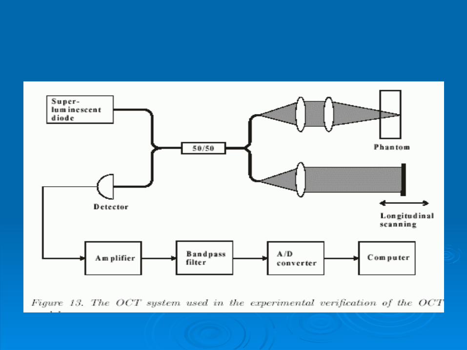
While standard electronic techniques are adequate for While standard electronic techniques are adequate for processing ultrasonic echoes that travel at the speed of processing ultrasonic echoes that travel at the speed of sound, interferometric techniques are required to extract sound, interferometric techniques are required to extract the reflected optical signals from the infrared light used the reflected optical signals from the infrared light used in OCT. in OCT.
The output, measured by an interferometer, is computer The output, measured by an interferometer, is computer processed to produce high-resolution, real time, cross processed to produce high-resolution, real time, cross sectional or 3-dimensional images of the tissue. This sectional or 3-dimensional images of the tissue. This powerful technology provides in situ images of tissues at powerful technology provides in situ images of tissues at near histological resolution without the need for excision near histological resolution without the need for excision or processing of the specimen. or processing of the specimen.

In addition to providing In addition to providing high-level high-level resolutionsresolutions for the evaluation of for the evaluation of microanatomic structures OCT is microanatomic structures OCT is inherently able to provide inherently able to provide information information regarding tissue compositionregarding tissue composition. Using . Using spectroscopy, users can evaluate the spectroscopy, users can evaluate the spectral absorption characteristics of spectral absorption characteristics of tissue while simultaneously determining tissue while simultaneously determining the orderliness of the tissue through the the orderliness of the tissue through the use of polarization imaginguse of polarization imaging


Advantages of OCTAdvantages of OCT
HIGH RESOLUTION:HIGH RESOLUTION: Current OCT systems have Current OCT systems have resolutions at 4-20 um compared to resolutions at 4-20 um compared to 110 um for high frequency 110 um for high frequency ultrasound. ultrasound.
TISSUE CHARACTERIZATION: TISSUE CHARACTERIZATION: Using information inherent to the Using information inherent to the returning photon signals, OCT can returning photon signals, OCT can perform both spectroscopic and perform both spectroscopic and polarization imaging to better polarization imaging to better evaluate the composition of tissues evaluate the composition of tissues and lesions.and lesions.
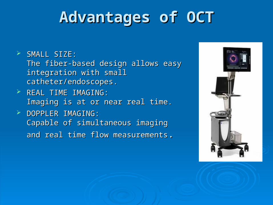
Advantages of OCTAdvantages of OCT
SMALL SIZE: SMALL SIZE: The fiber-based design allows easy The fiber-based design allows easy integration with small catheter/endoscopes.integration with small catheter/endoscopes.
REAL TIME IMAGING: REAL TIME IMAGING: Imaging is at or near real time.Imaging is at or near real time.
DOPPLER IMAGING: DOPPLER IMAGING: Capable of simultaneous imaging and real Capable of simultaneous imaging and real
time flow measurementstime flow measurements. .

Wigner transformationWigner transformation
2/,...1
,...1
)2
sin()()(2)2
cos()()(2
)2exp()()(2),(
2
2
2
2
2
2
2121
21
Kn
Kk
K
mnmkSmkSj
K
mnmkSmkS
mnjmkSmkSnkW
K
Km
K
Km
K
Km
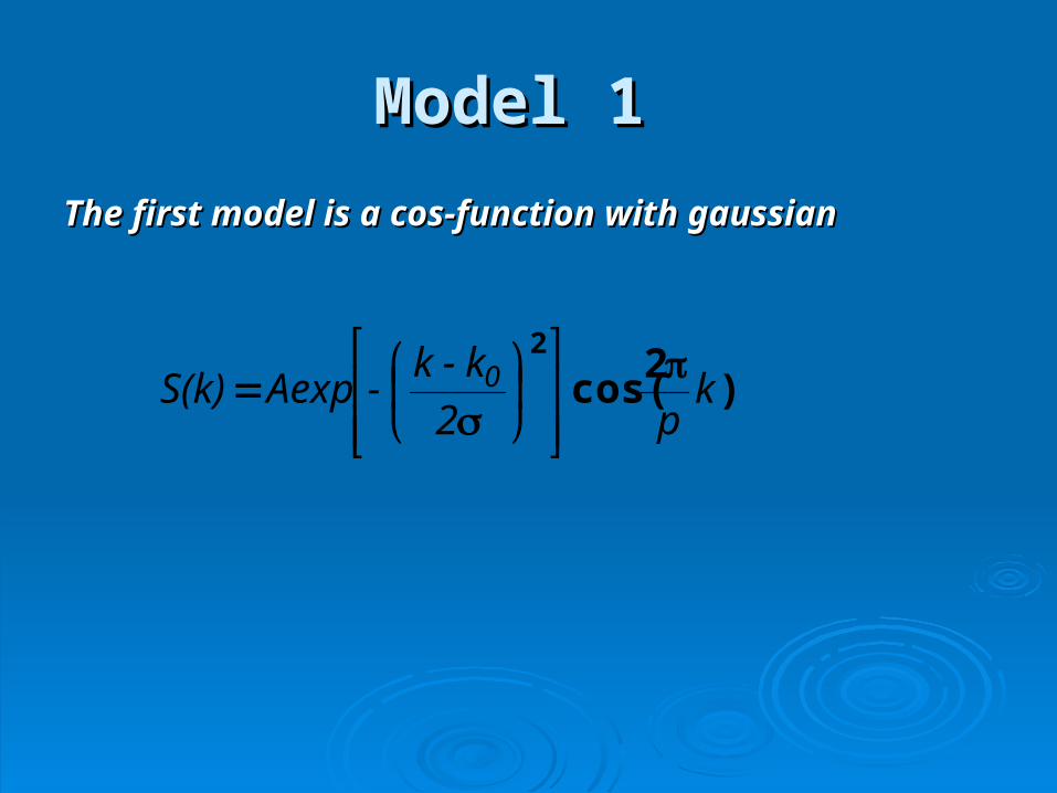
Model 1Model 1
The first model is a cos-function with gaussianThe first model is a cos-function with gaussian
)2
cos(2
kp2
k-k-Aexp S(k) 0

Wigner transformation of 2 signals with different Wigner transformation of 2 signals with different parameters (Re)parameters (Re)

Wigner transformation of 2 signals with same parameters Wigner transformation of 2 signals with same parameters (Re)(Re)

Wigner transformation of 2 signals with different Wigner transformation of 2 signals with different
parameters (Im)parameters (Im)
ПВ двух КФГО сигналов с разными параметрами (мнимая часть) как ПВ двух КФГО сигналов с разными параметрами (мнимая часть) как правило обнуляетсяправило обнуляется

Model 2Model 2
50,12
Aexp S(k)
k
Kk
p
Signal – Linear frequency modulation

Wigner transformation of 2 signals with same parameters Wigner transformation of 2 signals with same parameters (Re)(Re)

Wigner transformation of 2 signals with different Wigner transformation of 2 signals with different parameters (Re)parameters (Re)

Wigner transformation of 2 signals with different Wigner transformation of 2 signals with different parameters (Im)parameters (Im)

Thank you!!!Thank you!!!