Compromised Survival of Cerebellar Molecular Layer...
11
Report Compromised Survival of Cerebellar Molecular Layer Interneurons Lacking GDNF Receptors GFRa1 or RET Impairs Normal Cerebellar Motor Learning Graphical Abstract Highlights d The signals controlling survival of molecular layer interneurons (MLIs) are unclear d Whether MLIs are involved in normal cerebellar function was unclear d Purkinje cells express GDNF, and survival of MLIs depends on GDNF receptors GFRa1 and RET d Requirement of MLIs for cerebellar-dependent motor learning Authors Maria Christina Sergaki, Juan Carlos Lo ´ pez-Ramos, Stefanos Stagkourakis, Agne ` s Gruart, Christian Broberger, Jose ´ Marı´a Delgado-Garcı ´a, Carlos F. Iba ´n ˜ ez Correspondence [email protected] In Brief Sergaki et al. find that conditional mutant mice lacking GDNF receptor GFRa1 or RET lose a quarter of their cerebellar molecular layer interneurons (MLIs), resulting in compromised motor learning but not overall motor coordination. These results identify an endogenous survival mechanism for MLIs and reveal their unexpected vulnerability in the control of cerebellar-dependent motor learning Sergaki et al., 2017, Cell Reports 19, 1977–1986 June 6, 2017 ª 2017 The Author(s). http://dx.doi.org/10.1016/j.celrep.2017.05.030
Transcript of Compromised Survival of Cerebellar Molecular Layer...

Report
CompromisedSurvival of C
erebellarMolecular LayerInterneurons Lacking GDNF Receptors GFRa1 orRET Impairs Normal Cerebellar Motor LearningGraphical Abstract
Highlights
d The signals controlling survival of molecular layer
interneurons (MLIs) are unclear
d Whether MLIs are involved in normal cerebellar function was
unclear
d Purkinje cells express GDNF, and survival of MLIs depends
on GDNF receptors GFRa1 and RET
d Requirement of MLIs for cerebellar-dependent motor
learning
Sergaki et al., 2017, Cell Reports 19, 1977–1986June 6, 2017 ª 2017 The Author(s).http://dx.doi.org/10.1016/j.celrep.2017.05.030
Authors
Maria Christina Sergaki,
Juan Carlos Lopez-Ramos,
Stefanos Stagkourakis, Agnes Gruart,
Christian Broberger,
Jose Marıa Delgado-Garcıa,
Carlos F. Ibanez
In Brief
Sergaki et al. find that conditional mutant
mice lacking GDNF receptor GFRa1 or
RET lose a quarter of their cerebellar
molecular layer interneurons (MLIs),
resulting in compromised motor learning
but not overall motor coordination. These
results identify an endogenous survival
mechanism for MLIs and reveal their
unexpected vulnerability in the control of
cerebellar-dependent motor learning

Cell Reports
Report
Compromised Survival of Cerebellar Molecular LayerInterneurons Lacking GDNF Receptors GFRa1 or RETImpairs Normal Cerebellar Motor LearningMaria Christina Sergaki,1 Juan Carlos Lopez-Ramos,2 Stefanos Stagkourakis,1 Agnes Gruart,2 Christian Broberger,1
Jose Marıa Delgado-Garcıa,2 and Carlos F. Ibanez1,3,4,5,*1Department of Neuroscience, Karolinska Institute, Stockholm S-17177, Sweden2Division of Neurosciences, Pablo de Olavide University, Seville 41013, Spain3Department of Physiology, National University of Singapore, Singapore 117597, Singapore4Life Sciences Institute, National University of Singapore, Singapore 117456, Singapore5Lead Contact
*Correspondence: [email protected]
http://dx.doi.org/10.1016/j.celrep.2017.05.030
SUMMARY
The role of neurotrophic factors as endogenous sur-vival proteins for brain neurons remains contentious.In the cerebellum, the signals controlling survival ofmolecular layer interneurons (MLIs) are unknown,and direct evidence for the requirement of a full com-plement of MLIs for normal cerebellar function andmotor learning has been lacking. Here, we showthat Purkinje cells (PCs), the target of MLIs, expressthe neurotrophic factor GDNF during MLI develop-ment and survival of MLIs depends on GDNFreceptors GFRa1 and RET. Conditional mutantmice lacking either receptor lose a quarter of theirMLIs, resulting in compromised synaptic inhibitionof PCs, increased PC firing frequency, and abnormalacquisition of eyeblink conditioning and vestibulo-ocular reflex performance, but not overall motoractivity or coordination. These results identify anendogenous survival mechanism for MLIs and revealthe unexpected vulnerability and selective require-ment of MLIs in the control of cerebellar-dependentmotor learning.
INTRODUCTION
The importance of the cerebellum for motor learning has long
been recognized. Cerebellar circuits adjust and fine-tune motor
programs to allow movement accuracy through a trial-and-error
process that is critical for normal motor learning. However, the
cellular and molecular components underlying these functions
remain poorly characterized. Long-term depression (LTD) of
the synapse between granule cell parallel fibers and Purkinje
cell (PC) dendrites was initially hypothesized to be important
for cerebellar motor learning (Ito, 2000), but more recent studies
have indicated that motor learning can occur without cerebellar
LTD (Hirano, 2014; Schonewille et al., 2011). On the other hand,
the role of cerebellar molecular layer interneurons (MLIs) in
CelThis is an open access article under the CC BY-N
shaping the computational properties of cerebellar circuits
underlying plasticity and learning has lately received more atten-
tion (Jorntell et al., 2010). By performing MLI recordings on
behaving animals, a recent study uncovered a positive correla-
tion between MLI activity and classical eyeblink conditioning, a
common experimental paradigm of cerebellar motor learning
(ten Brinke et al., 2015). However, the direct involvement of
MLIs for the expression of cerebellar motor learning responses
remains to be demonstrated.
MLIs have been subdivided in basket and stellate cells ac-
cording to morphological criteria. Basket cells are located in
the deeper part of themolecular layer (ML) and form perineuronal
nets around the perikarya of PCs that end in a brush-shaped ter-
minal or ‘‘pinceau’’ on the initial segments of PC axons, which
allows them tomodulate PC spike output. Stellate cells are found
in the superficial part of the ML, target PC dendrites, and are
thought to regulate the excitatory synaptic inputs to PCs
(Sotelo, 2015). Deep and superficial MLIs also show differen-
tial expression of a subset of genes that is highly enriched
among cerebellar inhibitory interneurons (Schilling and Ober-
dick, 2009). Despite these differences, some investigators still
consider stellate and basket cells as belonging to the same pop-
ulation (Sotelo, 2015). In the mouse cerebellum, the different
GABAergic cell types are born during different time windows of
development: PCs are generated first, between embryonic day
(E) 10 and E12, granule layer interneurons (GLIs; i.e., Golgi cells)
between E16 and postnatal day (P) 0, and MLIs within the first
postnatal week (Carletti and Rossi, 2008). MLIs originate from
a single progenitor pool within the cerebellar ventricular zone
(VZ) that expresses the transcription factor Ptf1a (Hoshino
et al., 2005). Among MLIs, basket cells are born first, around
P2, and stellate cells later, around P10 (Yamanaka et al., 2004).
From the VZ, MLI progenitors migrate outward to reach the pro-
spective white matter (WM) of the embryonic cerebellum, where
they persist and continue dividing for another 2 days until they
turn on expression of GAD67, a key enzyme in GABA production,
and resume their migration toward the ML (Cameron et al., 2009;
Yamanaka et al., 2004; Zhang and Goldman, 1996). MLI migra-
tion continues within the ML for another 2–3 days until the end
of the second postnatal week (Yamanaka et al., 2004). MLIs
l Reports 19, 1977–1986, June 6, 2017 ª 2017 The Author(s). 1977C-ND license (http://creativecommons.org/licenses/by-nc-nd/4.0/).
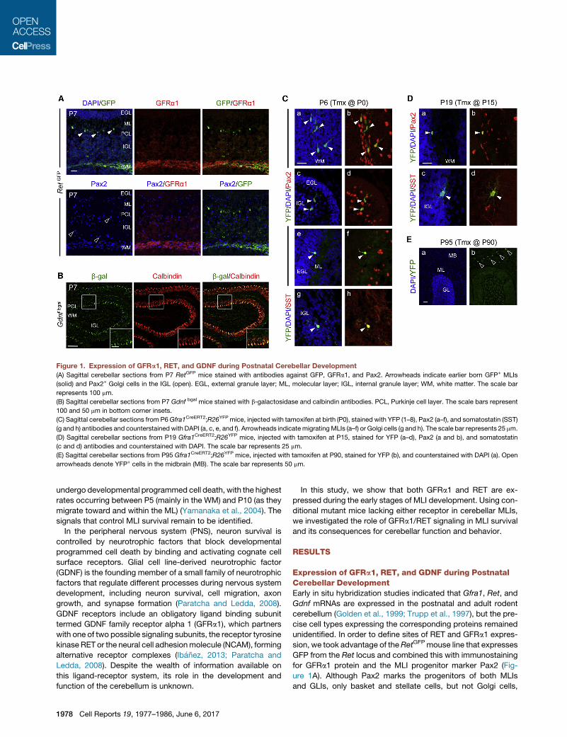
Figure 1. Expression of GFRa1, RET, and GDNF during Postnatal Cerebellar Development
(A) Sagittal cerebellar sections from P7 RetGFP mice stained with antibodies against GFP, GFRa1, and Pax2. Arrowheads indicate earlier born GFP+ MLIs
(solid) and Pax2+ Golgi cells in the IGL (open). EGL, external granule layer; ML, molecular layer; IGL, internal granule layer; WM, white matter. The scale bar
represents 100 mm.
(B) Sagittal cerebellar sections from P7 Gdnf bgal mice stained with b-galactosidase and calbindin antibodies. PCL, Purkinje cell layer. The scale bars represent
100 and 50 mm in bottom corner insets.
(C) Sagittal cerebellar sections from P6Gfra1CreERT2;R26YFP mice, injected with tamoxifen at birth (P0), stained with YFP (1–8), Pax2 (a–f), and somatostatin (SST)
(g and h) antibodies and counterstained with DAPI (a, c, e, and f). Arrowheads indicate migratingMLIs (a–f) or Golgi cells (g and h). The scale bar represents 25 mm.
(D) Sagittal cerebellar sections from P19 Gfra1CreERT2;R26YFP mice, injected with tamoxifen at P15, stained for YFP (a–d), Pax2 (a and b), and somatostatin
(c and d) antibodies and counterstained with DAPI. The scale bar represents 25 mm.
(E) Sagittal cerebellar sections from P95 Gfra1CreERT2;R26YFP mice, injected with tamoxifen at P90, stained for YFP (b), and counterstained with DAPI (a). Open
arrowheads denote YFP+ cells in the midbrain (MB). The scale bar represents 50 mm.
undergo developmental programmed cell death, with the highest
rates occurring between P5 (mainly in the WM) and P10 (as they
migrate toward and within the ML) (Yamanaka et al., 2004). The
signals that control MLI survival remain to be identified.
In the peripheral nervous system (PNS), neuron survival is
controlled by neurotrophic factors that block developmental
programmed cell death by binding and activating cognate cell
surface receptors. Glial cell line-derived neurotrophic factor
(GDNF) is the founding member of a small family of neurotrophic
factors that regulate different processes during nervous system
development, including neuron survival, cell migration, axon
growth, and synapse formation (Paratcha and Ledda, 2008).
GDNF receptors include an obligatory ligand binding subunit
termed GDNF family receptor alpha 1 (GFRa1), which partners
with one of two possible signaling subunits, the receptor tyrosine
kinase RET or the neural cell adhesionmolecule (NCAM), forming
alternative receptor complexes (Ibanez, 2013; Paratcha and
Ledda, 2008). Despite the wealth of information available on
this ligand-receptor system, its role in the development and
function of the cerebellum is unknown.
1978 Cell Reports 19, 1977–1986, June 6, 2017
In this study, we show that both GFRa1 and RET are ex-
pressed during the early stages of MLI development. Using con-
ditional mutant mice lacking either receptor in cerebellar MLIs,
we investigated the role of GFRa1/RET signaling in MLI survival
and its consequences for cerebellar function and behavior.
RESULTS
Expression of GFRa1, RET, and GDNF during PostnatalCerebellar DevelopmentEarly in situ hybridization studies indicated that Gfra1, Ret, and
Gdnf mRNAs are expressed in the postnatal and adult rodent
cerebellum (Golden et al., 1999; Trupp et al., 1997), but the pre-
cise cell types expressing the corresponding proteins remained
unidentified. In order to define sites of RET and GFRa1 expres-
sion, we took advantage of theRetGFPmouse line that expresses
GFP from the Ret locus and combined this with immunostaining
for GFRa1 protein and the MLI progenitor marker Pax2 (Fig-
ure 1A). Although Pax2 marks the progenitors of both MLIs
and GLIs, only basket and stellate cells, but not Golgi cells,

express parvalbumin (PV) when they mature. At P7, GFP and
GFRa1 signals were found to colocalize with Pax2 in the pro-
spective WM that contains MLI progenitors (Figure 1A). During
development, MLI progenitors travel through the granule and
PC layers to reach theML, after which they lose Pax2 expression
(Cameron et al., 2009). RET-GFP, but not GFRa1, was also found
in scattered cells in the ML (Figure 1A, solid arrowheads), which
likely correspond to earlier born MLIs that have already reached
the ML and turned off Pax2 expression. Neither GFRa1 nor RET
was found to be expressed in Pax2+ Golgi cells of the internal
granule layer (IGL), distinguished by their larger cell bodies
(Geurts et al., 2001;Weisheit et al., 2006) (Figure 1A, open arrow-
heads). RET-GFP expression persisted at P15 and P60 in PV+
MLIs located in the deeper part of the ML, where basket cells
are known to reside, but was absent elsewhere in the ML, sug-
gesting downregulation of RET expression in mature stellate
cells (Figure S1A). Processes emanating from these GFP+ cells
were seen enveloping the cell bodies of PCs, labeled with calbin-
din, forming characteristic perineuronal nets, a feature that dis-
tinguishes basket cells (Sotelo, 2015). No immunofluorescence
signal could be detected for GFRa1 at P15 or P60 (data not
shown). GDNF expression was visualized by immunohistochem-
istry using a Gdnfbgal mouse line expressing beta-galactosidase
from theGdnf locus or by in situ hybridization. GDNF expression
was found in PCs at all ages examined, from P7 to P60, and was
absent in WM cells (Figures 1A, S1A, and S1C).
The presence of GFRa1 in MLI progenitors at P7 and its
absence at later postnatal stages suggested transient expres-
sion of GFRa1 in cerebellar MLIs. In order to investigate this
further, we used a Gfra1CreERT2 mouse line expressing tamox-
ifen-inducible Cre recombinase (CreERT2) from the Gfra1 locus
(Sergaki and Ibanez, 2017). Gfra1CreERT2 mice were crossed
with the Rosa26YFP reporter line; offspring were injected with
tamoxifen at P0, P15, or P90 and analyzed at P6, P19, or P95,
respectively. YFP+ cells expressing Pax2 were found in the cer-
ebellum of P6 and P19 mice (Figures 1C and 1D) but not at P95
(Figure 1E), indicating that GFRa1 is expressed in both early- and
late-born MLI progenitors but not in mature MLIs localized at the
ML. At P6, YFP+ cells in the ML or close to the IGL displayed
morphological features of migrating MLIs, with a typical apical
dendrite (Figures 1Ca–1Cf, arrowheads). Larger YFP+ cells ex-
pressing somatostatin (SST), a marker of Golgi neurons (Geurts
et al., 2001), were also found in the IGL at P6 and P19 (Figures
1Cg and 1Ch). In summary, both RET and GFRa1 are expressed
in developing MLIs of the mouse cerebellum, but, whereas RET
is retained inmature basket cells, GFRa1 expression is restricted
to MLI progenitors. GFRa1, but not RET, is also expressed
in developing Golgi neurons, while GDNF was found in PCs at
all ages.
Increased Apoptosis in MLI Progenitors Lacking GFRa1or RETIn order to investigate the function of GFRa1 and RET inMLI pro-
genitors, we used conditional mutants for each of the receptors.
Because global knockout mice lacking either protein die at birth,
we eliminated GFRa1 and RET expression by crossing Gfra1fx/fx
or Retfx/fx mice with the Ptf1aCre driver line. Both mutant lines
(Ptf1aCre;Gfra1fx/fx and Ptf1aCre;Retfx/fx, respectively) survived
to adulthood and had normal gross cerebellar morphology and
architecture. Developmental cell death was investigated by im-
munostaining for activated caspase-3 at P5 in homozygous mu-
tants and fx/fx controls lacking Cre. Increased apoptosis was
observed among MLI progenitors in the folial WM in both mutant
lines (Figures 2A–2D). At P10, the number of Pax2+ MLIs (distin-
guished from Golgi cells by their smaller size and morphology)
was reduced in the folial WM aswell as throughout the cerebellar
cortex of both mutants (Figures 2E–2G and 2I–2K). On the other
hand, the number of large size Pax2+ neurons corresponding to
Golgi cells (Figures 2Eand2I, solid arrowheads)wasnot changed
in the mutants (Figures 2H and 2L). Conditional deletion of either
Gfra1 or Ret with the Gad67Cre driver, which turns on later than
Ptf1aCre, did not affect the number of Pax2+ MLIs in the P10 cer-
ebellum (Figures S2A–S2E), underscoring the requirement of
GFRa1 and RET receptors for survival of MLI progenitors before
they expressGAD67.Wealso investigatedwhether the reduction
in Pax2+MLIs observed in themutants could be due to defects in
proliferation or migration. At P5, the number of proliferating cells
in the folial WM, assessed 2 hr after a BrdU pulse, was not
different between genotypes (Figures S3A–S3D). At this age,
BrdU only marks MLI progenitors, because Golgi cells no longer
proliferate.Wenote that somecells showing immunoreactivity for
activated caspase-3 at P5 were also labeled with BrdU, indi-
cating that both proliferating and nonproliferating cells are dying
in the mutants (Figure S3E). To examine migration, we quantified
the number of Pax2/BrdU double-positive cells in different layers
of the cerebellar cortex at P10 (i.e., 5 days after the BrdU pulse).
Again, no difference was found between genotypes (Figures S3F
and S3G). Together, these data indicated that loss of GFRa1 or
RET in MLI progenitors specifically compromised their survival
but not proliferation or migration.
Loss of MLIs in the Cerebellum of Adult Gfra1 and Ret
Conditional MutantsAt 2 months of age, the cell density and overall thickness of the
ML in the cerebellum of Gfra1 and Ret conditional mutants was
significantly reduced, as assessed by DAPI staining (Figures
3A–3C). This reduction could be accounted for by a specific
loss ofMLIs, as determined by the density of PV+ cells in this layer
(Figures 3B–3E). Overall, 23%of all MLIs weremissing in the cer-
ebellum of 2-month-old Gfra1 (23.35 ± 7.03%) and Ret (22.72 ±
9.06%) conditional mutants (Figures 3B and 3C). No difference
was observed after deletion of Gfra1 with the Gad67Cre driver
(Figure 3F). A smaller but significant loss of PV+MLIswasalsode-
tected in 2-month-old Gdnf heterozygote mice (Figure 3G). To
determine whether basket and stellate cells were equally
affected by the loss of GFRa1, we subdivided the ML in 4 strata
of equal thickness and quantified cell density in each of them.
Stratum #4 is adjacent to the PC layer and thus likely contains
the majority of basket cells, while the remaining strata are ex-
pected to be enriched in stellate cells. However, we found no dif-
ference in cellular distribution betweenGfra1 conditionalmutants
and controls (Figures S3A and S3B). In agreement with this, the
density of both GFP+ (i.e., basket) and GFP� (i.e., stellate) sub-
populations of PV+ cells were diminished in the ML of cerebella
from 2-month-old Ptf1aCre;RetGFP/fx mutants compared with
controls (Figure 3H). Outside the cerebellum, overlap of Ptf1a
Cell Reports 19, 1977–1986, June 6, 2017 1979

Figure 2. Increased Apoptosis in MLI Progenitors Lacking GFRa1 or RET
(A and C) Cerebellar sections from P5 Gfra1 (A) or Ret (C) mutants and controls stained with cleaved-caspase-3 (Casp3) antibody. Arrows point to cleaved-
Casp3+ cells in the WM. White lines delineate the border of the folium. EGL, external granule layer; ML, molecular layer; WM, white matter. The scale bar
represents 50 mm.
(B and D) Quantification of cleaved-Casp3+ cells in the folial WM of control and Gfra1 (B) or Ret (D) mutants. Values are mean ± SEM. n = 6 mice per group.
(E and I) Cerebellar sections from P10Gfra1 (E) or Ret (I) mutants and controls stained for Pax2 and counterstained with DAPI. Migratory MLIs (open arrows) and
Golgi cells (solid arrows) are indicated. PCL, Purkinje cell layer; IGL, internal granule layer. The scale bar represents 50 mm.
(F, G, J, and K) Quantification of folial WM (F and J) or total (G and K) Pax2+ MLI progenitor densities in P10Gfra1 (F and G) or Ret (J and K) mutants and controls.
Values are mean ± SEM. n = 6 (F and G) or 5 (J and K) mice per group.
(H and L) Quantification of Pax2+ Golgi cells in the IGL of P10 Gfra1 (H) or Ret (L) mutants and controls. Values are mean ± SEM. n = 6 and 5 or 8 and 6 mice per
group, respectively.
expression with GFRa1 could be detected only in the embryonic
inferior olivary nucleus (Figure S3C). However, we did not
observe any difference in the number of calbindin-expressing
neurons in this area between adult controls andGfra1 conditional
mutants (Figures S3D and S3E).
Reduced Density of Dystroglycan Puncta and IncreasedFiring Rate in PCs of Adult Gfra1 and Ret ConditionalMutantsMLIs provide GABAergic innervation to PCs at the dendrites and
the axon initial segment. Dystroglycan is a postsynaptic marker
of synapses between MLIs and PC dendrites (Briatore et al.,
2010). The density of dystroglycan+ puncta in the ML of
2-month-old Gfra1 conditional mutants was significantly dimin-
ished compared with Gfra1fx/fx control mice (Figures 3I and 3J),
suggesting reduced GABAergic inhibition of PC dendrites. On
the other hand, we saw no change in the number of pinceau ter-
minals on the initial segment of the PC axon, as assessed by im-
munostaining for the specificmarker Kv1.2 (Wang et al., 1993), or
in the intensity of the Kv1.2 staining (Figures 3K–3M). Because
1980 Cell Reports 19, 1977–1986, June 6, 2017
axon collaterals from five to seven basket neurons contribute
to the formation of each pinceau terminal (Somogyi and Hamori,
1976), a loss of 23% of MLIs would not be expected to affect the
overall number of these terminals. In agreement with a reduction
in GABAergic inputs on PCs, electrophysiological recordings
from the somata of PCs revealed a consistent and significant in-
crease in the firing frequency of PCs in both Gfra1 and Ret mu-
tants (Figures 3N, 3Q, S3F, and S3G), which was accompanied
by decreased spontaneous inhibitory postsynaptic current
(sIPSC) frequency (Figures 3O, 3R, S3H, and S3I) and rightward
shift in the distribution of sIPSC inter-event intervals (Figures 3P
and 3S). sIPSC amplitudes were not affected (Figures S3J and
S3K). Taken together, these data demonstrated a marked
impairment in PC inhibition, resulting in increased PC output in
both receptor mutants due to reduced number of MLIs.
Deficient Motor Learning in Conditional Mutant MiceLacking GFRa1 or RET in MLIsIn order to assess the behavioral consequences of the cellular
and electrophysiological defects observed in the mutants, we

tested the performance of Gfra1 and Ret conditional mutant
mice in two paradigms that specifically evaluate cerebellum-
dependent motor learning. The eyeblink is a reflex in response
to corneal stimulation (Yang et al., 2015). In classical eyeblink
conditioning, animals learn to associate a conditioned stimulus
(CS), such as a tone, with an unconditioned stimulus (US),
such as a mild electric shock to the supraorbital nerve, which
evokes eyeblinks (Figure S4A). As a result of the CS-US associ-
ation during training, an eyeblink conditioned response (CR) to
CS presentations becomes progressively enhanced (Sanchez-
Campusano et al., 2009; Thompson and Steinmetz, 2009). Fig-
ures 4A and 4C show representative electromyographic (EMG)
recordings from Gfra1 and Ret mutants, respectively, obtained
during the seventh conditioning session (see Experimental Pro-
cedures for details). In this example, the response of the mutants
to the CS was considerably diminished, although both mutant
and control mice responded equally to the US in latency (e.g.,
7.6 ± 0.2 ms in Ret mutant versus 7.5 ± 0.3 ms in control) and
amplitude (0.91 ± 0.12 mV in Ret mutant versus 0.85 ±
0.10 mV in control). The learning curves revealed that whereas
control mice acquired classical conditioning with a progressive
increase in the percentage of CRs, both Gfra1 and Ret mutants
showed a markedly slower learning response throughout the
conditioning sessions (Figures 4B and 4D).
The vestibulo-ocular reflex (VOR) helpsmaintain the focus on a
visual image when the head turns (Figure S4B). The cerebellum
plays an important role in the control of gain and phase dynamics
of the VOR (Ito, 1998). VOR responses were measured in condi-
tional Gfra1 and Retmutants at 0.1, 0.3, and 0.6 Hz (Figures 4E–
4H). The mutants displayed a slower increase in VOR gain with
increasing frequency (Figures 4E and 4G) and a significant in-
crease in the number of saccades evoked by the decrease in
gain (Figures 4F and 4H). Together, these results revealed signif-
icant defects inmotor learning and performance ofGfra1 andRet
conditional mice in two behavioral paradigms controlled by cere-
bellar circuits. Interestingly, no defects were observed in Gfra1
and Ret conditional mutants in several other motor tests,
including the open field, rotarod and balance beam tests (Figures
S4D–S4G), indicating a selective impairment in motor learning
rather than motor coordination or activity per se.
DISCUSSION
The prospective cerebellar WM produces different types of
GABAergic interneurons at specific developmental stages from
a common progenitor pool (Leto et al., 2006). The signals and
molecular mechanisms that regulate this process are not well
understood. Cerebellar MLIs are known to provide GABAergic
innervation to PCs, but direct evidence for the necessity of a
full complement of MLIs for normal cerebellar function and
behavior, as well as the compensatory capacity of the MLI pop-
ulation, has been lacking. This study demonstrates the require-
ment of theGDNF/GFRa1/RET signaling pathway for the survival
of cerebellar MLIs during developmental programmed cell
death. Genetic disruption of this pathway resulted in the loss of
approximately a quarter of cerebellar MLIs. This was sufficient
to compromise synaptic inhibition of PCs and disrupt normal
PC discharge and motor learning behavior. Given that no other
cerebellar cell type apart from MLIs expresses RET, the effects
observed in Ret conditional mutants are cell-autonomous to
the MLIs. Although Golgi cells and PCs also transiently express
GFRa1 during their early development, deletion of GFRa1 does
not affect their number or distribution at postnatal stages (this
study and Sergaki and Ibanez, 2017). Given the nearly identical
phenotypes of Gfra1 and Ret mutants, the effects observed in
Gfra1 conditional mutants are also, in all likelihood, cell-autono-
mous to the MLIs.
A Full Complement of Cerebellar MLIs Is Required forNormal Cerebellum-Dependent Motor Learning but NotMotor CoordinationMLIs provide feedforward inhibition to PCs, thereby shaping PC
activation in response to granule cell input (Dizon and Khoda-
khah, 2011). In a previous study, optogenetic activation of
MLIs resulted in a brief suppression of spontaneous PC firing,
which was sufficient to cause small orofacial movements in
mice (Heiney et al., 2014). Although this result indicated a link
between MLI function and movement generation, it did not pro-
vide evidence for a role of MLIs in motor learning. Moreover, as
this was based on a gain-of-function strategy, the requirement
of MLIs was not established in those experiments. Other studies
reported that genetic ablation of the g2 subunit of the GABA-A
receptor in PCs resulted in irregular spike firing and impaired
ability to adapt the phase of the VOR (Wulff et al., 2009), as
well as defects in conditioned eyeblink responses (ten Brinke
et al., 2015), indirectly implicating MLI-mediated inhibition in
those functions. However, GABA-B receptor-mediated inhibi-
tion of PCs was unchanged in those mice (Wulff et al., 2009),
and deletion of synaptic GABA-A receptors in PCs would also
have disrupted any inhibition mediated by recurrent collaterals
of PC axons. By compromising the survival mechanisms of
MLIs, our study is the first to provide direct evidence for the
requirement of these neurons for normal PC activity and cere-
bellar-dependent motor learning. At the circuit level, our analysis
of synaptic contacts between MLIs and PCs revealed a reduc-
tion in GABAergic inputs to PC dendrites but not in pinceau ter-
minals on the axon initial segment in Gfra1 mutant mice. This
suggests that a decrease in dendritic GABAergic input may be
sufficient to affect PC firing frequency.Gfra1 and Ret conditional
mutants performed significantly worse than control mice in both
the eyeblink conditioning and VOR paradigms, showing slower
learning acquisition and failure to adjust gain responses. How-
ever, these mice performed normally in the rotarod and balance
beam tests, two classical assays of cerebellum-dependent
motor coordination. This indicates a selective impairment in
motor learning, rather than motor coordination or activity
per se, caused by a partial loss of cerebellar MLIs, and suggests
distinct underlying circuitry controlling motor learning and activ-
ity. The fact that mutant mice still retained more than 75% of
cerebellar MLIs underscores the importance of a full comple-
ment of cerebellar MLIs for normal cerebellum-dependent
motor learning, as loss of only a quarter of all cerebellar MLIs
was sufficient to cause abnormalities. In contrast, for example,
the basal ganglia can withstand the loss of more than two-thirds
of nigral dopaminergic neurons before Parkinsonian symptoms
emerge (Dauer and Przedborski, 2003). Our results therefore
Cell Reports 19, 1977–1986, June 6, 2017 1981

Figure 3. Loss of MLInterneurons Reduced GABAergic Synapses and Increased PC Firing Rate in the Cerebellum of Adult Gfra1 and Ret
Conditional Mutants
(A) Cerebellar sections from 2-month-old Gfra1 mutants and controls stained with DAPI. White lines denote cerebellar layers (as identified from PV staining, not
shown here). ML, molecular layer; PCL, Purkinje cell layer; GL, granule layer. The scale bar represents 50 mm.
(B and C) Quantification of DAPI+ and PV+ cells in the ML of 2-month-oldGfra1 (B) or Ret (C) mutants and controls. Values are mean ± SEM. n = 6 (B) or 5 (C) mice
per group, respectively.
(D and E) Cerebellar sections from 2-month-oldGfra1 (D) or Ret (E) mutants and controls stained with parvalbumin (PV) and counterstained with DAPI. The scale
bar represents 50 mm.
(legend continued on next page)
1982 Cell Reports 19, 1977–1986, June 6, 2017

reveal an unexpected vulnerability to the loss of even a minority
of the MLI population.
MLIs Depend on GDNF/GFRa1/RET Signaling forSurvival during Developmental Programmed Cell DeathCerebellar MLIs undergo developmental programmed cell death
between P5 and P10 (Yamanaka et al., 2004) but the mecha-
nisms that control this process have been unknown. Unlike the
situation in the PNS, the role of neurotrophic factors in the control
of programmed cell death in the brain remains controversial.
Although GDNF was initially discovered for its ability to promote
survival of midbrain dopaminergic neurons in vitro and upon
injury, mice lacking RET in dopaminergic neurons show either
no defects (Jain et al., 2006) or only a marginal neuronal loss at
very advanced age (Kramer et al., 2007). A report that initially
claimed the absolute requirement of GDNF for survival of brain
catecholaminergic neurons (Pascual et al., 2008) has more
recently been challenged (Kopra et al., 2015). To date, most of
the neurotrophic factors that promote survival of PNS neurons
have not shown similar effects in developing neurons of the
brain. This has led to the idea that other stimuli, such as neuronal
activity and neurotransmitter input, play a more important role in
regulating neuronal survival in the brain (Dekkers et al., 2013).
The results of the present study show that signaling by GFRa1
andRET is required for the survival of cerebellar MLIs during their
period of developmental programmed cell death. GDNF was ex-
pressed during the same time window by PCs (i.e., the target of
MLIs). As PC axons travel through the WM to reach neurons in
the deep nuclei, MLI progenitors may obtain GDNF from these
axons. Together with the loss of a fraction ofMLIs inGdnf hetero-
zygote mutant mice, these data suggest that a target-derived
neurotrophic circuit operates to control the survival of this
neuronal population during development. Although all MLI pro-
genitors were seen to express GFRa1 and RET, only about a
quarter of all MLIs were lost in Gfra1 and Ret mutants, suggest-
ing heterogeneous survival requirements in these cells. The fact
that MLIs were lost in equal proportion in all ML strata as well as
among basket and stellate cells, argues against the loss of a
particular MLI subtype in the mutants.
ConclusionsThis study demonstrates the dependence of cerebellar MLIs on
PC-derived GDNF for their survival during developmental pro-
(F) Quantification of DAPI+ cells in the ML of 2-month-old Gad67Cre;Gfra1fx/fx mu
(G) Quantification of PV+ cells in the ML of 2-month-old wild-type and heterozyg
(H) Quantification of GFP+/PV+ double-positive and GFP�/PV+ cells in theML of 2-
per group, respectively.
(I) Cerebellar sections from 2-month-oldGfra1mutants and controls stained with c
scale bars represent 25 mm (left) and 5 mm (insets).
(J) Quantification of dystroglycan+ puncta in the ML of 2-month-old Gfra1 mutan
respectively.
(K) Cerebellar sections from 2-month-old Gfra1mutants and controls stained with
(L and M) Quantification of the number of Kv1.2+ pinceau synapses (L) and Kv1.
Values are mean ± SEM. n = 3 mice per group, respectively. n.s., not significant
(N and Q) PC firing frequency in cerebella of 2-month-oldGfra1 (N) or Ret (Q) muta
PC. Mean and SEM are shown with parallel lines. n = 18 and 20 (N) or 5 and 7 (Q
(O and R) sIPSC frequency in PCs from Gfra1 (O) or Ret (R) mutants and control
(P and S) Cumulative frequency diagram of sIPSC inter-event interval in PCs from
grammed cell death and provides direct evidence for the require-
ment of a full complement of MLIs for cerebellum-dependent
motor learning. The unexpected vulnerability to the loss of a frac-
tion of MLIs reveals a previously unappreciated lack of redun-
dancy in the cerebellar circuitry that controls motor learning.
EXPERIMENTAL PROCEDURES
For detailed procedures, see Supplemental Experimental Procedures.
Ethics Statement
All animal experiments were approved by Stockholm North Ethical Committee
for Animal Research (protocols N27/15, N173/15, and N26/15).
Statistical Analysis
For image analysis, Student’s t test was used for evaluation of the statistical
significance of the results. For slice recordings, the two-sample Kolmo-
gorov-Smirnov (K-S2) test was used to compare pooled cumulative frequency
distributions. For behavioral studies, statistical analyseswere carried out using
the SPSS package (SPSS) for a statistical significance level of p < 0.05.
Histological Studies
For immunostaining, cerebellar sections were blocked for 1 hr in PBS contain-
ing 5% normal donkey serum and 0.1% Triton X-100. Incubation with primary
antibodies, diluted in blocking solution, was done overnight (o/n) at 4�C. Sec-tions were washed 3 3 10 min in PBS and then incubated with fluorescently
labeled secondary antibodies (diluted in blocking solution) and 1 mg/mL
DAPI (D1306; Sigma) for counterstaining for 2 hr at room temperature (RT).
The slides were finally washed 3 3 10 min in PBS and mounted with DAKO
fluorescent medium.
Genetic Fate Mapping and BrdU Labeling
For genetic fate mapping,Gfra1CreERT2;Rosa26YFP mice received a single sub-
cutaneous injection of 2 mg/30 g tamoxifen (Tmx; Sigma) dissolved in corn oil
(Sigma) containing 10% ethanol at P0, P15, and P90.
For BrdU labeling, pups were injected subcutaneously with 25 mg/kg BrdU
(Sigma) in PBS at P5. Embryos were collected 2 hr after injection for prolifera-
tion analysis or 5 days later for migration studies. For BrdU detection, sections
were incubated in 2 N HCl, 0.1% Triton X-100 at 37�C for 20 min, washed with
0.1 M sodium borate for 15 min, washed 2 3 5 min with PBS, and incubated
with rat anti-BrdU antibody.
Image Analysis
All fluorescent images were captured with a Carl Zeiss LSM710 confocal
microscope using ZEN 2009 software (Carl Zeiss), and cell counts were
made with ImageJ software (http://imagej.nih.gov/ij/). For caspase-3 analysis,
counts were made in the entire length of folial WM from six sagittal sections
(14 mm thick, one section every 140 mm) per animal from medial to lateral
planes. For Pax2 MLI and Golgi cell counts, two images, containing all layers
tants and controls. Values are mean ± SEM. n = 3 mice per group.
ous Gdnf mutant mice. Values are mean ± SEM. n = 6 mice per group.
month-oldRetmutants and controls. Values are mean ± SEM. n = 6 and 8mice
albindin and dystroglycan antibodies. Asterisk denotes cell bodies of PCs. The
ts and controls. Values represent mean ± SEM. n = 6 and 7 mice per group,
Kv1.2 and calbindin antibodies as indicated. The scale bar represents 50 mm.
2 staining intensity (M) in the PCL of 2 month old Gfra1 mutants and controls.
ly different (p > 0.05).
nts and controls. Each dot denotes mean from 300 s recording from individual
) PCs per group, respectively.
s. n = 9 and 11 or 6 and 5 PCs per group, respectively.
Gfra1 (P) or Ret (S) mutants and controls (Kolmogorov-Smirnov, p < 0.0001).
Cell Reports 19, 1977–1986, June 6, 2017 1983

Figure 4. Deficient Motor Learning in Adult Gfra1 and Ret Conditional Mutants
(A and C) EMG recordings from the Obicularis oculi (O.O.) muscle obtained in 2-month-old Gfra1 (A) and Ret (C) mutants and controls during the seventh
conditioning session. Schematics of conditioned stimulus (CS) and unconditioned stimulus (US) are shown at the top.
(B and D) Percentage of conditioned responses (CRs) during habituation, conditioning, and extinction sessions in 2-month-oldGfra1 (B) and Ret (D) mutants and
their respective controls. Values are mean ± SEM. n = 8 mice per group.
(E and G) Bode plots for gain VOR reflexes recorded in a lighted room from Gfra1 (E) and Ret (G) mutants and their respective controls. Values are mean ± SEM.
n = 8 mice per group. *p < 0.05 (two-way ANOVA).
(F and H) Mean number of compensatory saccades per cycle carried out by Gfra1 (F) and Ret (H) mutants and their respective controls. *p < 0.05, **p < 0.01
(two-way ANOVA).
1984 Cell Reports 19, 1977–1986, June 6, 2017

of the cerebellar cortex, were obtained from each folium, approximately at the
same location, and twomidsagittal sections (14 mm)were analyzed permouse.
For counts in adult tissue, DAPI+, PV+ cells or dystroglycan+ puncta were
analyzed in the ML and Kv1.2+ synapses on PC cell bodies.
Cerebellar Slice Recordings
Whole-cell recordings were made from PC somata primarily located in lobules
4–7 (Larsell, 1952) at near physiological temperature (34�C). The holding
potential in voltage-clamp recordings was 60 mV. Recordings of sIPSCs
were performed in voltage-clamp mode in the presence of 10 mM CNQX and
25 mM AP-5 to block ionotropic glutamatergic neurotransmission. All sIPSCs
recordings were concluded by application of 10 mM gabazine to confirm the
GABAergic nature of events by the complete loss of all synaptic currents.
Recordings were performed using a Multiclamp 700B amplifier, a DigiData
1440, and pClamp10.2 software (Molecular Devices).
Behavioral Studies
Classical conditioningwas achieved using a delay paradigm. TheUS consisted
of a cathodal, square pulse applied to the supraorbital nerve (500 ms, 3 3
threshold) at the end of the CS. A total of 2 habituation, 10 conditioning, and
5 extinction sessions were carried out for each animal. A conditioning session
consisted of 60 CS-US presentations and lasted 30min. For vestibular stimula-
tion, a single animal was placed on a homemade turning-table system. Eye po-
sitions for each frequency andanimalwereaveraged (10complete rotations33
recording sessions) for offline analysis of gain andphase (de JeuandDeZeeuw,
2012). The number of compensatory eye saccades was also quantified.
SUPPLEMENTAL INFORMATION
Supplemental Information includes Supplemental Experimental Procedures
and four figures and can be found with this article online at http://dx.doi.org/
10.1016/j.celrep.2017.05.030.
AUTHOR CONTRIBUTIONS
M.C.S. designed and performed all experiments (except electrophysiological
studies, VOR, and eyeblink conditioning) and prepared the first draft of the
manuscript and figures. J.C.L.-R., A.G., and J.M.D.-G. performed the VOR
and eyeblink conditioning experiments. S.S. and C.B. performed the electro-
physiological studies. C.F.I. contributed to the design of experiments and
interpretation of results and produced the final version of the manuscript
and figures.
ACKNOWLEDGMENTS
We thank Diana Fernandez Suarez for help with calbindin staining of adult
brainstem sections; Francoise Helmbacher (IBDML, Marseille, France) for
providing brain tissue fromGdnfbgal mice; Mart Saarma and Jaan-Olle Andres-
soo (University of Helsinki, Finland) for Gfra1fx/fx mice; and Annika Andersson,
Jose A. Santos-Naharro, and Jose M. Gonzalez-Martın for technical assis-
tance. Support was provided by grants from the Swedish Research Council
(2016-01538), the Knut and Alice Wallenbergs Foundation (Wallenberg
Scholars Program; KAW 2012.0270), and the National University of Singapore
(R-185-000-227-133 and -733 toC.F.I.); fromMINECO (BFU21014-56692-R to
A.G. and J.M.D.-G.); and from the European Research Council (ENDOSWITCH
261286) and the Swedish Research Council (to C.B.).
Received: February 5, 2017
Revised: March 29, 2017
Accepted: May 9, 2017
Published: June 6, 2017
REFERENCES
Briatore, F., Patrizi, A., Viltono, L., Sassoe-Pognetto, M., and Wulff, P. (2010).
Quantitative organization of GABAergic synapses in the molecular layer of the
mouse cerebellar cortex. PLoS ONE 5, e12119.
Cameron, D.B., Kasai, K., Jiang, Y., Hu, T., Saeki, Y., and Komuro, H. (2009).
Four distinct phases of basket/stellate cell migration after entering their final
destination (the molecular layer) in the developing cerebellum. Dev. Biol.
332, 309–324.
Carletti, B., and Rossi, F. (2008). Neurogenesis in the cerebellum. Neuroscien-
tist 14, 91–100.
Dauer, W., and Przedborski, S. (2003). Parkinson’s disease: mechanisms and
models. Neuron 39, 889–909.
de Jeu, M., and De Zeeuw, C.I. (2012). Video-oculography in mice. J. Vis. Exp.
65, e3971.
Dekkers, M.P.J., Nikoletopoulou, V., and Barde, Y.-A. (2013). Cell biology in
neuroscience: Death of developing neurons: new insights and implications
for connectivity. J. Cell Biol. 203, 385–393.
Dizon, M.J., and Khodakhah, K. (2011). The role of interneurons in shaping
Purkinje cell responses in the cerebellar cortex. J. Neurosci. 31, 10463–10473.
Geurts, F.J., Timmermans, J., Shigemoto, R., and De Schutter, E. (2001).
Morphological and neurochemical differentiation of large granular layer inter-
neurons in the adult rat cerebellum. Neuroscience 104, 499–512.
Golden, J.P., DeMaro, J.A., Osborne, P.A., Milbrandt, J., and Johnson, E.M.,
Jr. (1999). Expression of neurturin, GDNF, and GDNF family-receptor mRNA
in the developing and mature mouse. Exp. Neurol. 158, 504–528.
Heiney, S.A., Kim, J., Augustine, G.J., and Medina, J.F. (2014). Precise control
of movement kinematics by optogenetic inhibition of Purkinje cell activity.
J. Neurosci. 34, 2321–2330.
Hirano, T. (2014). Around LTD hypothesis in motor learning. Cerebellum 13,
645–650.
Hoshino, M., Nakamura, S., Mori, K., Kawauchi, T., Terao, M., Nishimura, Y.V.,
Fukuda, A., Fuse, T., Matsuo, N., Sone, M., et al. (2005). Ptf1a, a bHLH tran-
scriptional gene, defines GABAergic neuronal fates in cerebellum. Neuron
47, 201–213.
Ibanez, C.F. (2013). Structure and physiology of the RET receptor tyrosine ki-
nase. Cold Spring Harb. Perspect. Biol. 5, 1–10.
Ito, M. (1998). Cerebellar learning in the vestibulo-ocular reflex. Trends Cogn.
Sci. 2, 313–321.
Ito, M. (2000). Mechanisms ofmotor learning in the cerebellum. Brain Res. 886,
237–245.
Jain, S., Golden, J.P., Wozniak, D., Pehek, E., Johnson, E.M., Jr., and Mil-
brandt, J. (2006). RET is dispensable for maintenance of midbrain dopami-
nergic neurons in adult mice. J. Neurosci. 26, 11230–11238.
Jorntell, H., Bengtsson, F., Schonewille, M., and De Zeeuw, C.I. (2010). Cere-
bellar molecular layer interneurons - computational properties and roles in
learning. Trends Neurosci. 33, 524–532.
Kopra, J., Vilenius, C., Grealish, S., Harma, M.-A., Varendi, K., Lindholm, J.,
Castren, E., Voikar, V., Bjorklund, A., Piepponen, T.P., et al. (2015). GDNF is
not required for catecholaminergic neuron survival in vivo. Nat. Neurosci. 18,
319–322.
Kramer, E.R., Aron, L., Ramakers, G.M., Seitz, S., Zhuang, X., Beyer, K., Smidt,
M.P., and Klein, R. (2007). Absence of Ret signaling in mice causes progres-
sive and late degeneration of the nigrostriatal system. PLoS Biol. 5, e39.
Larsell, O. (1952). The morphogenesis and adult pattern of the lobules and fis-
sures of the cerebellum of the white rat. J. Comp. Neurol. 97, 281–356.
Leto, K., Carletti, B., Williams, I.M., Magrassi, L., and Rossi, F. (2006). Different
types of cerebellar GABAergic interneurons originate from a common pool of
multipotent progenitor cells. J. Neurosci. 26, 11682–11694.
Paratcha, G., and Ledda, F. (2008). GDNF andGFRalpha: a versatile molecular
complex for developing neurons. Trends Neurosci. 31, 384–391.
Pascual, A., Hidalgo-Figueroa, M., Piruat, J.I., Pintado, C.O., Gomez-Dıaz, R.,
and Lopez-Barneo, J. (2008). Absolute requirement of GDNF for adult cate-
cholaminergic neuron survival. Nat. Neurosci. 11, 755–761.
Sanchez-Campusano, R., Gruart, A., and Delgado-Garcıa, J.M. (2009).
Dynamic associations in the cerebellar-motoneuron network during motor
learning. J. Neurosci. 29, 10750–10763.
Cell Reports 19, 1977–1986, June 6, 2017 1985

Schilling, K., and Oberdick, J. (2009). The treasury of the commons: making
use of public gene expression resources to better characterize the molecular
diversity of inhibitory interneurons in the cerebellar cortex. Cerebellum 8,
477–489.
Schonewille, M., Gao, Z., Boele, H.-J., Veloz, M.F., Amerika, W.E., �Simek,
A.A.M., De Jeu, M.T., Steinberg, J.P., Takamiya, K., Hoebeek, F.E., et al.
(2011). Reevaluating the role of LTD in cerebellar motor learning. Neuron 70,
43–50.
Sergaki, M.C., and Ibanez, C.F. (2017). GFRa1 regulates Purkinje cell migra-
tion by counteracting NCAM function. Cell Rep. 18, 367–379.
Somogyi, P., and Hamori, J. (1976). A quantitative electron microscopic study
of the Purkinje cell axon initial segment. Neuroscience 1, 361–365.
Sotelo, C. (2015). Molecular layer interneurons of the cerebellum: develop-
mental and morphological aspects. Cerebellum 14, 534–556.
ten Brinke, M.M., Boele, H.-J., Spanke, J.K., Potters, J.-W., Kornysheva, K.,
Wulff, P., IJpelaar, A.C.H.G., Koekkoek, S.K.E., and De Zeeuw, C.I. (2015).
Evolving models of Pavlovian conditioning: cerebellar cortical dynamics in
awake behaving mice. Cell Rep. 13, 1977–1988.
Thompson, R.F., and Steinmetz, J.E. (2009). The role of the cerebellum in clas-
sical conditioning of discrete behavioral responses. Neuroscience 162,
732–755.
Trupp, M., Belluardo, N., Funakoshi, H., and Ibanez, C.F. (1997). Complemen-
tary and overlapping expression of glial cell line-derived neurotrophic factor
1986 Cell Reports 19, 1977–1986, June 6, 2017
(GDNF), c-ret proto-oncogene, and GDNF receptor-alpha indicates multiple
mechanisms of trophic actions in the adult rat CNS. J. Neurosci. 17, 3554–
3567.
Wang, H., Kunkel, D.D., Martin, T.M., Schwartzkroin, P.A., and Tempel, B.L.
(1993). Heteromultimeric K+ channels in terminal and juxtaparanodal regions
of neurons. Nature 365, 75–79.
Weisheit, G., Gliem, M., Endl, E., Pfeffer, P.L., Busslinger, M., and Schilling, K.
(2006). Postnatal development of the murine cerebellar cortex: formation and
early dispersal of basket, stellate and Golgi neurons. Eur. J. Neurosci. 24,
466–478.
Wulff, P., Schonewille, M., Renzi, M., Viltono, L., Sassoe-Pognetto, M.,
Badura, A., Gao, Z., Hoebeek, F.E., van Dorp, S., Wisden, W., et al. (2009).
Synaptic inhibition of Purkinje cells mediates consolidation of vestibulo-cere-
bellar motor learning. Nat. Neurosci. 12, 1042–1049.
Yamanaka, H., Yanagawa, Y., and Obata, K. (2004). Development of stellate
and basket cells and their apoptosis in mouse cerebellar cortex. Neurosci.
Res. 50, 13–22.
Yang, Y., Lei, C., Feng, H., and Sui, J.-F. (2015). The neural circuitry and
molecular mechanisms underlying delay and trace eyeblink conditioning in
mice. Behav. Brain Res. 278, 307–314.
Zhang, L., and Goldman, J.E. (1996). Developmental fates andmigratory path-
ways of dividing progenitors in the postnatal rat cerebellum. J. Comp. Neurol.
370, 536–550.