Comprehensive Treatment of Severe Periodontal and...
10
CaseReport ComprehensiveTreatmentofSeverePeriodontalandPeriimplant Bone Destruction Caused by Iatrogenic Factors Gregor-Georg Zafiropoulos , 1 Andreas Parashis , 2 Taha Abdullah, 3 Evangelos Sotiropoulos, 3 and Gordon John 4 1 College of Dental Medicine, University of Sharjah, Sharjah, UAE 2 College of Dentistry, Ohio State University, Columbus, OH, USA 3 College of Dental Medicine, Mohammed Bin Rashid University of Medicine and Health Sciences, Dubai, UAE 4 School of Dentistry, University of Duesseldorf, Duesseldorf, Germany Correspondence should be addressed to Gregor-Georg Zafiropoulos; ggzafi@gmx.de Received 20 September 2017; Accepted 3 December 2017; Published 30 January 2018 Academic Editor: Sukumaran Anil Copyright © 2018 Gregor-Georg Zafiropoulos et al. is is an open access article distributed under the Creative Commons Attribution License, which permits unrestricted use, distribution, and reproduction in any medium, provided the original work is properly cited. Dental implant success requires placement after periodontal therapy, with adequate bone volume, plaque control, primary stability, control of risk factors, and use of well-designed prostheses. is report describes the surgical and prosthetic management of a patient with severe iatrogenic periodontal/periimplant bone destruction. Methods. A 55-year-old female smoker with fixed partial dentures (FPDs) supported on teeth and implants presented with oral pain, swelling, bleeding, and a 10-year history of multiple implant placements and implants/prosthesis failures/replacements. Radiographs showed severe bone loss, subgingival caries, and periapical lesions. All implants and teeth were removed except implants #4 and #10 which served to retain an interim maxillary restoration. Bone defects were covered with nonresorbable dPTFE membranes. In the mandible, three new implants were placed and loaded immediately with a bar-retained temporary denture. Results. Seven months postoperatively, the bone defects were regenerated, and three additional mandibular implants were placed. All mandibular implants were splinted and loaded with a removable overdenture. Conclusions. In this case, periimplant infection and tissue destruction resulted from the lack of periodontal treatment/maintenance and failure to use evidence-based surgical and loading protocols. Combination therapy resolved the disease and the patient’s severe discomfort while providing im- mediate function and an aesthetic solution. 1. Background Nowadays, implant-supported restorations are generally accepted as a state-of-the-art treatment option. Many advances in materials and techniques, in surgical and loading protocols, in restorative design as well as a better understanding of the biological/mechanical concepts of osseointegration and of the importance of infection resolution before placement and maintenance, made implants more acceptable by the dental community. Furthermore, appropriate implant treatments are be- coming increasingly important also for the general dentists as the number of implants placed per year continues to increase. Gaviria et al. [1] analyzing data of the American Association of Oral and Maxillofacial Surgeons reported that approximately 100,000 to 300,000 dental implants are being placed every year. Also in Germany, the published data showed 200,000 placed implants in the year 2000, and according to statements of scientific societies, the recent number of placed implants is 1.2 million [2]. Periimplantitis, one of the main factors of implant failure, is an inflammatory condition involving the soft and hard tissue surrounding the implant. e 6th European Workshop on Periodontology considered bacterial plaque as the main etiological factor for periimplant tissue damage Hindawi Case Reports in Dentistry Volume 2018, Article ID 7174608, 9 pages https://doi.org/10.1155/2018/7174608
Transcript of Comprehensive Treatment of Severe Periodontal and...

Case ReportComprehensive Treatment of Severe Periodontal and PeriimplantBone Destruction Caused by Iatrogenic Factors
Gregor-Georg Zafiropoulos ,1 Andreas Parashis ,2 Taha Abdullah,3
Evangelos Sotiropoulos,3 and Gordon John4
1College of Dental Medicine, University of Sharjah, Sharjah, UAE2College of Dentistry, Ohio State University, Columbus, OH, USA3College of Dental Medicine, Mohammed Bin Rashid University of Medicine and Health Sciences, Dubai, UAE4School of Dentistry, University of Duesseldorf, Duesseldorf, Germany
Correspondence should be addressed to Gregor-Georg Zafiropoulos; [email protected]
Received 20 September 2017; Accepted 3 December 2017; Published 30 January 2018
Academic Editor: Sukumaran Anil
Copyright © 2018 Gregor-Georg Zafiropoulos et al. ,is is an open access article distributed under the Creative CommonsAttribution License, which permits unrestricted use, distribution, and reproduction in anymedium, provided the original work isproperly cited.
Dental implant success requires placement after periodontal therapy, with adequate bone volume, plaque control, primarystability, control of risk factors, and use of well-designed prostheses. ,is report describes the surgical and prostheticmanagement of a patient with severe iatrogenic periodontal/periimplant bone destruction. Methods. A 55-year-old femalesmoker with fixed partial dentures (FPDs) supported on teeth and implants presented with oral pain, swelling, bleeding, anda 10-year history of multiple implant placements and implants/prosthesis failures/replacements. Radiographs showed severebone loss, subgingival caries, and periapical lesions. All implants and teeth were removed except implants #4 and #10 whichserved to retain an interim maxillary restoration. Bone defects were covered with nonresorbable dPTFE membranes. In themandible, three new implants were placed and loaded immediately with a bar-retained temporary denture. Results. Sevenmonths postoperatively, the bone defects were regenerated, and three additional mandibular implants were placed. Allmandibular implants were splinted and loaded with a removable overdenture. Conclusions. In this case, periimplant infectionand tissue destruction resulted from the lack of periodontal treatment/maintenance and failure to use evidence-based surgicaland loading protocols. Combination therapy resolved the disease and the patient’s severe discomfort while providing im-mediate function and an aesthetic solution.
1. Background
Nowadays, implant-supported restorations are generallyaccepted as a state-of-the-art treatment option. Manyadvances in materials and techniques, in surgical andloading protocols, in restorative design as well as a betterunderstanding of the biological/mechanical concepts ofosseointegration and of the importance of infectionresolution before placement and maintenance, madeimplants more acceptable by the dental community.Furthermore, appropriate implant treatments are be-coming increasingly important also for the generaldentists as the number of implants placed per year
continues to increase. Gaviria et al. [1] analyzing data ofthe American Association of Oral and MaxillofacialSurgeons reported that approximately 100,000 to 300,000dental implants are being placed every year. Also inGermany, the published data showed 200,000 placedimplants in the year 2000, and according to statements ofscientific societies, the recent number of placed implantsis 1.2 million [2].
Periimplantitis, one of the main factors of implantfailure, is an inflammatory condition involving the soft andhard tissue surrounding the implant. ,e 6th EuropeanWorkshop on Periodontology considered bacterial plaqueas the main etiological factor for periimplant tissue damage
HindawiCase Reports in DentistryVolume 2018, Article ID 7174608, 9 pageshttps://doi.org/10.1155/2018/7174608
and also included poor oral hygiene and history ofperiodontitis as risk indicators [3]. Despite technological,surgical, and material advancements that contribute toenhanced implant survival and/or success, placing dentalimplants still requires thorough education, training, andcontinuous professional development in order to acquirethe knowledge of which materials, which surgical tech-niques, which type of loading, and which type of resto-rations are indicated in every clinical scenario. In otherwords, implants should be placed by well-trained, qual-ified clinicians [4].
,is report describes the surgical and prosthetic man-agement of a patient with severe iatrogenic periodontal andperiimplant bone destruction.
2. Case Presentation
A 55-year-old female, smoker (4–6 cigarettes/day), ingood general health presented in our clinic in May 2015with the chief complaint of strong and acute pain in botharches as well as generalized spontaneous bleeding andsuppuration (see Case Management). ,e patient did not
consent to intraoral photography at the initial visit. Shereported that the same dentist had performed all priortreatments.
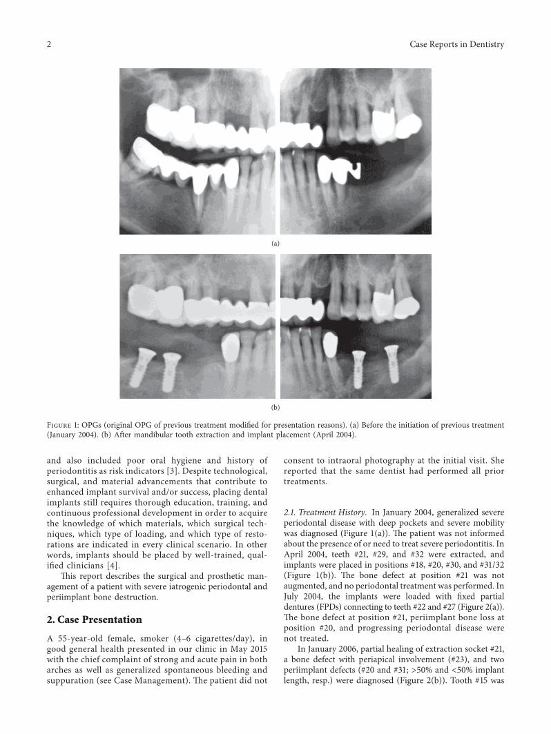
2.1. Treatment History. In January 2004, generalized severeperiodontal disease with deep pockets and severe mobilitywas diagnosed (Figure 1(a)). ,e patient was not informedabout the presence of or need to treat severe periodontitis. InApril 2004, teeth #21, #29, and #32 were extracted, andimplants were placed in positions #18, #20, #30, and #31/32(Figure 1(b)). ,e bone defect at position #21 was notaugmented, and no periodontal treatment was performed. InJuly 2004, the implants were loaded with fixed partialdentures (FPDs) connecting to teeth #22 and #27 (Figure 2(a)).,e bone defect at position #21, periimplant bone loss atposition #20, and progressing periodontal disease werenot treated.
In January 2006, partial healing of extraction socket #21,a bone defect with periapical involvement (#23), and twoperiimplant defects (#20 and #31; >50% and <50% implantlength, resp.) were diagnosed (Figure 2(b)). Tooth #15 was
(a)
(b)
Figure 1: OPGs (original OPG of previous treatment modified for presentation reasons). (a) Before the initiation of previous treatment(January 2004). (b) After mandibular tooth extraction and implant placement (April 2004).
2 Case Reports in Dentistry

extracted, an implant plan was made (as shown in theorthopantomograph (OPG)), and no further periodontal/periimplant treatment was performed. Between the end ofJanuary andOctober 2006, teeth #5–8, #10, #12, and #15 wereextracted; a composite veneered FPD was inserted with teeth#4, #9, and #11 as abutments; and an implant in position#15/16 was placed, but appeared to have only 50% bonecontact (Figure 3(a)). No further periodontal/periimplanttreatment was performed.
,e patient reported visiting the dental office often dueto pain, resulting in the fitting of a new maxillary restorationwith immediate implant placement and loading in No-vember 2006. ,e mandibular periimplant defects showedfurther progression (Figure 3(b)). A new implant in position #15was placed (compare with implant geometry on Figure 3(a)),tooth #12 was replaced with an implant, and additionalimplants were placed in positions #1, #4–6, and #8. ,enew implants had insufficient bone contact; the implant inposition #1 had only apical contact with bone. In the sub-sequent 2 years, the patient complained often about pain andvisited the dental office regularly. However, other than su-perficial cleaning, no periodontal/periimplant treatment wasperformed.
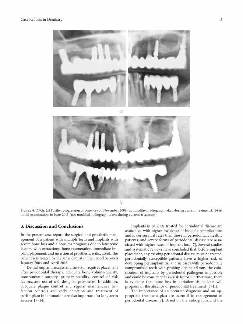
An OPG taken in November 2009 demonstrated furtherprogression of bone loss (Figure 4(a)). ,e patient reported
that the dentist in 2010 removed the mandibular FPDs,implants, and the majority of teeth and inserted anotherfixed restoration with immediate placement and loading,connecting the three implants with teeth #22 and #27. NoOPG showing this treatment or follow-up were available.,e patient visited the dental office regularly for cleaning andcomplained of new pain. In 2015, she was referred for peri-odontal consultation. Comparison of Figures 4(a) and 4(b)shows that the mandibular implants were explanted, and threenew implants were placed and loaded.
2.2. Case Management. Comprehensive dental and peri-odontal examinations were performed, and an OPG wasmade (Figure 4(b)). All maxillary and mandibular implantsand teeth showed radiographic severe bone loss, and teeth#9, #11, and #27 additionally showed subgingival caries andperiapical lesions. Periimplant pockets were 6–10mm deepwith spontaneous bleeding, soft-tissue swelling, and pain onpalpation.
After receiving oral and written descriptions of theproposed treatment, including surgical procedures, the pa-tient provided written informed consent. To address the acutecondition, mandibular periimplant abscesses were drainedthrough the pockets, and clindamycin (800mg/day) wasprescribed, due to the patient’s reported allergy to penicillin.
(a)
(b)
Figure 2: OPGs (original OPG of previous treatment modified for presentation reasons). (a) After mandibular implant loading (July 2004).(b) After extraction of tooth #15 (January 2006).
Case Reports in Dentistry 3

,e patient’s file and radiographs were retrieved from herformer dentist.
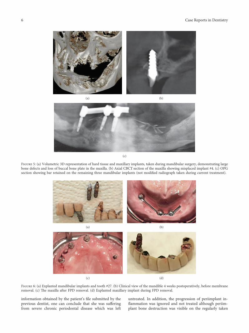
All mandibular and maxillary implants and teeth wereremoved, except implants #4 and #10 which served to tem-porarily retain an interim maxillary restoration. Duringsurgery and after removal of the mandibular teeth and im-plants and cleaning of the bone defects, a cone beam com-puted tomograph (CBCT) was made (Figures 5(a) and 5(b)).,e extraction sockets and periimplant bone defects werecleaned, and gentamicin-loaded collagen fleeces (Jason;Botiss Biomaterials, Zossen, Germany) were placed in thedefects [5]. Subsequently, the defects were covered withnonresorbable dense polytetrafluoroethylene membranes(dPTFE; Cytoplast Ti-250; Osteogenics Biomedical, Lub-bock, TX, USA) without additional bone grafting, aspreviously described [6]. Implants (K3Pro rapid; 3.5mmdiameter, 11mm length: Argon Dental, Bingen/R, Ger-many) were placed in positions #24, #26, and #30 andloaded the same day with a bar-retained removable tem-porary denture. ,e membranes were removed 4 weeks
postoperatively (Figures 5(c), 6(a), and b6(b)). ,e bar wasmilled of type 3 CrCo alloy (ZENOTEC NP; Wieland,Pforzheim, Germany), a metal base was constructed, andelastic plastic clips (Preci Matrice, CEKA, Waregem, Bel-gium) were used to retain the base over the bar.
On the same day, all remaining maxillary teeth andimplants, except #4 and #10, were extracted, periimplantlesions on #4 and #10 were treated (Figures 6(c) and 6(d)),and the maxilla was temporarily restored with a milled FPDfixed on the implants #4 and 10 using provisional cement(Implant Provisional; Alvelogro Inc., Snoqualmie, WA,USA) and a removable partial denture for the molar areas(Figure 7).
Seven months postoperatively, the bone defects wereregenerated, and three additional mandibular implants wereplaced in positions #22, #28, and #31/32 (K3Pro rapid;4.5mm diameter, 9 and 11 mm lengths, Argon Dental)(Figure 8(a)). All six mandibular implants were splinted witha milled bar and loaded as described previously (Figures 8(b)and 9).
(a)
(b)
Figure 3: OPGs (original OPG of previous treatment modified for presentation reasons). (a) After extraction of tooth #14 and implantplacement in position #15/16 (November 2006). (b) After maxillary restoration (November 2007).
4 Case Reports in Dentistry
3. Discussion and Conclusions
In the present case report, the surgical and prosthetic man-agement of a patient with multiple teeth and implants withsevere bone loss and a hopeless prognosis due to iatrogenicfactors, with extractions, bone regeneration, immediate im-plant placement, and insertion of prosthesis, is discussed. ,epatient was treated by the same dentist in the period betweenJanuary 2004 and April 2015.
Dental implant success and survival requires placementafter periodontal therapy, adequate bone volume/quality,nontraumatic surgery, primary stability, control of riskfactors, and use of well-designed prostheses. In addition,adequate plaque control and regular maintenance (in-fection control) and early detection and treatment ofperiimplant inflammation are also important for long-termsuccess [7–14].
Implants in patients treated for periodontal disease areassociated with higher incidence of biologic complicationsand lower survival rates than those in periodontally healthypatients, and severe forms of periodontal disease are asso-ciated with higher rates of implant loss [7]. Several studiesand systematic reviews have concluded that, before implantplacement, any existing periodontal disease must be treated,periodontally susceptible patients have a higher risk ofdeveloping periimplantitis, and in cases with periodontallycompromised teeth with probing depths >5mm, the colo-nization of implants by periodontal pathogens is possibleand could be considered as a risk factor. Furthermore, thereis evidence that bone loss in periodontitis patients willprogress in the absence of periodontal treatment [7–11].
,e importance of an accurate diagnosis and an ap-propriate treatment plan are essential in management ofperiodontal disease [7]. Based on the radiographs and the
(a)
(b)
Figure 4: OPGs. (a) Further progression of bone loss on November 2009 (not modified radiograph taken during current treatment). (b) Atinitial examination in June 2015 (not modified radiograph taken during current treatment).
Case Reports in Dentistry 5
information obtained by the patient’s file submitted by theprevious dentist, one can conclude that she was sufferingfrom severe chronic periodontal disease which was left
untreated. In addition, the progression of periimplant in-flammation was ignored and not treated although periim-plant bone destruction was visible on the regularly taken
(a) (b)
(c)
Figure 5: (a) Volumetric 3D representation of hard tissue and maxillary implants, taken during mandibular surgery, demonstrating largebone defects and loss of buccal bone plate in the maxilla. (b) Axial CBCT section of the maxilla showing misplaced implant #4. (c) OPGsection showing bar retained on the remaining three mandibular implants (not modified radiograph taken during current treatment).
(a) (b)
(c) (d)
Figure 6: (a) Explanted mandibular implants and tooth #27. (b) Clinical view of the mandible 4 weeks postoperatively, before membraneremoval. (c) ,e maxilla after FPD removal. (d) Explanted maxillary implant during FPD removal.
6 Case Reports in Dentistry

(a) (b)
(c)
Figure 7: (a, b) Maxillary temporary rehabilitation with FPD retained on implants #4 and #10 and removable denture for the molar areas.(c) OPG 4 weeks after surgery with the mandibular overdenture (not modified radiograph taken during current treatment).
(a)
(b)
Figure 8: Mandibular OPG sections eight months postoperatively (not modified radiograph taken during current treatment). (a) Afterplacement of three additional implants. (b) After bar mounting.
Case Reports in Dentistry 7
radiographs. ,e patient reported regular oral hygiene ap-pointments in the dental office but only supragingival de-bridement was performed.
Currently, there is not enough focus on the prevention ofperiimplant diseases, as compared to periodontal maintenance[7, 13]. It is well known that, in periodontitis susceptible patientstreated with dental implants, residual pockets represent a sig-nificant risk for the development of periimplantitis and implantloss. Moreover, patients in supportive periodontal treatmentdeveloping reinfections are at greater risk for periimplantitisand implant loss than periodontally stable patients [14].
An additional finding, after examining the patient’s file,was the absence of accurate radiographs of diagnostic quality
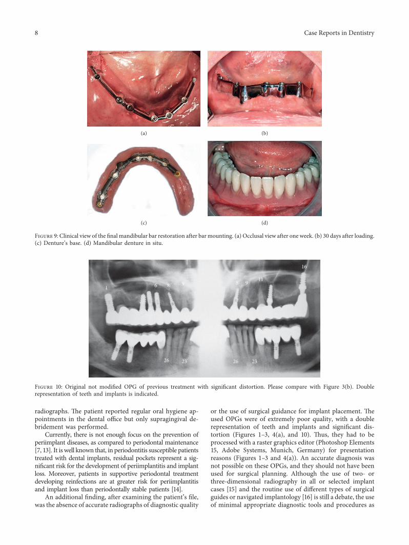
or the use of surgical guidance for implant placement. ,eused OPGs were of extremely poor quality, with a doublerepresentation of teeth and implants and significant dis-tortion (Figures 1–3, 4(a), and 10). ,us, they had to beprocessed with a raster graphics editor (Photoshop Elements15, Adobe Systems, Munich, Germany) for presentationreasons (Figures 1–3 and 4(a)). An accurate diagnosis wasnot possible on these OPGs, and they should not have beenused for surgical planning. Although the use of two- orthree-dimensional radiography in all or selected implantcases [15] and the routine use of different types of surgicalguides or navigated implantology [16] is still a debate, the useof minimal appropriate diagnostic tools and procedures as
(a) (b)
(c) (d)
Figure 9: Clinical view of the final mandibular bar restoration after barmounting. (a) Occlusal view after one week. (b) 30 days after loading.(c) Denture’s base. (d) Mandibular denture in situ.
1 6
26 2623 23
8 89 911
16
Figure 10: Original not modified OPG of previous treatment with significant distortion. Please compare with Figure 3(b). Doublerepresentation of teeth and implants is indicated.
8 Case Reports in Dentistry

well as medical and dental standards is mandatory fora successful result after implant placement.
Another treatment modality, which was repeatedly ap-plied in the presented case, was immediate implant place-ment and loading in infected and compromized periodontaltissues as well as the connection of teeth and implants.Furthermore, the restorations did not fit on the abutments(Figure 4(b)). ,ese could be additional factors for teeth andimplants loss. In the present case, an immediate implantplacement and eventually loading could be possible, only byfollowing established rules and clinical protocols as well asguidelines from the scientific literature. However, the lack ofknowledge has led to a disaster [4, 17, 18].
Combination therapy resolved the disease and the pa-tient’s severe discomfort while providing immediate func-tion and an aesthetic solution. Patient’s rehabilitation wasachieved by elimination of the infection, bone regeneration,and implant placement. In the mandible, three implantswere placed during the first surgery, splinted and loadedwith an overdenture, restoring function, and aesthetics. Inaddition, the bar-retained mandibular overdenture pro-tected the augmented areas from pressure during the healingperiod. In the maxilla, implants were removed, periimplantlesions in the remaining two implants were treated, and anaesthetic and functionally acceptable long-term provisionalrestoration was fabricated.
,e long-term periodontal and periimplant infectionand tissue destruction presented in this case resulted fromlack of periodontal and periimplant treatment as well asmaintenance and failure to use evidence-based diagnostic,surgical, and restorative procedures. Combination therapyresolved the disease and the patient’s severe discomfortwhile providing immediate function and an aestheticsolution.
Conflicts of Interest
,e authors declare that they have no conflicts of interest.
References
[1] L. Gaviria, J. P. Salcido, T. Guda, and J. L. Ong, “Current trendsin dental implants,” Journal of the Korean Association of Oraland Maxillofacial Surgeons, vol. 40, no. 2, pp. 50–60, 2014.
[2] S. Paleczek, Bruchfestigkeit provisorischer Bruecken gelagert aufImplantaten bzw. Implantaten und Zaehne, Doctoral ,esis,University of Regensburg, Regensburg, Germany, 2010.
[3] J. Lindhe, J. Meyle, and Group D of European Workshop onPeriodontology, “Peri-implant diseases: Consensus Report ofthe Sixth European Workshop on Periodontology,” Journal ofClinical Periodontology, vol. 35, no. 8, pp. 282–285, 2008.
[4] N. Harel, Z. Ormianer, E. Zecharia, and A. Meirowitz, “Con-sequences of experience and specialist training on the fabricationof implant-supported prostheses: a survey,” Journal of ProstheticDentistry, vol. 117, no. 6, pp. 743–748, 2016.
[5] O. Kilian, H. Hossain, I. Flesch et al., “Elution kinetics, anti-microbial efficacy, and degradation and microvasculature ofa new gentamicin-loaded collagen fleece,” Journal of BiomedicalMaterials Research Part B: Applied Biomaterials, vol. 90B, no. 1,pp. 210–222, 2009.
[6] O. Hoffmann, B. K. Bartee, C. Beaumont, A. Kasaj, G. Deli,and G. G. Zafiropoulos, “Alveolar bone preservation in ex-traction sockets using non-resorbable dPTFE membranes:a retrospective non-randomized study,” Journal ofPeriodontology, vol. 79, no. 8, pp. 1355–1369, 2008.
[7] N. Donos, L. Laurell, and N. Mardas, “Hierarchical decisionson teeth vs. implants in the periodontitis-susceptible patient:the modern dilemma,” Periodontology 2000, vol. 59, no. 1,pp. 89–110, 2012.
[8] M. A. Stokman, A. J. van Winkelhoff, A. Vissink,F. K. Spijkervet, and G. M. Raghoebar, “Bacterial colonizationof the peri-implant sulcus in dentate patients: a prospectiveobservational study,” Clinical Oral Investigations, vol. 21,no. 2, pp. 717–724, 2017.
[9] S. Eick, C. A. Ramseier, K. Rothenberger, U Bragger, D. Buser,and G. E. Salvi, “Microbiota at teeth and implants in partiallyedentulous patients. A 10-year retrospective study,” ClinicalOral Implants Research, vol. 27, no. 2, pp. 218–225, 2016.
[10] G. Kalykakis, G.-G. Zafiropoulos,M. Yildirim,H. Spiekermann,and R. J. Nisengard, “Clinical and microbiological status ofosseointegrated implants,” Journal of Periodontology, vol. 65,no. 8, pp. 766–770, 1994.
[11] H. Wennstrom and N. P. Lang, “Treatment planning forimplant therapy in the periodontally compromised patient,”in Textbook of Clinical Periodontology and Implant Dentistry,J. Lindhe, N. P. Lang, and T. Karring, Eds., pp. 675–686,Blackwell Munksgaard, Oxford, UK, 5th edition, 2008.
[12] A. Ramanauskaite and T. Tervonen, “,e efficacy of sup-portive peri-implant therapies in preventing peri-implantitisand implant loss: a systematic review of the literature,” Journalof Oral and Maxillofacial Research, vol. 7, no. 3, p. e12, 2016.
[13] B. E. Pjetursson, C. Helbling, H. P. Weber et al., “Peri-implantitis susceptibility as it relates to periodontal therapyand supportive care,” Clinical Oral Implants Research, vol. 23,no. 7, pp. 888–894, 2012.
[14] G. C. Armitage and P. Xenoudi, “Post-treatment supportivecare for the natural dentition and dental implants,”Periodontology 2000, vol. 71, no. 1, pp. 164–184, 2016.
[15] M. M. Bornstein, K. Horner, and R. Jacobs, “Use of cone beamcomputed tomography in implant dentistry: current concepts,indications and limitations for clinical practice and research,”Periodontology 2000, vol. 73, no. 1, pp. 51–72, 2017.
[16] M. Vercruyssen, T. Fortin, G. Widmann, R. Jacobs, andM. Quirynen, “Different techniques of static/dynamic guidedimplant surgery: modalities and indications,” Periodontology2000, vol. 66, no. 1, pp. 214–227, 2014.
[17] O. Hoffmann and G. G. Zafiropoulos, “Tooth-implant con-nection: a review,” Journal of Oral Implantology, vol. 38, no. 2,pp. 194–200, 2012.
[18] D. P. Tarnow, S. J. Chu, and P. D. Fletcher, “Clinical decisions:determining when to save or remove an ailing implant,”Compendium of Continuing Education in Dentistry, vol. 37,pp. 233–243, 2016.
Case Reports in Dentistry 9

DentistryInternational Journal of
Hindawiwww.hindawi.com Volume 2018
Environmental and Public Health
Journal of
Hindawiwww.hindawi.com Volume 2018
Hindawi Publishing Corporation http://www.hindawi.com Volume 2013Hindawiwww.hindawi.com
The Scientific World Journal
Volume 2018Hindawiwww.hindawi.com Volume 2018
Public Health Advances in
Hindawiwww.hindawi.com Volume 2018
Case Reports in Medicine
Hindawiwww.hindawi.com Volume 2018
International Journal of
Biomaterials
Scienti�caHindawiwww.hindawi.com Volume 2018
PainResearch and TreatmentHindawiwww.hindawi.com Volume 2018
Preventive MedicineAdvances in
Hindawiwww.hindawi.com Volume 2018
Hindawiwww.hindawi.com Volume 2018
Case Reports in Dentistry
Hindawiwww.hindawi.com Volume 2018
Surgery Research and Practice
Hindawiwww.hindawi.com Volume 2018
BioMed Research International Medicine
Advances in
Hindawiwww.hindawi.com Volume 2018
Hindawiwww.hindawi.com Volume 2018
Anesthesiology Research and Practice
Hindawiwww.hindawi.com Volume 2018
Radiology Research and Practice
Hindawiwww.hindawi.com Volume 2018
Computational and Mathematical Methods in Medicine
EndocrinologyInternational Journal of
Hindawiwww.hindawi.com Volume 2018
Hindawiwww.hindawi.com Volume 2018
OrthopedicsAdvances in
Drug DeliveryJournal of
Hindawiwww.hindawi.com Volume 2018
Submit your manuscripts atwww.hindawi.com