Compounds in Cultures as Measured Exchange Chromatography' · In the present study, ion exchange...
6
Acidic Free Amino Compounds Formed in Various Lactic Acid Starter Cultures as Measured by Ion Exchange Chromatography' 2 M. K. HAMDY, W. J. HARPER AND H. H. WEISER Departments of Dairy Technology and Bacteriology, The Instituie of Nutrition and Food Technology, The Ohio State University, Columbus, Ohio Received for publication February 4, 1955 The role of starter organisms in the ripening of various cheese varieties has been the subject of extensive study in recent years (Deane, 1951; Peterson et al., 1948; Baribo and Foster, 1952; and Van der Zant and Nelson, 1954a). The accumulation of various free amino compounds during cheese ripening has been attributed primarily to enzymes of bacterial origin (Peterson et al., 1948; Deane, 1951; Harper and Gould, 1952). Most of the previous studies have been limited to the organism Streptococcus lactis (Hansen, 1941; Morgan and Nelson, 1951). Baribo and Foster (1952) determined some of the characteristics of the endo- cellular proteinases of one strain each of S. lactis, Lactobacillus casei, and Micrococcus freudenreichii. Van der Zant and Nelson (1953a and 1953b) investigated the proteolytic activity of S. lactis and a cell-free extract of this organism prepared by sonic vibration. Recently, Van der Zant and Nelson (1954a, 1954b) reported using paper chromatography to investigate the presence of amino acids and peptides in skimmilk inoculated with S. lactis, and to study some of the characteristics of the endocellular peptidases of this organism. In the present study, ion exchange chromatography was applied to investigate the proteolytic activity as measured by the presence of various acidic free amino acids of several lactic acid starter organisms; napiely, Lactobacillus bulgaricus, Lactobacillus lactis, and Strepto- coccus thermophilus. EXPERIMENTAL METHODS Cultures. Lactobacillus bulgaricus strains V4, V10, V12, V29, V71, R and R3, L. lactis strains 8 to 15, KM, V104 and V1o9, S. thermophilus strains S, T3, T4, and T5 were used in the present study. These cultures were obtained from the Dairy Products Section, Washington Utilization Research Branch, U. S. Department of Agriculture. They represent isolates from starters used in the I Supported in part by funds from the Research and Market- ing Act of 1946 through the Dairy Products Section, Washing- ton Utilization Research Branch, U.S.D.A., the Institute of Nutrition and Food Technology, The Ohio State University, The Ohio Dairy Products Fund, and The Keenan Fund. 2 Scientific Paper 14-54. The Department of Dairy Tech- nology, The Ohio State University. Ohio Agr. Exp. Sta. Journal Article 59-54. 221 manufacture of Provolone and Romano cheeses. They were cultivated through serial passage until they grew actively at 42 C within 12 hours and then were used to inoculate 700 ml of sterile skimmilk using two per cent inoculum. Unless otherwise mentioned, the cultures were incubated for 12 hours at 42 C and then stored at 10 C for various lengths of time. Application of ion exchange chromatography. The proteolytic activity of these organisms on the skimmilk was measured by determining qualitatively and quanti- tatively the presence of eleven free acidic a-amino compounds using Moore and Stein's (1951) ion exchange chromatographic method as modified by Hamdy et al. (1955). The acids studied in the order of their elution were: cysteic acid, serine phosphate, taurine, aspartic acid, threonine, serine, asparagine, glutamine, glutamic acid, proline, and glycine. Preparation of sample for ion exchange chromatography. Fifty-ml samples were withdrawn aseptically from the milk cultures with a sterile pipette and placed in 250- ml centrifuge bottles with 100 ml of distilled water containing 20 per cent ethyl alcohol by volume. The mixture was stirred vigorously with a mechanical stirrer for five minutes, and then cooled at 8 C for approximately two hours. This permitted precipitation of the nonsoluble protein fractions, after which the sample was centrifuged and then filtered through a fine sintered glass filter. The precipitates were washed twice with 50 ml distilled water. The filtrate (about 200 ml) was condensed at a low temperature under vacuum to 8 to 10 ml. One ml of this concentrate was adjusted to pH 3.0 and used for the ion exchange chromatography. The chromatographic separation. The sample was adsorbed on a 110 x 0.9-cm sulfonated polystyrene resin column operated in the sodium form. The column was then mounted on an automatic constant volume (1.0 ml) fraction collector. Two aliquot portions (0.5 ml) of pH 4.0 sodium citrate buffer were used to wash the acids in the resin bed. The chromatogram was de- veloped by displacing these adsorbed amino acids using about 150 ml of pH 4.0 sodium citrate buffer. The effluents were collected in 1.0-ml fractions in small 10-ml glass vials, and the elution was concluded after collecting 150 fractions. Analysis of effluent fractions. The presence and the on May 7, 2019 by guest http://aem.asm.org/ Downloaded from
Transcript of Compounds in Cultures as Measured Exchange Chromatography' · In the present study, ion exchange...
Acidic Free Amino Compounds Formed in Various Lactic AcidStarter Cultures as Measured by Ion Exchange Chromatography' 2
M. K. HAMDY, W. J. HARPER AND H. H. WEISER
Departments of Dairy Technology and Bacteriology, The Instituie of Nutrition and Food Technology,The Ohio State University, Columbus, Ohio
Received for publication February 4, 1955
The role of starter organisms in the ripening ofvarious cheese varieties has been the subject of extensivestudy in recent years (Deane, 1951; Peterson et al.,1948; Baribo and Foster, 1952; and Van der Zant andNelson, 1954a). The accumulation of various free aminocompounds during cheese ripening has been attributedprimarily to enzymes of bacterial origin (Petersonet al., 1948; Deane, 1951; Harper and Gould, 1952).Most of the previous studies have been limited to
the organism Streptococcus lactis (Hansen, 1941;Morgan and Nelson, 1951). Baribo and Foster (1952)determined some of the characteristics of the endo-cellular proteinases of one strain each of S. lactis,Lactobacillus casei, and Micrococcus freudenreichii. Vander Zant and Nelson (1953a and 1953b) investigatedthe proteolytic activity of S. lactis and a cell-freeextract of this organism prepared by sonic vibration.Recently, Van der Zant and Nelson (1954a, 1954b)reported using paper chromatography to investigate thepresence of amino acids and peptides in skimmilkinoculated with S. lactis, and to study some of thecharacteristics of the endocellular peptidases of thisorganism.
In the present study, ion exchange chromatographywas applied to investigate the proteolytic activity asmeasured by the presence of various acidic free aminoacids of several lactic acid starter organisms; napiely,Lactobacillus bulgaricus, Lactobacillus lactis, and Strepto-coccus thermophilus.
EXPERIMENTAL METHODS
Cultures. Lactobacillus bulgaricus strains V4, V10, V12,V29, V71, R and R3, L. lactis strains 8 to 15, KM, V104 andV1o9, S. thermophilus strains S, T3, T4, and T5 were usedin the present study. These cultures were obtained fromthe Dairy Products Section, Washington UtilizationResearch Branch, U. S. Department of Agriculture.They represent isolates from starters used in the
I Supported in part by funds from the Research and Market-ing Act of 1946 through the Dairy Products Section, Washing-ton Utilization Research Branch, U.S.D.A., the Institute ofNutrition and Food Technology, The Ohio State University,The Ohio Dairy Products Fund, and The Keenan Fund.
2 Scientific Paper 14-54. The Department of Dairy Tech-nology, The Ohio State University. Ohio Agr. Exp. Sta.Journal Article 59-54.
221
manufacture of Provolone and Romano cheeses. Theywere cultivated through serial passage until they grewactively at 42 C within 12 hours and then were used toinoculate 700 ml of sterile skimmilk using two per centinoculum. Unless otherwise mentioned, the cultureswere incubated for 12 hours at 42 C and then stored at10 C for various lengths of time.
Application of ion exchange chromatography. Theproteolytic activity of these organisms on the skimmilkwas measured by determining qualitatively and quanti-tatively the presence of eleven free acidic a-aminocompounds using Moore and Stein's (1951) ion exchangechromatographic method as modified by Hamdy et al.(1955). The acids studied in the order of their elutionwere: cysteic acid, serine phosphate, taurine, asparticacid, threonine, serine, asparagine, glutamine, glutamicacid, proline, and glycine.
Preparation of sample for ion exchange chromatography.Fifty-ml samples were withdrawn aseptically from themilk cultures with a sterile pipette and placed in 250-ml centrifuge bottles with 100 ml of distilled watercontaining 20 per cent ethyl alcohol by volume. Themixture was stirred vigorously with a mechanicalstirrer for five minutes, and then cooled at 8 C forapproximately two hours. This permitted precipitationof the nonsoluble protein fractions, after which thesample was centrifuged and then filtered through a finesintered glass filter. The precipitates were washed twicewith 50 ml distilled water. The filtrate (about 200 ml)was condensed at a low temperature under vacuum to8 to 10 ml. One ml of this concentrate was adjusted topH 3.0 and used for the ion exchange chromatography.
The chromatographic separation. The sample wasadsorbed on a 110 x 0.9-cm sulfonated polystyrene resincolumn operated in the sodium form. The column wasthen mounted on an automatic constant volume (1.0ml) fraction collector. Two aliquot portions (0.5 ml) ofpH 4.0 sodium citrate buffer were used to wash theacids in the resin bed. The chromatogram was de-veloped by displacing these adsorbed amino acidsusing about 150 ml of pH 4.0 sodium citrate buffer.The effluents were collected in 1.0-ml fractions in small10-ml glass vials, and the elution was concluded aftercollecting 150 fractions.
Analysis of effluent fractions. The presence and the
on May 7, 2019 by guest
http://aem.asm
.org/D
ownloaded from
HAMDY, HARPER AND WEISER
position of the a-amino acids were determined by spot-ting, with a capillary tube, a small amount of each ofthe effluent fractions on Whatman No. 1 filter paper.
The spots were completely air dried and sprayed withthe ninhydrin-n-propanol solution of Moore and Stein(1948), then placed in a 100 C oven for 3 to 5 minutesto develop the color. After establishing the position ofthe amino acids and determining their idenrtity bycomparison with the location of pure amino acids(Hamdy et al., 1955), the vials containing the specifica-amino compound were combined and then adjustedto a known volume, and the concentration determinedusing the photometric ninhydrin method of Moore andStein (1948). The concentration of the specific aminoacids in the sample was then calculated using standardcurves for each acid as described by Hamdy et al.(1955). In all amino acids determinations, duplicateexperiments were made and the average results are re-
ported.
RESULTSThis study was divided into two general phases.
The first phase consisted of an investigation of theproteolytic activity of single strain cultures of L. lactis,L. bulgaricus and S. thermophilus. For the second phase
of work, mixed strain cultures of the three organismswere used.
Single strain studies. L. lactis V104, L. bulgaricus V4,and S. thermophilus T5 were used. These strains were
grown alone and in various combinations with eachother in skimmilk for 12 hours at 42 C and then storedat 10 C. Selection of these specific strains was madeprimarily on their ability to produce comparableamounts of acid, with a range in pH values of 5.1 to5.3 after 12 hours incubation. This permitted a studyof the effect of one species on another without sup-
pressing the growth of any of the individual organisms.The results of the analyses of the three different
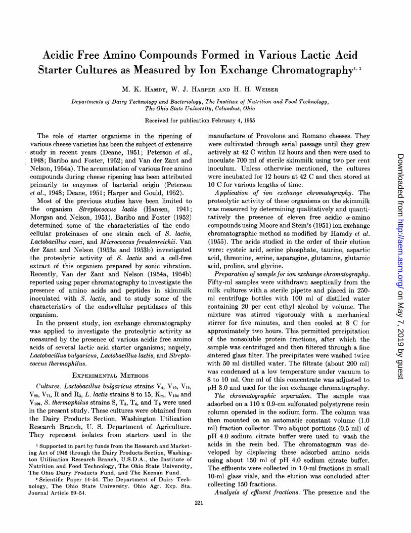
lactic acid producing cultures at the end of 12 hours ofincubation and after 0.5, 3, 7, 18, and 28 days ofstorage are shown in table 1. As noted, neither cysteicacid or proline could be detected in any of the culturesat any given time. Of the other amino compounds,only serine phosphate was present in all cultures atall times. In general, L. lactis V104 showed the greatestconcentration of amino acids at any given time, where-as very few of the amino acids could be found in milkinoculated with S. thermophilus T5. The effect ofstorage time on the accumulation of the various acidsis considerable, and the variations in this effect should
TABLE 1. The acidic free amino compound content of various lactic acid starter cultures*
Concentration of Amino Compounds in mg/1000 ml of Fernented Milkt
Age of Culture Age of Culture ~Serine. Aspartic Threonine Serine Asparagine Glutamicphosphate
Taurine acid| Glutamine acid Glycine
Lactobacillus lactis V 104
12 hours incubation ......................... 41.7 0 + + + 0 + 012 hours storage ................. ............ 68.3 0 0 7.9 10.1 52.9 03 days storage ............................. 121.1 2.2 0 0 | 0 146.0 41.2 07 days storage ............................. 75.2 17.6 0 0.2 21.4 43.2 39.0
18 days storage ............................. 157.9 + 0 139.2 37.7 0 56.628 days storage ............................. 159.5 0 0 0 8.0 98.0 0 28.1
Streptococcus thermophilus Ti
12 hours incubation ......................... 31.9 0 0 0 0 0 + +12 hours storage ............................ 45.6 + 0 0 0 0 + +3 days storage ............................. 52.2 0 0 0 0 0 0 07 days storage ............................. 49.5 0 0 0 0 0 0 0
18 days storage ............................. 49.4 0 0 0 0 0 0 028 days storage ............................. 45.9 0 0 0 0 0 0 0
Lactobacillus bulgaricus V4
12 hours incubation ......................... 36.3 0 0.8 0 0 0 + 012 hours storage ............... ............. 43.9 3.6 1.9 18.2 0 + +3 days storage ............................. 97.9 + 0 12.1 0 + 25.27 days storage ................ ............. 104.4 0 0 0 9.4 35.8 + +18 days storage ................ ............. 97.4 + 0 0 3.8 15.6 0 13.528 days storage ........... 146.2 0 0 0 8.0 31.3 0 +
* Acid present but in less than 0.5 mg/1000 ml concentration.t Cysteic acid and proline were not present.$ Threonine and serine could not always be separated, and asparagine and glutamine were not separated in this trial.
[VOL. 3222
on May 7, 2019 by guest
http://aem.asm
.org/D
ownloaded from
AMINO COMPOUNDS IN LACTIC ACID CULTURES
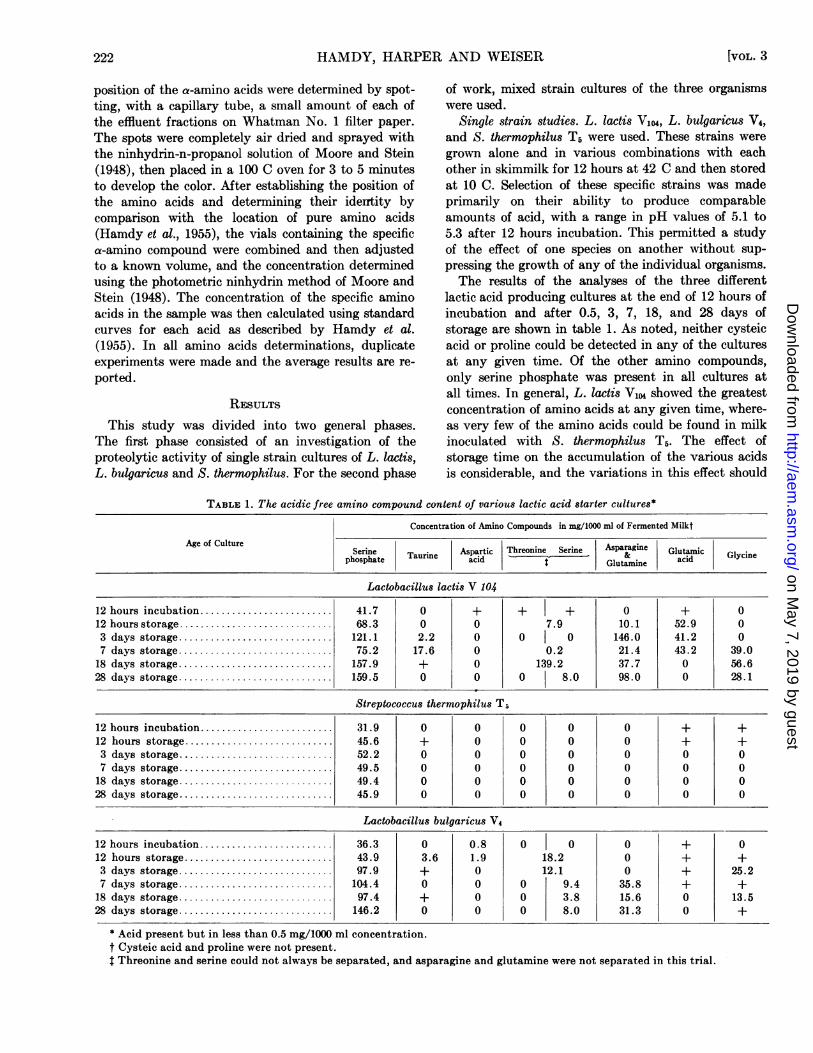
TABLE 2. The acidic free amino compound content of various lactic acid starter cultures
Concentration of Amino Compounds in mg/1000 ml of Fermented Milk*Age of Culture
Serine Taurine Aspartic Threonine Serine Asparagine Glutamic Glycinephosphate acid & Glutamine acid
Lactobacillus lactis V104 and Lactobacillus bulgaricus V4
12 hours storage .100.3 8.7 0 0 0 0 35.3 03 days storage .......................... 93.9 14.7 0 0 0 199.1 0 07 days storage .......................... 113.5 0 0 0 0 0 0 29.1
18 days storage .......................... 205.1 3.5 0 0 0 38.0 0 51.328 days storage . 102.3 +t 0 0 0 44.7 0 55.4
Lactobacillus bulgaricus V4 and Streptococcus thermophilus T5
12 hours storage ......................... 82.0 0 0 0 0 0 0 03 days storage .......................... 102.4 0 0 0 0 0 0 07 days storage .84.3 1.5 0 0 0 0.89 0 67.03
18 days storage .......................... 77.3 0 0 0 0 + 0 028 days storage .......................... 84.3 0 0 0 0 0 0 0
Lactobacillus lactis V104 and Streptococcus thermophilus T5
12 hours storage ........... 12.5 0 0 0 0 0 5.1 03 days storage .31.9 0 0 0 0 57.8 8.8 07 days storage .......................... 71.0 0 0 0 0 60.3 86.4 0
18 days storage .......................... 236.9 + 0 0 0 0 0 53.528 days storage .218.7 11.4 22.6 0 0 0 0 97.7
Lactobacillus lactis V104, Lactobacillus bulgaricus V4 and Streptococcus thermophilus T5
12 hours storage ......................... 62.6 + 0 7.4 0 0 28.53 days storage .77.3 + 0 14.3 19.5 0 65.67 days storage . 96.3 + 0 0 5.4 25.8 0 2.6
18 days storage . 200.4 3.1 0 0 28.6 74.0 0 74.028 days storage .......... 99.0 0 0 0 38.1 56.0 0 34.1
* Cysteic acid and proline were not present.t Acid present but in less than 0.5 mg/1000 ml concentration.
be noted. For example, aspartic and glutamic acidstended to be highest after 12 hours of storage, then todecrease, whereas the amides, asparagine and gluta-mine, and glycine increased during the storage period.Other compounds, such as serine phosphate andthreonine-serine increased and decreased during thestorage.The various single strains of organisms were also
grown in combination with one another and stored inthe same manner. As shown in table 2, there is a defi-nite effect of one species on another as indicated bythe change in the pattern of free amino compoundsproduced. For example, glycine, which was producedin very small amounts or not at all, was present inmuch higher concentrations in the mixed cultures.
Glutamic acid was produced by L. lactis V104 andby L. bulgaricus V4 Up to the 18th and 7th day ofstorage respectively, yet upon growth and storage ofthese cultures in combination with one another theglutamic acid was not detected except at 12 hoursstorage. Serine and threonine also were produced bythese strains growing alone, but their presence couldnot be found when L. lactis and L. bulgaricus were
grown together. The same effect was noted when L.bulgaricus V4 was grown with S. thermNphilus T5. Uponthe growth of these organisms together, most of theacids produced by L. bulgaricus V4 were not detected.The combination of all three organisms resulted in
an entirely different amino acid pattern than whenany two were grown in combination. For example,both threonine and serine were detected after incuba-tion and subsequent storage. Glutamic acid was notfound at any time, whereas asparagine and glutaminewere present after 3 days of storage. Glycine waspresent in all samples at all periods of storage, whichwas not true when any two of the organisms weregrown together.Mixed strain studies. L. bulgaricus strains V4, VI0,
V12, V29, V71, R and R3; L. lactis strains Km, 8-15, V104and Viog; S. thermophilus strains S, T3, T4, and T5 wereused in this study. These mixed strains were grownalone and in various combinations in skimmilk to in-vestigate their proteolytic activity, indicated by theliberation of amino acids, and the effect of one specieson the other as previously noticed in the single strainsstudy. The cultures were grown for 24 hours at 42 C,
1955] 223
on May 7, 2019 by guest
http://aem.asm
.org/D
ownloaded from
HAMDY; HARPER AND WEISER
TABLE 3. The acidic free amino acid content of various mixed cultures of lactic acid starter cultures*
Concentration of Acids in mg/10O ml of Fermented MilktAge of Culture
Serine phosphate Taurine Aspartic acid Serine Asparagine Glutamic acid
Lactobacillus lactis (mixed strains)
2days ... 41.6 0 0 0 0 50.5912 days ... 18.6 -0 0 0 0 79.1240 days ... 59.8 0 0 0 0 26.4
Streptococcus thermophilus (mixed strains)
2 days ...... 68.9 0 0 0 0 140.712 days ... 97.8 5.92 4.20 0 0 53.940 days ...... 172.0 0 0 19.56 0 38.4
Lactobacillus bulgaricus (mixed strains)
2 days ... 27.0 0 0 0 0 012 days ... 39.6 0 14.10 0 0 75.340 days ... 76.4 0 0 87.8 97.4 185.6
* Cultures grown in skimmilk for 24 hours at 42 C, stored at 10 C.t Cysteic acid, threonine, glutamine, and proline were not detected in any culture.
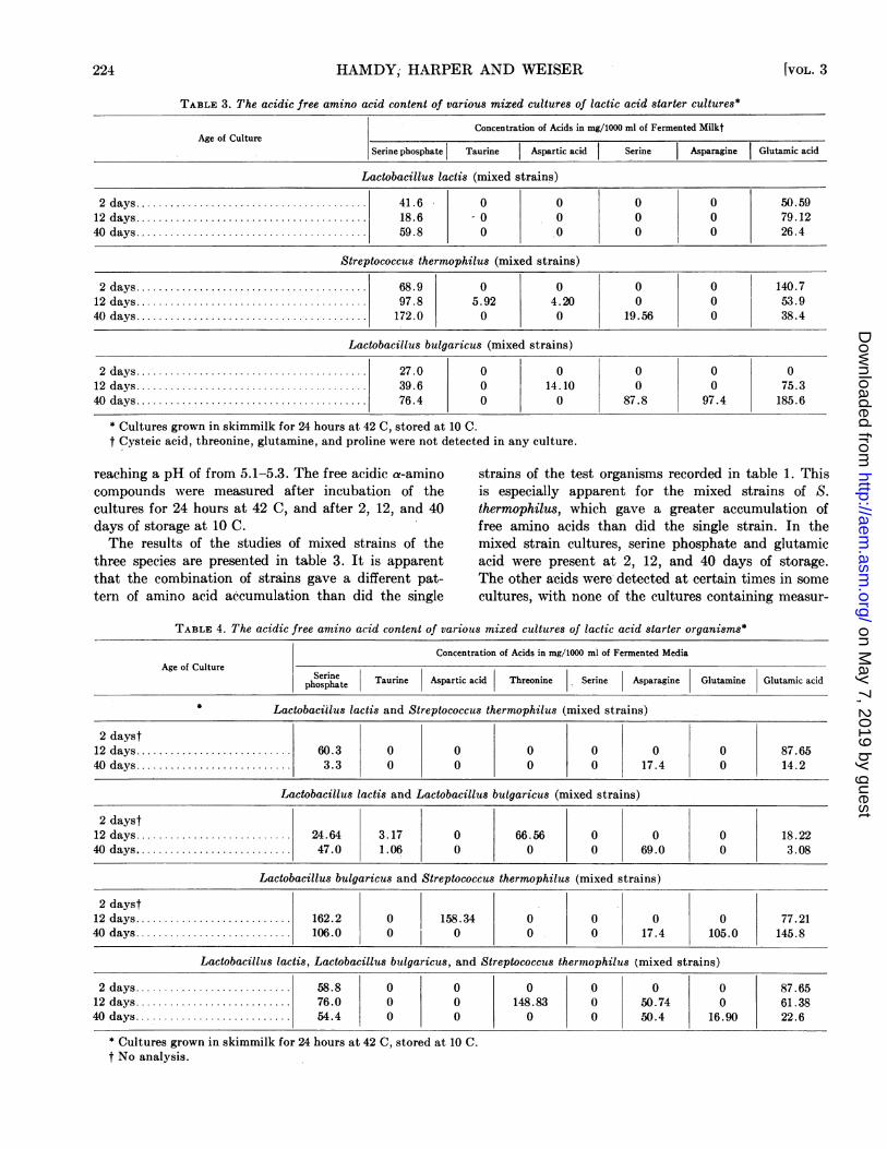
reaching a pH of from 5.1-5.3. The free acidic a-amino strains of the test organisms recorded in table 1. Thiscompounds were measured after incubation of the is especially apparent for the mixed strains of S.cultures for 24 hours at 42 C, and after 2, 12, and 40 thermophilus, which gave a greater accumulation ofdays of storage at 10 C. free amino acids than did the single strain. In theThe results of the studies of mixed strains of the mixed strain cultures, serine phosphate and glutamic
three species are presented in table 3. It is apparent acid were present at 2, 12, and 40 days of storage.that the combination of strains gave a different pat- The other acids were detected at certain times in sometern of amino acid accumulation than did the single cultures, with none of the cultures containing measur-
TABLE 4. The acidic free amino acid content of various mixed cultures of lactic acid starter organisms*
Concentration of Acids in mg/1000 ml of Fermented MediaAge of Culture
phoSerine Taurine Aspartic acid Threonine Serine Asparagine Glutamine Glutamic acid
Lactobacillus lactis and Streptococcus thermophilus (mixed strains)
2 dayst2 days .60.3 0 0 0 0 0 0 87.6540 days ......... 3.3 0 0 0 0 17.4 0 14.2
Lactobacillus lactis and Lactobacillus bulgaricus (mixed strains)
2 dayst12 days ..... 24.64 3.17 0 66.56 0 0 0 18.2240 days ... 47.0 1.06 0 0 0 69.0 0 3.08
Lactobacillus bulgaricus and Streptococcus thermophilus (mixed strains)
2 dayst12 days .| 162.2 0 158.34 0 0 0 0 77.2140 days ... 106.0 0 0 0 0 17.4 105.0 145.8
Lactobacillus lactis, Lactobacillus bulgaricus, and Streptococcus thermophilus (mixed strains)
2 days ... 58.8 0 0 0 0 0 0 87.6512 days ... 76.0 0 0 148.83 0 50.74 0 61.3840 days ... 54.4 0 0 0 0 50.4 16.90 22.6
* Cultures grown in skimmilk for 24 hours at 42 C, stored at 10 C.t No analysis.
[VOL. 3224
on May 7, 2019 by guest
http://aem.asm
.org/D
ownloaded from
AMINO COMPOUNDS IN LACTIC ACID CULTURES
able concentrations of cysteic acid, threonine, gluta-mine or proline.The results of the various species grown in combina-
tion are shown in table 4. As was true for single straincultures, there was a pronounced effect of one specieson another. Threonine was not produced by any ofthe species grown alone, but the mixture of L. lactisand L. bulgaricus resulted in a rather high concentra-tion of threonine at 12 days of storage. Glutaminewas found only in the mixed culture of L. bulgaricusand S. thermophilus and in the mixture of these specieswAith L. lactis but not with any species growing alone.
Control. A control consisting of only sterilized skim-milk media, incubated and stored at 10 C for the samelenigth of time as in the other experiments, showed thepresence of no acidic and amino compounds otherthan serinie phosphate. This was detectable after in-cubation, then increased in concentration from 9.11to 14.4 to 39.9 followed by a decrease to 18.2 to 14.2and finally to 6.6 mg of serine phosphate per 1000 mlof skimmilk media at 12 hours, 3, 7, 18, 28, and 40days respectively. No viable microorganism could bedetected in one-ml samples of sterilized milk. At thepresent time, there is no apparent explanation for thisinteresting observation.
DiscUSSIONThe data presented in this paper suggest that the
proteolytic activities of the starter organisms play animportant part in bringing about the specific changesthat take place in the ripening of Italian cheeses,Provolone and Romano. In this study, the proteolyticactivities of these cultures were measured in terms ofeleven free acidic a-amino compounds in skimmilk.The proteolytic activity in cheese, in general, is
due to the action of multiple enzyme systems fromdifferent sources: namely, the milk, rennet paste, andthe microflora present. The results of this study sug-gest that each type or strain of lactic acid starter or-ganism may produce different proteolytic end productswhen growing alone than when growing in combina-tion with other strains of the same or different species.Single culture studies revealed that L. lactis V104 andL. bulgaricus V4 were more active in their proteolyticactivities than S. thermophilus T5, while in mixedculture studies, the S. thermophilus showed more ac-tivity than both the L. bulgaricus and the L. lactis.The noticeable influence of one organism or group oforganisms on one another, to bring about definitechanges of the amino acid pattern as indicated in thisinvestigation, may be explained on the basis of: (a)synergism between species or strains to bring aboutchanges that neither of the species or strains couldproduce alone; (b) blocking of some reactions that leadto the liberation of certain amino acids; or (c) metabio-sis, whereby the product of one organism may become
a substrate for the other organism. Nurmikko (1952)has shown that symbiotic interrelationships do existbetween different strains of lactic acid bacteria in amedium of known chemical composition.
Serine phosphate was produced in skimmilk mediaby all the lactic acid starter culture organisms underinvestigation, either growing alone or in combinationwith others. This acidic amino acid may have a bio-logical significance in these cultures, but this is notclear at this time. It was also found in some sterileskimmilk samples initially and upon incubation at42 C and storage at 10 C. The presence of serine phos-phate may be due to chemical changes resulting fromthe sterilization of the skimmilk. The concentration ofthe serine phosphate in the sterile skimmilk controlsample was almost one-fifth its concentration in anyof the inoculated culture media. The presence of freeamino acids has been reported by Block (1951) and byVan der Zant and Nelson (1954a) in a protein-freefraction of skimmilk. These investigators did notreport the presence of serine phosphate acid in theirstudies. The role of serine phosphate is under investi-gation at the present time with the hope of securingmore information about its liberation, utilization andsignificance.
SUMMAIRYThe content of free acidic amino compounds in the
protein-free fraction of skimmilk incubated at 42 Cwith Italian cheese starter organisms for 12 to 24 hoursincubation and storage at 10 C for various lengths oftime was investigated by ion exchange chromatog-raphy. The results showed an increase in amino acidsin mixed cultures of various strains of Lactobacilluslactis, Lactobacillus bulgaricus, and Streptococcusthermophilus. Serine phosphate was present in allcultures due to bacterial action. It was also observedin some of the sterile skimmilk samples due to theeffect of heat.
REFERENCESBARIBO, L. E., AND FOSTER, E. M. 1952 The intracellular
proteinases of certain organisms from cheese and theirrelationship to the proteinases in cheese. J. Dairy Sci.,35, 149-160.
BLOCK, R. J. 1951 Some amino acids, peptides and amines inmilk, concentrated milks and cheese. J. Dairy Sci., 34,1-10.
DEANE, D. D. 1951 Preliminary studies of the effect ofacido-proteolytic organisms and temperatures of curing onthe ripening of cheddar cheese made from pasteurizedmilk. J. Dairy Sci., 34, 776-783.
HAMDY, M. K., HARPER, W. J., AND WEISER, H. H. 1955 Amodified procedure for the separation of acidic aminocompounds using a sulfonated polystyrene resin. J.Dairy Sci., 38, 147-154.
HANSEN, P. A. 1941 A study in cheese ripening: The in-fluence of autolyzed cells of Streptococcus cremoris andStreptococcus lactis on the development of Lactobacilluscasei. J. Dairy Sci., 24, 969-976.
19551 225
on May 7, 2019 by guest
http://aem.asm
.org/D
ownloaded from
HAMDY, HARPER AND WEISER
HARPER, W. J., AND GOULD, I. A. 1952 Italian cheese ripen-ing. Butter, Cheese, Milk Products J., 43, No. 8, 22-24,44, 46.
MOORE, S., AND STEIN, W. H. 1948 Photometric ninhydrinmethod for use in the chromatography of amino acids.J. Biol. Chem., 176, 367-388.
MOORE, S., AND STEIN, W. H. 1951 Chromatography ofamino acids on sulfonated polystyrene resins. J. Biol.Chem., 192, 663-678.
MORGAN, M. E., AND NELSON, F. E. 1951 The distribution ofcertain amino acids in soluble fractions of milk cultures ofStreptococcus lactis. J. Dairy Sci., 34, 446-456.
NURMIKKO, V. 1952 Chemical factors affecting associationsof lactic acid bacteria. Acta. Chem. Scand., 6, 1258-1282.
PETERSON, M. H., JOHNSON, M. J., AND PRICE, W. V. 1948
Proteinase content of cheddar cheese during making andripening. J. Dairy Sci., 31, 55-61.
VAN DER ZANT, W. C., AND NELSON, IF. E. 1953a Proteolysisby Streptococcus lactis grown in milk with and withoutcontrolled pH. J. Dairy Sci., 36, 1104-1111.
VAN DER ZANT, W. C., AND NELSON, F. E. 1953b Character-istics of an endocellular proteolytic enzyme system ofStreptococcus lactis. J. Dairy Sci., 36, 1212-1222.
VAN DER ZANT, W. C., AND NELSON, F. E. 1954a Aminoacids and peptides in the protein-free fraction of milkbefore and after incubation with Streptococcus lactis.J. Dairy Sci., 37, 790-794.
VAN DER ZANT, W. C., AND NELSON, F. E. 1954b Character-istics of some endocellular peptidases of Streptococcuslactis. J. Dairy Sci., 37, 795-804.
Supplementation of Media with Yeast Culturefor Counting Anaerobic Spores"2
A. T. ADAMS3 AND JOHN C. AYRES
Food Processing Laboratory, Iowa State College, Ames, Iowa
Received for publication February 9, 1955
Long incubation times frequently are necessary forobtaining accurate dilution counts of spores of anaero-bic bacteria in meat products. According to Morrisonand Rettger (1930) and to Knaysi (1945, 1948) slowgermination often is due to limiting factors in themedium rather than to inherent microbial charac-teristics. The various environmental factors affectingspore germination were discussed by McClung (1944),Olsen and Scott (1946), Hills (1949a, b; 1950), Halvor-son (1950), Powell (1950, 1951) Wynne (1952),Stewart and Halvorson (1953, 1954), and Church et al.(1954), and suggest the complex requirements neces-sary for an ideal subculture medium. Wynne andFoster (1948) showed that a pork infusion thioglycol-late medium containing soluble starch considerablyreduced the germination time of spores of severalanaerobic species. The shortcomings of this mediumwere enumerated by Burke et al. (1950) in their discus-sion of the incidence of putrefactive anaerobic sporesin meat products.
I Journal Paper No. J-2470 of the Iowa Agricultural Ex-periment Station, Ames, Iowa. Project No. 1017.
2 This paper reports research undertaken in cooperationwith the Quartermaster Food and Container Institute for theArmed Forces, and has been assigned Number 471 in the seriesof papers approved for publication. The views or conclusionscontained in this report are those of the authors. They are notto be construed as necessarily reflecting the views or indorse-ment of the Department of Defense.
3 Present address: The Rath Packing Company, Waterloo,Iowa.
Since Brown et al. (1939), Oxford et al. (1940), andWeizman and Rosenfeld (1939) showed that extractsof dried yeast favored the development of certainanaerobic bacteria, the possibility was considered thatthe germination time of anaerobic spores also might befavorably affected by the addition of yeast cultures tosubculture media.The purpose of this investigation was to determine
the efficacy of yeast-supplemented media in reducingthe incubation time required for accurate enumerationof anaerobic spores and to devise an efficient mediumwithout the shortcomings of pork infusion mediumfor enumerating spores of anaerobic bacteria in meatproducts.
EXPERIMENTAL METHODSSpore suspensions of Clostridium sporogenes, Clos-
tridium novyi, Clostridium parabotulinum Type A andB, Clostridium tetanomorphum, and Clostridium speciesP. A. 3679 were prepared according to the methodsproposed by Stumbo et al. (1950).
Test cultures of Saccharomyces cerevisiae (IowaState College culture collection reference number92-1-B) were prepared in the following manner: 5 mlof a stock yeast suspension (ca 107 cells per ml) wereinoculated into sterile medium consisting of: Bactopeptone 3 g; dextrose, 30 g; K2HPO, 1 g; (NH4)2SO4,1 g; and H20, 250 ml. After agitation for 18 hours ona rotary shaker at room temperature (20 to 25 C), theculture was neutralized with NaOH. The use of pure
226 [VOL. 3
on May 7, 2019 by guest
http://aem.asm
.org/D
ownloaded from