Component Position: How I put it in the Safe Zone - Radlink€¦ · Component Position: How I put...
16
Component Position: How I put it in the Safe Zone Robert L. Barrack, M.D. Washington University School of Medicine St. Louis, Missouri USA Orthopedics today, HAWAII January, 2014
Transcript of Component Position: How I put it in the Safe Zone - Radlink€¦ · Component Position: How I put...

Component Position: How I put it in the Safe Zone
Robert L. Barrack, M.D.
Washington University School of Medicine
St. Louis, Missouri USA
Orthopedics today, HAWAII January, 2014

Malchau et al; Charnley Award, 2010
Accurate, reproducible
positioning of the
acetabular component
greatest challenge:
optimal positioning
< 50% among best
surgeons
BUT, only one of half
dozen variable

Optimizing hip arthroplasty:
• Cup alignment entirely too narrow focus
• Current state of medical
knowledge/technology indicates clinical
results (and our goals) should extend far
beyond cup alignment to all aspects of
orientation of THA components: limb
length, offset, sizing, seating,
unrecognized fracture (to name a few)

Two approaches
• Virtual reality: utilization of advanced
technology that provide surrogates to
direct visualization of components: issues
with accuracy, validation, cost
• Intraoperative imaging: the gold
standard, but must meet rigid criteria of
maintaining efficiency: cannot add too
much time/cost or compromise sterility
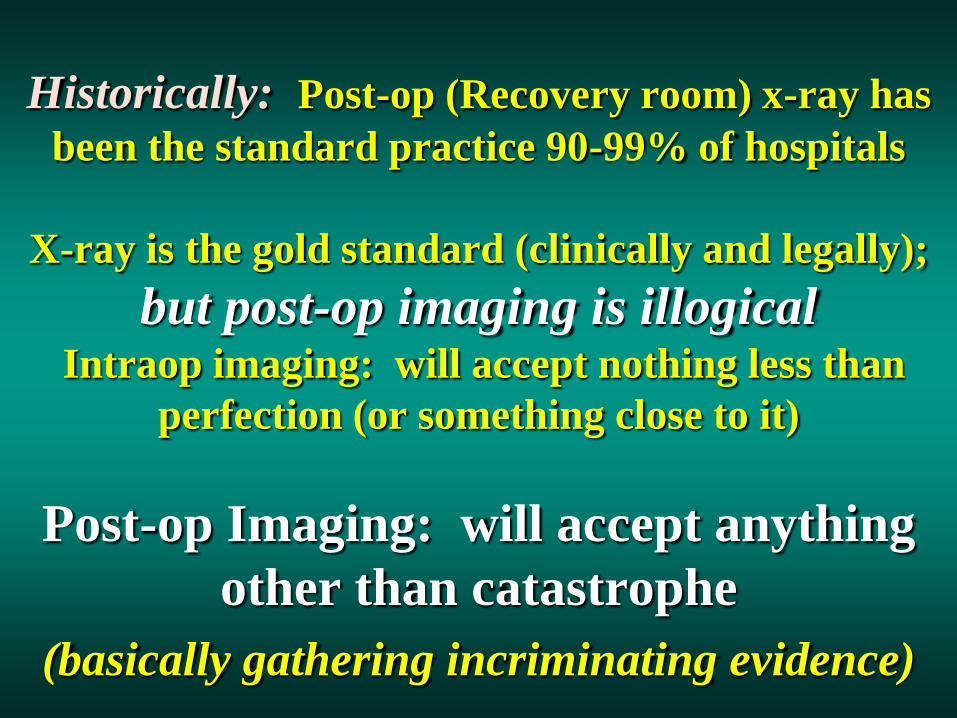
Historically: Post-op (Recovery room) x-ray has
been the standard practice 90-99% of hospitals
X-ray is the gold standard (clinically and legally);
but post-op imaging is illogical Intraop imaging: will accept nothing less than
perfection (or something close to it)
Post-op Imaging: will accept anything
other than catastrophe
(basically gathering incriminating evidence)

Why is intraop imaging not
been adopted in THA
• Adds too much time
• Disrupts work flow at a time when efficiency is
paramount
• Quality of images not adequate to take
measurements of interest to adequate degree of
accuracy (3-5º, 2-4 mm)
• Must be able to be an iterative process to
assess, make changes, reassess and document
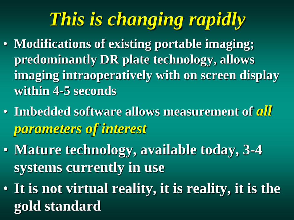
This is changing rapidly
• Modifications of existing portable imaging;
predominantly DR plate technology, allows
imaging intraoperatively with on screen display
within 4-5 seconds
• Imbedded software allows measurement of all
parameters of interest
• Mature technology, available today, 3-4
systems currently in use
• It is not virtual reality, it is reality, it is the
gold standard

Advantages of Digital Imaging
• Higher quality images
• Faster service speed.
• Minimal impact on O.R. workflow.
• Eventual reduction in operating costs.
• Eliminate the processing of chemicals,
processing dark room and film storage room.
• Eliminate outliers/returns to O.R.; may allow
return to hard/hard bearings (C-O-C)
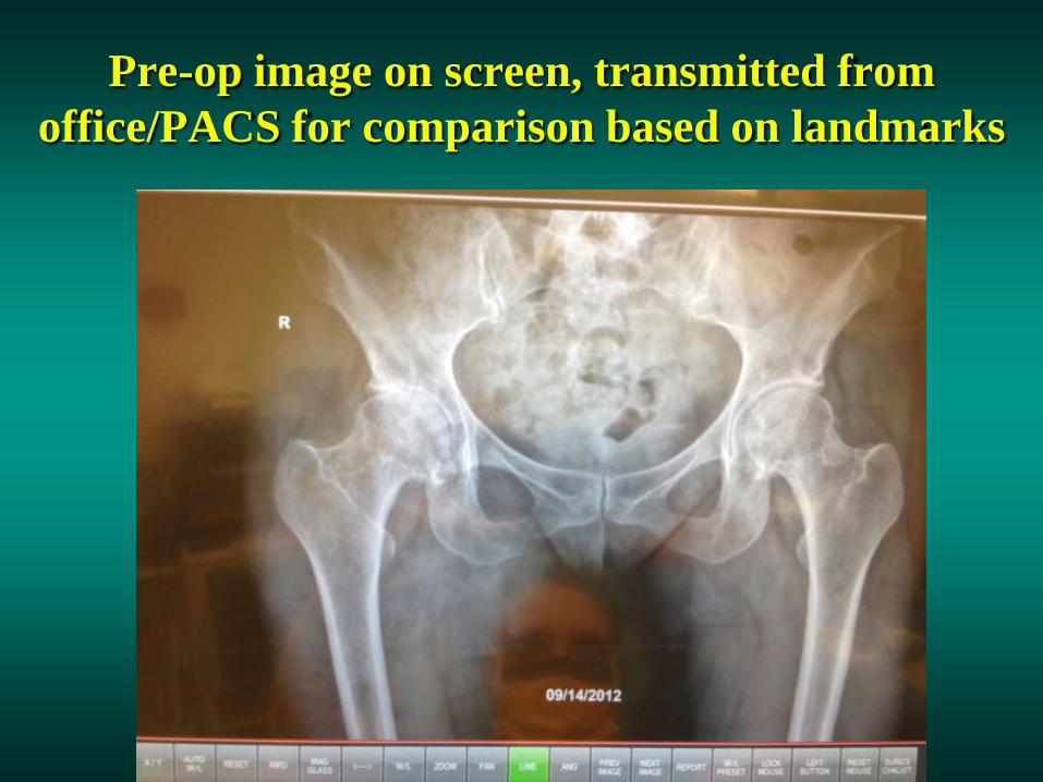
Pre-op image on screen, transmitted from
office/PACS for comparison based on landmarks

Image with trials in place; verify pelvic tilt/rotation;
reshoot if necessary
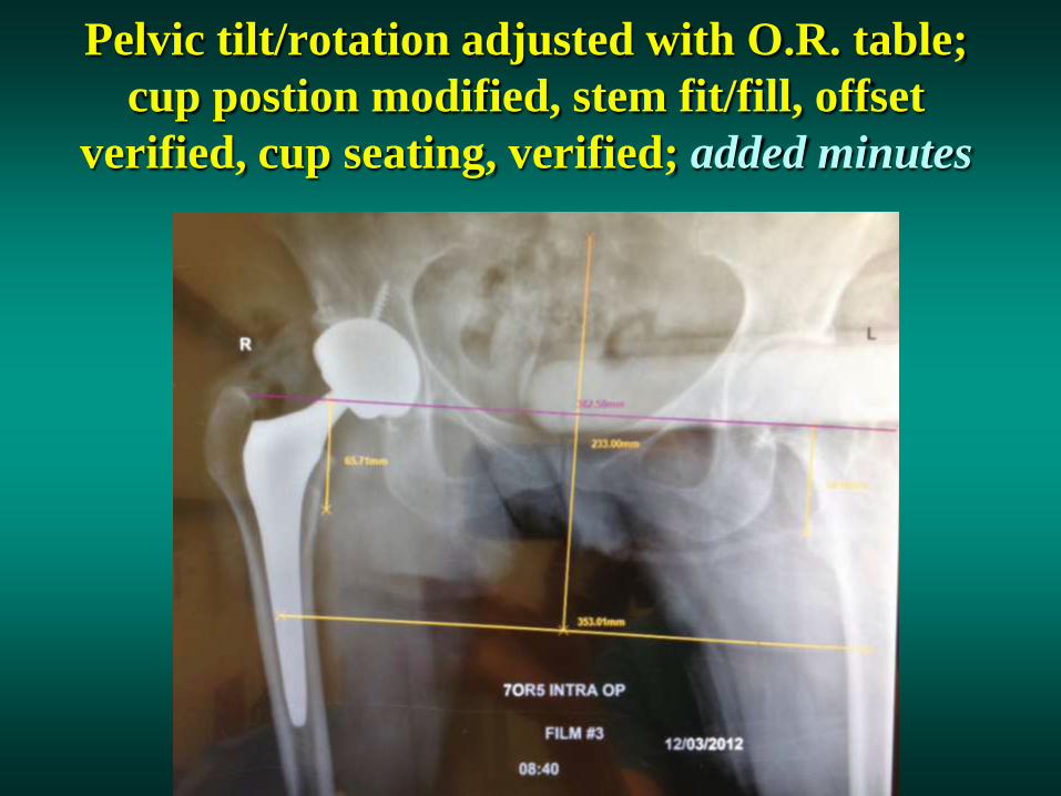
Pelvic tilt/rotation adjusted with O.R. table;
cup postion modified, stem fit/fill, offset
verified, cup seating, verified; added minutes

1º left hip; plan to lengthen;
anticipate right revision THA

Verify pelvic rotation/tilt: based on pelvic
inlet dimensions and mid-sacral to pubic line,
rotate table to reproduce pre-op and/or
computer modification of angles

Conclusion
• Advances in technology (DR, digital
radiography) have made intraoperative digital
imaging a practical/feasible strategy to avoid
outliers that increase complications and
compromise results
• Rapidly evolving technology, current status
effective in consistently optimizing THA
component placement
• Excellent teaching tool; rapidly embraced at our
center; successfully eliminated outliers/surprises