Complications of laser dermatologic...
15
Lasers in Surgery and Medicine 38:1–15 (2006) Complications of Laser Dermatologic Surgery Andrea Willey, MD, 1 R. Rox Anderson, MD, 2 Jose L. Azpiazu, MD, 3 Abnoeal D. Bakus, PhD, 4 Richard J. Barlow, FRCP, 5 Jeffrey S. Dover, MD, FRCPC, 6,7 Jerome M. Garden, MD, 4 Suzanne L. Kilmer, MD, 8 Nerea Landa, MD, 3 Dieter Manstein, MD, 2 E. Victor Ross, MD Jr., 9 Neil Sadick, MD, FACP, FAACS, 10 Emil A. Tanghetti, MD, 11,12 Dina Yaghmai, MD, 4 and Brian D. Zelickson, MD 1,13,14 * 1 Department of Dermatology, University of Minnesota, Minneapolis, Minnesota 55455 2 Wellman Center of Photomedicine, Department of Dermatology, Harvard Medical School Boston, Massachusetts 02114 3 Dermitek Clinic of Dermatology, Laser and Aesthetic Surgery, Bilbao, Basque Country, Spain 4 Departments of Dermatology and Biomedical Engineering, Northwestern University, Chicago, Illinois 60611 5 St. John’s Institute of Dermatology, St. Thomas’ Hospital, London, England SE1 7EH 6 Yale University School of Medicine, New Haven, Connecticut 06520 7 SkinCare Physicians, Chestnut Hill, Massachusetts 02467 8 Laser and Skin Surgery Center of Northern California, Sacramento, California 95816 9 Naval Medical Center San Diego, Department of Dermatology, San Diego, California 92134 10 Weill Medical College of Cornell University, Department of Dermatology, New York, New York 10021 11 University of California, Davis, Medical Center, Sacramento, California 95819 12 Center for Dermatology and Laser Surgery, Sacramento, California 95819 13 Abbott Northwestern Hospital Center for Cosmetic Care, Edina, Minnesota 14 Skin Specialists, Ltd. Minneapolis, Minnesota 55402 Background and Objective: Innovations in lasers, light and radiofrequency devices have allowed for improved therapeutic efficacy and safety and the ability to treat patients with an ever-increasing number of medical and aesthetic indications. Safety remains a primary concern and the timely communication of complications and their management is vital to insure that treatments be as safe as possible. The purpose of this report on the Proceedings of the First International Laser Surgery Morbidity Meeting is to provide laser experts the opportunity to present and discuss complications that their patients have experienced and how they were successfully managed. Methods: Laser experts were invited to present complica- tions of laser, light, and radiofrequency treatments that their patients have experienced and to discuss the potential mechanisms leading to the complications their manage- ment and final outcomes. Results: Nineteen unique cases are presented and the clinical management of each case discussed. Eighteen sets of pre- and post-operative photos are presented. Conclusion: This report shows that even experts, with extensive experience using light-based therapies, can and do have patients who develop complications. Sound clinical judgment, and knowing how to avoid complications and their timely post-operative management, is essential to insure optimal therapeutic outcome. Lasers Surg. Med. 38:1–15, 2006. ß 2006 Wiley-Liss, Inc. Key words: complications; safety INTRODUCTION Laser dermatologic surgery has rapidly advanced since the first device was introduced more than four decades ago [1]. Continued innovations in optical technology and refinement of existing devices have allowed for new developments to meet growing consumer demands for effective and safe laser therapies [2]. Such innovations include: (1) the expanding use of specific wavelengths, pulse durations and cooling strategies; (2) introduction of non-ablative rejuvenation techniques, including radiofre- quency, intense pulsed light and other light sources; and (3) combinations of laser, light, and radiofrequency tech- nologies, all of which have provided dermatologic laser surgeons with the ability to treat patients with an ever- increasing number of medical and aesthetic indications. Constant, however, has been the emphasis on safety in the application of both new and old laser technologies. In the hands of knowledgeable and well-trained practitioners, complication rates with the use of lasers remain low. However, when complications do occur, timely communica- tion of the occurrence and skillful management are *Correspondence to: Brian D. Zelickson, MD, Department of Dermatology, University of Minnesota, 420 Delaware Street SE, MMC 98, Minneapolis, MN 55455, 612-863-3001. E-mail: [email protected] Accepted 16 November 2005 Published online 23 January 2006 in Wiley InterScience (www.interscience.wiley.com). DOI 10.1002/lsm.20286 ß 2006 Wiley-Liss, Inc.
Transcript of Complications of laser dermatologic...

Lasers in Surgery and Medicine 38:1–15 (2006)
Complications of Laser Dermatologic Surgery
Andrea Willey, MD,1 R. Rox Anderson, MD,2 Jose L. Azpiazu, MD,3 Abnoeal D. Bakus, PhD,4
Richard J. Barlow, FRCP,5 Jeffrey S. Dover, MD, FRCPC,6,7 Jerome M. Garden, MD,4
Suzanne L. Kilmer, MD,8 Nerea Landa, MD,3 Dieter Manstein, MD,2 E. Victor Ross, MD Jr.,9
Neil Sadick, MD, FACP, FAACS,10 Emil A. Tanghetti, MD,11,12 Dina Yaghmai, MD,4
and Brian D. Zelickson, MD1,13,14*
1Department of Dermatology, University of Minnesota, Minneapolis, Minnesota 554552Wellman Center of Photomedicine, Department of Dermatology, Harvard Medical School Boston,Massachusetts 021143Dermitek Clinic of Dermatology, Laser and Aesthetic Surgery, Bilbao, Basque Country, Spain4Departments of Dermatology and Biomedical Engineering, Northwestern University, Chicago, Illinois 606115St. John’s Institute of Dermatology, St. Thomas’ Hospital, London, England SE1 7EH6Yale University School of Medicine, New Haven, Connecticut 065207SkinCare Physicians, Chestnut Hill, Massachusetts 024678Laser and Skin Surgery Center of Northern California, Sacramento, California 958169Naval Medical Center San Diego, Department of Dermatology, San Diego, California 9213410Weill Medical College of Cornell University, Department of Dermatology, New York, New York 1002111University of California, Davis, Medical Center, Sacramento, California 9581912Center for Dermatology and Laser Surgery, Sacramento, California 9581913Abbott Northwestern Hospital Center for Cosmetic Care, Edina, Minnesota14Skin Specialists, Ltd. Minneapolis, Minnesota 55402
Background and Objective: Innovations in lasers, lightand radiofrequency devices have allowed for improvedtherapeutic efficacy and safety and the ability to treatpatients with an ever-increasing number of medical andaesthetic indications. Safety remains a primary concernand the timely communication of complications and theirmanagement is vital to insure that treatments be as safe aspossible. The purpose of this report on the Proceedings ofthe First International Laser Surgery Morbidity Meeting isto provide laser experts the opportunity to present anddiscuss complications that their patients have experiencedand how they were successfully managed.Methods: Laser experts were invited to present complica-tions of laser, light, and radiofrequency treatments thattheir patients have experienced and to discuss the potentialmechanisms leading to the complications their manage-ment and final outcomes.Results: Nineteen unique cases are presented and theclinical management of each case discussed. Eighteen setsof pre- and post-operative photos are presented.Conclusion: This report shows that even experts, withextensive experience using light-based therapies, can anddo have patients who develop complications. Sound clinicaljudgment, and knowing how to avoid complications andtheir timely post-operative management, is essential toinsure optimal therapeutic outcome. Lasers Surg. Med.38:1–15, 2006. � 2006 Wiley-Liss, Inc.
Key words: complications; safety
INTRODUCTION
Laser dermatologic surgery has rapidly advanced sincethe first device was introduced more than four decades ago[1]. Continued innovations in optical technology andrefinement of existing devices have allowed for newdevelopments to meet growing consumer demands foreffective and safe laser therapies [2]. Such innovationsinclude: (1) the expanding use of specific wavelengths,pulse durations and cooling strategies; (2) introduction ofnon-ablative rejuvenation techniques, including radiofre-quency, intense pulsed light and other light sources; and(3) combinations of laser, light, and radiofrequency tech-nologies, all of which have provided dermatologic lasersurgeons with the ability to treat patients with an ever-increasing number of medical and aesthetic indications.Constant, however, has been the emphasis on safety in theapplication of both new and old laser technologies. In thehands of knowledgeable and well-trained practitioners,complication rates with the use of lasers remain low.However, when complications do occur, timely communica-tion of the occurrence and skillful management are
*Correspondence to: Brian D. Zelickson, MD, Department ofDermatology, University of Minnesota, 420 Delaware Street SE,MMC 98, Minneapolis, MN 55455, 612-863-3001.E-mail: [email protected]
Accepted 16 November 2005Published online 23 January 2006 in Wiley InterScience(www.interscience.wiley.com).DOI 10.1002/lsm.20286
� 2006 Wiley-Liss, Inc.

essential to insure that the use of lasers in medicine andsurgery remains as safe as possible. The purpose of theFirst International Laser Surgery Morbidity Meeting heldat Minnesuing Acres Lake Nebagamon, WI, on June 18–20,2004 was to provide laser experts around the world theopportunity to present and discuss complications that theirpatients experienced and how they were successfullymanaged post treatment.
METHODS
Laser experts were invited to present complications oflaser treatments that their patients have experienced inrecent years and to discuss the potential mechanismsleading to the complication and its successful clinicalmanagement. Complications were reported using a stan-dard format, including a description of the patient,indication for treatment, device used, treatment para-meters (Table 1), complications that occurred, their man-agement, final outcomes, and a brief discussion of the causeof the complication and potential methods of prevention.Nineteen unique cases are presented and the clinicalmanagement of each case discussed.
RESULTS
Summary of Cases Presented and Discussion
Pulsed Dye Laser (PDL)Case 1: Pulsed dye laser (PDL)-induced scars due
to intermittent cryogen spray cooling (CSC) devicefailure. Three pediatric patients presented on the sameday for pulsed dye laser (PDL) therapy of their facial portwine stains (PWS) birthmarks. The patients were treatedwith a PDL in conjunction with cryogen spray cooling (CSC)under general anesthesia using the following treatmentparameters: l, 595 nm; F, 9–14 J/cm2; tp, 1.5 milliseconds;SS, 7 mm; CSC tc, 30–60 milliseconds; td, 20 milliseconds.Within a few days post-operatively, all three patientsdeveloped scattered �7 mm round erosions, equivalent insize and shape to the laser treatment spot diameter,followed several weeks later by hemorrhagic crusting andatrophic scars.
The complication occurred as a result of intermittentfailure of the CSC device caused by bubbles present in thesupply line between the cryogen canister and laser handpiece. Several weeks post-operatively, the cryogen bubbledetector in the hand piece was found to be defective, whichresulted in the intermittent loss of CSC. The complication is
not easily avoidable given the absence of signs of devicemalfunction during treatment.Millisecond Pulsed 1,064-nm Nd:YAG LaserCase 2: Scarring following long pulsed 1,064 nm
Nd:YAG laser treatment of port wine stain (PWS). Thepatient was a 55-year-old male who presented with a PWSwith long-standing blebs on the neck and ear. The patient’sPWS was treated with a Nd:YAG laser using the followingtreatment parameters: l, 1,064 nm; F, 100 J/cm2; tp, 40milliseconds; SS, 7 mm; Zimmer air cooling. Intra-opera-tively, the patient developed immediate tissue grayingwithin the test spot (Fig. 1A), which resulted in a white scar(Fig. 1B). Intravascular coagulation was observed withouttissue graying immediately following the laser pulse usingthe following treatment parameters: l, 1,064 nm; F, 60 J/cm2; tp, 40 milliseconds; SS, 10 mm. The PWS was thentreated with a PDL at the following treatment parameters:l, 595 nm; F, 10 J/cm2; tp, 40 milliseconds; SS, 10 mm;Zimmer air cooling (set at unit 4) followed 1 minute later bytreatment with a Nd:YAG laser using the test parameters(F, 60 J/cm2; tp, 40 milliseconds; SS, 10 mm).
The complication occurred as a result of thermal injuryassociated with the high fluence used to achieve photo-coagulation with deeply penetrating 1,064 nm wavelength.Placement of test spots to determine the minimal purpuricthreshold [7] or use of initial treatment with a PDL to allowfor reduced fluences of the 1,064 nm laser may minimizethis complication.Case 3: Ulceration and scarring following long pulsed
1,064 nm Nd:YAG laser treatment of a proliferatinghemangioma. The patient was a 3-month-old female witha rapidly growing hemangioma on the left cheek withcombined superficial and deep components (Fig. 2A). Thepatient’s hemangioma was treated with a Nd:YAG laserusing the following treatment parameters: l, 1,064 nm;F, 56 J/cm2; tp, 3 milliseconds; SS, 10 mm; RR, 1 Hz; CSC tc,50 milliseconds; td 50 milliseconds. One day post-opera-tively, four blisters developed in the areas of greatestapparent vascularity (Fig. 2B) which crusted over and weretender for several days. The deep component of thehemangioma decreased at least one-third in size over thefollowing month. Superficial crusting was noted on onespot for almost a month as the lesion continued to regress(Fig. 2C). The crusting was initially managed withpetrolatum ointment. Three areas, including the area ofprolonged crusting, healed with hypopigmentation andslight scarring. To avoid further blistering, subsequenttreatments were continued with a PDL using the followingtreatment parameters: l, 595 nm; F, 7.5 J/cm; tp, 1.5milliseconds; SS, 10 mm; RR, 1 Hz; CSC tc, 30 milliseconds;td, 30 milliseconds. The hemangioma slowly decreased insize by an additional 25% after the next three PDLtreatments.
The complication occurred as a result of deep thermalinjury associated with the use of highly penetrating nearinfrared wavelengths. Starting at a lower fluence may haveminimized this complication. Only experienced lasersurgeons should use 1,064 nm lasers for congenitalvascular lesions in children and in adults.
TABLE 1. Symbols Representing Treatment
Parameters
l Wavelength
F Fluence
tp Pulse duration
SS Spot size
RR Repetition rate
tc Cryogen spurt duration
td Delay between cryogen spurt and laser pulse
2 WILLEY ET AL.

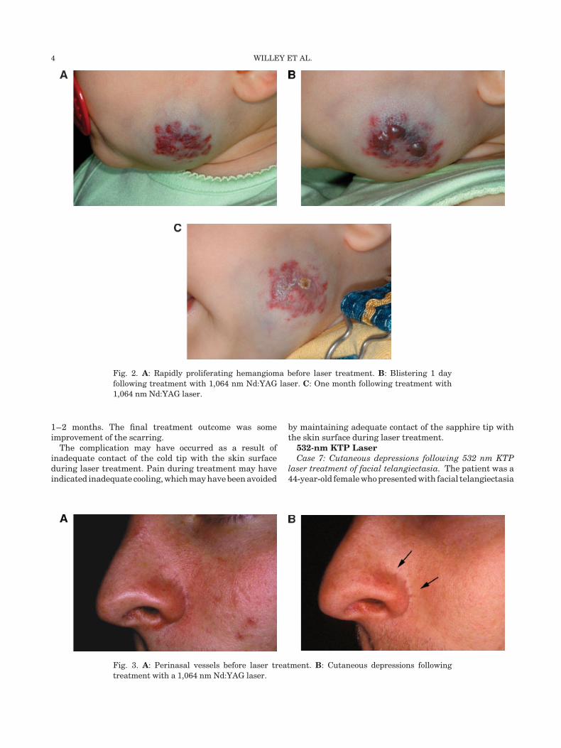
Case 4: Textural changes following long pulsed 1,064 nmNd:YAG laser treatment of perinasal vessels. The patientwas a 27-year-old Caucasian male who presented withperinasal telangiectasia (Fig. 3A). The patient’s perinasalblood vessels were treated with a Nd:YAG laser using thefollowing treatment parameters: l, 1,064 nm; F, 380 J/cm2;tp, 13 milliseconds; SS, 1.5 mm; sapphire tip parallel contactcooling. The treatment was uneventful and resulted inlesion clearing; however, 2 months post-operatively thepatient developed cutaneous indentations within thetreated areas (Fig. 3B). The complication was managed bywatchful waiting. Spontaneous resolution of these types ofindentations may occur after a period of 6–10 months. Non-ablative dermal remodeling may be attempted to improvethe indentations with infrared or visible light lasers. If theindentations do not resolve, ablative laser resurfacing alsomay be employed.
The complication occurred as a result of heat diffusionand thermal injury to tissue surrounding vessels. Thiscomplication may have been avoided by using lowerfluences, however, even with the appropriate selection oflaser treatment parameters, as in this case, patients candevelop cutaneous indentations.Case 5: Scarring following long pulsed 1,064 nm
Nd:YAG laser treatment of excess facial hair. The patientwas a 17-year-old female who presented with excess facialhair. The patient’s excess hair was treated with a Nd:YAGlaserusingthefollowingtreatmentparameters:l, 1,064nm;F, 100 J/cm2; tp, 50 milliseconds; SS, 10 mm hexagonal;sapphire tip contact cooling 58C on the pre-auricular cheek.Two days post-operatively, the patient developed blistering
at the treatment sites with subsequent scarring (Fig. 4A).The complication was managed by treatment of thefacial scars with a Candela VBeam PDL using the followingtreatment parameters: l, 595 nm; F, 6–7 J/cm2; tp,10 milliseconds; SS, 10 mm; CSC tc, 30 milliseconds; td,30 milliseconds for two sessions without improvement. Thefinal treatment outcome was hypertrophic scarring thatpersisted at 4 years follow-up (Fig. 4B).
The complication occurred as a result of excessiveheating due to overlapping pulses or failure of the cool-ing system due to inadequate contact of the cooled tip.Avoiding overlapping pulses, checking the tip for adequatecooling prior to laser treatment, insuring good contactbetween the cooled tip and the skin surface may haveavoided this complication. Future considerations toimprove the scarring include Er:YAG or fractional laserskin resurfacing.
Case 6: Ulceration and scarring following long pulsed1,064 nmNd:YAG laser treatment of leg veins. The patientwas a 67-year-old female who presented with ectatic legveins. The patient’s leg veins were treated with a Nd:YAGlaser for twelve treatments over a 2-year period using thefollowing treatment parameters: l, 1,064 nm; F, 270 J/cm2;tp, 50 milliseconds; SS, 3 mm; RR, 2 Hz; contact cooling.Intra-operatively, during the 12th treatment session,the patient experienced pain with subsequent ulcerationand scarring at the treatment sites (Fig. 5A). Sevenmonths later, the scarring was treated with a PDL usingthe following parameters: l, 595 nm; F, 6.5–7 J/cm2; tp, 6–10 milliseconds; SS, 10 mm; CSC tc, 30 milliseconds; td,30 milliseconds, for a total of eight sessions separated by
Fig. 1. A: PWS with immediate graying of test spot treated with a 1,064 nm Nd:YAG laser.
B: PWS with scarring of test spot and improvement of remaining lesion following treatment
with 595 nm PDL and 1,064 nm Nd:YAG lasers.
LASER DERMATOLOGIC SURGERY 3
1–2 months. The final treatment outcome was someimprovement of the scarring.
The complication may have occurred as a result ofinadequate contact of the cold tip with the skin surfaceduring laser treatment. Pain during treatment may haveindicated inadequate cooling, which may have been avoided
by maintaining adequate contact of the sapphire tip withthe skin surface during laser treatment.532-nm KTP LaserCase 7: Cutaneous depressions following 532 nm KTP
laser treatment of facial telangiectasia. The patient was a44-year-old female who presented with facial telangiectasia
Fig. 2. A: Rapidly proliferating hemangioma before laser treatment. B: Blistering 1 day
following treatment with 1,064 nm Nd:YAG laser. C: One month following treatment with
1,064 nm Nd:YAG laser.
Fig. 3. A: Perinasal vessels before laser treatment. B: Cutaneous depressions following
treatment with a 1,064 nm Nd:YAG laser.
4 WILLEY ET AL.

(Fig. 6A). The patient’s telangiectasia was treated with aKTP laser using the following treatment parameters: l, 532nm; F, 15 J/cm2; tp, 40 milliseconds; SS, 5 mm; withsapphire tip contact cooling, followed by a second pass usingthe following treatment parameters: F, 9 J/cm2; tp, 10milliseconds; SS, 4 mm. Post-operatively, the patientdeveloped erythema and swelling in the treated areas,followed 1 month later by hyperpigmentation over thelarger vessels. The final treatment outcome was cutaneousdepression and scarring at 3 months (Fig. 6B).
The complication occurred as a result of overheating oflarger vessels with inadequate time allowed for heatdissipation required for larger vessels. The lack of blister-ing indicates adequate cooling of the sapphire tip. Thiscomplication may have been avoided by using caution in
treating larger vessels and increasing the time intervalbetween successive passes.
Intense Pulse LightCase 8: Inadequate cooling with intense pulsed light
(IPL) treatment of melasma. The patient was a 40-year-oldfemale who presented with mild melasma. The patient’smelasma was treated with an IPL device using thefollowing treatment parameters: l, 520–1,200 nm; F, 21J/cm2; tp, 20 milliseconds; SS, 12�12 mm2; button 4. A coldaluminum roller was applied to the skin approximatelyafter every 15 pulses on the right forehead. Intra-operatively, the patient experienced moderate to severepain, and developed immediate erythema, gray discolora-tion and blistering, with crusting over the next 4 days(Fig. 7A). At the same visit, subsequent treatment of the leftforehead using the same parameters with more frequentcooling (use of roller every five pulses) resulted in no pain orcomplications (Fig. 7B).
The complication was managed by early application ofantibiotic ointment. To ‘‘feather’’ the perimeter of theeroded sites, the surrounding hyperpigmentation wasretreated with conservative IPL parameters and adequatecooling 2 months later. The final treatment outcome wasmild hypopigmentation that persisted at the site of deepestinjury 10 months after treatment (Fig. 7C).
The complication occurred as a result of infrequentapplication of the cold aluminum roller and could have beenavoided by more frequent application of the cold rollingsystem and/or by suspending treatment to assess the skinsurface. The pain response alone should have alerted thetreating physician that there might be excessive heatgeneration due to inadequate skin cooling. In the absence ofintegration, cooling is operator dependent, and successfultreatment requires careful real-time attention to the painresponse as well as the skin surface color. Erythema thatsharply mirrors the IPL footprint, graying of the skin, orfrank blistering indicates excessive fluence or inadequatecooling.
Fig. 4. A: Blistering and erythema 48 hours following laser epilation with a 1,064 nm Nd:YAG
laser. B: Persistent scars 4 years following laser treatment.
Fig. 5. Ulceration following treatment of leg veins with a
1,064 nm Nd:YAG laser.
LASER DERMATOLOGIC SURGERY 5

Intense Pulsed Light and Frequency Doubled532-nm Nd:YAG LaserCase 9: Dyschromia following sequential IPL and
frequency doubled Nd:YAG for photoaging. The patientwas a 42-year-old female (skin type II) who presented withdiffuse Fitzpatrick Class II photo aging manifested by mildwrinkling, increased vascularity and pigment dyschromia.The patient’s wrinkling, increased vascularity, and dys-chromia were treated with a Q-switched Nd:YAG laserusing the following treatment parameters: l, 532 nm;F, 1.1J/cm2; SS, 4 mm; RR; 10 Hz. On the same day, the patient’sclinical manifestations were treated with an IPL deviceusing the following treatmentparametersl, 560–1,200 nm;F, 28 J/cm2; SS, 10 �35 mm2; tp, 2.4 milliseconds pass one,4.2 milliseconds pass two. Post-operatively, the patientdeveloped IPL ‘‘foot-printing’’ the size of the IPL crystal andpersistent pigment dyschromia over Nd:YAG treated sites(Fig. 8). The complication was managed by applicationof increasing concentrations of a topical cream consistingof 5%–20% glycolic acid with 4% hydroquinone appliednightly, and biweekly microdermabrasions alternatingwith 20%–30% salicylic acid peels. The final outcome wasmarked improvement noted 4.5 months after treatment.
The complication occurred as a result of thermal injuryfollowing treatment with two pigment specific lasers inthe same day, which may increase the risk of skinhypersensitivity and pigment dyschromia. Performing theprocedures on separate days may have minimized thiscomplication.
Combination Radiofrequency and BroadSpectrum LightCase 10: Dyschromia following treatment of facial
photoaging. The patient was a 47-year-old female (skintype II) who presented with diffuse Fitzpatrick Class IIphoto aging manifested by mild wrinkling, increasedvascularity and pigment dyschromia. The patient wastreated with a combination bi-polar radiofrequency energyand broad spectrum light device using the followingtreatment parameters: l, 580–980 nm; F, 20 J/cm3 RFenergy, and 15 J/cm2 broad spectrum light; two to threepasses to the face without sequelae. A second treatmentwas performed 1 month later using the following treatmentparameters: F, 25 J/cm3 RF energy and 25 J/cm2 broad-spectrum light. Post-operatively after the second treat-ment, the patient developed footprinting and persistentdyschromia at the treated sites (Fig. 9). The complicationwas managed with topical application of 2% hydrocortisonecream with 4% hydroquinone in addition to biweeklymicrodermabrasions, alternating with 20%–30% salicylicpeels and 5% glycolic acid and nightly 2% hydroquinonecream. The final treatment outcome was gradual improve-ment of the dyschromia over 6 months.
The complication occurred as a result of an ex-cessive increase in fluence leading to thermal injury.Using conservative treatment parameters in early treat-ment sessions, performing a test spot, and slowly in-creasing fluences by 10% may have minimized thiscomplication.
Fig. 6. A: Facial vessels before laser treatment. B: Cutaneous depression 3 months following
treatment with a Q-switched Nd:YAG laser.
6 WILLEY ET AL.

CO2 and Erbium:YAG Laser ResurfacingCase 11: Actinic bronzing in ‘‘Skipped Areas’’ following
CO2 resurfacing for photoaging. The patient was a 52-year-old Caucasian female who presented with mild photodamage. The patient underwent uneventful full face laserskin resurfacing using a standard pulsed CO2 laser andthe following treatment parameters: l, 10.6 mm; F, 300 mJ;
SS, 2.25 mm. A single pass was applied with a computerpattern scanner (CPG) density of 6 (35% overlap betweenspots within the scan). Re-epithelialization was completein 10 days. Erythema diminished significantly over 2–4 weeks; diffuse erythema persisted for 12 weeks.Eighteen months post-operatively the patient developedbrown ‘‘stripes’’ on her cheeks that were coincident with‘‘skip’’ areas between the initial individual square CPGscans (Fig. 10A,B).
The complication was managed with treatment of theactinically bronzed skip areas with a long pulsed 532-nmKTP laser without improvement. Full-face laser resurfa-cing with the treatment parameters previously used isplanned. The complication occurred as a result of inad-vertently skipped areas between scans at the time ofresurfacing and could have been avoided by meticulousattention to laying down perfectly contiguous scans (usingmagnifying loupes during resurfacing). In the absence ofcomplete skin coverage, the physician can use a ‘‘freehand’’hand piece with independent pulses to ‘‘fill in’’ skip areasbetween scans at the time of initial resurfacing.
Case 12: Enterobacter cloacae infection following CO2
laser resurfacing. The patient is a 38-year-old female whopresented increased tenderness under one eye 1 weekfollowing CO2 laser resurfacing. Yellowish discharge witherythema and swelling consistent with an infection werenoted. Cultures were taken and the complication wasmanaged with dilute vinegar soaks, and the oral antibioticprescribed was changed from cephalexin to ciprofloxacin500 mg twice daily. Bacterial culture grew enterobactercloacae sensitive to ciprofloxacin. The patient’s symptomsimproved within the next 2 days.
Fig. 7. A: One day after IPL treatment: the patient’s right side
shows effect of inadequate cooling; the left side was treated
with same parameters but with more aggressive cooling.
B: Healing wound 2 weeks following IPL burn. C: Focal
hypopigmentation 10 months following IPL burn. Note the
melasma has returned.
Fig. 8. Dyschromia following treatment with IPL and 532 nm
frequency-doubled Nd:YAG laser.
LASER DERMATOLOGIC SURGERY 7

The patient had children ages 2 and 5 years. Neitherchild was still in diapers, however children are typically notas careful with hand washing and hands may be contami-nated with fecal flora. Practitioners should be careful towarn mothers of young children (as well as those with pets)to be aware of this potential contamination and to alwayswash their hands before touching their face to help controlspread of bacteria. If a patient presents with post-operative
pain, erythema, or discharge it is imperative to culture non-healing areas for fungus, viruses, and bacteria.Case 13: Pseudomonas aeruginosa infection following
CO2 laser resurfacing leading to extensive scarring. Thepatient was a 50-year-old female who presented 1 year afterCO2 laser skin resurfacing with full face and anterolateralneck hypertrophic scarring and dyspigmentation (Fig. 11).The patient had been treated with three full passes witha CO2 laser using the following treatment parameters: l,10.6 mm; F, 200 mJ; tp, 80–520 milliseconds; SS, 4–9 mm.The entire area was treated with identical settings. Post-operative wound care included open wound healingwith saline soaks and aquaphor healing ointment. Duringthe first week post-operatively, the patient developedincreasing pain and purulent discharge; cultures grewPseudomonas aeruginosa. Antimicrobial therapy wasinitiated and the infection subsided. However, healingand full re-epithelialization was delayed for several weeks,with subsequent hypertrophic scarring and dyspigmenta-tion. The scarring was treated with a PDL using thefollowing treatment parameters: l, 595 nm;F, 7.5 J/cm2; tp,0.45 milliseconds; SS, 7 mm. In addition, the complicationwas managed with pressure dressings, intralesionalsteroids, potent topical steroids, and daily occupationaltherapy. The final outcome was extensive hypertrophicscarring and dyspigmentation.
The complication occurred as a result of overly aggressiveCO2 laser skin resurfacing and delayed diagnosis of a post-operative wound infection, both of which contributed to thefinal outcome. This complication could have been prevented
Fig. 9. Dyschromia following treatment with bi-polar radio-
frequency energy and broad spectrum light.
Fig. 10. A, B: Streaks of retained pigment (brown) in ‘‘skip
areas’’ 18 months following single pass CO2 laser resurfacing.
Fig. 11. Extensive hypertrophic scarring following CO2 laser
resurfacing complicated by infection.
8 WILLEY ET AL.

by performing two rather than three passes, varying thetreatment parameters depending on the facial area treated,avoiding the neck except in occasional circumstances withvery gentle low fluences and one pass only. Earlierdiagnosis may have limited the extent of scarring, butwould not have eliminated the risk in this individual. Thetreatment of the scarring with the PDL, pressure dressingsand topical and intralesional steroids was appropriate.Case 14: Herpes simplex infection and staphylococcal
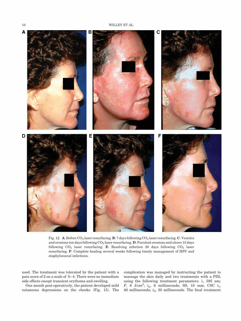
ecthyma followingCO2 laser resurfacing. The patient was a40-year-old female who presented with photo aging (Fig.12A). The patient underwent CO2 laser resurfacing withuneventful healing until the 8th post-operative day whenshe presented with a number of vesicles and erosions on theright cheek (Fig. 12B). Oral valacyclovir 500 mg twice a daywas started 2 days pre-operatively and taken until post-operative day 7. Ten days post-operatively increasingvesicles and erosions developed (Fig. 12C). A Tzanckpreparation and viral cultures for herpes simplex virus(HSV) were positive. The patient was admitted to hospitaland treated with intravenous acyclovir. Fifteen days post-operatively, after 7 days of intravenous acyclovir and 2 daysfollowing hospital discharge, the patient presented withpurulent erosions in many of the previously eroded areasthat coalesced into superficial purulent ulcers (Fig. 12D).Wound cultures grew Staphylococcus aureus and oraldicloxacillin 500 mg four times daily was initiated. Twentydays post-operatively, the infection appeared to be resolving(Fig. 12E). The final treatment outcome was completehealing with no textural change several weeks followingtimely management of HSV and staphylococcal infections,and a very happy patient who remained remarkablyoptimistic throughout the post-operative course (Fig. 12F).
The complication occurred as a result of facial HSVinfection that developed immediately after discontinuationof antiviral therapy and upon facial re-epithelialization.Intravenous acyclovir effectively treated the HSV infection,but by not continuing the dicloxacillin, a bacterial superinfection developed. This complication may have beenavoided by extending the antiviral prophylactic therapy,and contemporaneous antibiotic upon diagnosing the HSVinfection may have prevented the secondary bacterialinfection. Early diagnosis, appropriate therapy, and apositive patient attitude helped to prevent scarring in thiscase.Case 15: Hypopigmentation and scarring following CO2
resurfacing for exogenous ochronosis. The patient was a60-year-old Southern African black female who presentedwith exogenous ochronosis predominantly affecting hermalar areas and upper lip. Test spots were performed witha CO2 laser using the following treatment parameters: l,10.6 mm; 40 W, 200 mm hand piece; 6 mm square pattern,using single, double, and triple passes. After 12 months, thetest areas resolved to form a single uniformly hypopigmen-ted patch which was nevertheless cosmetically pleasing tothe patient (Fig. 13A). Test spots were also performed witha Q-Switched 694 nm Ruby laser and showed no lesseningof the ochronosis. The patient was treated with single passCO2 laser resurfacing using the above treatment para-
meters. Post-operatively, the patient developed markedhyperpigmentation of lesional skin and hypopigmentationof other resurfaced areas, the latter characterized bydelayed and incomplete re-pigmentation. In addition, at12–16 weeks there was incipient, erythematous scarringon the right cheek and upper lip (Fig. 13B).
The complication was managed by treatment of scarringwith a PDL using the following treatment parameters: l,585 nm; F, 7 J/cm2, tp, 1.5 milliseconds; SS, 7 mm; CSC tc,30 milliseconds; td, 30 milliseconds delay. The finaltreatment outcome was improvement of the ochronosis,however, with persistent patterning and hypopigmenta-tion at 2 years. The complication may have occurred as aresult of compromised healing in the presence of dermalchanges associated with exogenous ochronosis that ledto exaggerated fibrosis and consequent hypopigmentationand scarring. Avoiding ablative resurfacing in thepresence of exogenous ochronosis may have prevented thecomplication.
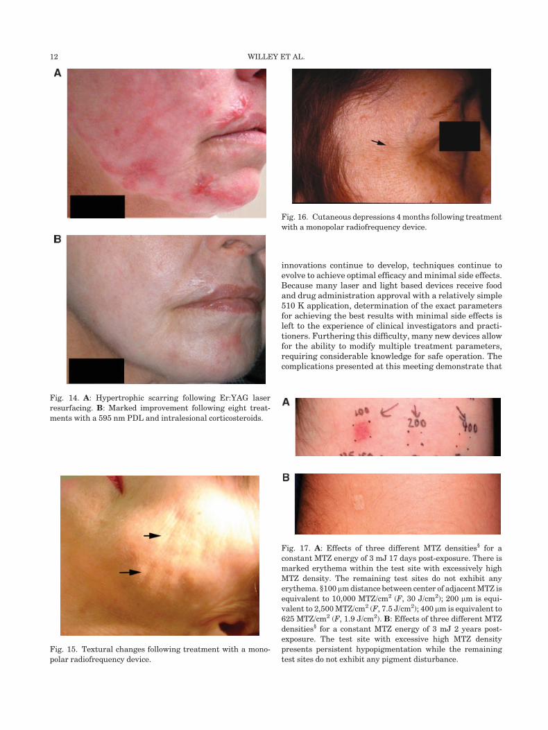
Case 16: Hypertrophic scarring following Erbium:YAGlaser resurfacing. The patient was a 51-year-old femalereferred for evaluation and treatment of hypertrophicscarring that resulted from variable pulsed Er:YAG laserresurfacing (Fig. 14A). In areas of ongoing healing, anerythematous, almost eczematous plaque was evident.Other areas had thickened hypertrophic red scars. Allareas were treated with a PDL using the followingparameters: l, 595 nm; F, 7.0–7.5 J/cm2; tp, 6 milliseconds;SS, 10 mm; CSC tc, 30 milliseconds; td, 30 milliseconds. Thethickened areas were also injected with intralesionaltriamcinolone at concentrations of 5–20 mg/ml dependingon the thickness of the scars. Eight laser treatments wereperformed at 1–2 month intervals, with more frequenttreatments earlier on. As the progression of new scarringslowed, longer intervals between treatments were allowed.The final treatment outcome was marked improvement inscarring, with some persistent scars in the areas of greatesthypertrophy 2 years after initiation of therapy (Fig. 14B).
The complication occurred as a result of aggressiveablation and a tendency toward hypertrophic scarring.Careful attention to any non-healing areas, which are asign of potential scarring, could have minimized thiscomplication. An erythematous, eczematous region unre-lated to a contact allergic or irritant dermatitis may be aprecursor to scarring. If topical modalities do not improvethe area, the PDL may be used to help halt the scarprogression. In some cases, several treatments may beneeded to obtain the best possible cosmetic result.
Monopolar Radiofrequency DeviceCase 17: Textural changes following radiofrequency
treatment facial tissue laxity. The patient was a 57-year-old Caucasian presented with mild facial laxity. Thepatient was treated with a monopolar radiofrequencydevice. A full-face procedure was performed with noanesthesia using a grid and the following treatmentparameters: SS, 1�1 cm2; lower face F, 124 J/cm2 the firstpass and then 106–89 J/cm2 the second pass; upper neckand forehead F, 106–89 J/cm2; and selected double passesof the sub-mental and jowl areas. A total of 200 pulses were
LASER DERMATOLOGIC SURGERY 9
used. The treatment was tolerated by the patient with apain score of 2 on a scale of 0–4. There were no immediateside effects except transient erythema and swelling.
One month post-operatively, the patient developed mildcutaneous depressions on the cheeks (Fig. 15). The
complication was managed by instructing the patient tomassage the skin daily and two treatments with a PDLusing the following treatment parameters: l, 595 nm;F, 6 J/cm2; tp, 6 milliseconds; SS, 10 mm; CSC tc,40 milliseconds; td, 30 milliseconds. The final treatment
Fig. 12. A: Before CO2 laser resurfacing.B: 7 days following CO2 laser resurfacing.C: Vesicles
and erosions ten days following CO2 laser resurfacing.D: Purulent erosions and ulcers 15 days
following CO2 laser resurfacing. E: Resolving infection 20 days following CO2 laser
resurfacing. F: Complete healing several weeks following timely management of HSV and
staphylococcal infections.
10 WILLEY ET AL.

outcome was 80% improvement in the skin indentations at12 months follow-up.
Although, biopsies were not obtained, the complicationmost likely occurred as a result of selective heating of thesubcutaneous fat layer by radiofrequency energy. Despitethe appropriate level of patient discomfort on a subjectivepain scale using no anesthesia during the two passes, theprocedure produced cutaneous depressions. Using lowerfluences with multiple passes may help to eliminate thistype of complication.Case 18: Textural changes following radiofrequency
treatment of periorbital tissue laxity. The patient was a56-year-old female who presented with periorbital tissuelaxity. The patient was treated with a monopolar radio-frequency device using the following treatment para-meters: F, 14.5 J/cm2; SS, 1�1 cm2; RR, 5 min. Twomonths post-operatively, the patient developed cutaneousdepressions in the both temple areas (Fig. 16). Thecomplication was managed by watchful waiting. Thecomplication occurred as a result of excessive heating byradiofrequency energy and may have been minimized byuse of lower energy with multiple passes.Fractional Resurfacing Prototype DeviceCase 19: Persistent hypopigmentation following evalua-
tion of microscopic thermal zone densities after fractionallaser skin resurfacing. The patient was a 36-year-old male(skin type II) who was exposed to test spots for evaluation ofthe effects of different microscopic thermal zone (MTZ)densities for a fractional resurfacing prototype device
emitting at 1,450 nm and an energy per MTZ of 3 mJ. Thetest site treated with 10,000 MTZ/cm2 (100 mm betweenMTZ); F, 30 J/cm2 developed immediate whitening,followed by marked erythema persistent for several weeksafter exposure, while the test sites with lower MTZdensities of 2,500 MTZ/cm2 (200 mm between MTZ); F,7.5 J/cm2 and 625 MTZ/cm2 (400 mm between MTZ); F,1.9 J/cm2 were well-tolerated (Fig. 17A). The final treat-ment outcome was persistent hypopigmentation at the testsite with the highest MTZ density (10,000 MTZ/cm2), whilethe lower MTZ densities did not exhibit any dyspigmenta-tion 2 years post-operatively (Fig. 17B).
The complication occurred with excessively high MTZdensities (approximately five times the total MTZ densityas applied with the currently available fractional resurfa-cing device after multiple passes). Such high densitiesresult in confluent bulk heating of the skin and as they lackof undamaged tissue between individual MTZs, they arenot consistent with the concept of ‘‘fractional’’ resurfacinganymore. This complication can be prevented by strictlyavoiding the use of excessively high MTZ densities withfractional resurfacing devices.
DISCUSSION
Innovations in lasers, light and radiofrequency deviceshave allowed for improved therapeutic efficacy and safetyand the ability to treat patients with an ever-increasingnumber of medical and aesthetic indications. [2]. As such
Fig. 13. A: Patient with exogenous ochronosis showing uniform hypopigmentation following
test spots using one, two, and three passes with a CO2 laser. B: Incomplete facial
repigmentation, together with scarring on the lip and cheek 16 weeks after resurfacing.
LASER DERMATOLOGIC SURGERY 11
innovations continue to develop, techniques continue toevolve to achieve optimal efficacy and minimal side effects.Because many laser and light based devices receive foodand drug administration approval with a relatively simple510 K application, determination of the exact parametersfor achieving the best results with minimal side effects isleft to the experience of clinical investigators and practi-tioners. Furthering this difficulty, many new devices allowfor the ability to modify multiple treatment parameters,requiring considerable knowledge for safe operation. Thecomplications presented at this meeting demonstrate that
Fig. 14. A: Hypertrophic scarring following Er:YAG laser
resurfacing. B: Marked improvement following eight treat-
ments with a 595 nm PDL and intralesional corticosteroids.
Fig. 15. Textural changes following treatment with a mono-
polar radiofrequency device.
Fig. 16. Cutaneous depressions 4 months following treatment
with a monopolar radiofrequency device.
Fig. 17. A: Effects of three different MTZ densities§ for a
constant MTZ energy of 3 mJ 17 days post-exposure. There is
marked erythema within the test site with excessively high
MTZ density. The remaining test sites do not exhibit any
erythema. §100 mm distance between center of adjacent MTZ is
equivalent to 10,000 MTZ/cm2 (F, 30 J/cm2); 200 mm is equi-
valent to 2,500 MTZ/cm2 (F, 7.5 J/cm2); 400 mm is equivalent to
625 MTZ/cm2 (F, 1.9 J/cm2). B: Effects of three different MTZ
densities§ for a constant MTZ energy of 3 mJ 2 years post-
exposure. The test site with excessive high MTZ density
presents persistent hypopigmentation while the remaining
test sites do not exhibit any pigment disturbance.
12 WILLEY ET AL.
even experts with decades of experience can and do havepatients with complications associated with light-basedtreatments, emphasizing the importance of timely andskillful management.
One of the greatest advances in laser surgery in recentyears has been the development of skin cooling technolo-gies, which protect the epidermis from heat injury andallow for the use of higher fluences [3,4]. Failure of skincooling, whether due to device or operator error, poses risksof thermal injury: misalignment of the cryogen spray tip,angling of the hand piece, and inadequate duration ofcryogen spray have been described [5]. Misalignment of thecryogen spray tip causes crescent-shaped hypopigmenta-tion [5], unlike the round scars in Case 1. Intermittentfailure of the cryogen spray due to air bubbles in the linebetween the cryogen canister and the laser hand piece is arare complication that may lead to PDL-induced scarring,especially when fluences greater than 10 J/cm2 are used.Awareness of this type of device malfunction is importantgiven that the associated complications may be inconspic-uous at the time of treatment.
All methods of skin cooling (evaporative, contact, orconvective) may have unique complications if improperlyutilized. Infrequent application of aluminum rollers (Case8), failure of water chilling, inadequate contact, and/orretrograde motion of copper tips can result in inadequatecooling with increased pain and undesirable tissue reac-tions at the time of treatment. These immediate reactionscan herald post-operative blistering, ulceration, and scar-ring; careful real-time attention to the patient’s painresponse and the skin surface color immediately aftertreatment is required to insure adequate skin cooling(Cases 6 and 8). Additional measures include familiaritywith the specific device, feeling the cooled tip prior toinitiation of therapy, appropriate use of pre, parallel, andpost-cooling methods, optimal duration of cryogen spray fora given fluence and spot diameter [6], and increasingfluences slowly with the initiation of cooling methods.
Because absorption of both oxyhemoglobin and melaninis reduced in the near infrared region of the electromag-netic spectrum, significantly higher fluences are requiredfor photocoagulation of deeper vessels. The use of propercooling methods and avoidance of overlapping pulses isessential with long-pulsed 1,064 nm lasers to preventinjury associated with inadequate dissipation of heat(Cases 5 and 6). Immediate blanching of target vesselswith no change in the overlying epidermis should beobserved to insure appropriate treatment parameters areused. In the absence of epidermal injury, cutaneousdepressions can occur 1–2 months following laser treat-ment due to diffusion of heat beyond the target vessel(Cases 4 and 7).
Recent work evaluating the efficacy of long pulsed1,064 nm lasers for the treatment of PWS demonstrated asteep fluence-response relationship much like that observedin Case 2 [7]. In addition, great variation in the minimalpurpuric threshold (MTP) observed, suggests that the skinresponse changes rapidly at fluences greater than theMPT. Determination of MPT is recommended with use of
the 1,064 nm Nd:Yag laser to treat PWS. In addition, Blacket al. demonstrated increased absorption during coagulationwith the 1,064 nm laser associated with the formation of met-hemoglobin that may lead to ‘‘runaway’’ heat deposition andburns, suggesting the safety profile of long pulsed 1,064 nmlasers may be improved by initial treatment with a 532 nmlaser followed by a 1,064 nm laser at lower fluences asdemonstrated in Case 2 [8,9]. The use of longer wavelengthsand higher fluences to treat vascular lesions may be bestreserved for laser experts familiar with the complexities ofthese lesions and devices (Cases 2,3, and 6).
Innovations in the treatment of photo aging havegreatly expanded in recent years to include broad-spectrum IPL sources that allow for simultaneous treat-ment of vascular and pigmentary changes. Early compli-cations with IPL devices have been reduced with the use ofcooling methods, selective filters [10–12], light guidepositioning, lower fluences for the neck and chest,reducing fluence with progression of treatment, cautionin darker and tanned skin, and treating only cosmeticunits to avoid irregular treatment delineation. Additivethermal injury may occur with the use of combinations oflaser, light, and radiofrequency therapies (Cases 9 and10), which may increase the risk of epidermal injury. Withcareful attention to the time allowed between passes, useof lower fluences and multiple passes may improve efficacyand safety.
Complications associated with CO2 resurfacing havebeen well described [13–19]. The use of aggressivetechniques, including use of high energy densities, multiplepasses, and overlapping spots or scans can increase the riskof hypertrophic scarring and infections (Cases 13 and 16).The management of infection risk has been controversial,and various regimens of prophylaxis against viral, bacter-ial, fungal infections, and post-operative wound care havebeen recommended [20–39]. Ideally, antimicrobial prophy-laxis should be tailored to individual risks (Case 12) and thetimely management can minimize adverse outcomes (Cases12, 13, and 14). Further, Case 15 raises the possibility thatexogenous ochronosis may interfere with normal woundhealing suggesting that ochronosis may be a relativecontraindication for ablative resurfacing.
It is important to treat signs of potential scar formationearly. Increased or prolonged erythema or eczematouschanges can be a sign of potential trouble. Potent topicalcorticosteroids and treatment with the PDL may improvescarring (Cases 13 and 16); intralesional steroids may berequired for hypertrophic scars. The patient should befollowed closely and treated at 3–6 week intervals depend-ing on scar progression. If there is any question as to thebest way to proceed, the patient should be referred to a skinlaser expert for management.
Growing demand for non-invasive rejuvenation of photoaged skin has led to the expansion of non-ablativetechniques [40], including monopolar radiofrequencydevices that induce collagen remodeling and selectiveheating of the fibrous septae [41–46] and fractionalresurfacing devices that induce spatially selective dermalinjury with minimal epidermal damage [47]. The use of
LASER DERMATOLOGIC SURGERY 13
radiofrequency devices with excessive energies and over-lapping pulses may lead to cutaneous depressions thatmay or may not be associated with excessive pain (Cases17 and 18). Even so, excessive pain during treatment is animportant indicator of thermal injury. Cutaneous depres-sions may spontaneously improve over 6–12 months.Gentle dermal massage, non-ablative remodeling, anddermal fillers may be beneficial. Avoiding inadvertentoverlapping of volumetric areas and the use of lowerenergies with multiple passes may improve efficacy andminimize the risk of unintended thermal injury.
A recently developed fractional resurfacing device(FraxelTM) may offer a promising alternative to non-ablativeresurfacing methods [47]. A microscopic pattern of laser-induced epidermal and dermal thermal injury providespartial epidermal replacement and collagen remodeling withminimal downtime. Optimal pattern densities allow forsufficient spacing of MTZs and therefore avoid confluenceof thermal injury. The operator should avoid very high MTZdensities and should also provide sufficient time for the skinto cool down between multiple passes. This is especiallyimportant when small areas are covered with multiplepasses. In order to provide a relatively homogeneous MTZdensity over the entire treatment area, the operatorshould keep the hand piece perpendicular to the skinsurface and should carefully track the number of passesperformed within each area. Immediate skin whiteningduring treatment should be strictly avoided as it indicatesexcessive thermal injury that may be associated withpersistent hypopigmentation (Case 19).
CONCLUSION
Complications such as scarring, dyspigmentation, andtextural changes can occur with any laser, light, orradiofrequency based treatment, even in the hands of themost experienced laser surgeons. Such complications mayresult from diverse causes, including device malfunction,poor patient selection, operator error, post-treatmentmismanagement, or simple misfortune. Many recentadvances in laser surgery take advantage of energy sourcesand treatment parameters that do not strictly adhere to theprinciples of selective photothermolysis and requireincreasing knowledge and skill of the laser surgeon. Ascontinued innovations attempt to improve upon existingtechniques and technologies, awareness of potential com-plications, early recognition when problems do arise, andknowledge of how to skillfully manage complications isessential to insure patient safety.
REFERENCES
1. Anderson RR, Parrish JA. Selective photothermolysis: Pre-cise microsurgery by selective absorption of pulsed radiation.Science 1983;220:524–527.
2. Alam M, Dover JS, Arndt KA. Energy delivery devicesfor cutaneous remodeling. Arch Dermatol 2003;139:1351–1360.
3. Nelson JS, Milner TE, Anvari B, Tanenbaum BS, Kimel S,Svaasand LO, Jacques SL. Dynamic epidermal coolingduring pulsed laser treatment of port-wine stain. A n ewmethodology with preliminary clinical evaluation. ArchDermatol 1995;131:695–700.
4. Kelly KM, Nanda VS, Nelson JS. Treatment of port-winestain birthmarks using the 1.5-msec pulsed dye laser at highfluences in conjunction with cryogen spray cooling. DermatolSurg 2002;28:309–313.
5. Kelly KM, Svaasand LO, Nelson JS. Optimization of lasertreatment safety in conjunction with cryogen spray cooling.Arch Dermatol 2003;139:1372–1373.
6. Aguilar G, Wang GX, Nelson JS. Dynamic behavior ofcryogen spray cooling: Effects of spurt duration and spraydistance. Lasers Surg Med 2003;32:152–159.
7. Yang MU, Yaroslavsky AN, Farinelli WA, Flotte TJ,Rius-Diaz F, Tsao SS, Anderson RR. Long-pulsed neodymiu-m:yttrium-aluminum-garnet laser treatment for port winestains. J Am Acad Dermatol 2005;52:480–490.
8. Black JF, Wade N, Barton JK. Mechanistic comparison ofblood undergoing laser photocoagulation at 532 and 1,064nm. Lasers Surg Med 2005;36:155–165.
9. Barton JK, Franginease G, Pummer H, Black JF. Coopera-tive phenomena in two-pulse, two-color laser photocoagula-tion of cutaneous blood vessels. Photochem Photobiol2001;73:642–650.
10. Weiss RA, Sadick NS. Epidermal cooling crystal collar devicefor improved results and reduced side effects on legtelangiectasias using intense pulsed light. Dermatol Surg2000;26:1015–1018.
11. Bjerring P, Christiansen K, Troilius A. Intense pulsed lightsource for treatment of facial telangiectasias. J Cosmet LaserTher 2001;3:169–173.
12. Bjerring P, Christiansen K, Troilius A, Dierickx C. Facialphoto rejuvenation using two different intense pulsed light(IPL) wavelength bands. Lasers Surg Med 2004;34:120–126.
13. McBurney EI. Side effects and complications of laser therapy.Dermatol Clin 2002;20:165–176.
14. Nanni CA, Alster TS. Complications of carbon dioxide laserresurfacing. An evaluation of 500 patients. Dermatol Surg1998;24:315–320.
15. Nanni CA, Alster TS. Complications of cutaneous lasersurgery. A review. Dermatol Surg 1998;24:209–219.
16. Sriprachya-Anunt S, Fitzpatrick RE, Goldman MP, SmithSR. Infections complicating pulsed carbon dioxide laserresurfacing for photoaged facial skin. Dermatol Surg1997;23:527–535.
17. Bernstein LJ, Kauvar AN, Grossman MC, Geronemus RG.The short- and long-term side effects of carbon dioxide laserresurfacing. Dermatol Surg 1997;23:519–525.
18. Rendon-Pellerano MI, Lentini J, Eaglstein WE, Kirsner RS,Hanft K, Pardo RJ. Laser resurfacing: Usual and unusualcomplications. Dermatol Surg 1999;25:360–366.
19. Sullivan SA, Dailey RA. Complications of laser resurfacingand their management. Ophthal Plast Reconstr Surg 2000;16:417–426.
20. Alster TS. Cutaneous resurfacing with CO2 and erbium:YAG lasers: Preoperative, intraoperative, and postoperativeconsiderations Plast Reconstr Surg 1999;103:619–632.
21. Walia S, Alster TS. Cutaneous CO2 laser resurfacinginfection rate with and without prophylactic antibiotics.Dermatol Surg 1999;25:857–861.
22. Goldman MP. Regarding CO2 laser resurfacing infectionrates. Dermatol Surg 2000;26:402–404.
23. Friedman PM, Geronemus RG. Antibiotic prophylaxis inlaser resurfacing patients. Dermatol Surg 2000;26:695–697.
24. Alster TS. Against antibiotic prophylaxis for cutaneous laserresurfacing. Dermatol Surg 2000;26:697–698.
25. Sriprachya-Anunt S, Fitzpatrick RE, Goldman MP, SmithSR. Infections complicating pulsed carbon dioxide laserresurfacing for photoaged facial skin. Dermatol Surg 1997;23:527–535.
26. Ross EV, Amesbury EC, Barile A, Proctor-Shipman L,Feldman BD. Incidence of postoperative infection or positiveculture after facial laser resurfacing: A pilot study, a casereport, and a proposal for a rational approach to antibioticprophylaxis. J Am Acad Dermatol 1998;39:975–981.
27. Manuskiatti W, Fitzpatrick RE, Goldman MP, Krejci-Papa N. Prophylactic antibiotics in patients undergoing
14 WILLEY ET AL.

laser resurfacing of the skin. J Am Acad Dermatol 1999;40:77–84.
28. Bellman B, Brandt FS, Holtmann M, Bebell WR. Infectionwith methicillin-resistant Staphylococcus aureus after car-bon dioxide resurfacing of the face. Successful treatment withminocycline, rifampin, and mupiricin ointment. DermatolSurg 1998;24:279–282.
29. Lowe NJ, Lask G, Griffin ME. Laser skin resurfacing. Pre-and posttreatment guidelines. Dermatol Surg 1995;21:1017–1019.
30. Newman JP, Fitzgerald P, Koch RJ. Review of closed dress-ings after laser resurfacing. Dermatol Surg 2000;26:562–571.
31. Beeson WH, Rachel JD. Valacyclovir prophylaxis for herpessimplex virus infection or infection recurrence following laserskin resurfacing. Dermatol Surg 2002;28:331–336.
32. Gilbert S, McBurney E. Use of valacyclovir for herpes simplexvirus-1 (HSV-1) prophylaxis after facial resurfacing: Arandomized clinical trial of dosing regimens. Dermatol Surg2000;26:50–54.
33. Alster TS, Nanni CA. Famciclovir prophylaxis of herpessimplex virus reactivation after laser skin resurfacing.Dermatol Surg 1999;25:242–246.
34. Fulton JE, Jr. Complications of laser resurfacing. Methods ofprevention and management. Dermatol Surg 1998;24:91–99.
35. Ruiz-Esparza J, Barba Gomez JM, Gomez de la Torre OL.Wound care after laser skin resurfacing. A combination ofopen and closed ethods using a new polyethylene mask.Dermatol Surg 1998;24:79–81.
36. Batra RS, Ort RJ, Jacob C, Hobbs L, Arndt KA, Dover JS.Evaluation of a silicone occlusive dressing after laser skinresurfacing. Arch Dermatol 2001;137:1317–1321.
37. Onouye T, Menaker G, Christian M, Moy R. Occlusivedressing versus oxygen mist therapy following CO2 laserresurfacing. Dermatol Surg 2000;26:572–576.
38. Weiss RA, Goldman MP. Interpenetrating polymer networkwound dressing versus petrolatum following facial CO2 laserresurfacing: A bilateral comparison. Dermatol Surg2001;27:449–451.
39. Suarez M, Fulton JE, Jr. A novel occlusive dressing for skinresurfacing. Dermatol Surg 1998;24(5):567–570.
40. Kim KH, Geronemus RG. Nonablative laser and lighttherapies for skin rejuvenation. Arch Facial Plast Surg2004;6:398–409.
41. Zelickson BD, Kist D, Bernstein E, Brown DB, KsenzenkoS, Burns J, Kilmer S, Mehregan D, Pope K. Histo-logical and ultrastructural evaluation of the effects ofa radiofrequency-based nonablative dermal remodelingdevice: A pilot study. Arch Dermatol 2004;140:204–209.
42. Fritz M, Counters JT, Zelickson BD. Radiofrequency treat-ment for middle and lower face laxity. Arch Facial Plast Surg2004;6:370–373.
43. Alster TS, Tanzi E. Improvement of neck and cheek laxitywith a nonablative radiofrequency device: A lifting experi-ence. Dermatol Surg 2004;30:503–507.
44. Fitzpatrick R, Geronemus R, Goldberg D, Kaminer M, KilmerS, Ruiz-Esparza J. Multicenter study of noninvasive radio-frequency for periorbital tissue tightening. Lasers Surg Med2003;33:232–242.
45. Narins DJ, Narins RS. Non-surgical radiofrequency facelift. JDrugs Dermatol 2003;2:495–500.
46. Hsu TS, Kaminer MS. The use of nonablative radiofrequencytechnology to tighten the lower face and neck. Semin CutanMed Surg 2003;22:115–123.
47. Manstein D, Herron GS, Sink RK, Tanner H, Anderson RR.Fractional photothermolysis: A new concept for cutaneousremodeling using microscopic patterns of thermal injury.Lasers Surg Med 2004;34:426–438.
LASER DERMATOLOGIC SURGERY 15