Complications of Diabetes Mellitus Dr Rodney Itaki Lecturer Anatomical Pathology Discipline...
17
Complications of Diabetes Mellitus Dr Rodney Itaki Lecturer Anatomical Pathology Discipline University of Papua New Guinea School of Medicine & Health Sciences Division of Pathology
-
Upload
cora-gilbert -
Category
Documents
-
view
219 -
download
4
Transcript of Complications of Diabetes Mellitus Dr Rodney Itaki Lecturer Anatomical Pathology Discipline...

Complications of Diabetes Mellitus
Dr Rodney ItakiLecturer
Anatomical Pathology Discipline
University of Papua New GuineaSchool of Medicine & Health SciencesDivision of Pathology
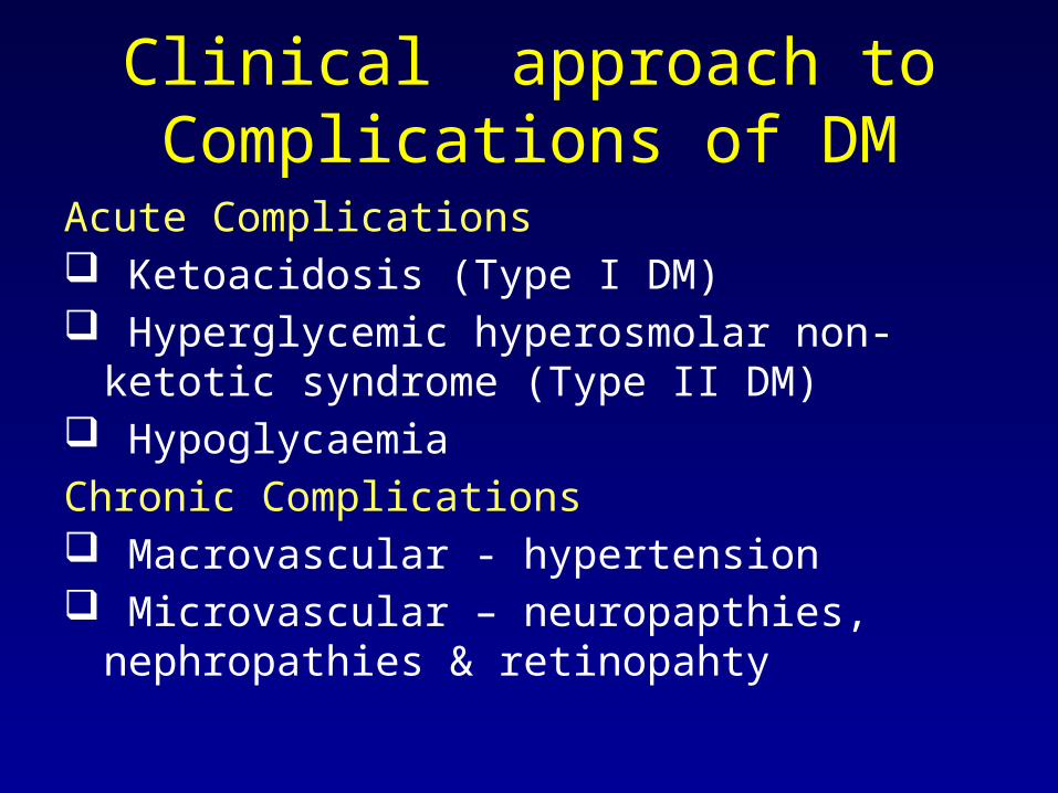
Clinical approach to Complications of DM
Acute Complications Ketoacidosis (Type I DM) Hyperglycemic hyperosmolar non-ketotic
syndrome (Type II DM) HypoglycaemiaChronic Complications Macrovascular - hypertension Microvascular – neuropapthies,
nephropathies & retinopahty

Pathological Approach to Complications of DM
• Microangiopathy – small vessel disease• Retinopathy• Nephropathy• Neuropathy
• Underlying cause is hyperglycemia

Pathogenesis
• Nonenzymatic Glycosylation• Formation of irreversible
glycosylation products with protein (e.g collagen) in blood vessels and interstitial tissues.
• Rather than dissociating, undergo series of slow series of chemical rearrangement that is irreversible forming Advanced Glycosylation End Products (AGE).
• AGE resistant to enzymatic proteolysis

Non-enzymatic Glycosylation of Proteins
Ref: Robins Pathological Basis of Diseases, 6th Ed.

Biological Properties of AGE
Ref: Robins Pathological Basis of Diseases, 6th Ed.

Disturbance in Polyol Pathway
• Occurs in nerves, lens, kidneys & blood vessels. Do not need insulin for glucose intake.
• Osmotic cell injury
• Impairs ion pumps - Schwann cells (peripheral & autonomic neurophathy), pericytes in retinal capillaries. (retinal microaneurysm)
Ref: Robins Pathological Basis of Diseases, 6th Ed.

Morphology of Diabetes Complications• Pancreas: Most changes & more distinctive in
Type I than Type II DM.– Reduction in number and size of islets– Leukocyte infiltration of islets (T lymphocytes
mostly).– Eosinophil infiltrates
• Amyloid replacement of islets in Type II. Islets obliterated in advanced Type II DM.
• Vascular system: atherosclerosis of all vessels. Hyaline thickening of the wall of arterioles causing narrowing of lumen.
• Diabetic microangiopathy: Diffuse thickening of BM of capillaries (skin, skeletal muscle, retina, renal glomeruli, renal medulla, renal tubules, Bowman’s capsule, peripheral nerves, and placenta). Hyaline material forming concentric layers – type IV collagen. Despite thickened BM, capillaries more leaky than normal to plasma proteins.

Morphology of Diabetes Complications
• Diabetic nephropathy: glomerular lesions, renal vascular lesions (arteriosclerosis) & pyelonephritis, including necrotizing papillitis.– Glomerular lesions – capillary BM
thickening, diffuse glomerularsclerosis, nodular glomerularsclerosis.
– Renal vascular lesions – part of systemic involvement of blood vessels. Both afferent and efferent arterioles.
– Pyelonephritis – due to acute or chronic inflammation. Begins in the interstitial tissue and spread to affect tubules.
– Papillary necrosis more common in diabetics.

Morphological Complications of Diabetes
• Diabetic Ocular complications: take the form of retinopathy, cataract formation and glaucoma.
• Retinopathy – backgound (nonproliferative) & proliferative rentinopathy
• Background retinopathy– BM thickening,– pericyte degeneration. Loss results in microaneurysms– capillary microaneurysms (hemorrhages)– Microvascular obstructions and non-perfusion of
capillaries in posterior fundus that lack pericytes and endothelium. Cause hypoxia(cotton-wool spots).
– Arteriolar hyalinization causing BM thickening.– Gradual increase in retinal vein caliber as response to
ischaemia. Form venous loops and beading in veins.

Morpholigical Complications of Diabetes
• Proliferative retinopathy – characterised by neovascularisation and fibroplasia. – Neovascularisation occurs in response to
hypoxia of retina.– ischemic retinal cells produce VEGF (vascular
endothelial growth factor) inducing angiogenesis from larger veins and arterial vessels.
– new vessels incompletely formed & poorly supported and move with eye movements resulting in haemorrhages.
– Fibroplasia results later & contribute to retinal detachment
• Diabetic Neuropathy: central and peripheral nervous system. Pattern is of peripheral, symmetric neuropathy of lower limbs affecting both motor and sensory function.

Morphological complications of diabetes
• Peripheral neuropathy – axonal neuropathy. Some segmental demyelination.
• Endoneurial arterioles show hyalinization, BM thickening.
• Neuropathies catergorised as: distal symmetric sensory (or sensorimoto) neuropathy, autonomic neuropathy & focal (or multifocal) asymmetric neuropathy.
• Symmetric neuropathy affecting distal sensory and motor nerves common neuropathy. Autonomic neuropathy also common.
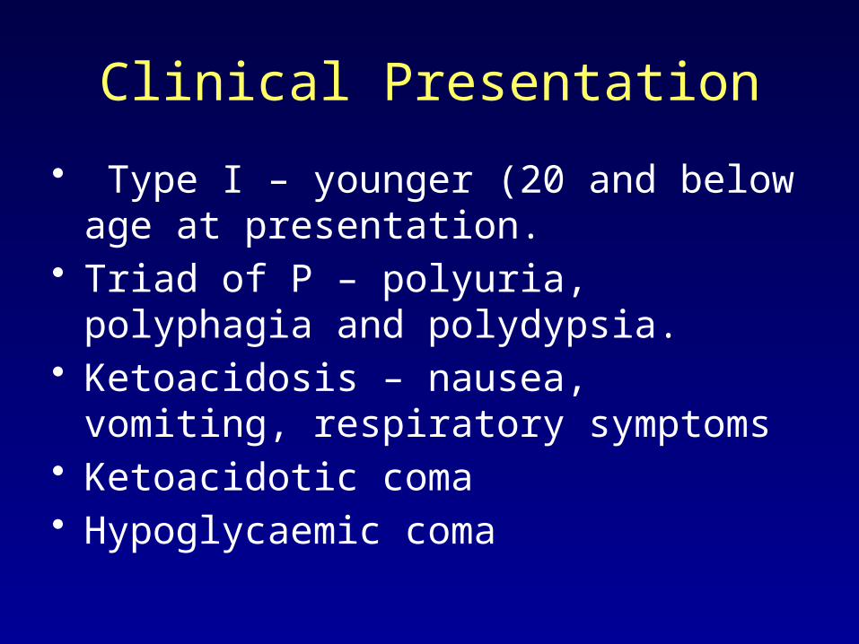
Clinical Presentation
• Type I – younger (20 and below age at presentation.
• Triad of P – polyuria, polyphagia and polydypsia.
• Ketoacidosis – nausea, vomiting, respiratory symptoms
• Ketoacidotic coma• Hypoglycaemic coma
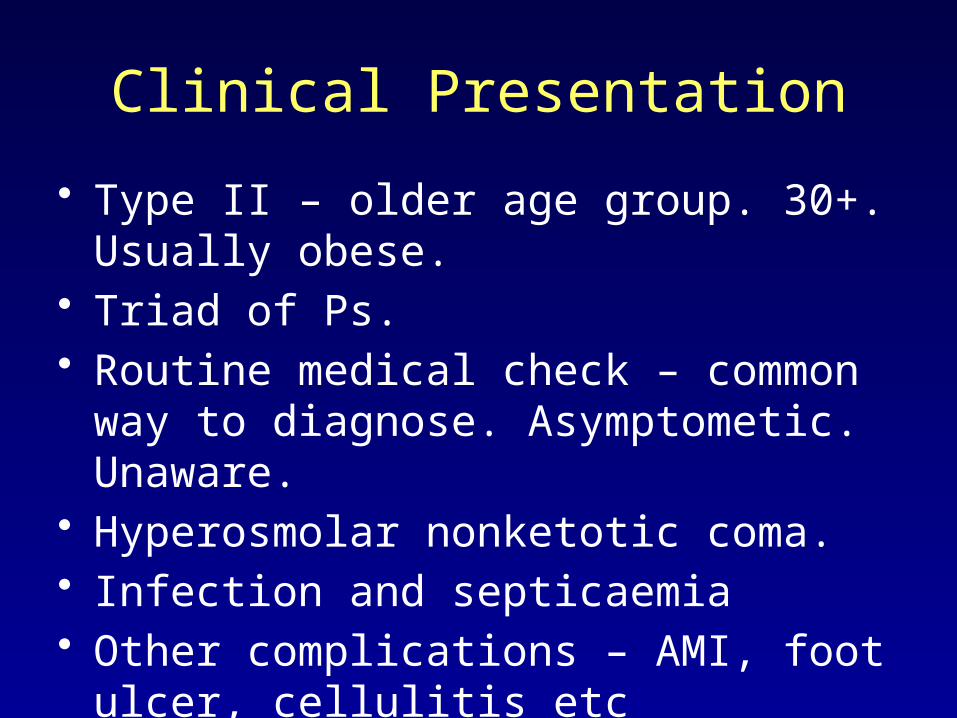
Clinical Presentation
• Type II – older age group. 30+. Usually obese.
• Triad of Ps. • Routine medical check – common
way to diagnose. Asymptometic. Unaware.
• Hyperosmolar nonketotic coma.• Infection and septicaemia• Other complications – AMI, foot
ulcer, cellulitis etc
Laboratory Diagnosis
• Elevated blood glucose: non-fasting fasting >11.1 mmol/L; Fasting of >7.1 mmol/L.
• Oral glucose tolerance test.• HBA1C – monitoring of control of
blood glucose level.

Prognosis
• Most patients die from– AMI– Chronic renal failure– CVA– Infections.

End
Main Reference: Robins Pathological Basis of
Diseases, 6th Ed. Chapter on The Pancreas.
Download PPT notes on: www.pathologyatsmhs.wordp
ress.com