Complete Hydatidiform Mole with Co-existing Live Fetus: A ...singleton molar pregnancy, and elective...
4
ORIGINAL ARTICLE Complete Hydatidiform Mole with Co-existing Live Fetus: A Case Series Maimoona Ahmed 1 • Geeta Kolar 2 • Suseela Vavilala 3 • Sunil Jaiman 4 Received: 2 October 2015 / Accepted: 23 November 2015 / Published online: 10 December 2015 Ó Society of Fetal Medicine 2015 Abstract This review was undertaken to evaluate the maternal and fetal risk associated with complete hydatidi- form mole with co-existing fetus (CHMF) and to assess the feasibility for continuing such pregnancies. Twin gesta- tions with CHMF were reviewed from the hospital database from 2005 to 2014 at our center. Diagnosis was based on ultrasonography and confirmed post-delivery, histopatho- logically. Amniocentesis for karyotype was done for the live fetuses. Serum b-hCG levels were followed till they normalized. Six cases of CHMF were salvaged from the archives. Three had live normal babies (50 %), pregnancy was terminated in two cases for excessive bleeding (33.3 %) and one miscarried (16.7 %). One fetus exhibited 47, XXY Klinefelter syndrome while rest showed normal karyotype. Two patients required blood transfusion, one was complicated with severe pre-eclampsia whereas none showed gestational trophoblastic neoplasm. CHMF is a rare condition that can be diagnosed by ultrasonography. Continuation of such a pregnancy is an acceptable option and expectant management instead of therapeutic abortion can be pursued after weighing the possibility of fetal sur- vival against maternal risk. Counseling of the couple and family plays a crucial role. Associated spectrum of maternal and fetal complications mandates close pre- and post-natal surveillance. Keywords Twin pregnancy Á Co-existing hydatidiform mole Introduction Twin pregnancy with complete hydatidiform mole and co- existing live fetus is a rare clinical entity. The diagnosis is usually made on obstetric ultrasound examination with the typical appearance of a complete mole with a live fetus in the other sac. The controversy lies in the management of such cases due to the higher incidence of maternal complications like hemorrhage, early onset severe pre-eclampsia, thyrotox- icosis, and the risk of persistent trophoblastic disease and fetal complications such as abnormal karyotype, abortion, and preterm birth [1]. The tumultuous course due to aforemen- tioned complications, coupled with these pregnancies being associated with advanced maternal age and use of assisted reproductive techniques (indicating years of childlessness), would probably seem as the proverbial sword of Damocles for most [2]. However, despite the tribulations, termination of such pregnancies need not be the sole option. Materials and Methods The cases of twin gestation with complete hydatidiform mole with co-existing fetus were reviewed over a period of 10 years from 2005 till 2014 at Fernandez hospital, Hyderabad, which is a tertiary referral center. The infor- mation was compiled from the patient database. The diagnosis was based on ultrasonography (Fig. 1). & Maimoona Ahmed [email protected] 1 High Risk Pregnancy and Perinatology, Fernandez Hospital, Hyderabad, India 2 Department of Fetal Medicine, Fernandez Hospital, Hyderabad, India 3 Department of Fetal Medicine, Fernandez Hospital, Hyderabad, India 4 Department of Perinatology, Fernandez Hospital, Hyderabad, India 123 J. Fetal Med. (December 2015) 2:171–174 DOI 10.1007/s40556-015-0067-6
Transcript of Complete Hydatidiform Mole with Co-existing Live Fetus: A ...singleton molar pregnancy, and elective...
ORIGINAL ARTICLE
Complete Hydatidiform Mole with Co-existing Live Fetus:A Case Series
Maimoona Ahmed1 • Geeta Kolar2 • Suseela Vavilala3 • Sunil Jaiman4
Received: 2 October 2015 / Accepted: 23 November 2015 / Published online: 10 December 2015
� Society of Fetal Medicine 2015
Abstract This review was undertaken to evaluate the
maternal and fetal risk associated with complete hydatidi-
form mole with co-existing fetus (CHMF) and to assess the
feasibility for continuing such pregnancies. Twin gesta-
tions with CHMF were reviewed from the hospital database
from 2005 to 2014 at our center. Diagnosis was based on
ultrasonography and confirmed post-delivery, histopatho-
logically. Amniocentesis for karyotype was done for the
live fetuses. Serum b-hCG levels were followed till they
normalized. Six cases of CHMF were salvaged from the
archives. Three had live normal babies (50 %), pregnancy
was terminated in two cases for excessive bleeding
(33.3 %) and one miscarried (16.7 %). One fetus exhibited
47, XXY Klinefelter syndrome while rest showed normal
karyotype. Two patients required blood transfusion, one
was complicated with severe pre-eclampsia whereas none
showed gestational trophoblastic neoplasm. CHMF is a
rare condition that can be diagnosed by ultrasonography.
Continuation of such a pregnancy is an acceptable option
and expectant management instead of therapeutic abortion
can be pursued after weighing the possibility of fetal sur-
vival against maternal risk. Counseling of the couple and
family plays a crucial role. Associated spectrum of
maternal and fetal complications mandates close pre- and
post-natal surveillance.
Keywords Twin pregnancy � Co-existing hydatidiform
mole
Introduction
Twin pregnancy with complete hydatidiform mole and co-
existing live fetus is a rare clinical entity. The diagnosis is
usually made on obstetric ultrasound examination with the
typical appearance of a complete mole with a live fetus in the
other sac. The controversy lies in the management of such
cases due to the higher incidence of maternal complications
like hemorrhage, early onset severe pre-eclampsia, thyrotox-
icosis, and the risk of persistent trophoblastic disease and fetal
complications such as abnormal karyotype, abortion, and
preterm birth [1]. The tumultuous course due to aforemen-
tioned complications, coupled with these pregnancies being
associated with advanced maternal age and use of assisted
reproductive techniques (indicating years of childlessness),
would probably seem as the proverbial sword ofDamocles for
most [2]. However, despite the tribulations, termination of
such pregnancies need not be the sole option.
Materials and Methods
The cases of twin gestation with complete hydatidiform
mole with co-existing fetus were reviewed over a period of
10 years from 2005 till 2014 at Fernandez hospital,
Hyderabad, which is a tertiary referral center. The infor-
mation was compiled from the patient database. The
diagnosis was based on ultrasonography (Fig. 1).
& Maimoona Ahmed
1 High Risk Pregnancy and Perinatology, Fernandez Hospital,
Hyderabad, India
2 Department of Fetal Medicine, Fernandez Hospital,
Hyderabad, India
3 Department of Fetal Medicine, Fernandez Hospital,
Hyderabad, India
4 Department of Perinatology, Fernandez Hospital, Hyderabad,
India
123
J. Fetal Med. (December 2015) 2:171–174
DOI 10.1007/s40556-015-0067-6

Amniocentesis for fetal karyotype was done for the live
fetuses. Pregnancy was terminated in cases with severe
maternal or fetal complications. Following delivery, the
diagnosis was confirmed on histopathological examination.
Serum b-hCG levels were followed up till they normalized.
Results
There were total 47,256 deliveries at our hospital in the
study period. Of these, six (0.01 %) cases were of twin
gestation with complete hydatidiform mole and co-existing
fetus. The clinical presentation of the cases and the out-
come are summarized in Table 1. The mean maternal age
was 27.3 years. Mean gestational age at diagnosis was
20 weeks whereas mean gestational age at delivery was
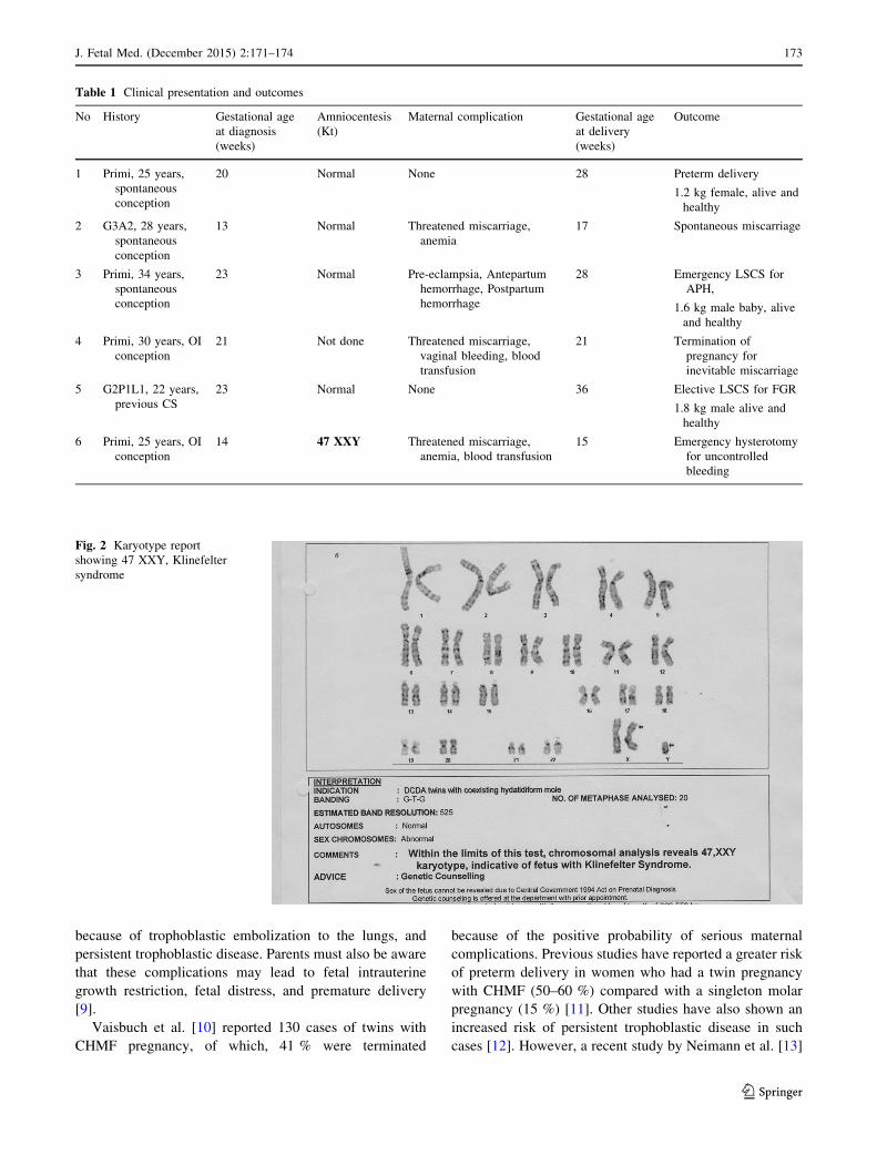
24.5 weeks. Four mothers had live fetuses with a normal
karyotype, one had a fetus with 47, XXY Klinefelter syn-
drome (Fig. 2) while in one case, which had presented with
inevitable miscarriage, karyotype could not be done.
Of the six cases, one (16.7 %) had spontaneousmiscarriage;
in two cases (33.3 %), pregnancywas terminated for excessive
bleeding; and three patients (50 %) delivered live and normal
babies but all three had preterm deliveries. Cesarean section
rate was 66.7 %. All the six cases had weekly follow-up of
serum b-hCG levels and showed falling trend. No case had
persistent gestational trophoblastic neoplasm. Histopathologi-
cal examination confirmed the presence of complete hydatidi-
form mole along with the normal placenta.
Discussion
The incidence of complete hydatidiform mole with co-
existing fetus has been reported as one in 20,000 to one in
1,00,000 pregnancies [3], while the incidence at our
institute, over the past 10 years, has been mammoth one in
7692 pregnancies. This disproportionately higher incidence
could possibly be explained by our hospital being a tertiary
referral center with a specialized fetal medicine unit.
Molar pregnancy can be divided on the basis of cyto-
genetics, histopathology, and morphology as partial mole
or complete mole. A complete mole has a diploid kary-
otype, no embryo and amnion, and uniform changes of
placental villi and trophoblasts. A partial mole, on the other
hand, has a triploid karyotype, the presence of an embryo
and only focal changes of placental villi and trophoblasts
[4, 5]. A complete hydatidiform mole with a co-existing
live fetus is a separate special entity and should be dif-
ferentiated from a partial mole [6]. This is done on the
basis of ultrasonography. An early scan showing two ges-
tational sacs or a later scan demonstrating the intertwin
membrane, confirms the diagnosis of complete mole with a
co-existing fetus. The typical ultrasonographic findings of a
molar pregnancy consist of a complex cystic pattern with a
‘snowstorm’ appearance [7]. Clinically, the patient may
present with hyperemesis, hyperthyroidism, vaginal spot-
ting or even heavy bleeding, pregnancy-induced hyper-
tension, and larger-than-gestational age uterus. However,
these conditions are not always present. Though ultra-
sonography is deemed the cornerstone for the diagnosis,
confirmation requires histopathological examination.
Prenatal testing of the fetal karyotype is essential in
deciding continuation and prognosis of the pregnancy and
to differentiate between a partial mole and CHMF. A tri-
ploid karyotype indicates a partial mole in which the fetus
would be severely malformed and growth restricted. A
diploid fetal karyotype indicates a viable fetus with a
normal placenta co-existing alongside a twin molar pla-
centa. Most cases in literature have reported the co-existing
live normal fetus [8]. In such cases, the pregnancy can be
allowed to continue since it has a considerable chance to
result in a normal live neonate. Two cases in literature have
reported the co-existing live fetus with anencephaly [8]. In
the present case series, karyotyping was done in five cases,
of which, four were normal and one with 46, XXY kary-
otype Klinefelter syndrome. None of the cases had any
anatomical abnormalities. In the present study, the mean
age of diagnosis was 20 weeks. With the introduction of
the nuchal scan (11 to 13?6 weeks), early diagnosis is
possible in the first trimester itself and the couple can have
the option of a safer termination of pregnancy, should that
be their choice.
Parents who choose to continue a twin pregnancy with
CHMF should be counseled about the risk of possible
maternal complications associated with molar pregnancy
such as early-onset pre-eclampsia, hyperemesis gravi-
darum, hyperthyroidism, vaginal bleeding, anemia, devel-
opment of theca lutein ovarian cysts, respiratory distressFig. 1 Ultrasonography image showing the two separate sacs, one
with complete mole and other with live fetus
172 J. Fetal Med. (December 2015) 2:171–174
123
because of trophoblastic embolization to the lungs, and
persistent trophoblastic disease. Parents must also be aware
that these complications may lead to fetal intrauterine
growth restriction, fetal distress, and premature delivery
[9].
Vaisbuch et al. [10] reported 130 cases of twins with
CHMF pregnancy, of which, 41 % were terminated
because of the positive probability of serious maternal
complications. Previous studies have reported a greater risk
of preterm delivery in women who had a twin pregnancy
with CHMF (50–60 %) compared with a singleton molar
pregnancy (15 %) [11]. Other studies have also shown an
increased risk of persistent trophoblastic disease in such
cases [12]. However, a recent study by Neimann et al. [13]
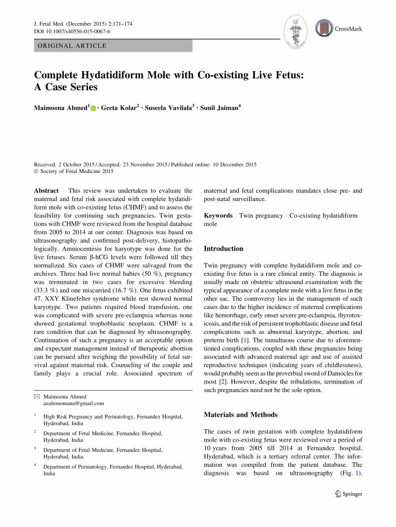
Table 1 Clinical presentation and outcomes
No History Gestational age
at diagnosis
(weeks)
Amniocentesis
(Kt)
Maternal complication Gestational age
at delivery
(weeks)
Outcome
1 Primi, 25 years,
spontaneous
conception
20 Normal None 28 Preterm delivery
1.2 kg female, alive and
healthy
2 G3A2, 28 years,
spontaneous
conception
13 Normal Threatened miscarriage,
anemia
17 Spontaneous miscarriage
3 Primi, 34 years,
spontaneous
conception
23 Normal Pre-eclampsia, Antepartum
hemorrhage, Postpartum
hemorrhage
28 Emergency LSCS for
APH,
1.6 kg male baby, alive
and healthy
4 Primi, 30 years, OI
conception
21 Not done Threatened miscarriage,
vaginal bleeding, blood
transfusion
21 Termination of
pregnancy for
inevitable miscarriage
5 G2P1L1, 22 years,
previous CS
23 Normal None 36 Elective LSCS for FGR
1.8 kg male alive and
healthy
6 Primi, 25 years, OI
conception
14 47 XXY Threatened miscarriage,
anemia, blood transfusion
15 Emergency hysterotomy
for uncontrolled
bleeding
Fig. 2 Karyotype report
showing 47 XXY, Klinefelter
syndrome
J. Fetal Med. (December 2015) 2:171–174 173
123

in 2007 revealed that the risk of preterm delivery after a
diploid mole with a viable fetus is similar to that after a
singleton molar pregnancy, and elective early termination
of such pregnancy, because of the risk of preterm delivery
alone, should not be recommended. They also concluded
that the risk of persistent trophoblastic disease after a
diploid mole with co-existing fetus pregnancy is similar to
that after a singleton molar pregnancy, and expectant
management instead of therapeutic abortion can be
pursued.
Sebire et al. [14] reviewed 77 total cases. Of these, 53
decided to continue the pregnancy, 23 spontaneously
aborted before 24 weeks, 28 pregnancies lasted more than
28 weeks resulting in 20 livebirths (40 %). There was no
statistically significant difference in the occurrence of
persistent trophoblastic disease in the women who termi-
nated the pregnancy in the first trimester and those who
continued their pregnancy, thus showing that the risk of
persistent trophoblastic disease does not increase with
advancing gestational age. Piura et al. [15] reviewed 24
studies that reported 30 cases of CHMF resulting in a live
birth documented in detail. Cesarean section was reported
due to fetal or maternal complications in 14 of 30 cases
(46.7 %).
The management of complete mole after evacuation or
delivery is similar whether there is a co-existing fetus or
not. Uterine evacuation is followed by b-hCG monitoring.
Chest radiography is mandatory. Additional imaging
investigations may be needed as directed by symptoms. b-hCG is performed weekly until ascertainment of normal
values for two consecutive weeks; then monthly up to
one year. Effective means of contraception are recom-
mended to avoid pregnancy for at least 6–12 months.
Chemotherapy is indicated if b-hCG levels are persistent or
rising, or metastasis to lungs or other sites appear. Hys-
terectomy may be needed for life-threatening hemorrhage
or in a patient with rising b-hCG titres, but no evidence of
metastasis, who does not desire fertility or refuses
chemotherapy [16].
Conclusion
Twin pregnancy with complete hydatidiform mole and co-
existent live fetus is a rare condition that can be diagnosed
by obstetric ultrasound. Termination versus expectant
management should be decided after karyotype and
detailed anatomical survey of the live fetus. Decision to
continue the pregnancy should be taken after weighing the
possibility of fetal survival against maternal risk. Coun-
seling of the couple and family plays a crucial role in such
cases. Those who choose to continue the pregnancy, the
management has to be in a tertiary care hospital with strict
vigilance on maternal, fetal, and neonatal condition. Long
term follow-up with serum b-hCG levels is mandatory.
Compliance with ethical standards
Conflict of interest None.
References
1. Matsui H, Sekiya S, Hando T, Wake N, Tomoda Y. Hydatidiform
mole coexistent with a twin live fetus: a national collaborative
study in Japan. Hum Reprod. 2000;15:608–11.
2. Montes-de-Oca-Valero F, Macara L, Shaker A. Twin pregnancy
with a complete hydatidiform mole and co-existing fetus fol-
lowing in-vitro fertilization. Hum Reprod. 1999;14:2905–7.
3. Malhotra N, Deka D, Takkar D, Kochar S, Goel S, Sharma MC.
Hydatiform mole with coexisting live fetus in dichorionic twin
gestation. Eur J Obstet Gynecol Reprod Biol. 2001;94:301–3.
4. Szulman AE, Surti U. The syndromes of hydatidiform mole.
(I) Cytogenetic and morphologic correlations. Am J Obstet
Gynecol. 1978;131:665–71.
5. Szulman AE, Surti U. The syndromes of hydatidiform mole. (II)
Morphologic evolution of the complete and partial mole. Am J
Obstet Gynecol. 1978;132:20–7.
6. Chen Fang-Ping. Molar pregnancy and living normal fetus
coexisting until term: prenatal biochemical and sonographic
diagnosis. Hum Reprod. 1997;12(4):853–6.
7. Ferraz TJ, Bartosch CM, Ramalho CM, Carvalho FA, Carvalho
BC, Brandao OG, Montenegro NA. Complete mole in a
dichorionic twin pregnancy after intracytoplasmic sperm injec-
tion. Revista Brasileira de Ginecologia e Obstetricia.
2013;35(1):39–43.
8. Sarah A, et al. Metastatic gestational trophobalstic disease fol-
lowing a complete hydatidiform mole coexistent with an
anencehalic fetus diagnosed at 10 weeks gestation. J Ultrasound
Med. 2008;27:1533–6.
9. RCOG. The management of gestational trophoblastic disease.
Green-top guideline no. 38. London: RCOG; 2010.
10. Vaisbuch E, Ben-Arie A, Dgani R, Perlman S, Sokolovsky N,
Hagay Z. Twin pregnancy consisting of a complete hydatidiform
mole and co-existent fetus: report of two cases and review of
literature. Gynecol Oncol. 2005;98(1):19–23.
11. Steller MA, Genest DR, Bernstein MR, Lage JM, Goldstein DP,
Berkowitz RS. Natural history of twin pregnancy with complete
hydatidiform mole and coexisting fetus. Obstet Gynecol.
1994;83(1):35–42.
12. Bruchim I, Kidron D, Amiel A, Altaras M, Fejgin MD. Complete
hydatidiformmole and a coexistent viable fetus: report of two cases
and review of the literature. Gynecol Oncol. 2000;77(1):197–202.
13. Niemann I, Sunde L, Petersen LK. Evaluation of the risk of
persistent trophoblastic disease after twin pregnancy with diploid
hydatidiform mole and coexisting normal fetus. Am J Obstet
Gynecol. 2007;197(1):45-e1–5.
14. Sebire NJ, Foskett M, Paradinas FJ, Fisher RA, Francis RJ, Short
D, et al. Outcome of twin pregnancies with complete hydatidi-
form mole and healthy co-twin. Lancet. 2002;359(9324):2165–6.
15. Piura B, Rabinovich A, Hershkovitz R, Maor E, Mazor M. Twin
pregnancy with a complete hydatidiform mole and surviving co-
existent fetus. Arch Gynecol Obstet. 2008;278(4):377–82.
16. Makary Raafat, et al. Twin gestation with complete hydatidiform
mole and a coexisting live fetus: case report and brief review of
literature. Obstet Med. 2010;3:30–2.
174 J. Fetal Med. (December 2015) 2:171–174
123