Complete genome sequence and phylogenetic analysis of hepatitis B virus isolated from turkish...
6
Journal of Medical Virology 76:476–481 (2005) Complete Genome Sequence and Phylogenetic Analysis of Hepatitis B Virus Isolated From Turkish Patients With Chronic HBV Infection Gu ¨ lendam Bozdayi, 1 * A. Resat Tu ¨ rkyilmaz, 2 Ramazan Idilman, 3 Ersin Karatayli, 2 Seyyal Rota, 1 Cihan Yurdaydin, 2,3 and A. Mithat Bozdayi 2,3 1 Department of Microbiology, School of Medicine, Gazi University, Ankara, Turkey 2 Institute of Hepatology, Ankara University, Ankara, Turkey 3 Department of Gastroenterology, School of Medicine, Ankara University, Ankara, Turkey Hepatitis viruses are the leading causes of chro- nic liver disease resulting in chronic hepatitis, cirrhosis, and hepatocellular carcinoma in the world and also in Turkey. Although Turkey has an intermediate rate of hepatitis B virus (HBV) infect- ion with a prevalence reported as 5%, a complete HBV genome sequence has not been published. In this study, the molecular characterization and phylogenetic analysis are described of 11 com- plete HBV genomes isolated from 11 naı¨ve patients (5 male, 6 female; ages: 18–54 years old, median 35 years old) with chronic HBV infection. Of 11 patients, 7 and 4 were HBeAg positive/anti-HBe negative and HBeAg negative/ anti-HBe positive, respectively. All patients had no co-infection with HCV, HDV, or HIV. HBV DNA was extracted from the sera of the patients. The complete genome was amplified by PCR and cloned into a TA vector. The PCR products were sequenced directly and the complete HBV gen- ome sequences were determined. Ten HBV genomes were 3182 base pairs in length. There was a 183 bp deletion (between nucleotides 2987 – 3169) in pre-S region in one HBeAg positive patient. There were two pre-core stop codons (G1896A) in two HBeAg negative and three core promoter dual mutations (T1762/A1764) in one HBeAg positive and two HBeAg negative pati- ents’ HBV genomes. Phylogenetic analysis of all complete genomes yielded that all Turkish sequences were clustered in genotype D branch (ten in subgenotype D1 and one in subgenotype D2). The analysis of S gene amino acid sequences revealed that surface gene subtypes of one and ten HBV strains were subtype ayw3 and ayw2, respectively. This study indicates that Turkish patients with chronic hepatitis B infection show very little genotypic heterogeneity. Genotype D of HBV DNA and subtype ayw2 of surface gene represent almost the whole Turkish patient population infected with HBV. J. Med. Virol. 76:476–481, 2005. ß 2005 Wiley-Liss, Inc. KEY WORDS: HBV DNA; complete genome; Turkish patients; phylogenetic analysis; genotype; subgeno- type; subtype INTRODUCTION Viral hepatitis is a worldwide health problem. Hepatitis B virus (HBV) may cause chronic infection, which may further result in cirrhosis and hepatocellular carcinoma. It is estimated that up to a third of the world population has been infected with HBV during their lifespan and the majority recover after acute or subacute infection. However, a substantial proportion develops chronic infection. Phylogenetic analysis of these viruses based on divergence of the complete genomes revealed that HBV has eight main genotypes from A to H and four main HBsAg subtypes ayw, adr, adw, and ayr [Okamoto et al., 1988; Norder et al., 1992; Takahashi et al., 1998; Stuyver et al., 2000; Arauz-Ruiz et al., 2002]. The distribution of the genotypes of these viruses varies in different geographical parts of the world. Genotype A *Correspondence to: Gu ¨ lendam Bozdayi, MD, PhD, Depart- ment of Medical Microbiology, Gazi University, School of Medicine, Beis ,evler, 06100, Ankara, Turkey. E-mail: [email protected] Accepted 5 April 2005 DOI 10.1002/jmv.20386 Published online in Wiley InterScience (www.interscience.wiley.com) ß 2005 WILEY-LISS, INC.
-
Upload
guelendam-bozdayi -
Category
Documents
-
view
216 -
download
4
Transcript of Complete genome sequence and phylogenetic analysis of hepatitis B virus isolated from turkish...
Journal of Medical Virology 76:476–481 (2005)
Complete Genome Sequence and PhylogeneticAnalysis of Hepatitis B Virus Isolated FromTurkish Patients With Chronic HBV Infection
Gulendam Bozdayi,1* A. Resat Turkyilmaz,2 Ramazan Idilman,3 Ersin Karatayli,2
Seyyal Rota,1 Cihan Yurdaydin,2,3 and A. Mithat Bozdayi2,3
1Department of Microbiology, School of Medicine, Gazi University, Ankara, Turkey2Institute of Hepatology, Ankara University, Ankara, Turkey3Department of Gastroenterology, School of Medicine, Ankara University, Ankara, Turkey
Hepatitis viruses are the leading causes of chro-nic liver disease resulting in chronic hepatitis,cirrhosis, and hepatocellular carcinoma in theworld and also in Turkey. AlthoughTurkey has anintermediate rate of hepatitis B virus (HBV) infect-ion with a prevalence reported as 5%, a completeHBV genome sequence has not been published.In this study, the molecular characterization andphylogenetic analysis are described of 11 com-plete HBV genomes isolated from 11 naıvepatients (5 male, 6 female; ages: 18–54 yearsold, median 35 years old) with chronic HBVinfection. Of 11 patients, 7 and 4 were HBeAgpositive/anti-HBe negative and HBeAg negative/anti-HBe positive, respectively. All patients hadno co-infection with HCV, HDV, or HIV. HBV DNAwas extracted from the sera of the patients. Thecomplete genome was amplified by PCR andcloned into a TA vector. The PCR products weresequenced directly and the complete HBV gen-ome sequences were determined. Ten HBVgenomes were 3182 base pairs in length. Therewas a 183 bp deletion (between nucleotides2987–3169) inpre-S region inoneHBeAgpositivepatient. There were two pre-core stop codons(G1896A) in two HBeAg negative and three corepromoter dual mutations (T1762/A1764) in oneHBeAg positive and two HBeAg negative pati-ents’ HBV genomes. Phylogenetic analysis of allcomplete genomes yielded that all Turkishsequences were clustered in genotype D branch(ten in subgenotype D1 and one in subgenotypeD2). The analysis of Sgene aminoacid sequencesrevealed that surface gene subtypes of one andten HBV strains were subtype ayw3 and ayw2,respectively. This study indicates that Turkishpatients with chronic hepatitis B infection showvery little genotypic heterogeneity. Genotype D
of HBV DNA and subtype ayw2 of surfacegene represent almost the whole Turkish patientpopulation infected with HBV. J. Med. Virol.76:476–481, 2005. � 2005 Wiley-Liss, Inc.
KEY WORDS: HBV DNA; complete genome;Turkish patients; phylogeneticanalysis; genotype; subgeno-type; subtype
INTRODUCTION
Viral hepatitis is a worldwide health problem.Hepatitis B virus (HBV) may cause chronic infection,which may further result in cirrhosis and hepatocellularcarcinoma. It is estimated that up to a third of the worldpopulation has been infected with HBV during theirlifespan and the majority recover after acute or subacuteinfection. However, a substantial proportion developschronic infection.
Phylogenetic analysis of these viruses based ondivergence of the complete genomes revealed thatHBV has eight main genotypes from A to H and fourmain HBsAg subtypes ayw, adr, adw, and ayr [Okamotoet al., 1988; Norder et al., 1992; Takahashi et al., 1998;Stuyver et al., 2000; Arauz-Ruiz et al., 2002]. Thedistribution of the genotypes of these viruses varies indifferent geographical parts of the world. Genotype A
*Correspondence to: Gulendam Bozdayi, MD, PhD, Depart-ment of Medical Microbiology, Gazi University, School ofMedicine, Beis,evler, 06100, Ankara, Turkey.E-mail: [email protected]
Accepted 5 April 2005
DOI 10.1002/jmv.20386
Published online in Wiley InterScience(www.interscience.wiley.com)
� 2005 WILEY-LISS, INC.
is mainly detected in Northwestern Europe, NorthAmerica, and Africa, whereas genotype B and C arefound in Southeastern Asian populations. Genotype D isthe commonest of genotype in the world and thepredominant one in Mediterranean basin. Genotype Eand F are seen in East Africa and the New World,respectively. Genotype G is a recently determinedgenotype in a few patients in France, America, andGermany. Genotype H was reported in patients fromCentral America. Genotypes A and F have been dividedinto two subgenotypes [Kramvis et al., 2002; Norderet al., 2003]. In addition, it was reported recently thateach of B, C, and D genotypes could also be dividedinto four subgenotypes showing different geographicaldistribution [Norder et al., 2004].
It seems that the diversity of HBV genotypes maybe related to different clinical patterns of infections[Mayerat et al., 1999; Orito et al., 2001], liver diseaseseverity [Kao et al., 2000a; Ding et al., 2001], develop-ment of cirrhosis and hepatocellular carcinoma [Kaoet al., 2000b; Fujie et al., 2001; Tsubota et al., 2001], viralpersistence, and response to antiviral treatment [Kaoet al., 2000b]. It has been suggested that some of themolecular virological patterns, such as existence ofbasal core promoter and pre-core stop codon mutationsare mainly related to certain HBV genotypes [Chanet al., 1999; Takahashi et al., 1999; Grandjacques et al.,2000; Orito et al., 2001].
Determination of the genotypes and subtypes of theHBV provides epidemiological data, which can contri-bute further to vaccination and antiviral treatmentstrategies, diagnostic development, and prediction ofthe course of the disease. Although Turkey has anintermediate rate of HBV infection with a reportedprevalence around 5%, which accounts for 3.5 millionpeople in the whole population [Uzunalimoglu et al.,2001], a complete HBV genome sequence has not beenpublished yet. The aim of this study is to describe themolecular characterization and phylogenetic analysis of11 complete HBV genomes isolated from naıve patientswith chronic HBV infection.
MATERIALS AND METHODS
Patients
Eleven naıve patients with chronic HBV infectionreceiving no antiviral treatment, (5 males and 6 females;age�SD; 38� 12 years, median: 35 years old) wereenrolled in the study. All patients had no co-infectionwith HCV, HDV, or HIV.
Hepatitis Serology and HBV DNA Assay
HBsAg, anti-HBs, HBeAg, anti-HBe, anti-HCV, andanti-HIV were determined by the microparticle enzymeimmunoassay method and anti-HDV by the enzymeimmunoassay method (Abbott Laboratories, IL). HBVDNA levels were tested by using a commercial liquid-hybridization assay (Digene, MD), with a lower limit ofdetection of 5 pg/ml.
Viral DNA Extraction
DNA was extracted from 150 ml of serum using theproteinase K method. Briefly, 150 ml of serum was mixedwith 300 ml of Tris hydrochloride buffer (13.3 mM,pH 8.0) including 6.7 mM EDTA, 0.67% (wt/vol) sodiumdodecyl sulfate, and proteinase K (133 mg/ml), andincubated at 708C for 3 hr. DNA was extracted withphenol-chloroform, and precipitated with ethanol in thepresence of carrier tRNA (10 mg/ml). The precipitatewas dissolved in 20 ml of 10 mM Tris Hydrochloridebuffer (pH 8.0) supplemented with 1 mM EDTA.In parallel, a commercially available extraction kit(QIAamp DNA Blood Mini Kit, Qiagen, Hilden, Germany)was also used according to the instruction of themanufacturer.
HBV DNA Complete Genome Amplification
A method described previously was used to amplifythe complete genomes of HBV DNA isolated from thepatients. Briefly, the primers HBVP1:50 CCG GAA AGCTTG AGC TCT TCT TTT TCA CCT CTG CCT AAT CA 30
and HBVP2: 50 CCG GAA AGC TTG AGC TCT TCA AAA
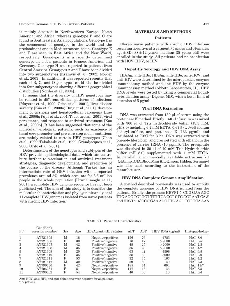
TABLE I. Patients’ Characteristics
PtaGeneBank
acession number Sex Age HBeAg/anti-HBe status ALT AST HBV DNA (pg/ml) Histopat-hology
1 AY721605 M 18 Negative/positive 156 76 4783 HAI: 8/02 AY721606 F 30 Positive/negative 18 17 >2000 HAI: 6/53 AY721607 M 42 Positive/negative 45 25 >2000 HAI: 2/34 AY721608 M 35 Positive/negative 36 23 >2000 HAI: 4/25 AY721609 M 20 Positive/negative 63 42 4598 HAI: 6/56 AY721610 F 35 Positive/negative 38 32 5009 HAI: 0/07 AY721611 F 53 Positive/negative 32 35 163 HAI: 4/28 AY721612 M 32 Positive/negative 59 39 30 HAI: 2/39 AY796030 F 42 Negative/positive 185 74 694 HAI: 11/710 AY796031 F 51 Negative/positive 117 113 36 HAI: 8/511 AY796032 F 54 Negative/positive 40 30 10 HAI: 6/4
Anti-HCV, anti-HIV, and anti-delta tests were negative for all patients.aPt, patient.
Complete Genome of HBV in Turkish Patients 477

AGT TGC ATG GTG CTG G 30 having Sap1 recognitionsite flanks were used in a long PCR reaction includinghigh fidelity taq polymerase [Gunther et al., 1998]. Thesensitivity of long PCR was 1�103 copy/ml in our hands.The PCR products were also cloned into TA vectors(Topo TA cloning kit, Invitrogen, CA) according toinstructions of manufacturer for further analysis. Wedesigned new primers (HBVseq1480: 50 GAC TCT CTCGTC CCC TTC 30-nt 1480–1497 and HBVseq2146: 50
TAG ACG CTG GAT CTT CCA 30-nt 2146–2129) for theamplification and the confirmation of the sequences ofthe hybridization sites of HBVP1 and HBVP2 primers.
Direct Sequencing of PCR Products
Following the clean up of PCR products, directsequencing was performed using primers; HBVP1,HBVP2, HBVseq1480, HBVseq2146, HBV 676–699: 50
TTT ACT AGT GCC ATT TGT TCA GTG 30, HBV 66–90:50 GCT CCA GTT CAG GAA CAG TAA ACC C 30,HBV2796–2826: 50 CAC CTG CAG CCT CAT TTT GTGGGT CAC CAT A 30, HBV 2357–2380: 50 GGC AGG TCCCCT AGA AGA AGA ACT 30, HBV 2432–2408: 50 ATTGAG ATC TTC TGC GAC GCG GCG A 30, and the dyeterminator cycle sequencing kit (Applied Biosystems,Foster City, CA). The reaction products were run on theABI 310 automated sequencer (Applied Biosystems,Foster City, CA).
Phylogenetic Analysis
Following the alignment of the 50 complete sequences(36 for genotype D and 14 for the other genotypes)representative for all genotypes of HBV from the data-base of GenBANK/EMBL/DDBJ and 11 sequences obt-ained from the sera of the patients with HBV infection,phylogenetic comparison was done by distance matrix/UPGMA analysis using Kimura 2-parameter by MEGA2software package program available at http://www.megasoftware.net/ [Kumar et al., 2001]. In addition tothe complete genome, phylogenetic analysis of S, preS,core, and X gene sequences were also done separately.
RESULTS
Patients
Of 11 patients, 7 were HBeAg positive and 4 wereHBeAg negative/anti-HBe positive. Characteristics ofthe patients are given in Table I. All patients hadhistopathologically proven chronic B hepatitis and hadreceived no antiviral treatment. The patients wereenrolled prospectively in the study. The HBeAg statusof the patients (seven HBeAg positive patients vs. fouranti-HBe positive patients) was coincidental which doesnot reflect the real HBeAg status incidence in Turkey(generally accepted as 30% HBeAg positive patients vs.70% anti-HBe positive patients).
Nucleotide and Amino Acid Analyses
The genome lengths of HBV isolates were consistently3182 base pairs except one genome in which there was a T
AB
LE
II.
Com
pari
sion
ofIs
olate
dH
BV
Gen
omes
inT
erm
sof
Gen
ome
Len
gth
,Gen
otyp
e,S
ubty
pe,
an
dth
eP
art
icu
lar
Seq
uen
ces
inS
urf
ace
Gen
ean
dC
ore
Pro
mot
eran
dP
reco
reR
egio
n
Pt
Gen
Ban
kace
ssio
nn
um
ber
HB
VD
NA
Len
gth
(nt)
Del
etio
n(n
t)G
enot
yp
eS
ubty
pe
X02496
Su
rface
gen
eco
don
nu
mber
pc7
1
Cor
ep
rom
oter
mu
tati
ons
nt_
RN
Ap
cA
(-26)T
,n
t_R
NA
pc
G(-
24)T
Pre
-cor
est
opco
don
mu
tati
ons
nt_
pcG
83A
,n
t_p
cG86A
69
C122
R125
T127
P134
Y160
K195
V
1A
Y721605
3182
No
D1
ayw
2—
——
——
——
TN
oN
o2
AY
721606
3182
No
D1
ayw
2—
——
——
——
TN
oN
o3
AY
721607
3182
No
D1
ayw
2—
——
——
——
TN
oN
o4
AY
721608
3182
No
D1
ayw
2—
——
——
——
Tn
t_R
NA
pc
A(-
26)T
No
5A
Y721609
3182
No
D1
ayw
2S
top
Cod
on—
——
——
—T
No
No
6A
Y721610
2999
183
(in
pre
-S)
D1
ayw
2S
top
Cod
on—
——
——
—T
No
No
7A
Y721611
3182
No
D1
ayw
2S
top
Cod
on—
——
——
—T
nt_
RN
Ap
cA
(-26)T
nt_
RN
Ap
cG
(-24)T
No
8A
Y721612
3182
No
D1
ayw
2—
——
——
——
TN
oN
o9
AY
796030
3182
No
D1
ayw
2—
——
——
——
TN
on
t_p
cG83A
10
AY
796031
3182
No
D2
ayw
3—
——
T—
——
Tn
t_R
NA
pc
A(-
26)T
nt_
RN
Ap
cG
(-24)T
No
11
AY
796032
3182
No
D1
ayw
2—
——
——
——
Tn
t_R
NA
pc
A(-
26)T
nt_
RN
Ap
cG
(-24)T
nt_
pcG
83A
An
ewn
omen
clatu
resy
stem
,p
rop
osed
by
Bart
hol
omeu
szan
dS
chaef
eran
dbase
don
nu
cleo
tid
ean
dam
ino
aci
dn
um
ber
sst
art
ing
at
each
open
read
ing
fram
ew
as
use
dto
defi
ne
the
nu
cleo
tid
en
um
ber
san
dth
egen
es.
478 Bozdayi et al.

183 base pairs deletion in pre-S1 gene (nucleotides2987–3169). In all patients, when compared to othergenotypes, there was a 33 nucleotide deletion in the pre-S1 region, which also corresponds to the spacer region ofHBV polymerase gene and is characteristic of genotypeD. Two HBeAg negative/anti-HBeAg positive patientshad nt_pcG83A [G1896A in the old nomenclature;proposed by Bartholomeusz and Schaefer, 2004] precore stop codon mutation, which is the major mutationof the precore gene resulting in formation of a non-functional premature protein. The nucleotide at pc71(T1858 in the old nomenclature) was a thymine in allpatients which plays a crucial role in alteration ofguanine to adenine at nt_pcG83 and nt_pcG86 to createa more stable stem loop structure. Dual core promotermutations, nt_RNApc A(-26)T (T1762A in old nomen-clature) and nt_RNApc G(-24)T (G1764A in old nomen-clature) were detected in HBV genomes isolated fromone HBeAg positive and two HBeAg negative/anti-HBeAg positive patients, which may reduce the tran-scription rate of the pre-core/core mRNA. In addition,one HBeAg positive patient had an nt_RNApc A(-26)Tmutation (Table II). In three HBV isolates, a stop codonmutation at codon 69 in S gene was detected (Pt6, Pt7,Pt8). One of these three HBV isolates (Pt7), was alsoassociated with a 183 base pair deletion in pre-S1 gene.Although the sequences of the viruses isolated fromthese patients were supplied from the DNA sequences ofcomplete genome PCR products, ten clones from Pt6, sixclones from Pt7 and nine clones from Pt8 were alsosequenced. The sequences of the clones from Pt6 yieldedstop codon at codon 69 in five clones and wild typesequences in the remaining five clones. Of six clonesbelonging to Pt7, sequences of three clones werecarrying the 183 base pairs deletion in pre-S1 gene andthe same stop codon mutation, two clones had only thestop codon mutation at codon 69 with no deletion, andone clone displayed wild type sequence. In Pt8, whileseven clones had stop codon mutation at codon 69, twoclones consisted of wild type sequences. There were noother mutation patterns residing in any epitopes on S,pre-S1, pre-S2, and core gene. The analysis of S genebased on the presence of Arg122, Thr125, Pro127, andLys160 residues determined by sequence data in 11patients revealed that ten and one HBV strains weresubtype ayw2 and ayw3, respectively (Table II), whichdisperses throughout in genotype D.
Fig. 1. Following the alignment of the 36 complete sequencesrepresentative for genotype D and 14 complete sequences representa-tive for the other genotypes of HBV from the database of GenBANK/EMBL/DDBJ and 11 sequences obtained from the sera of the patientswith HBV infection, phylogenetic tree obtained by distance matrix/UPGMA comparison (with Kimura-2 correction) after bootstrapping1000 replicates of sequence segment from the complete genome of HBV(nt 1–3182). The sequences of complete genome of HBV from 11Turkish patients are available at GenBank with accession numbersfrom AY721605 to AY721612 and from AY796030 to AY796032. In thisanalysis, three Turkish HBV isolates [two unpublished, AY661792 andAY661793 and one published, M32138 by Tong et al., 1990] retrievedfrom the GenBank were also included.
Complete Genome of HBV in Turkish Patients 479

Phylogenetic Analyses
By comparing the sequences of HBV isolated fromTurkish patients with 50 complete sequences of HBVretrieved from the GenBank database, representing allother existing genotypes, the sequences of all 11 patientswere consistent with that of genotype D (Fig. 1). Of 11Turkish HBV DNA sequences, 10 and 1 sequences wereclustered in genotype D1 and genotype D2, respectively,by phylogenetic analysis of 36 complete genotype Dgenome sequences mostly included in a recent reportallowing the distinction of four subgenotypes, D1–D4[Norder et al., 2004]. Nucleotide divergences of Turkishgenotype D were closely related to the genotype D1(0.0019� 0.001 nucleotide/genome) (Table III). Onecomplete genome genotyped as D2 and the ten completegenomes genotyped as D1 were found to be subtypeayw3and ayw2, respectively.
Phylogenetic analysis of the pre-S1, S region, pre-core/core, and X gene sequences of 11 isolated HBV DNAgenomes and all other genotype D sequences retrievedfrom the GenBank database confirmed that all Turkishsequences were clustered mostly in the genotype DTurkish branch. Each topology was inline with the dataobtained from complete genome sequences. Recombina-tion of the HBV genes belonging to different genotypeswere not observed in any separate analysis of all openreading frames of HBV. The sequences of the completegenome of HBV from 11 Turkish patients are availableat GenBank with accession numbers from AY721605 toAY721612 and AY796030 to AY796032.
DISCUSSION
Genotyping is the genetic characterization of agenome, which can classify the genomes based onnucleotide substitutions, deletions or insertions, anddiscriminate one individual strain from another. Thisinformation constitutes the molecular virological char-acteristics of the strain and may be useful clinically. Inthis study, we genotyped the complete genomes of HBVDNA isolated from 11 naıve patients with chronic HBVinfection. The data obtained in this study indicate thatTurkish patients with chronic B hepatitis do not show
genotypic diversity. All HBV sequences yielded mole-cular characteristics of genotype D genomes by beingsimilar in length except one having an unusual deletionin pre-S1 gene and showing a fingerprint deletion in pre-S1. All the HBV isolates from Turkish patients wereclustered in Genotype D branch with high bootstrapvalues. Likewise, all patients but one with chronichepatitis B had the ayw2 subtype. In subgenotypinganalysis, 10 HBV isolates subtyped as ayw2 and oneHBV isolate subtyped as ayw3 were determined as D1and D2, respectively. No recombination event wasobserved for any gene in the sequences of Turkishpatients, which is seemingly related to the existence of aunique genotype. This genotype and subtype homoge-neity is inline with data from other Mediterraneancountries and our own published material [Chan et al.,1999; Tahan et al., 2003; Bozdayi et al., 2004; Leblebi-cioglu and Eroglu, 2004; Yalcin et al., 2004].
None of the HBeAg positive patients had pre-core stopcodon mutation and two of three HBeAg negativepatients had a stop codon mutation at nt_pcG83A. Corepromoter mutations were also detected in two HBeAgpositive patients. The deletion in pre-S1 and stop codonmutations at S gene created a defective virus, thefunctions of which might be complemented by the wildtype virus existing in the viral pool. The co-existence ofwild type and defective viral strains was clearlydemonstrated by sequencing of the clones belonging toHBV isolates having stop codon mutations at codon 69 inthe S gene and deletion in pre-S1. The 183 base pairspre-S1 deletion is found frequently in persistent viralinfection. This deletion removes the promoter of thesmall envelope gene and results in generation of adeleted large envelope protein causing accumulation ofnucleocapsids containing viral DNA. Complementationwith the wild type small protein allows the mutantvirion formation [Melegari et al., 1997].
On the other hand, the findings in genetic diversity ofTurkish HBV strains may not be as expected, consider-ing Turkey’s geographical setting which has served as abridge in many migration events throughout history andcomposition of its population. In addition to theuniformity of the genotype, the nucleotide divergence
TABLE III. Nucleotide Divergences Between Turkish HBV Genomes (D-TR) and all Other Sequences Retrieved From theGenBank Database
Genotype D-TR D1 D2 D3 D4 A B C E F G H
D-TR 0.001 0.002 0.002 0.003 0.006 0.005 0.005 0.005 0.006 0.007 0.006D1 0.019 0.002 0.002 0.003 0.006 0.005 0.005 0.005 0.006 0.007 0.006D2 0.026 0.027 0.002 0.003 0.005 0.005 0.005 0.005 0.006 0.007 0.006D3 0.034 0.035 0.038 0.003 0.005 0.005 0.005 0.004 0.006 0.007 0.006D4 0.044 0.045 0.047 0.049 0.005 0.005 0.005 0.004 0.006 0.007 0.006A 0.098 0.099 0.098 0.100 0.100 0.005 0.005 0.006 0.006 0.007 0.006B 0.109 0.108 0.109 0.112 0.110 0.095 0.005 0.006 0.006 0.007 0.007C 0.105 0.104 0.107 0.108 0.104 0.098 0.102 0.005 0.006 0.007 0.006E 0.075 0.076 0.076 0.079 0.077 0.099 0.111 0.103 0.007 0.007 0.006F 0.139 0.139 0.141 0.144 0.141 0.140 0.143 0.135 0.137 0.007 0.007G 0.143 0.143 0.145 0.145 0.142 0.143 0.149 0.143 0.145 0.085 0.007H 0.116 0.116 0.116 0.118 0.119 0.115 0.129 0.131 0.112 0.148 0.152
Nucleotide divergences of Turkish genotype D were closely related to the genotype D1.
480 Bozdayi et al.
in the Turkish genotype D patients seems to be less thanin other genotype D sequences, allowing postulatingthat the spread of HBV could be later than migrationevents in this population. The HBV genotype D is alsoprevalent in neighboring countries of the Middle Eastand the Mediterranean basin, and in Central Asia fromwhere the Turkish variant of genotype D has beenpossibly imported centuries ago [Alestig et al., 2001;Saudy et al., 2003]. It is also interesting that, in additionto two patients from Iran and Egypt, two patients fromLatvia and Russia were residing along with Turkishpatients in the clade analysis of complete genome andseparate analysis of each open reading frame of HBV.
In conclusion, 11 complete genomic DNA of HBVisolates from Turkish patients with chronic hepatitis Brepresent a rather homogenous genotypic diversity.These results are inline with epidemiological studiesfrom the Mediterranean region and may be representa-tive for neighboring countries. These sequences ofTurkish HBV genomes may contribute to the informa-tion on the genetic diversity of HBV worldwide.
REFERENCES
Alestig E, Hannoun C, Horal P, Lindh M, 2001. Hepatitis B virusgenotypes in Mongols and Australian Aborigines. Arch Virol 12:2321–2329.
Arauz-Ruiz P, Norder H, Robertson BH, Magnius LO. 2002. GenotypeH: A new Amerindian genotype of hepatitis B virus revealed inCentral America. J Gen Virol 83:2059–2073.
Bartholomeusz A, Schaefer S. 2004. Hepatitis B virus genotypes:Comparison of genotyping methods. Rev Med Virol 14:3–16.
Bozdayi AM, Aslan N, Bozdayi G, Turkyilmaz AR, Sengezer T, Wend U,Erkan O, Aydemir F, Zakirhodjaev S, Orucov S, Bozkaya H, GerlichW, Karayalcin S, Yurdaydin C, Uzunalimoglu O. 2004. Molecularepidemiology of hepatitis B, C and D viruses in Turkish patients.Arch Virol 149:2115–2129.
Chan HL, Hussain M, Lok AS. 1999. Different hepatitis B virusgenotypes are associated with different mutations in the corepromoter and precore regions during hepatitis B e antigenseroconversion. Hepatology 29:976–984.
Ding X, Mizokami M, Yao G, Xu B, Orito E, Ueda R, Nakanishi M. 2001.Hepatitis B virus genotype distribution among chronic hepatitis Bvirus carriers in Shanghai, China. Intervirology 44:43–47.
Fujie H, Moriya K, Shintani Y, Yotsuyanagi H, Iino S, Koike K. 2001.Hepatitis B virus genotypes and hepatocellular carcinoma inJapan. Gastroenterology 120:1564–1565.
Grandjacques C, Pradat P, Stuyver L, Chevallier M, Chevallier P,Pichoud C, Maisonnas M, Trepo C, Zoulim F. 2000. Rapid detectionof genotypes and mutations in the pre-core promoter and the pre-core region of hepatitis B virus genome: Correlation with viralpersistence and disease severity. J Hepatol 33:430–439.
Gunther S, Sommer G, Von Breunig F, Iwanska A, Kalinina T,Sterneck M, Will H. 1998. Amplification of full-length hepatitis Bvirus genomes from samples from patients with low levels ofviremia: Frequency and functional consequences of PCR-intro-duced mutations. J Clin Microbiol 36:531–538.
Kao JH, Wu NH, Chen PJ, Lai MY, Chen DS. 2000a. Hepatitis Bgenotypes and the response to interferon therapy. J Hepatol 33:998–1002.
Kao JH, Chen PJ, Lai MY, Chen DS. 2000b. Hepatitis B genotypescorrelate with clinical outcomes in patients with chronic hepatitisB. Gastroenterology 118:554–559.
Kramvis A, Weitzmann L, Owiredu WK, Kew MC. 2002. Analysis of thecomplete genome of subgroup A0 hepatitis B virus isolates fromSouth Africa. J Gen Virol 83:835–839.
Kumar S, Tamura K, Jakobsen IB, Nei M. 2001. MEGA2 molecularevolutionary genetics analysis software. Bioinformatics 17:1244–1245.
Leblebicioglu H, Eroglu C, Members of the Hepatitis Study Group.2004. Acute hepatitis B virus infection in Turkey: Epidemiology andgenotype distribution. Clin Microbiol Infect 10:537–541.
Mayerat C, Mantegani A, Frei PC. 1999. Does hepatitis B virus (HBV)genotype influence the clinical outcome of HBV infection? J ViralHepat 6:299–304.
Melegari M, Scaglioni PP, Wands JR. 1997. The small envelope proteinis required for secretion of a naturally occurring hepatitis B virusmutant with pre-S1 deleted. J Virol 71:5449–5454.
Norder H, Hammas B, Lofdahl S, Courouce AM, Magnius LO. 1992.Comparison of the amino acid sequences of nine different serotypesof hepatitis B surface antigen and genomic classification ofthe corresponding hepatitis B virus strains. J Gen Virol 73:1201–1208.
Norder H, Arauz-Ruiz P, Blitz L, Pujol FH, Echevarria JM, MagniusLO. 2003. The T(1858) variant predisposing to the precore stopmutation correlates with one of two major genotype F hepatitis Bvirus clades. J Gen Virol 84:2083–2087.
Norder H, Courouce AM, Coursaget P, Echevarria JM, Lee SD,Mushahwar IK, Robertson BH, Locarnini S, Magniusa LO. 2004.Genetic diversity of hepatitis B virus strains derived worldwide:Genotypes, subgenotypes, and HBsAg subtypes. Intervirol 47:289–309.
Okamoto H, Tsuda F, Sakugawa H, Sastrosoewignjo RI, Imai M,Miyakawa Y, Mayumi M. 1988. Typing hepatitis B virus byhomology in nucleotide sequence: Comparison of surface antigensubtypes. J Gen Virol 69:2575–2583.
Orito E, Mizokami M, Sakugawa H, Michitaka K, Ishikawa K, Ichida T,Okanoue T, Yotsuyanagi H, Iino S. 2001. A case-control study forclinical and molecular biological differences between hepatitis Bviruses of genotypes B and C. Japan HBV Genotype ResearchGroup. Hepatology 33:218–223.
Saudy N, Sugauchi F, Tanaka Y, Suzuki S, Aal AA, Zaid MA, Agha S,Mizokami M. 2003. Genotypes and phylogenetic characterization ofhepatitis B and delta viruses in Egypt. J Med Virol 70:529–536.
Stuyver L, De Gendt S, Van Geyt C, Zoulim F, Fried M, Schinazi RF,Rossau R. 2000. A new genotype of hepatitis B virus: Completegenome and phylogenetic relatedness. J Gen Virol 81:67–74.
Tahan V, Ozdogan O, Tozun N. 2003. Epidemiology of viral hepatitis inthe Mediterranean basin. Rocz Akad Med Bialymst 48:11–17.
Takahashi K, Akahane Y, Hino K, Ohta Y, Mishiro S. 1998. Hepatitis Bvirus genomic sequence in the circulation of hepatocellularcarcinoma patients: Comparative analysis of 40 full-length isolates.Arch Virol 143:2313–2326.
Takahashi K, Ohta Y, Kanai K, Akahane Y, Iwasa Y, Hino K, Ohno N,Yoshizawa H, Mishiro S. 1999. Clinical implications of mutations C-to-T1653 and T-to-C/A/G1753 of hepatitis B virus genotype Cgenome in chronic liver disease. Arch Virol 144:1299–1308.
Tong SP, Li JS, Vitvitski L, Trepo C. 1990. Active hepatitis B virusreplication in the presence of anti-HBe is associated with viralvariants containing an inactive pre-C region. J Virol 176:596–603.
Tsubota A, Arase Y, Ren F, Tanaka H, Ikeda K, Kumada H. 2001.Genotype may correlate with liver carcinogenesis and tumorcharacteristics in cirrhotic patients infected with hepatitis B virussubtype adw. J Med Virol 65:257–265.
Uzunalimoglu O, Yurdaydin C, Cetinkaya H, Bozkaya H, Sahin T,Colakoglu S, Tankurt E, Sarioglu M, Ozenirler S, Akkiz H, Tozun N,Degertekin H, Okten A. 2001. Risk factors for hepatocellularcarcinoma in Turkey. Dig Dis Sci 46:1022–1028.
Yalcin K, Degertekin H, Bahcecioglu IH, Demir A, Aladag M, YildirimB, Horasanli S, Ciftci S, Badur S. 2004. Hepatitis B virus genotype Dprevails in patients with persistently elevated or normal ALT levelsin Turkey. Infection 32:242–249.
Complete Genome of HBV in Turkish Patients 481