Compensatory regulation of Na+ absorption by Na+/H+ exchanger
12
RESEARCH Open Access Compensatory regulation of Na + absorption by Na + /H + exchanger and Na + -Cl - cotransporter in zebrafish (Danio rerio) Wei-Jen Chang 1,2,3 , Yi-Fang Wang 1,4 , Huei-Jyun Hu 1,4 , Jung-Hsuan Wang 1,5 , Tsung-Han Lee 2,3 and Pung-Pung Hwang 1* Abstract Introduction: In mammals, internal Na + homeostasis is maintained through Na + reabsorption via a variety of Na + transport proteins with mutually compensating functions, which are expressed in different segments of the nephrons. In zebrafish, Na + homeostasis is achieved mainly through the skin/gill ionocytes, namely Na + /H + exchanger (NHE3b)-expressing H + -ATPase rich (HR) cells and Na + -Cl - cotransporter (NCC)-expressing NCC cells, which are functionally homologous to mammalian proximal and distal convoluted tubular cells, respectively. The present study aimed to investigate whether or not the functions of HR and NCC ionocytes are differentially regulated to compensate for disruptions of internal Na + homeostasis and if the cell differentiation of the ionocytes is involved in this regulation pathway. Results: Translational knockdown of ncc caused an increase in HR cell number and a resulting augmentation of Na + uptake in zebrafish larvae, while NHE3b loss-of-function caused an increase in NCC cell number with a concomitant recovery of Na + absorption. Environmental acid stress suppressed nhe3b expression in HR cells and decreased Na + content, which was followed by up-regulation of NCC cells accompanied by recovery of Na + content. Moreover, knockdown of ncc resulted in a significant decrease of Na + content in acid-acclimated zebrafish. Conclusions: These results provide evidence that HR and NCC cells exhibit functional redundancy in Na + absorption, similar to the regulatory mechanisms in mammalian kidney, and suggest this functional redundancy is a critical strategy used by zebrafish to survive in a harsh environment that disturbs body fluid Na + homeostasis. Keywords: Na + -Cl - cotranspoter, Na + /H + exchanger, Ionocyte, Acid acclimation Introduction Sodium is the dominant cation in vertebrate body fluids, and therefore sodium regulation is one of most import- ant tasks in maintaining body fluid homeostasis in vertebrates. In mammalian kidney, about two-thirds of sodium reabsorption occurs in the proximal tubule, and is achieved mainly by apically-expressed Na + /H + ex- changers (NHE). The remaining sodium is reabsorbed by the distal convoluted tubules and the collecting ducts, via Na + -Cl - cotransporters (NCC) and epithelial sodium channels (ENaC), respectively [1]. Hormonal control of sodium reabsorption occurs primarily in the distal convoluted tubules and the collecting ducts [2], suggesting that while NHE performs the majority of sodium re- absorption, this process is fine-tuned by NCC and ENaC in the mammalian kidney. Sodium transport along the nephron can be regulated through controlling the abun- dance, activity, and subcellular distribution of transporters [3]. Previous studies have demonstrated that a high-salt diet provokes subcellular redistribution and decreased abundance of NCC, but does not affect the abundance or activity of NHE3 [3-5]. In addition, the hormones angio- tensin II and aldosterone, which are the major factors that determine the sodium reabsorption rate, positively regu- late NCC activity via a WNK4 (with-no-lysine kinase 4)- dependent pathway [6,7]. Taken together, dietary salt change, activates the functional regulation of NCC in * Correspondence: [email protected] 1 Institute of Cellular and Organismic Biology, Academia Sinica, Taipei, Taiwan Full list of author information is available at the end of the article © 2013 Chang et al.; licensee BioMed Central Ltd. This is an Open Access article distributed under the terms of the Creative Commons Attribution License (http://creativecommons.org/licenses/by/2.0), which permits unrestricted use, distribution, and reproduction in any medium, provided the original work is properly cited. Chang et al. Frontiers in Zoology 2013, 10:46 http://www.frontiersinzoology.com/content/10/1/46
Transcript of Compensatory regulation of Na+ absorption by Na+/H+ exchanger
Chang et al. Frontiers in Zoology 2013, 10:46http://www.frontiersinzoology.com/content/10/1/46
RESEARCH Open Access
Compensatory regulation of Na+ absorption byNa+/H+ exchanger and Na+-Cl- cotransporter inzebrafish (Danio rerio)Wei-Jen Chang1,2,3, Yi-Fang Wang1,4, Huei-Jyun Hu1,4, Jung-Hsuan Wang1,5, Tsung-Han Lee2,3
and Pung-Pung Hwang1*
Abstract
Introduction: In mammals, internal Na+ homeostasis is maintained through Na+ reabsorption via a variety of Na+
transport proteins with mutually compensating functions, which are expressed in different segments of thenephrons. In zebrafish, Na+ homeostasis is achieved mainly through the skin/gill ionocytes, namely Na+/H+
exchanger (NHE3b)-expressing H+-ATPase rich (HR) cells and Na+-Cl- cotransporter (NCC)-expressing NCC cells,which are functionally homologous to mammalian proximal and distal convoluted tubular cells, respectively. Thepresent study aimed to investigate whether or not the functions of HR and NCC ionocytes are differentiallyregulated to compensate for disruptions of internal Na+ homeostasis and if the cell differentiation of the ionocytesis involved in this regulation pathway.
Results: Translational knockdown of ncc caused an increase in HR cell number and a resulting augmentation ofNa+ uptake in zebrafish larvae, while NHE3b loss-of-function caused an increase in NCC cell number with aconcomitant recovery of Na+ absorption. Environmental acid stress suppressed nhe3b expression in HR cells anddecreased Na+ content, which was followed by up-regulation of NCC cells accompanied by recovery of Na+
content. Moreover, knockdown of ncc resulted in a significant decrease of Na+ content in acid-acclimated zebrafish.
Conclusions: These results provide evidence that HR and NCC cells exhibit functional redundancy in Na+
absorption, similar to the regulatory mechanisms in mammalian kidney, and suggest this functional redundancy is acritical strategy used by zebrafish to survive in a harsh environment that disturbs body fluid Na+ homeostasis.
Keywords: Na+-Cl- cotranspoter, Na+/H+ exchanger, Ionocyte, Acid acclimation
IntroductionSodium is the dominant cation in vertebrate body fluids,and therefore sodium regulation is one of most import-ant tasks in maintaining body fluid homeostasis invertebrates. In mammalian kidney, about two-thirds ofsodium reabsorption occurs in the proximal tubule, andis achieved mainly by apically-expressed Na+/H+ ex-changers (NHE). The remaining sodium is reabsorbedby the distal convoluted tubules and the collecting ducts,via Na+-Cl- cotransporters (NCC) and epithelial sodiumchannels (ENaC), respectively [1]. Hormonal controlof sodium reabsorption occurs primarily in the distal
* Correspondence: [email protected] of Cellular and Organismic Biology, Academia Sinica, Taipei, TaiwanFull list of author information is available at the end of the article
© 2013 Chang et al.; licensee BioMed CentralCommons Attribution License (http://creativecreproduction in any medium, provided the or
convoluted tubules and the collecting ducts [2], suggestingthat while NHE performs the majority of sodium re-absorption, this process is fine-tuned by NCC and ENaCin the mammalian kidney. Sodium transport along thenephron can be regulated through controlling the abun-dance, activity, and subcellular distribution of transporters[3]. Previous studies have demonstrated that a high-saltdiet provokes subcellular redistribution and decreasedabundance of NCC, but does not affect the abundance oractivity of NHE3 [3-5]. In addition, the hormones angio-tensin II and aldosterone, which are the major factors thatdetermine the sodium reabsorption rate, positively regu-late NCC activity via a WNK4 (with-no-lysine kinase 4)-dependent pathway [6,7]. Taken together, dietary saltchange, activates the functional regulation of NCC in
Ltd. This is an Open Access article distributed under the terms of the Creativeommons.org/licenses/by/2.0), which permits unrestricted use, distribution, andiginal work is properly cited.
Chang et al. Frontiers in Zoology 2013, 10:46 Page 2 of 12http://www.frontiersinzoology.com/content/10/1/46
mammalian kidney through an aldosterone-dependentand WNK4-mediated mechanism [8].Sodium transporters expressed along the nephron are
able to compensate for each other in sodium absorptionmechanisms. In nhe3 knockout mice, the levels ofsodium-phosphate cotransporter (NaPi2) and the 70 kDaENaC γ-subunit were found to be increased [9]. Moreover,the abundance of the 70 kDa ENaC γ-subunit was alsoreported to be increased in ncc knockout mice [9]. Loss ofNCC activity in mice resulted in only a subtle perturbationof Na+ homeostasis, which was presumed to arise from acompensatory increase in ENaC activity [10,11]. Compen-sation through differential regulation of multiple Na+ up-take pathways is essential for internal Na+ homeostasis inmammals, and from an evolutionary physiological per-spective, it may be conserved in other osmoregulatory ver-tebrates. However, there is little available information onthis process in other vertebrates.Teleosts, as an aquatic vertebrate, maintain body fluid
homeostasis of Na+ (and other ions) through active ab-sorption of this ion from freshwater (FW) environmentswith Na+ levels less than 1 mM. The Na+ uptake mecha-nisms in fish, mainly conducted in the gills or skin (duringembryonic stages), are analogous to those in mammaliankidney, in terms of transporter expression and function inthe ionocytes [12-15]. Zebrafish has recently proved to bea competent model for molecular physiological studies onion- and osmo-regulation in fish, because of its advantagesin terms of gene manipulation and availability of genomedatabases [15-18]. According to the recently proposedzebrafish ion regulation model, there are four subtypes ofionocyte, as follows: Na+-K+-ATPase-rich (NaR) cells,H+-ATPase-rich (HR) cells, Na+-Cl- cotransporter (NCC)-expressing cells, and K+-secreting (KS) cells; these havebeen identified to express distinct sets of ion transporters,and thus to conduct different ions [15,18,19]. Of theseionocytes, HR cells, which co-express NHE3b andH+-ATPase in the apical membrane, were identified to bethe major cell type responsible for Na+ absorption and H+
secretion in zebrafish [20-23]. On the other hand, NCCcells, which apically express NCC, play a minor role inNa+ uptake in zebrafish [24]; this was demonstrated bytwo lines of evidence: sodium green, an Na+ fluorescenceprobe, accumulates only in HR cells, and metolazone, anNCC-specific inhibitor, does not impair sodium green ac-cumulation in HR cells [21].In zebrafish HR cells, certain co-expressed transporters
and enzymes (vacuolar H+-ATPase, ammonia transporter(Rhcg1), 2 carbonic anhydrase (CA2-like and CA15a), andanion exchanger (AE1b)) were found to affect the activityand function of NHE3b [22,25-29]. The activity ofelectroneutral NHE3b in the apical membrane of zebrafishHR cells depends on the gradients of Na+ and H+ acrossthe cell membrane [30], and therefore acidic FW
environments do not favor NHE3b activity. Exposure topH 4 FW was found to suppress nhe3b mRNA expressionin the zebrafish gill [23]. Notably, the same acid treatmentwas reported to cause an initial decrease in body Na+ con-tent, and thereafter a recovery of content accompanied byincreased Na+ uptake [31]. This raises the possibility thatan alternative pathway, not involving NHE3b, may play acompensatory role in Na+ homeostasis in zebrafish underenvironmental acid stress.An ortholog of mammalian NCC, zSlc12a10.2, was
reported to be specifically expressed in an ionocyte typedistinct from NHE3-expressing HR-type cells in zebrafishskin/gills; this NCC-type ionocyte was subjected to func-tional analysis, revealing the presence of an apical Na+ andCl- uptake pathway [24]. In addition, expression of nhe3band ca15a was found to be up-regulated in NCCmorphants [24]. Based on these results, we hypothesizedthat Na+ absorption in zebrafish skin/gill is similar to thatin mammalian kidneys; NHE3 performs the majority ofNa+ uptake, and the other transporters, such as NCC, playa supplementary role; furthermore, we hypothesized thatthese Na+ transport proteins would be able to compensatefor one another in the regulation of body fluid Na+
homeostasis.Herein, we tested the hypothesis that compensatory
regulation of Na+ uptake mechanisms is conserved in tele-osts. The experiments were designed to determine whetherncc expression is up-regulated when nhe3b expression isdecreased (and vice versa), and whether NCC is requiredfor Na+ absorption in acid-acclimated zebrafish.
ResultsNCC loss-of-function induces differentiation of HR cellsCompensatory expression of Na+ transporters was previ-ously observed in NCC null mice [9]. In order to identifywhether similar compensation occurs in zebrafish, thedevelopment of HR cells was observed in ncc mor-phants. As shown in Figure 1A-C, knockdown of NCCtranslation enhanced differentiation of HR cells in4- and 5 dpf larvae. The Na+ content remained at wildtype levels in 2- and 3 dpf morphants, but was signifi-cantly increased in 5- and 6 dpf larvae (Figure 1D). Ini-tially, NCC loss-of-function does not significantly affectNa+ accumulation, but by later stages, Na+ accumulationis enhanced. This may be due to an accompanying in-crease of HR cells, which express the major Na+-uptaketransporter, NHE3b. This suggests that an increase ofHR cells (and thus NHE3b expression) may compensatefor the loss of NCC in terms of Na+ uptake.
Knockdown of gcm2 results in increased NCC celldifferentiation and subsequent recovery of Na+ contentThe transcription factor Gcm2 is known to control thedifferentiation of HR cells, which express NHE3b

A B
DC
Figure 1 Knockdown of ncc increases HR cell number and Na+ accumulation. (A, B): H+-ATPase (H-pump) immunostaining signals (arrow)in 4-dpf wild type larvae (WT) and ncc morphants. Scale bar = 100 μm. (C, D): Comparisons of HR cell density (C) and whole body Na+ content(D) between wild type larvae (white bars) and ncc morphants (gray bars) at different developmental stages (n = 8 in C and 6 in D). Mean ± SD.*p < 0.05; **p < 0.01; ***p < 0.001 (Student’s t-test).
Chang et al. Frontiers in Zoology 2013, 10:46 Page 3 of 12http://www.frontiersinzoology.com/content/10/1/46
[23,32,33]. Blocking HR cell differentiation with gcm2MOs resulted in an increase of NCC cells (Figure 2A-C).This increase in NCC cells may be related to an increasein the ability of morphants to absorb Na+. Compared to4 dpf wild type larvae, Na+ influx was significantly in-creased in gcm2 morphants, but was dramatically de-creased in foxi3a/3b morphants (Figure 2D). Thetranscription factors Foxi3a and Foxi3b control the dif-ferentiation of NaR and HR cells [34,35], and were alsofound to be required for the differentiation of NCC cells(Additional file 1: Figure S1). Closer inspection of thegcm2 morphants revealed an initial decrease of Na+ con-tent at 3 dpf, which was restored to wild type levels at 4dpf; by 5 dpf, the Na+ content of the morphants hadexceeded that of the wild type (Figure 2E). Taken to-gether, it appears that loss of HR cells impairs the Na+
uptake function, and subsequently stimulates NCC celldifferentiation resulting in a compensatory increase inNa+ accumulation.
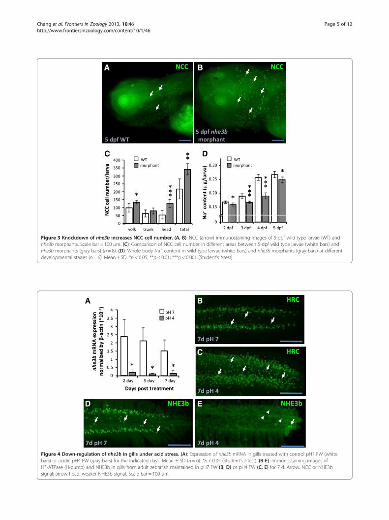
Knockdown of nhe3b results in increased NCC celldifferentiation and subsequent recovery of Na+ contentA similar compensatory effect was also observed innhe3b morphants. NCC cells were increased in 5-dpfnhe3b morphants as compared to wild type larvae, and
the increase was mainly observed in the head region ofthe morphants (Figure 3A-C). Knockdown of nhe3b sig-nificantly decreased Na+ accumulation in the larvaefrom as early as 2 dpf, but levels had begun to recoverby 5 dpf (Figure 3D). As for gcm2 knockdown, down-regulation of nhe3b translation impairs Na+ uptake,which is thereafter compensated by enhanced NCC celldifferentiation and Na+ absorption ability.
Expression of nhe3b in adult gills is inhibited by acidicenvironmentsNext, we investigated compensation of Na+ uptake func-tion during acclimation to an acidic environment. Themajority of nhe3b mRNA expression in adult gills wassuppressed during acclimation to an acidic environment(Figure 4A). Immunostaining revealed that HR cells weremarkedly increased, while NHE3b signals were signifi-cantly decreased, after acid acclimation (Figure 4B-E).Therefore, nhe3b mRNA and NHE3b protein are bothdecreased in zebrafish gills after acid treatment.
NCC cell differentiation in adult gills is up-regulated afteracid acclimationThe NCC cell density in gill filaments was not changedafter acid acclimation for 7 d (Figure 5A-C); however,

A B
EDC
Figure 2 Loss of HR cells by gcm2 knockdown increases NCC cell number and Na+ absorption ability. (A, B): Immunostaining images ofNCC cells (arrow) in 4-dpf wild type larvae (WT) and gcm2 morphants. Scale bar = 100 μm. (C): Comparison of NCC cell density between wild typelarvae (white bars) and gcm2 morphants (gray bars) at different developmental stages (n = 8). (D): Na+ influx in 4-dpf wild types, gcm2 morphants,and foxi3a/3b morphants (n = 6). (E): Whole body Na+ content in wild type larvae (white bars) and gcm2 morphants (gray bars) at differentdevelopmental stages (n = 6). Mean ± SD. *p < 0.05 (Student’s t-test).
Chang et al. Frontiers in Zoology 2013, 10:46 Page 4 of 12http://www.frontiersinzoology.com/content/10/1/46
the total number of NCC cells in both the filaments andlamellae was increased (Figure 5A-B, E). In summary,NHE3b is down-regulated and NCC is up-regulated inadult gills during acclimation to acidic environments.
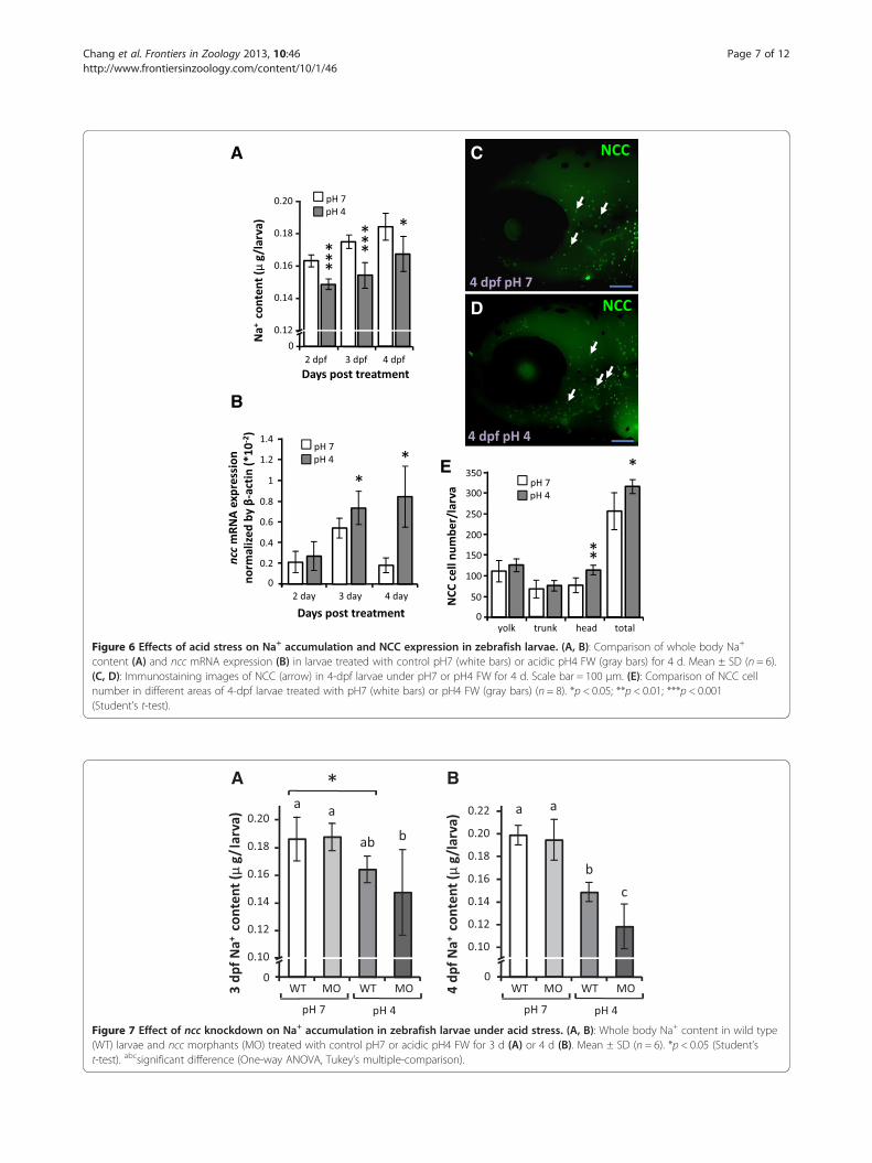
Acid acclimation of larvae results in increased NCC celldifferentiation and a gradual recovery of Na+ contentWhole body Na+ content was significantly decreased inacid-acclimated larvae as compared to controls raised inpH 7 FW during the first 2~3 d (Figure 6A); however,the difference between test and control groups declinedby 4 dpf, suggesting a recovery of Na+ absorption abilityin the acid-acclimated larvae. In addition, expression ofncc mRNA in larvae was significantly increased after3~4 d acid treatment (Figure 6B), and NCC cell numberin the head region, and thus larval skin overall, was alsoincreased after acid acclimation (Figure 6C-E). This sug-gests that the recovery of Na+ accumulation may haveresulted from increased ncc expression and NCC celldifferentiation during acid acclimation.
NCC is required for compensatory regulation of Na+
uptake under acid stressThe whole body Na+ contents of wild type larvae andncc morphants were compared after treatment withpH 7 (control) or pH 4 FW. The Na+ contents of wildtype and ncc morphants maintained in pH 7 FW werenot significantly different (Figure 7A, B). However, theNa+ content in 3 dpf ncc morphants was lower (albeitnot significantly) than that in wild type larvae upon acidstress; this decrease was greater and significant at 4 dpf(Figure 7A, B), suggesting that NCC is necessary for thecompensatory regulation of Na+ absorption during acidacclimation.
DiscussionLike the mammalian kidney, zebrafish skin and gills havebeen proposed to perform Na+ uptake via a variety ofNa+ transporters, including ENaC, NHE3 and NCC[14-16,36]. Certain pharmacological experiments havesuggested a role for ENaC in Na+ uptake mechanisms inzebrafish [21]; however, a sequence homologous to
A B
DC
Figure 3 Knockdown of nhe3b increases NCC cell number. (A, B): NCC (arrow) immunostaining images of 5-dpf wild type larvae (WT) andnhe3b morphants. Scale bar = 100 μm. (C): Comparison of NCC cell number in different areas between 5-dpf wild type larvae (white bars) andnhe3b morphants (gray bars) (n = 8). (D): Whole body Na+ content in wild type larvae (white bars) and nhe3b morphants (gray bars) at differentdevelopmental stages (n = 6). Mean ± SD. *p < 0.05; **p < 0.01; ***p < 0.001 (Student’s t-test).
A B
C
D E
Figure 4 Down-regulation of nhe3b in gills under acid stress. (A): Expression of nhe3b mRNA in gills treated with control pH7 FW (whitebars) or acidic pH4 FW (gray bars) for the indicated days. Mean ± SD (n = 6). *p < 0.05 (Student’s t-test). (B-E): Immunostaining images ofH+-ATPase (H-pump) and NHE3b in gills from adult zebrafish maintained in pH7 FW (B, D) or pH4 FW (C, E) for 7 d. Arrow, NCC or NHE3bsignal; arrow head, weaker NHE3b signal. Scale bar = 100 μm.
Chang et al. Frontiers in Zoology 2013, 10:46 Page 5 of 12http://www.frontiersinzoology.com/content/10/1/46

A
B
C D
Figure 5 NCC is up-regulated in gills under acid stress. (A, B):Immunostaining images of NCC (arrow) in gills filaments under pH7or pH4 FW for 7d. (C, D): Comparisons of NCC cell density in gillfilaments (C) and of NCC cell number in both gill lamella andfilament (D) between pH7 (white bars) and pH4 FW (gray bars) for7d. Means ± SD (n = 8). *p < 0.05 (Student’s t-test).Scale bar = 100 μm.
Chang et al. Frontiers in Zoology 2013, 10:46 Page 6 of 12http://www.frontiersinzoology.com/content/10/1/46
ENaC has not yet been identified in zebrafish [14,15].Yan et al. [23] identified NHE3b in HR cells, and ana-lyzed changes in nhe3b mRNA expression induced bylow-Na+ FW in the cells. Esaki et al. [21] demonstratedthat treatment with EIPA, an NHE inhibitor, blockedboth sodium green (a fluorescent Na+ probe) accumula-tion and 24Na+ influx in zebrafish larvae [21], suggestingthat NHE3 is a major transporter for Na+ uptake inzebrafish. However, a recent study by Kumai and Perry(2011) did not observe a significant effect of EIPA on Na+
influx in zebrafish larvae, bringing the role of NHE3binto question. Further investigation is required to recon-cile the inconsistencies between these pharmacologicalstudies. Taking an alternate approach, Shih et al. [29]adopted non-invasive electrophysiological methods to
detect Na+ uptake activity in intact zebrafish larvae, andobserved a decrease in Na+ uptake after EIPA treatmentor nhe3b knockdown. The present nhe3b knockdown ex-periments resulted in a severe decrease in Na+ contentin zebrafish morphants (Figure 3), further reinforcingthe aforementioned perception of NHE3b as a majortransporter for Na+ absorption. Compared to NHE3,NCC plays a relatively subtle and regulatory role inmaintaining Na+ and fluid volume homeostasis in mam-mals [10,11,37]. In zebrafish, treatment with metolazone(an NCC inhibitor) was found to suppress Na+ uptake[24]. Besides, sodium green specifically accumulated inHR cells, but not NCC cells [21]. These suggest thatzebrafish ionocytes resemble their equivalents in themammalian kidney, in that NHE3b is the dominanttransport protein for maintaining Na+ homeostasis,whereas NCC plays a subtle role. This conclusion is fur-ther supported by the present study; NHE3b loss-of-function resulted in a drastic reduction in Na+ content,but no such effect was observed in the ncc morphants(Figures 1, 3). Interestingly, ectoderm derived-zebrafishionocytes and mesoderm derived-mammalian kidneycells exhibit similar functions and/or regulatory mecha-nisms, and these physiological processes appear to be con-served during vertebrate evolution from fish to mammals.In mammals, Na+ homeostasis is achieved through
compensatory regulation of Na+ absorption, which isachieved through differential expression of the relevantNa+ transport proteins in the kidneys [9]. Knockout ofnhe3 or ncc in mouse resulted in up-regulation ofγ-ENaC, to compensate for disruptions in Na+ homeo-stasis [9]. A low salt diet, which impairs body fluid Na+
homeostasis, resulted in up-regulated ncc expression,but did not affect expression of other Na+ transport pro-teins [3,38]. Here, we knocked down distinct Na+ trans-porters to demonstrate that zebrafish Na+ uptakemechanisms exhibit compensatory regulation throughdifferential control of the expression and function of therelevant Na+ transport proteins, a process analogous, interms of some major transporters, to that of Na+ re-absorption in the mammalian nephron. Knockdown ofgcm2 (encoding a transcription factor which positivelyregulates HR cell differentiation) results in loss ofNHE3b-expressing HR cells [32,33] and an increase inNCC cell number [39] (Figure 2) in zebrafish larvae.Similarly, loss-of-function of NHE3b induced an increaseof NCC cell number (Figure 3). On the other hand, thenumber of HR cells was found to increase in nccmorphants (Figure 1). Differential regulation of larvalHR and NCC cell densities was accompanied by changesin Na+ content or influx, reflecting disruptions in Na+
uptake function (Figures 1, 2 and 3) (and thus the bodyfluid Na+ homeostasis) and subsequent homeostatic ad-justment. Taken together, we propose that if body fluid
A C
D
B
E
Figure 6 Effects of acid stress on Na+ accumulation and NCC expression in zebrafish larvae. (A, B): Comparison of whole body Na+
content (A) and ncc mRNA expression (B) in larvae treated with control pH7 (white bars) or acidic pH4 FW (gray bars) for 4 d. Mean ± SD (n = 6).(C, D): Immunostaining images of NCC (arrow) in 4-dpf larvae under pH7 or pH4 FW for 4 d. Scale bar = 100 μm. (E): Comparison of NCC cellnumber in different areas of 4-dpf larvae treated with pH7 (white bars) or pH4 FW (gray bars) (n = 8). *p < 0.05; **p < 0.01; ***p < 0.001(Student’s t-test).
A B
Figure 7 Effect of ncc knockdown on Na+ accumulation in zebrafish larvae under acid stress. (A, B): Whole body Na+ content in wild type(WT) larvae and ncc morphants (MO) treated with control pH7 or acidic pH4 FW for 3 d (A) or 4 d (B). Mean ± SD (n = 6). *p < 0.05 (Student’st-test). abcsignificant difference (One-way ANOVA, Tukey’s multiple-comparison).
Chang et al. Frontiers in Zoology 2013, 10:46 Page 7 of 12http://www.frontiersinzoology.com/content/10/1/46
Chang et al. Frontiers in Zoology 2013, 10:46 Page 8 of 12http://www.frontiersinzoology.com/content/10/1/46
Na+ homeostasis is disturbed by decreased expression/function of either NHE3b or NCC, this will stimulate acompensatory increase in the expression/function of theother protein, thereby restoring equilibrium.Compensatory regulation of NHE3b and NCC in
zebrafish Na+ homeostasis appears to be of physiologicalsignificance, based on analyses of these transport pathwaysduring acclimation to environmental stress, such as acidicFW, which is known to disturb Na+ homeostasis inaquatic animals. In a recent study, recovery of Na+ contentand increased Na+ uptake were observed in acid-acclimated zebrafish, and NHE3 was suggested to play amajor role in this compensatory process, based onpharmacological and loss-of-function experiments [27];however, no data on NHE3b expression was available tosupport this conclusion. On the other hand, an earlierstudy concluded that zebrafish nhe3b expression may bedown-regulated because the H+ gradient is not favorablefor the operation of electronetural NHE3b [23]. This raisesthe possibility that Na+ transport proteins other thanNHE3b are critical for Na+ homeostasis in acid-acclimatedzebrafish. The present study has demonstrated that NCCis required for this regulatory mechanism of Na+ homeo-stasis under acid stress. Down-regulation of NHE3b andconcomitant up-regulation of NCC were observed notonly at the transcription level, but also in terms of cellnumber (Figures 4, 5 and 6). Notably, the timing of nccmRNA up-regulation is consistent with that of Na+ uptakerecovery upon acid acclimation (Figure 6), indicating thatNCC, instead of NHE3b, is the major participant in thiscompensatory process. As such, the two Na+ transporters,NHE3b and NCC, in the skin/gills of zebrafish have func-tional redundancy in regulating Na+ absorption. In a simi-lar manner to acid-acclimated larvae, nhe3b morphantsexhibited an initial decrease in Na+ content at 2 dpf, and asubsequent partial recovery at 4 dpf, accompanied by anincrease in NCC cell number (Figures 3, 6). This rein-forces our conclusion that the initial loss of Na+ in acid-treated larvae results from down-regulation of NHE3b,and the impaired Na+ content and/or decreased level ofNHE3b may trigger an increase in NCC cell number andthe concomitant compensatory recovery in Na+ content.Na+ content reflects changes in both influx and efflux.
One may consider passive Na+ efflux as one regulatoryprocess for body fluid Na+ homeostasis; however, it ap-pears not to be the case in acid-acclimated zebrafish. Ina recent study on the Na+ content and fluxes in acid-acclimated zebrafish, the recovery of Na+ content wasprimarily resulted from regulating Na+ uptake, ratherthan Na+ efflux [31]. Since NHE3b (Slc9a3b) and NCC(Slc12a10.2) are specifically expressed in the respectiveHR and NCC ionocytes in zebrafish skin and gills[17,23,24], the differential regulations of the 2 trans-porters found in the present study (see above) appear to
reflect the majority of the Na+ uptake changes associatedwith the compensatory recovery in Na+ content. Subse-quent loss-of-function experiments further reinforce thephysiological significance of the differential regulationsof the 2 transporters in body fluid Na+ homeostasis dur-ing acid acclimation. Knockdown of ncc did not signifi-cantly affect larval Na+ content, but the same treatmentunder acid stress resulted in a severe reduction of Na+
content as compared to wild type (Figure 7). This indi-cates that NCC plays a minor or subtle role in normalFW conditions, but replaces attenuated NHE3b as themajor player in zebrafish Na+ homeostasis during accli-mation to an acidic environment.Teleost gill (or skin during the embryonic stages) is an
organ with physiological plasticity, being able to modifyits morphology and functions to cope with environmen-tal changes. Modifications of the gills are stress- andspecies- dependent, and include alterations in the size,number, expression pattern, and apical morphology ofionocytes and the lamellar surface area [12,14,40-42].The ionocytes that appear in the gill lamella werethought to migrate from the gill filaments, and this wassuggested as a regulatory mechanism in response toenvironmental changes [12,43]. Such migration of iono-cytes is now thought to be unlikely, as ionocyte progeni-tor cells were recently observed in not only the filament,but also the lamellar epithelia in zebrafish gills [44]. Dif-ferent types of ionocyte differentiate from the same poolof epithelial stem cells [34,35]. Acid acclimation wasfound to increase proliferating stem cells in zebrafishlarval skin [45], followed by a resulting increase in thecell densities of all ionocyte types [45] (Figures 4, 5 and6). The increase in ionocytes may have led to these cellscompeting for the limited surface area in the larval skin(head, yolk and trunk) or adult gills (filaments and la-mellae). The present observations in both the larval skinand adult gills of zebrafish provide explicit evidence tosupport this concept. The increase in NCC cells in acid-acclimated larvae was restricted to the head region,with no increase observed in the yolk or trunk regions(Figure 6). Generally, NaR and HR cells are predomin-antly localized to the yolk and the skin of the trunk inorganisms in normal fresh water [34]; under acidic con-ditions, NaR and HR cells increase and cover most ofthe surface area of these regions [45], thereby occludingNCC cells (Figure 6). The hypothesis that HR and NCCcells compete for skin surface area is also supported byour loss-of-function experiments. Loss-of-function ofGcm2 resulted in a 2–2.7 fold increase in NCC celldensity throughout larval skin (Figure 2), while the add-itional NCC cells were mainly confined to the head re-gion (but not the trunk region) in nhe3b morphants(Figure 3). This subtle difference may be because HRcells with attenuated nhe3b expression continued to
Chang et al. Frontiers in Zoology 2013, 10:46 Page 9 of 12http://www.frontiersinzoology.com/content/10/1/46
occupy the surface of the trunk region in the nhe3bmorphants. In the case of adult gills, NCC cell density inthe filaments, where all ionocyte types primarily localizein normal FW, was unaffected by acid acclimation(Figure 5), on account of competition with increased HRand NaR cells at these regions (Figure 4); however, thetotal number of NCC cells in acid-acclimated gills in-creased due to the additional NCC cells that extended tothe lamellae (Figure 5) [32]. In addition, the width of gillfilaments increased after acid acclimation (Figures 4, 5),and the resulting increase in gill surface area may haveprovided more space for ionocyte proliferation.Cell differentiation is considered to be an important
strategy for maintaining body fluid homeostasis, and itappears to be conserved from zebrafish to mammals.One example is the intercalated cells that regulate H+
and HCO3- transport in the mammalian kidney. Changes
in extracellular pH directly induce conversion of β inter-calated cells to α intercalated cells, through hensin(DMBT1)-mediated cell differentiation [46,47]. A similarresponse was also observed in zebrafish, which regulatethe proliferation and differentiation of ionocytes in theskin and gills. Hypothermia is known to increaseionocyte number (and thus ionocyte function), throughextending cell lifespan by reducing the cell turnover rateand simultaneously triggering cell differentiation of pre-existing undifferentiated progenitors [44]. On the otherhand, acidic environments activate proliferation of epi-dermal stem cells, resulting in an increase of matureionocytes [32,45]. The present finding that increasedNCC cell number is accompanied by a recovery of Na+
content suggests that stress-induced NCC cell differenti-ation is associated with functional regulation of Na+ up-take. Regulation of the differentiation of NaR and HRcells depends on the differential expression of Foxi3aand Foxi3b [21,34,35], and Gcm2 is specifically requiredfor the differentiation of HR cells [32,33]. This study hasfurther demonstrated that Foxi3a and -3b are also re-quired for NCC cell differentiation (Additional file 1:Figure S1), while Gcm2 may negatively regulate thisprocess. As described above, both acid acclimation andGcm2 loss-of-function resulted in an increase of NCCcells. Notably, the newly-recruited NCC cells were con-fined to the head region (where HR cells do not appear)in acid-acclimated larvae with a concomitant increase ofgcm2 expression, while in gcm2 morphants, the add-itional NCC cells occupied the yolk/yolk extension (towhich HR cells are normally localized). Taken together,it seems that HR and NCC cells may originate from thesame precursor pool, and cell fate may be determined byGcm2. Under acid stress, up-regulation of gcm2 triggersHR cell differentiation in the yolk/yolk extension [32],while the ionocyte progenitors in the head region mayreceive signals other than Gcm2 to differentiate into
NCC cells. Further studies are required to determine theidentity of such signals, and to investigate differences inthe level of the Gcm2 signal between the yolk sac andhead regions in zebrafish larvae under acidic stress.
ConclusionsNHE3b-expressing HR cells and NCC-expressing NCCcells in zebrafish skin/gills are analogous to the proximaltubular cells and distal convoluted tubular cells in mam-malian kidneys, respectively, in terms of ion transporterexpression and Na+ absorption function [15]. This evo-lutionary conservation from zebrafish to mammals ap-pears to extend to the functional regulation of thoseionocytes. Expression and function of the two types ofionocyte and their major transporters, NHE3b and NCC,are differentially regulated, in a mutual compensatorypattern, to cope with physiological or environmentalstresses that disturb body fluid Na+ homeostasis. Thesefindings provide new insights into this process in fish aswell as in mammals. Although much remains to bestudied in the future, zebrafish, with its advantages inthe availability of molecular/cellular/physiological ap-proaches, is an emerging and competent model forin vivo studies of the functional regulation of epitheliatransport for body fluid homeostasis.
Materials and methodsAnimalsThe wild type AB strain of zebrafish (Danio rerio) wasobtained from stocks held at the Institute of Cellularand Organismic Biology, Academia Sinica. Zebrafishwere kept in an aquatic tank at 28.5°C under a photo-period of 14 h of light/10 h of darkness. Fertilized eggswere collected from mating pairs within 30 min afterfertilization, and were incubated in local tap water (FW)at 28.5°C. The major ion concentrations of FW are:[Na+], 0.5 mM; [K+], 0.3 mM; [Ca2+], 0.2 mM; [Mg2+],0.16 mM; and [Cl-], 0.5 mM. Embryos were collectedwithin 30 min after fertilization, and incubated in a Petridish until the desired developmental stage. The experi-mental protocols were approved by the Academia SinicaInstitutional Animal Care and Utilization Committee(approval no.: RFiZOOHP2010081).
Acclimation experimentsFW (control, pH 7.1 ~ 7.4) and acidic FW (pH 4.00 ~4.05) were prepared to determine the effects of an acidicmedium, following previously described methods [23].The acidic medium was made by adding H2SO4 to FW;the concentrations of other ions in acidic FW were thesame as those in normal FW. Adult zebrafish were accli-mated for 7 d to either acidic FW or FW, and all showednormal swimming behavior with no mortality during theacclimation period. During the experiments, prepared
Chang et al. Frontiers in Zoology 2013, 10:46 Page 10 of 12http://www.frontiersinzoology.com/content/10/1/46
acidic FW stock was continuously pumped into the ex-perimental tank bottom with an electric pump to main-tain a stable pH. The embryos were transferred to thePetri dishes containing FW or acidic FW immediatelyafter fertilization, and were incubated for up to 5 d withdaily renewal of the media. Temperature, pH, and thelight–dark cycle were controlled within a stable range.The ion concentrations were determined by atomic ab-sorption spectrometry (U-2000, Hitachi, Tokyo, Japan).Gills and larvae were sampled and/or fixed for subse-quent analysis at different times post acclimation, de-pending on the experiment.
Quantitative real-time PCR (qPCR)Expression levels of target gene mRNA were measured byqRT-PCR with a LightCycler real-time PCR system (Roche,Penzberg, Germany). Each PCR consisted of 30 ng ofcDNA, 500 nM of each primer, and LightCycler 480 SYBRGreen Master (Roche Applied System) in a final volume of10 μl. The primer sets for qPCR analysis were as follows:nhe3b (F 5′-GCGAAACCCACCCTGGCAAAC-3′, R 5′-GGCGAAGGAGTCTGTGGAGCG-3′); ncc (F 5′-GACCCAAGGTGGAGAGGACG-3′, R 5′-CAGTTGATACCGATACTCAGC-3′). PCR products were subjected to amelting-curve analysis, and representative samples wereelectrophoresed to verify that only a single product waspresent. Control reactions were conducted with sterilewater to determine background and genomic DNA con-tamination levels. The standard curve of each gene wasconfirmed to be in a linear range with β-actin as an in-ternal control.
ImmunocytochemistrySamples were fixed with 4% paraformaldehyde inphosphate-buffered saline (PBS; 1.4 mM NaCl, 0.2 mMKCl, 0.1 mM Na2HPO4, and 0.002 mM KH2PO4; pH 7.4)solution for 1 h at 4°C. After being rinsed with PBS, sam-ples were incubated with 3% bovine serum albumin (BSA)for 1 h to block nonspecific binding. Afterward, sampleswere incubated with polyclonal antibodies against the A-subunit of zebrafish H+-ATPase (HA) (synthetic peptide:AEMPADSGYPAYLGARLA), the C-terminal domain(APHKPNGKPADPNRDH) of zebrafish NHE3b, or theN-terminal domain (IKKSRPSLDVLRNPPDD) of zebrafishNCC-like 2 (slc12a10.2) (diluted 1:200; customization pro-duced by Genomics, Taipei, Taiwan), at 4°C overnight.Samples were further incubated with goat anti-rabbit IgGconjugated with Alexa Fluor 488 (Molecular Probes, 1:200diluted with PBS) at room temperature for 2 h. Observa-tion and image acquisition were carried out using an up-right fluorescence microscope (Axioplan 2 Imaging, CarlZeiss, Oberkochen, Germany). HR cells are mainly distrib-uted at the surface of the yolk and yolk extension, whileNCC cells are evenly dispersed over the whole body skin,
including the head, trunk, yolk sac, and yolk extension re-gions in zebrafish larvae under normal FW conditions. Forcomparison of cell density, the HR and NCC cells in 10–12 unit areas (100 × 100 μm2 each) of yolk sacs werecounted and averaged for each individual (Additional file2: Figure S2A). The total number of NCC cells in each re-gion (yolk sac/extension, trunk and head, Additional file 2:Figure S2B) were also counted. HR and NCC cells aremainly distributed in the gill filaments of adult zebrafishgills under the normal FW condition. The middle partswithin second pairs of the adult gills were sampled forimmunostaining, and NCC cells within a given length(250 μm in the distal edge of the filament) of tworandomly-selected filaments for each individual werecounted and averaged (Additional file 2: Figure S2C, C-1).Total NCC cell numbers in the selected filaments and la-mellae (within the given length 250 μm) were counted andsummed together (Additional file 2: Figure S2C-2). Thespecificities of the antibodies against zebrafish NHE3band NCC-like 2 were confirmed by western blot analysis(Additional file 3: Figure S3).
Translational knockdown with antisense morpholinooligomersMorpholino oligonucleotides (MOs) were obtained fromGene Tools (Philomath, OR, USA). The MOs were asfollows: foxi3a (+44 to +68, against 5′UTR), 5′-TCTTCCCGTTTCTCTTTGTTGAAGG-3′; foxi3b (+64 to +88,against 5′UTR), 5′-CTCGATCCTGAGGGTGCTCCAGTTG-3′; gcm2 (+320 to +344, against ATG), 5′-AAACTGATCTGAGGATTTGGACATG-3′; slc12a10.2(NCC; against 92–116 nucleotides within the codingregion) 5′-TTGCCAAAATCAGCCTCTCCCATAT-3′; andslc9a3b (NHE3b; +90 to +114 against ATG), 5′-AGTAGAAAACGCCATATCAGAACGC-3′. The MOs werediluted with 1X Danieau injection buffer (0.4 mM MgSO4;0.6 mM CaCl2; 0.7 mM KCl; 58 mM NaCl; 5 mM Hepes,pH 7.6) and injected using an IM-300 microinjection sys-tem (Narishigi Scientific Instrument Laboratory, Tokyo,Japan) into 1 ~ 2-cell-stage embryos at concentrations of 1,2, or 4 ng/embryo. The maximal concentration that causedno obvious toxic effects on embryogenesis was 2 ng/em-bryo, and this was thus used in the following experiments.Phenol red at 0.1% was used as a visualizing indicator. Mostof the morphants had normal size as the wild type embryos.Few morphants that had abnormal phenotype were ex-cluded from following studies. The specificities and effect-iveness of ncc and nhe3b morpholinos were confirmed bywestern blot analysis (Additional file 3: Figure S3).
Western blot analysisForty larvae were pooled and homogenized as one sample.Protein samples were separated via 10% SDS-PAGE elec-trophoresis as the amount 40 μg/well. After transferred
Chang et al. Frontiers in Zoology 2013, 10:46 Page 11 of 12http://www.frontiersinzoology.com/content/10/1/46
the proteins onto the polyvinylidene difluoride membrane(Millipore), the blots were incubated with antibodiesagainst NHE3b and NCC-like 2 diluted 1:450 and 1:800,respectively. The membranes were further incubated withan alkaline-phosphatase-conjugated goat anti-rabbit IgG(dilute 1:2500, Jackson Laboratories) and then developedwith 5-bromo-4-chloro-3-indolyphosphate/nitro- bluetetrazolium. Antibody that against β-actin was used asinternal control.
24Na+ influx analysisTracer media were prepared by adding appropriateamounts of 24NaHCO3 (prepared in a 1-mW Tsing HuaOpen-Pool Reactor, Nuclear Science and TechnologyDepartment Center, National Tsing Hua University,Hsinchu, Taiwan) to normal FW to give a final workingspecific activity of 20,000-36,000 cpm.mol-1. Fifteen lar-vae (one group) were transferred to the tracer medium,and incubated for 4 h. After incubation, larvae werewashed several times with isotope-free fresh water. Fif-teen larvae were pooled to form one sample, and the24Na+ absorbed within the sample was analyzed using agamma counter (model B5002; Packard, Meriden, CT).Unidirectional Na+ influx was calculated using the fol-lowing formula: Jin = Qlarva.X
-1out.t
-1, where Jin is the influx(pmol.larva-1.h-1), Qlarva is the radioactivity of the larva(CPM/individual) at the end of the incubation, Xout isthe specific activity of the incubation medium (cpm /pmol), and t is the incubation time (h).
Detection of whole body Na+ contentTwenty zebrafish larvae were pooled as one sample andsubjected to digestion with 13.1 N HNO3 at 60°C over-night. Digested solutions were diluted with double-deionized water, and the total Na+ content was measuredwith an atomic absorption spectrophotometer. Sodiumstandard solution (Merck, Darmstadt, Germany) was usedto mark the standard curves.
Statistical analysisValues were presented as the mean ± standard deviation(SD), and were compared using Student’s t-test and/orone-way analysis of variance (ANOVA).
Additional files
Additional file 1: Figure S1. Double knockdown of foxi3a and foxi3bblocks NCC cell differentiation in 3-dpf morphants (MO). Arrows indicateimmunostaining for NCC in wild type embryos (WT). Scale bar = 100 μm.
Additional file 2: Figure S2. Sampling and quantifying methods forcell number and cell density. A: The 12 selected areas for measuring NCCand HR cell densities in the embryonic yolk sac surface. Unit area:100 × 100 μm2. B: Regions of embryonic skin: head (I), yolk (II), and trunk(III). Scale bar = 100 μm. C: The middle part of 2nd gill arch for samplingand immunostaining. C-1, Areas of the gill filament (250 μm in total
length) selected for cell density measurements. C-2, The area (filamentand lamella within 250 μm in length) selected for determining totalcell number.
Additional file 3: Figure S3. Specificity and effectiveness of ncc andnhe3b morpholinos. Western blot analysis was conducted to detect theprotein expression of NHE (A) and NCC (B). Three-dpf wild type larvae(WT), nhe3b morphants (NHE MO), and ncc morphants (NCC MO) weresampled for the analysis. Arrows indicate the protein with the predictedsize. β-actin was used as internal control.
Competing interestsThe authors declare that they have no competing interests.
Authors’ contributionsWJC and YFW conceived of the study. WJC and PPH drafted the manuscript.WJC designed the experiments, collected data, and analyzed the results. HJHassisted in quantitative PCR assay and statistical analysis. JHW contributed toimmunostaining and image collection. THL and PPH supervised theexperiments. All authors read and approved the final manuscript.
AcknowledgmentsWe thank CH Lin, YC Lee and YC Tung for their technical and secretarialassistance, and the Nuclear Science and Technology Development Center,National Tsing Hua University, for support with the 24Na+ influx analysis. Weacknowledge the National Science Council of Taiwan for the supportinggrants.
Author details1Institute of Cellular and Organismic Biology, Academia Sinica, Taipei, Taiwan.2Molecular and Biological Agricultural Science Program, Taiwan InternationalGraduate Program, Academia Sinica, Taipei, Taiwan. 3Graduate Institute ofBiotechnology and Department of Life Science, National Chung-HsingUniversity, Taichung, Taiwan. 4Institute of Fishery Science, National TaiwanUniversity, Taipei, Taiwan. 5Institute of Zoology, National Taiwan University,Taipei, Taiwan.
Received: 1 April 2013 Accepted: 2 August 2013Published: 7 August 2013
References1. Jacquillet G, Rubera I, Unwin RJ: Potential role of serine proteases in
modulating renal sodium transport in vivo. Nephron Physiol 2011,119:22–29.
2. Reinhold SW, Kruger B, Barner C, Zoicas F, Kammerl MC, Hoffmann U,Bergler T, Banas B, Kramer BK: Nephron-specific expression of componentsof the renin-angiotensin-aldosterone system in the mouse kidney.J Renin Angiotensin Aldosterone Syst 2012, 13:46–55.
3. Yang LE, Sandberg MB, Can AD, Pihakaski-Maunsbach K, McDonough AA:Effects of dietary salt on renal Na+ transporter subcellular distribution,abundance, and phosphorylation status. Am J Physiol Renal Physiol 2008,295:F1003–F1016.
4. Song J, Hu X, Shi M, Knepper MA, Ecelbarger CA: Effects of dietary fat,NaCl, and fructose on renal sodium and water transporter abundancesand systemic blood pressure. Am J Physiol Renal Physiol 2004,287:F1204–F1212.
5. Sandberg MB, Maunsbach AB, McDonough AA: Redistribution of distaltubule Na+-Cl- cotransporter (NCC) in response to a high-salt diet.Am J Physiol Renal Physiol 2006, 291:F503–F508.
6. van der Lubbe N, Lim CH, Meima ME, van Veghel R, Rosenbaek LL, Mutig K,Danser AH, Fenton RA, Zietse R, Hoorn EJ: Aldosterone does not requireangiotensin II to activate NCC through a WNK4-SPAK-dependentpathway. Pflugers Arch 2012, 463:853–863.
7. Castaneda-Bueno M, Cervantes-Perez LG, Vazquez N, Uribe N, Kantesaria S,Morla L, Bobadilla NA, Doucet A, Alessi DR, Gamba G: Activation of therenal Na+:Cl- cotransporter by angiotensin II is a WNK4-dependentprocess. Proc Natl Acad Sci U S A 2012, 109:7929–7934.
8. Lai L, Feng X, Liu D, Chen J, Zhang Y, Niu B, Gu Y, Cai H: Dietary saltmodulates the sodium chloride cotransporter expression likely throughan aldosterone-mediated WNK4-ERK1/2 signaling pathway. Pflugers Arch2012, 463:477–485.
Chang et al. Frontiers in Zoology 2013, 10:46 Page 12 of 12http://www.frontiersinzoology.com/content/10/1/46
9. Brooks HL, Sorensen AM, Terris J, Schultheis PJ, Lorenz JN, Shull GE, KnepperMA: Profiling of renal tubule Na+ transporter abundances in NHE3 andNCC null mice using targeted proteomics. J Physiol 2001, 530:359–366.
10. Schultheis PJ, Lorenz JN, Meneton P, Nieman ML, Riddle TM, Flagella M,Duffy JJ, Doetschman T, Miller ML, Shull GE: Phenotype resemblingGitelman’s syndrome in mice lacking the apical Na+-Cl- cotransporter ofthe distal convoluted tubule. J Biol Chem 1998, 273:29150–29155.
11. Ledoussal C, Lorenz JN, Nieman ML, Soleimani M, Schultheis PJ, Shull GE:Renal salt wasting in mice lacking NHE3 Na+/H+ exchanger but not inmice lacking NHE2. Am J Physiol Renal Physiol 2001, 281:F718–F727.
12. Hwang PP, Lee TH: New insights into fish ion regulation andmitochondrion-rich cells. Comp Biochem Physiol A Mol Integr Physiol 2007,148:479–497.
13. Evans DH: Freshwater fish gill ion transport: August Krogh tomorpholinos and microprobes. Acta Physiol (Oxf ) 2011, 202:349–359.
14. Hwang PP, Lee TH, Lin LY: Ion regulation in fish gills: recent progress inthe cellular and molecular mechanisms. Am J Physiol Regul Integr CompPhysiol 2011, 301:R28–R47.
15. Hwang PP, Chou MY: Zebrafish as an animal model to study ionhomeostasis. Pflügers Arch Eur J Physiol 2013. in press.
16. Hwang PP: Ion uptake and acid secretion in zebrafish (Danio rerio).J Exp Biol 2009, 212:1745–1752.
17. Hwang PP, Perry SF: Ionic and acid–base regulation. In Fish physiology.Zebrafish. 29th edition. Edited by Perry SF, Ekker M, Farrell AP, Brauner CJ.San Diego, CA: Academic; 2010:311–343.
18. Chang WJ, Hwang PP: Development of zebrafish epidermis. Birth DefectsRes C Embryo Today 2011, 93:205–214.
19. Dymowska AK, Hwang PP, Goss GG: Structure and function of ionocytes inthe freshwater fish gill. Respir Physiol Neurobiol 2012, 184:282–292.
20. Lin LY, Horng JL, Kunkel JG, Hwang PP: Proton pump-rich cell secretesacid in skin of zebrafish larvae. Am J Physiol Cell Physiol 2006,290:C371–C378.
21. Esaki M, Hoshijima K, Kobayashi S, Fukuda H, Kawakami K, Hirose S:Visualization in zebrafish larvae of Na+ uptake in mitochondria-rich cellswhose differentiation is dependent on foxi3a. Am J Physiol Regul IntegrComp Physiol 2007, 292:R470–R480.
22. Horng JL, Lin LY, Huang CJ, Katoh F, Kaneko T, Hwang PP: Knockdown ofV-ATPase subunit A (atp6v1a) impairs acid secretion and ion balance inzebrafish (Danio rerio). Am J Physiol Regul Integr Comp Physiol 2007,292:R2068–R2076.
23. Yan JJ, Chou MY, Kaneko T, Hwang PP: Gene expression of Na+/H+
exchanger in zebrafish H+ -ATPase-rich cells during acclimation to low-Na+ and acidic environments. Am J Physiol Cell Physiol 2007,293:C1814–C1823.
24. Wang YF, Tseng YC, Yan JJ, Hiroi J, Hwang PP: Role of SLC12A10.2, a Na-Clcotransporter-like protein, in a Cl uptake mechanism in zebrafish (Daniorerio). Am J Physiol Regul Integr Comp Physiol 2009, 296:R1650–R1660.
25. Lin TY, Liao BK, Horng JL, Yan JJ, Hsiao CD, Hwang PP: Carbonic anhydrase2-like a and 15a are involved in acid–base regulation and Na+ uptake inzebrafish H+-ATPase-rich cells. Am J Physiol Cell Physiol 2008,294:C1250–C1260.
26. Shih TH, Horng JL, Hwang PP, Lin LY: Ammonia excretion by the skin ofzebrafish (Danio rerio) larvae. Am J Physiol Cell Physiol 2008,295:C1625–C1632.
27. Kumai Y, Perry SF: Ammonia excretion via Rhcg1 facilitates Na+ uptake inlarval zebrafish, Danio rerio, in acidic water. Am J Physiol Regul IntegrComp Physiol 2011, 301:R1517–R1528.
28. Lee YC, Yan JJ, Cruz SA, Horng JL, Hwang PP: Anion exchanger 1b, but notsodium-bicarbonate cotransporter 1b, plays a role in transport functionsof zebrafish H+-ATPase-rich cells. Am J Physiol Cell Physiol 2011,300:C295–C307.
29. Shih TH, Horng JL, Liu ST, Hwang PP, Lin LY: Rhcg1 and NHE3b areinvolved in ammonium-dependent sodium uptake by zebrafish larvaeacclimated to low-sodium water. Am J Physiol Regul Integr Comp Physiol2012, 302:R84–R93.
30. Parks SK, Tresguerres M, Goss GG: Theoretical considerations underlyingNa+ uptake mechanisms in freshwater fishes. Comp Biochem Physiol CToxicol Pharmacol 2008, 148:411–418.
31. Kumai Y, Bahubeshi A, Steele S, Perry SF: Strategies for maintaining Na+
balance in zebrafish (Danio rerio) during prolonged exposure to acidicwater. Comp Biochem Physiol A Mol Integr Physiol 2011, 160:52–62.
32. Chang WJ, Horng JL, Yan JJ, Hsiao CD, Hwang PP: The transcription factor,glial cell missing 2, is involved in differentiation and functionalregulation of H+-ATPase-rich cells in zebrafish (Danio rerio). Am J PhysiolRegul Integr Comp Physiol 2009, 296:R1192–R1201.
33. Esaki M, Hoshijima K, Nakamura N, Munakata K, Tanaka M, Ookata K,Asakawa K, Kawakami K, Wang W, Weinberg ES, Hirose S: Mechanism ofdevelopment of ionocytes rich in vacuolar-type H+-ATPase in the skin ofzebrafish larvae. Dev Biol 2009, 329:116–129.
34. Hsiao CD, You MS, Guh YJ, Ma M, Jiang YJ, Hwang PP: A positiveregulatory loop between foxi3a and foxi3b is essential for specificationand differentiation of zebrafish epidermal ionocytes. PLoS One 2007,2:e302.
35. Janicke M, Carney TJ, Hammerschmidt M: Foxi3 Transcription factors andnotch signaling control the formation of skin ionocytes from epidermalprecursors of the zebrafish embryo. Dev Biol 2007, 307:258–271.
36. Kumai Y, Perry SF: Mechanisms and regulation of Na+ uptake byfreshwater fish. Respir Physiol Neurobiol 2012, 184:249–256.
37. Schultheis PJ, Clarke LL, Meneton P, Miller ML, Soleimani M, Gawenis LR,Riddle TM, Duffy JJ, Doetschman T, Wang T, et al: Renal and intestinalabsorptive defects in mice lacking the NHE3 Na+/H+ exchanger.Nat Genet 1998, 19:282–285.
38. Holtback U, Aperia A, Celsi G: Hight salt alone does not influence thekinetics of the Na+-H- antipoter. Acta Physiol Scand 1993, 148:55–61.
39. Shono T, Kurokawa D, Miyake T, Okabe M: Acquisition of glial cells missing2 enhancers contributes to a diversity of ionocytes in zebrafish. PLoS One2011, 6:e23746.
40. Hiroi J, Kaneko T, Tanaka M: In vivo sequential changes in chloride cellmorphology in the yolk-sac membrane of mozambique tilapia(Oreochromis mossambicus) embryos and larvae during seawateradaptation. J Exp Biol 1999, 202(Pt 24):3485–3495.
41. Chou MY, Hung JC, Wu LC, Hwang SP, Hwang PP: Isotocin controls ionregulation through regulating ionocyte progenitor differentiation andproliferation. Cell Mol Life Sci 2011, 68:2797–2809.
42. Nilsson GE, Dymowska A, Stecyk JA: New insights into the plasticity of gillstructure. Respir Physiol Neurobiol 2012, 184:214–222.
43. Hirai N, Tagawa M, Kaneko T, Seikai T, Tanaka M: Distributional changes inbranchial chloride cells during freshwater adaptation in Japanese seabass Lateolabrax japonicus. Zool Sci 1999, 16:43–49.
44. Chou MY, Hsiao CD, Chen SC, Chen IW, Liu ST, Hwang PP: Effects ofhypothermia on gene expression in zebrafish gills: upregulation indifferentiation and function of ionocytes as compensatory responses.J Exp Biol 2008, 211:3077–3084.
45. Horng JL, Lin LY, Hwang PP: Functional regulation of H+-ATPase-rich cellsin zebrafish embryos acclimated to an acidic environment. Am J PhysiolCell Physiol 2009, 296:C682–C692.
46. Al-Awqati Q, Gao XB: Differentiation of intercalated cells in the kidney.Physiology (Bethesda) 2011, 26:266–272.
47. Al-Awqati Q: Terminal differentiation in epithelia: the role of integrins inhensin polymerization. Annu Rev Physiol 2011, 73:401–412.
doi:10.1186/1742-9994-10-46Cite this article as: Chang et al.: Compensatory regulation of Na+
absorption by Na+/H+ exchanger and Na+-Cl- cotransporter in zebrafish(Danio rerio). Frontiers in Zoology 2013 10:46.
Submit your next manuscript to BioMed Centraland take full advantage of:
• Convenient online submission
• Thorough peer review
• No space constraints or color figure charges
• Immediate publication on acceptance
• Inclusion in PubMed, CAS, Scopus and Google Scholar
• Research which is freely available for redistribution
Submit your manuscript at www.biomedcentral.com/submit