Compensatory orthodontic treatment for maxillary ... · PDF filethe final stages of crown...
11
Compensatory orthodontic treatment for maxillary deficiency: A 4-year follow-up Giordani Santos Silveira, a Johnny Holanda de Gauw, a Alexandre Trindade Motta, b and Jos e Nelson Mucha c Niter oi, Rio de Janeiro, Brazil In this article, we report the orthodontic treatment of a boy (age 12 years 9 months) who had a midface deficiency, a concave facial profile with maxillary retrusion, a complete crossbite (anterior and posterior), and the maxillary right canine retained in the alveolus. Rapid maxillary expansion was performed followed by complete orthodontic treatment with fixed appliances combined with Class III elastics and anterior vertical elastics. Time was allowed to elapse until growth was virtually over before removing the fixed appliances (at age 18 years 4 months), and no retainer of any type was used. As a result of treatment, significant improvement was noted in his facial appear- ance, with a proper maxillomandibular relationship, total correction of the maxillary atresia, and satisfactory over- jet and overbite. The results remained stable at the 4-year follow-up. Therefore, it can be argued that the use of Class III elastics combined with rapid maxillary expansion has a beneficial effect in the treatment of transverse and sagittal maxillary deficiency in growing patients. Excellence in how the treatment was finished and discon- tinuation of treatment and control in the final stages of growth contributed to the stability of the final results. (Am J Orthod Dentofacial Orthop 2014;146:227-37) D espite a lower prevalence of Angle Class III malocclusion compared with Class I and Class II in the population, treating the former poses a daunting challenge to the orthodontist, especially in patients with skeletal involvement. 1,2 The therapeutic options available for correction of Class III malocclusion in growing patients include the use of a chincup, 3 expansion and reverse pull of the maxilla, 4,5 functional orthopedic appliances, 6,7 headgear for the mandibular arch, 8 and Class III elastics. 9 Inducing a clockwise rotation of the mandible to cor- rect a Class III relationship and improve the facial profile by moving the chin downward and backward is a rather common strategy today. 9,10 There are, however, 2 caveats to this approach. First, it should not be used in patients with a high mandibular plane angle, increased lower anterior face height, and open bite. These features are common in skeletal Class III malocclusions. 11 Second, the stability of treated patients is more intimately related to therapeutic approaches that achieve an adequate overbite without producing mandibular clockwise rotation. 9,11-13 This case report describes the orthodontic treatment performed on a boy with maxillary sagittal and trans- verse deficiencies between the ages of 12 and 18 years. Rapid maxillary expansion (RME) was used, followed by fixed orthodontic appliances with Class III and vertical elastics. The results remained stable after 4 years without any kind of retention. DIAGNOSIS AND ETIOLOGY A boy, aged 12 years 9 months, came to the Depart- ment of Orthodontics of Fluminense Federal University in Brazil accompanied by his mother and seeking ortho- dontic care. His major complaints were retrusion of the upper lip, lack of maxillary teeth, dental misalignment, and compromised phonation. The medical history showed that the patient was taking carbamazepine (200 mg) to prevent seizures; this did not contraindicate orthodontic therapy. The extraoral examination disclosed a slightly concave profile with deficiency in the midface and retru- sion of the upper lip relative to the lower lip. In the front view, there were symmetry, passive lip seal, normal smile line, and deviation of the maxillary teeth to the right (Fig 1). There was some deviation in the mandibular closing pattern and clicks in the temporomandibular From the Department of Orthodontics, Fluminense Federal University, Niter oi, Rio de Janeiro, Brazil. a Postgraduate student. b Associate professor. c Professor and chair. All authors have completed and submitted the ICMJE Form for Disclosure of Potential Conflicts of Interest, and none were reported. Address correspondence to: Alexandre Trindade Motta, Av. Gilberto Amado, 1102/301 Rio de Janeiro, RJ, Brazil 22621-232; e-mail, [email protected]. Submitted, September 2013; revised and accepted, October 2013. 0889-5406/$36.00 Copyright Ó 2014 by the American Association of Orthodontists. http://dx.doi.org/10.1016/j.ajodo.2013.10.023 227 CASE REPORT
Transcript of Compensatory orthodontic treatment for maxillary ... · PDF filethe final stages of crown...

CASE REPORT
Compensatory orthodontic treatment formaxillary deficiency: A 4-year follow-up
Giordani Santos Silveira,a Johnny Holanda de Gauw,a Alexandre Trindade Motta,b and Jos�e Nelson Muchac
Niter�oi, Rio de Janeiro, Brazil
FromRio deaPostgbAssocProfeAll auPotenAddre1102/Subm0889-Copyrhttp:/
In this article, we report the orthodontic treatment of a boy (age 12 years 9months) who had amidface deficiency,a concave facial profile with maxillary retrusion, a complete crossbite (anterior and posterior), and the maxillaryright canine retained in the alveolus. Rapidmaxillary expansion was performed followed by complete orthodontictreatment with fixed appliances combined with Class III elastics and anterior vertical elastics. Time was allowedto elapse until growth was virtually over before removing the fixed appliances (at age 18 years 4 months), and noretainer of any type was used. As a result of treatment, significant improvement was noted in his facial appear-ance, with a proper maxillomandibular relationship, total correction of themaxillary atresia, and satisfactory over-jet and overbite. The results remained stable at the 4-year follow-up. Therefore, it can be argued that the use ofClass III elastics combined with rapid maxillary expansion has a beneficial effect in the treatment of transverseand sagittal maxillary deficiency in growing patients. Excellence in how the treatment was finished and discon-tinuation of treatment and control in the final stages of growth contributed to the stability of the final results. (Am JOrthod Dentofacial Orthop 2014;146:227-37)
Despite a lower prevalence of Angle Class IIImalocclusion compared with Class I and Class IIin the population, treating the former poses a
daunting challenge to the orthodontist, especially inpatients with skeletal involvement.1,2 The therapeuticoptions available for correction of Class IIImalocclusion in growing patients include the use of achincup,3 expansion and reverse pull of the maxilla,4,5
functional orthopedic appliances,6,7 headgear for themandibular arch,8 and Class III elastics.9
Inducing a clockwise rotation of the mandible to cor-rect a Class III relationship and improve the facial profileby moving the chin downward and backward is a rathercommon strategy today.9,10 There are, however, 2caveats to this approach. First, it should not be used inpatients with a high mandibular plane angle, increasedlower anterior face height, and open bite. These featuresare common in skeletal Class III malocclusions.11 Second,
the Department of Orthodontics, Fluminense Federal University, Niter�oi,Janeiro, Brazil.raduate student.ciate professor.ssor and chair.thors have completed and submitted the ICMJE Form for Disclosure oftial Conflicts of Interest, and none were reported.ss correspondence to: Alexandre Trindade Motta, Av. Gilberto Amado,301 Rio de Janeiro, RJ, Brazil 22621-232; e-mail, [email protected], September 2013; revised and accepted, October 2013.5406/$36.00ight � 2014 by the American Association of Orthodontists./dx.doi.org/10.1016/j.ajodo.2013.10.023
the stability of treated patients is more intimately relatedto therapeutic approaches that achieve an adequateoverbite without producing mandibular clockwiserotation.9,11-13
This case report describes the orthodontic treatmentperformed on a boy with maxillary sagittal and trans-verse deficiencies between the ages of 12 and 18 years.Rapid maxillary expansion (RME) was used, followedby fixed orthodontic appliances with Class III and verticalelastics. The results remained stable after 4 years withoutany kind of retention.
DIAGNOSIS AND ETIOLOGY
A boy, aged 12 years 9 months, came to the Depart-ment of Orthodontics of Fluminense Federal Universityin Brazil accompanied by his mother and seeking ortho-dontic care. His major complaints were retrusion of theupper lip, lack of maxillary teeth, dental misalignment,and compromised phonation. The medical historyshowed that the patient was taking carbamazepine(200 mg) to prevent seizures; this did not contraindicateorthodontic therapy.
The extraoral examination disclosed a slightlyconcave profile with deficiency in the midface and retru-sion of the upper lip relative to the lower lip. In the frontview, there were symmetry, passive lip seal, normal smileline, and deviation of the maxillary teeth to the right(Fig 1). There was some deviation in the mandibularclosing pattern and clicks in the temporomandibular
227
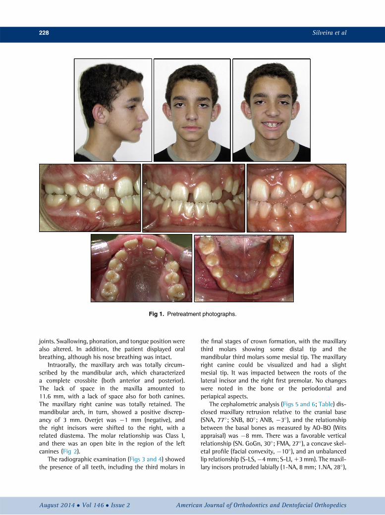
Fig 1. Pretreatment photographs.
228 Silveira et al
joints. Swallowing, phonation, and tongue position werealso altered. In addition, the patient displayed oralbreathing, although his nose breathing was intact.
Intraorally, the maxillary arch was totally circum-scribed by the mandibular arch, which characterizeda complete crossbite (both anterior and posterior).The lack of space in the maxilla amounted to11.6 mm, with a lack of space also for both canines.The maxillary right canine was totally retained. Themandibular arch, in turn, showed a positive discrep-ancy of 3 mm. Overjet was �1 mm (negative), andthe right incisors were shifted to the right, with arelated diastema. The molar relationship was Class I,and there was an open bite in the region of the leftcanines (Fig 2).
The radiographic examination (Figs 3 and 4) showedthe presence of all teeth, including the third molars in
August 2014 � Vol 146 � Issue 2 American
the final stages of crown formation, with the maxillarythird molars showing some distal tip and themandibular third molars some mesial tip. The maxillaryright canine could be visualized and had a slightmesial tip. It was impacted between the roots of thelateral incisor and the right first premolar. No changeswere noted in the bone or the periodontal andperiapical aspects.
The cephalometric analysis (Figs 5 and 6; Table) dis-closed maxillary retrusion relative to the cranial base(SNA, 77�; SNB, 80�; ANB, �3�), and the relationshipbetween the basal bones as measured by AO-BO (Witsappraisal) was �8 mm. There was a favorable verticalrelationship (SN. GoGn, 30�; FMA, 27�), a concave skel-etal profile (facial convexity, �10�), and an unbalancedlip relationship (S-LS,�4mm; S-LI,13mm). The maxil-lary incisors protruded labially (1-NA, 8 mm; 1.NA, 28�),
Journal of Orthodontics and Dentofacial Orthopedics

Fig 2. Pretreatment dental casts.
Fig 3. Pretreatment periapical radiographs.
Silveira et al 229
and the mandibular incisors had a moderate labial incli-nation (1-NB, 6 mm; 1.NB, 26�; IMPA, 94�) withoutsigns of Class III compensation. The patient was going
American Journal of Orthodontics and Dentofacial Orthoped
through a stage of active growth; therefore, the inter-maxillary sagittal relationship was likely to worsenwithout intervention.
ics August 2014 � Vol 146 � Issue 2

Fig 4. Pretreatment panoramic radiograph.
Fig 5. Pretreatment lateral cephalometric radiograph.
Fig 6. Pretreatment cephalometric tracing and measure-ments.
230 Silveira et al
TREATMENT OBJECTIVES
The treatment objectives were to (1) improve thefacial appearance by protruding the midface (maxilla)anteriorly and thus correct the unbalanced relationshipbetween the upper and lower lips; (2) correct the poste-rior and anterior crossbites; (3) improve the dental rela-tionships, especially of the molars and incisors, toachieve proper occlusion with appropriate overjet andoverbite; (4) make space for the maxillary canines; (5)achieve a mutually protected functional occlusion withstable and simultaneous occlusal contacts of all teethin central relationship and eccentric contacts guidedby the anterior teeth; (6) eliminate the signs of temporo-mandibular joint dysfunction; and (7) ensure that thepatient breathed mostly through the nose.
TREATMENT ALTERNATIVES
Three treatment options were considered: 2orthodontic-orthopedic approaches and 1 orthodontic-
August 2014 � Vol 146 � Issue 2 American
surgical approach. The latter would only be carried outif correction by the first 2 alternatives failed. RME usinga Haas type of appliance was the first planned interven-tion, regardless of the treatment to be undertaken in duecourse.14,15 It was decided that the first step would be tocorrect the maxillary atresia, so RME was the procedureof choice. Thereafter, spaces would be created for all theteeth in the maxilla, especially for the right canine.Therefore, the factors that distinguished thesetherapeutic options had to do with the approaches forsagittal intermaxillary correction.
The first option involved maxillary reverse pull andthen placing fixed appliances on both arches, since thisis the option most often reported in the literature tocorrect this kind of problem.4,5,11,16,17
The secondoption comprised theuse ofClass III elasticssupported on the last tooth of the maxillary braces and onhooks placed between the lateral incisors and canines inthemandibular arch, in addition toanterior square elastics.This would be themethod used for anteroposterior correc-tion, after the period of retention of the expansion.18
The third option would require orthognathic surgeryafter skeletal maturity if the craniofacial growth provedunfavorable or the patient was not compliant during theprevious phases of orthodontic treatment.
A team of orthodontists (students and an instructor)decided that the use of Class III elastics would providethe same mechanical components and action lines asreverse-pull forces combined with a chincup, and wouldtherefore be more desirable because it is a totally intrao-ral approach. Thus, the second option was adoptedbecause of the likelihood of achieving greater patient
Journal of Orthodontics and Dentofacial Orthopedics

Table. Cephalometric measurements
Measurement Normal Pretreatment Posttreatment Follow-upAge 12 y 9 mo 18 y 4 mo 22 y 7 moSkeletal patternSNA (�) 82 77 80 80SNB (�) 80 80 80 80ANB (�) 2 �3 0 0Facial convexity (�) 0 �10 �4 �4Y-axis (�) 59 60 62 63Facial angle (�) 87 89 90 91SN.GoGn (�) 32 30 30 31AO-BO (mm) �1 �8 �5 �6
Dental patternIMPA (�) 90 94 93 921.NA (�) 22 28 30 291-NA (mm) 4 8 7 71.NB (�) 25 26 24 231-NB (mm) 4 6 5 51.1 (�) 130 125 127 129
ProfileUpper lip, S line (mm) 0 �4 �2 �1Lower lip, S line (mm) 0 3 1 1
Silveira et al 231
compliance with intraoral elastics over the extraoralappliances suggested in the first option.
TREATMENT PROGRESS
A Haas palatal separator was installed with orthodon-tic bands on the maxillary first premolars and firstmolars.14,15,19 The screw was activated twice a day (aquarter turn each time) in the morning and afternoon,until overcorrection of the posterior crossbite wasobtained: ie, the internal slopes of the palatal cusps ofthe maxillary posterior teeth touched the internal slopesof the buccal cusps of the mandibular posterior teeth.This stage was reached after 28 days of activation, with14 mm of screw opening; subsequently, the screw wasstabilized with ligature wire and acrylic resin. Accordingto the treatment plan, the expander would be kept inplace as a retainer for 6 to 8 months. It was noted thatthe bite opened slightly, and the anteroposteriorrelationship improved.
The Haas appliance was removed after 8 months ofretention, and standard edgewise 0.022 3 0.028-infixed appliances were placed on all teeth. Orthodonticleveling of the maxillary and mandibular arches wasperformed with nickel-titanium 0.014-in and 0.018-instainless steel archwires. After creating space forthe maxillary right canine with a compressed nickel-titanium open-coil spring, a stainless steel0.019 3 0.026-in archwire with omega stops placedclose to the maxillary first molar tubes (to preserve thespace that had been obtained) was fabricated with
American Journal of Orthodontics and Dentofacial Orthoped
delta-shaped hooks placed between the lateral incisorsand the canines. This archwire was made expandableto maintain the expansion achieved with the RME. Sur-gical exposure was indicated, an orthodontic button wasbonded, and the canine was pulled with elastomericchains to its proper position in the arch.
Similarly, a mandibular stainless steel 0.0193 0.026-in archwire was installed with delta-shaped hooks placedbetween the lateral incisors and the canines, and withomega loops placed 1 mm from the molar tubes. Individ-ualized first- and third-order bends were carefully incor-porated while ensuring that the original form of themandibular arch was preserved, especially in relation tothe intercanine width. Initially, Class III elastics wereextended from the mandibular deltas to the tubes onthe second molars to obtain proper overjet. Subse-quently, anterior vertical elastics attached to the delta-shaped hooks (between the lateral incisors and themaxillary and mandibular canines) reinforced the ClassIII elastics to achieve proper overbite. The applied forceranged from 250 to 300 g. The patient was asked touse the elastics as long as possible in the 24 hours ofthe day. The same elastic was used in square fashion inthe anterior region. It was placed on the 4 deltas inboth arches.
To check that stability had been achieved despite thepossibility of residual growth, the orthodontic appliancewas retained but elastic mechanics was periodically dis-continued. This sequence was repeated twice before theocclusion was definitively stabilized and growth hadslowed.
ics August 2014 � Vol 146 � Issue 2

Fig 7. Posttreatment facial and intraoral photographs.
232 Silveira et al
The fixed appliance was removed without placementof a retainer in either arch.18 Total treatment time was67 months.
TREATMENT RESULTS
It is noteworthy that during the most active phase oforthodontic treatment, the patient's hygiene started lag-ging, requiring prophylactic and therapeutic interven-tions by the orthodontist. As can be seen in Figure 7,the occlusal surfaces of 5 teeth had to be restored.Furthermore, the presence of characteristic spots ofenamel demineralization was noted in some teeth aswell as some moderate gingival inflammation.
The posttreatment records (Figs 7-11) show that thegoals were achieved. There was significant improvementin the facial appearance, especially in the disharmonybetween the upper and lower lips, and a balanced
August 2014 � Vol 146 � Issue 2 American
smile with adequate maxillary incisor exposure wasobtained.
A proper relationship was established between the 2arches, with full correction of the maxillary atresia,occlusal contacts with ideal molar and canine occlu-sions, proper overjet and overbite, and correct and coin-cident midlines. An anterior functional guide wasobtained in the eccentric movements of the mandible;this proved efficient for the canines, and there were nomore clicks in the temporomandibular joints and nomandibular shift. The patient's breathing spontaneouslybecame predominantly nasal without the need fortreatment.
The horizontal position of the mandibular thirdmolars, whose radiographs suggested a dangerous prox-imity of their crowns to the distal roots of the secondmolars (Figs 9 and 10), prompted the extraction of all
Journal of Orthodontics and Dentofacial Orthopedics

Fig 8. Posttreatment dental casts.
Fig 9. Posttreatment periapical radiographs.
Silveira et al 233
third molars. By means of radiographic analysis andsuperimposition of the tracings (Figs 12 and 13;Table), a greater displacement of the maxilla vs themandible was visualized, which resulted in the
American Journal of Orthodontics and Dentofacial Orthoped
improvement of the relationship between the arches(ANB, 0�; angle of convexity, �4�), flaring of the maxil-lary anterior teeth (1-NA, 7 mm; 1.NA 30�), retrusionand retroclination of the mandibular incisors (1-NB,
ics August 2014 � Vol 146 � Issue 2

Fig 10. Posttreatment panoramic radiograph.
Fig 11. Posttreatment lateral cephalometric radiograph.
Fig 12. Posttreatment cephalometric tracing and mea-surements.
234 Silveira et al
5 mm; 1.NB, 24�), and maintenance of the mandibularplane angle (SN-GoGn, 30�; FMA, 27�).
Figures 14 and 15 show the patient at 4 yearsposttreatment. As can be observed, the resultsremained stable in maintaining both facial balance andintermaxillary dental relationships. From an intra-archperspective, dental alignment and dental arch formalso remained stable. Radiographic analysis showed sta-bility in the maxillomandibular relationship, the positionof the mandibular anterior teeth, and the mandibularplane angle (Table).
DISCUSSION
According to the clinical examination and the facialand cephalometric analyses, the cause of this patient'smalocclusion was an underdeveloped maxilla, bothtransversely and sagittally. The mandible was well posi-tioned in the anteroposterior and vertical directions.
August 2014 � Vol 146 � Issue 2 American
Since there was no evidence of mandibular prognathismin the examinations and family history reports, theprognosis was favorable. It was obvious that correctionwould require orthopedic intervention in the maxillabecause the patient was in an active growth phase.14,15,19
The classic therapy for maxillary retrusion in patientsduring growth—a palate-splitting appliance followed bya facemask or chincup—was not used for this patient. Thiswas decided in light of the patient's compliance and thepossibility that mesial dental movement might occurinstead of skeletal effects, which might result in furtherreduction of an already limited space for themaxillary ca-nines. The fact that anteroposterior elastic force wasapplied after insertion of the canines in the arch provedadvantageous, since when reverse pull is used, the ante-roposterior force is applied immediately after RME.
Another argument that also influenced this decisionwas the risk of an increase in lower anterior facial heightthat could result from clockwise rotation of themandible;this tendency had been previously associated with maxil-lary reverse pull that, in this case, would invariably impartsome instability.16,17,20,21 Ferro et al9 reported that thestability of Class III corrections is directly related to themaintenance of the vertical position of the mandible,corroborating what some other authors also advo-cated.7,12,13 As described by Bj€ork and Skieller,22 anteriormandibular rotation is a natural tendency in late adoles-cent growth, and one must therefore work for and notagainst facial growth to ensure stable results.9 Otherwise,when correction is obtained by further rotation of themandible, the physiologic pattern of forward rotationtends to manifest itself, and relapse occurs.9
Journal of Orthodontics and Dentofacial Orthopedics

Fig 13. Cephalometric superimpositions: pretreatment vs posttreatment.
Silveira et al 235
Also favorable in this case was the result achievedafter anteroposterior palatal expansion. The maxillaryand mandibular incisors were practically in an edge-to-edge relationship. The anterior displacement of themaxillary complex caused by RME is well documentedin the literature,19,23,24 despite conflicting resultsinvolving its long-term stability.25,26
It is noteworthy that by creating space for the maxil-lary right canine with the aid of the fixed appliance inconjunction with an open spring, some buccal flaringof themaxillary incisor crownswas expected. This adverseeffect was partly offset by the use of third-order bendsplaced in the orthodontic archwires to prevent the crownsfrom inclining even farther. The bendswere alsomeant toproduce some labial bodily movement.
Moreover, the correction of the anteroposterior prob-lem with the use of Class III elastics was likely to causeclockwise rotation of the mandible by incorporating anextrusive component in the molars.27 One strategy tolimit this side effect is the simultaneous use of a chin-cup—preferably vertical14 or combined with verticalelastics.27
In comparing the pretreatment and posttreatmentrecords, the changes observed in the SNA (increase of 3�)and ANB (increase of 3�) angles, with the SNB angle unal-tered (80�), suggest maxillary advancement and mainte-nance of the mandibular position. On the other hand, itwould bemore reasonable to admit that in this case a den-toalveolar sagittal correction was achieved using inter-maxillary Class III elastic mechanics, which caused themaxillary teeth to protrude and the mandibular teeth to
American Journal of Orthodontics and Dentofacial Orthoped
retract, because of the changes in the values of 1-NA,1.NA, 1-NB, and 1.NB mentioned above.
One major concern regarded the end of the growthperiod and the fear that the patient's condition mightrelapse. Elastic use was periodically discontinued andresumed until we felt reassured that little or no growthremained. This explains the long time of active treatment(67 months).
The decision not to use any retainer was based on thepremise that correction of anterior or posterior crossbitesdoes not require retention, because the occlusion itselfplays the role of a retainer if proper tooth contacts areestablished.18,28 Although this patient did not exhibitany significant signs of prognathism, when someresidual mandibular growth does occur, often afterage 18, the absence of a retainer would allow aspontaneous lingual offset of the mandibular incisorsand a buccal offset of the maxillary incisors, withoutcompromising the relationship between them. A properoverjet and overbite, in addition to the absence ofclockwise mandibular rotation after treatment,certainly contributed to the patient's stability 5 yearsafter removal of the fixed appliance without the use ofa retainer.
CONCLUSIONS
The favorable results achieved in this case reportshow that it is possible to correct transverse and sagittalmaxillary deficiencies during the active growth phase bymeans of RME followed by Class III and anterior verticalelastics supported on hooks placed on the fixed
ics August 2014 � Vol 146 � Issue 2

Fig 14. Facial and intraoral photographs at the 4-year follow-up.
236 Silveira et al
appliance. The stability that was observed 4 years afterthe end of treatment without retention suggests thatevaluating whether some remaining growth should beexpected, and ensuring appropriate relationships andocclusal contacts, proper overjet and overbite, andoptimal function without increasing the mandibularplane angle during treatment, all combine in maintain-ing the long-term results.
ACKNOWLEDGMENTS
We thank the following residents of the orthodonticsprogram of Fluminense Federal University who contrib-uted to the treatment reported in this article: Ana LuizaLuz Fernandes da Silva, Eduardo Kant Colunga Rothier,Larissa Bustamante Capucho Brand~ao, and LucianaHosken Souto.
August 2014 � Vol 146 � Issue 2 American
REFERENCES
1. Massler M, Frankel JM. Prevalence of malocclusion in childrenaged 14 to 18 years. Am J Orthod 1951;37:751-68.
2. Grando G, Young AA, Vedovello Filho M, Vedovello SA, Ramirez-Ya~nez GO. Prevalence of malocclusions in a young Brazilianpopulation. Int J Orthod Milwaukee 2008;19:13-6.
3. Deguchi T, McNamara JA Jr. Craniofacial adaptations inducedchincup therapy in Class III patients. Am J Orthod DentofacialOrthop 1999;115:175-82.
4. Macdonald KE, Kapust AJ, Turley PK. Cephalometric changes afterthe correction of Class III maloccclusion with maxillary expansion/facemask therapy. Am J Orthod Dentofacial Orthop 1999;116:13-24.
5. Da Silva Filho OG, Magro AC, Capelozza Filho L. Early treatmentof Class III malocclusion with rapid maxillary expansion andmaxillary protraction. Am J Orthod Dentofacial Orthop 1998;113:196-203.
6. Garattini G, Levrini L, Crozzoli P, Levrini A. Skeletal and dentalmodifications produced by the bionator III appliance. Am J OrthodDentofacial Orthop 1998;114:40-4.
Journal of Orthodontics and Dentofacial Orthopedics

Fig 15. Dental casts at the 4-year follow-up.
Silveira et al 237
7. Loh MK, Kerr WJ. The function regulator III: effects and indica-tions for use. Br J Orthod 1985;12:153-7.
8. Mora DR, Oberti G, Ealo M, Baccetti T. Camouflage of moder-ate Class III malocclusions with extraction of lower second mo-lars and mandibular cervical headgear. Prog Orthod 2007;8:300-7.
9. Ferro A, Nucci LP, Ferro F, Gallo C. Long-term stability of skeletalClass III patients treated with splints, Class III elastics and chincup.Am J Orthod Dentofacial Orthop 2003;123:423-34.
10. He S, Gao J, Wamalwa P, Wang Y, Zou S, Chen S. Camouflagetreatment of skeletal Class III malocclusion with multiloop edge-wise arch wire and modified Class III elastics by maxillary mini-implant anchorage. Angle Orthod 2013;83:630-40.
11. Macey-Dare LV. The early management of Class III malocclusionsusing protraction headgear. Dent Update 2000;27:508-13.
12. Battagel JM, Orton HS. Class III malocclusion: the post-retentionfindings following a non-extraction approach. Eur J Orthod1993;15:45-55.
13. Stensland A, Wisth PJ, Boe OE. Dentofacial changes in childrenwith negative overjet treated by a combined orthodontic andorthopaedic approach. Eur J Orthod 1988;10:39-51.
14. Haas AJ. Long-term posttreatment evaluation of rapid palatalexpansion. Angle Orthod 1980;50:189-217.
15. Haas AJ. Palatal expansion; just the beginning of dentofacialorthopedics. Am J Orthod 1970;57:219-55.
16. Cevidanes L, Baccetti T, Franchi L, McNamara JA Jr, De Clerck H.Comparison of two protocols for maxillary protraction: boneanchors versus face mask with rapid maxillary expansion. AngleOrthod 2010;80:799-806.
American Journal of Orthodontics and Dentofacial Orthoped
17. Ara�ujoEA, Ara�ujoCV.Non-surgical approaches to Class IIImalocclu-sions treatment. Rev Dent Press Ortodon Ortop 2008;13:129-57.
18. Riedel RA. The review of the retention problem. Angle Orthod1960;30:179-99.
19. Haas AJ. Rapid expansion of themaxillary dental arch and nasal cav-ity by opening the midpalatal suture. Angle Orthod 1961;31:73-9.
20. Kama JD, Ozer T, Baran S. Orthodontic and orthopaedic changesassociated with treatment in subjects with Class III malocclusions.Eur J Orthod 2006;28:496-502.
21. Figueiredo MA, Siqueira DF, Bommarito S, Scanavini MA. Ortho-dontic compensation in skeletal Class III malocclusion: a casereport. World J Orthod 2007;8:385-96.
22. Bj€ork A, Skieller V. Facial development and tooth eruption. Animplant study at the age of puberty. Am J Orthod 1972;62:339-83.
23. Chung CH, Font B. Skeletal and dental changes in the sagittal,vertical, and transverse dimensions after rapid palatal expansion.Am J Orthod Dentofacial Orthop 2004;126:569-75.
24. Ramoglu SI, Sari Z. Maxillary expansion in the mixed dentition:rapid or semi-rapid? Eur J Orthod 2010;32:11-8.
25. Chang JY, Mc Namara JA Jr, Herberger TA. A longitudinal study ofskeletal side effects induced by rapid maxillary expansion. Am JOrthod Dentofacial Orthop 1997;112:330-7.
26. Reed N, Ghosh J, Nanda RS. Comparison of treatment outcomeswith banded and bonded rapid palatal expansion appliances. AmJ Orthod Dentofacial Orthop 1999;116:31-40.
27. Tweed CH. Clinical orthodontics, Volume 2. St Louis: C. V. Mosby;1966. p. 946.
28. Kaplan H. The logic of modern retention procedures. Am J OrthodDentofacial Orthop 1988;93:325-40.
ics August 2014 � Vol 146 � Issue 2