Compendium of urinalysis Urine test strips and … · Compendium of urinalysis Urine test strips...
90
Compendium of urinalysis Urine test strips and microscopy COBAS, COBAS U, LIFE NEEDS ANSWERS,, ACCU-CHEK, CHEMSTRIP, COMBUR-TEST, COMBUR 10 TEST, MICRAL-TEST, URISYS, URISYS 1100 and URISYS 2400 are trademarks of Roche. All other trademarks are the property of their respective owners. ©2010 Roche Roche Diagnostics Ltd. CH-6343 Rotkreuz Switzerland www.roche.com xxxxxxxxxxx a 0810 - x.x XX
Transcript of Compendium of urinalysis Urine test strips and … · Compendium of urinalysis Urine test strips...

Compendium of urinalysisUrine test strips and microscopy
COBAS, COBAS U, LIFE NEEDS ANSWERS,, ACCU-CHEK, CHEMSTRIP, COMBUR-TEST, COMBUR 10 TEST, MICRAL-TEST, URISYS, URISYS 1100 and URISYS 2400 are trademarks of Roche.All other trademarks are the property of their respective owners.
©2010 Roche
Roche Diagnostics Ltd.CH-6343 RotkreuzSwitzerlandwww.roche.com
xxxx
xxxx
xxx
a 0
810
- x.
x X
X

1
Main disease indicationsUrinary Tract Infection
Interesting factsAre you aware of that …
More than 500 million people – 10% •of the world’s population – have some form of kidney damage 1
Urinary tract infections are the sec-•ond most common type of infection in the human body 2
One in 20 deaths is caused by diabetes; •8,700 deaths every day; six every min-ute 3
By 2030, almost 23.6 million people will •die from cardiovascular disease, mainly heart disease and stroke 4

22 3
Content
1 Main disease indication Urinary tract infection 8Kidney disease 10Diabetes 14
2 From urine fortune telling to real time diagnosis History of urinalysis 18 Application areas for urine test strips 20Pre-analytical treatment and test procedure 22
3 Characteristics of urine test strips from Roche Composition and benefit of the test strip 28Parameters of urine test strips 32Detection of microalbuminuria with micral-test 56
4 Drug interferences in urine Influencing factors 60
5 Automated urinalysis Urine test strip systems 64
6 Urine microscopy in differential diagnosis Microscope 70
7 Urine particles and formed elements Blood cells 74 White blood cells 74 Red blood cells 76Epithelial cells 78 Squamous epithelial cells 78 Renal tubular cells 79 Transitional epithelial cells 80 Atypical cells 81Casts 82 Hyaline casts 82 Granular casts 84 Pigmented casts 85 Waxy casts 86 Red blood cell casts 87 White blood cell casts 88 Epithelial cell casts 88 Fatty casts 89 Cylindroids 90 Rare casts 90 Pseudo casts 91

4 54
Pathogens 92 Trichomonads 92 Fungi 93 Bacteria 94Crystals 96 Calcium oxalate 96 Uric acid 97 Amorphous urate 98 Amorphous phosphate 98 Calcium phosphate 99 Calcium carbonate 99 Magnesium ammonium phosphate 99Rare crystal 100 Dicalcium phosphate 100 Dimagnesium phosphate 100 Tyrosine 101 Leucine 101 Hippuric acid 101 Cystine 102 Ammonium biurate 102Drug crystals 104Other constituents 106 Fat globules and cholesterol crystals 106 Spermatozoa 106 Contamination 107 Vaginal secretion 107 Skin 107 Feces 107 Clothing 107 Sample vessels 108 Air 108 Latex gloves 108 Glass splinters 108
8 Diagnostic Renal disease 112 Glomerulonephritis 112 Acute pyelonephritis 112 Endocarditis 112 Nephrotic syndrome 113 Acute tubular necrosis 113Urogenital disease 114 Bacterial cystitis 114 Vaginitis/urethritis/balanitis 114
9 Appendix References 118Storage, influence and interference factors 122Color change in urine 124Drug interference factors 126Microscopic urine sediment figures 130

66 7
Main disease indications
Urine is a key health barometer for many
diseases, mainly UTIs, kidney disease and
diabetes. The analysis of urine can reveal se-
rious diseases, which do not show symptoms
in their early but treatable stages and causes
severe damage if they remain undetected.
1

88
Urinary tract infection (UTI)
The urinary apparatus is made up of:•Twokidneys•Twoureters•Theurinarybladder•Theurethra
Each day, the human body filters 170 liters of primary urine through the kidneys. Primary urine consists of water, salt and dissolved low-molecular blood constituents.The water and all substances needed by the organism are largely reabsorbed. The re-maining “worthless” substance is known as urine. The amount of urine finally excreted through the urethra each day is approxi-mately 1.5 liters.
What is a UTI?Most UTIs are caused by bacteria entering the urinary tract (urethra) and multiplying rapidly. The origin of bacteria, especially E. coli, is usually the intestines or anus.
•UTIcanmeananythingfromamildinflam-mation of the bladder to severe kidney in-fections, and should be treated as soon as possible
•Bacteria can spread and infect the bladderand kidneys. The only way to stop this is early detection and the prescription of antibiotics
Urinary tract infection (UTI)
Fig. 1: The urinary apparatus
Two kidneys
Two ureters
The urinary bladder
The urethra
9
Main disease indicationsUrinary tract infection
Symptoms and causes of UTI Symptoms can vary, and sometimes there may be none at all. However, the most common are as follows:•Aburningsensationwhenurinating•Afrequenturgetourinate,butonlyinsmall
amounts•The urine may be discolored or cloudy,
strong or foul-smelling•Backorabdominalpain•Generalfeelingsofillnessorfever
Causes of UTI include:•Sexualintercourse•Poorhygiene•Genetic makeup e.g. the proximity of the
anus to the urethra
Risk groups•WomenaremuchmorelikelytogetaUTI•Peoplewithdiabeteshaveahigherriskof
developing a UTI due to changes in the im-mune system
•Anyotherdisorderthatsuppressestheim-mune system raises the risk of a UTI
Did you know?Research by the National Institutes of Health
suggests that recurrent UTIs may be caused
by the ability of bacteria to attach to cells
lining the urinary tract.
A growth of E. coli bacteria in the vagina
can also result from spermicidal foam on
condoms.

10
The kidneys are the most important excre-tion organ in the human organism. Every 24 hours around 1,500 liters of blood flowthrough the kidneys.
If a UTI is not treated promptly, bacteria may travel further up the ureters to multiply and infect the kidneys.
Function of the kidneys:•Filter blood to excrete water, electrolytes
and metabolic end products (e.g. urea)•Help to control blood pressure and blood
pH•Produce the important enzyme renin and
glycoprotein-hormone EPO•Helpmaintainhealthybones•Excretebloodconstituents(e.g.glucose)•Maintainwaterandelectrolytebalance•Regulatewatercontentofintracellularand
extracellularfluid
What does kidney disease imply?Kidney diseases are disorders that affect the kidneys. In many cases the tiny filtering units (nephrons) are attacked and their abi-lity to filter properly is limited. Gradual loss of kidney function is called chronic kidney disease (CKD), and people with CKD may develop permanent kidney failure as it pro-gresses. This may have fatal consequences unless a dialysis machine is used or a kid-ney transplant is performed. CKD occurs gradually and is often silent, going unde-tected for years. People with CKD may also have a high risk of suffering from a stroke or heart attack.
11
Main disease indicationsKidney disease
Nephrons – the trigger for CKDEach kidney has between one and three mil-lion nephrons. Each nephron can be sub-divided further into glomerular and tubular sections.
In the nephron, a glomerulus – a tiny blood vessel – intertwines with a tubule which collects urine. The glomerulus acts as a fil-tering unit and determines which substan-ces are excreted as waste and which are reabsorbed into the blood to nourish the body’s cells, such as water, essential nut-rients, salts and minerals. Afterwards, the tubules collect and concentrate waste and excrete it into the urine.
Damaged nephrons can lead to CKD and leave kidneys unable to remove wastes. The damage can occur slowly and over many years. As blood filtering becomes impaired, urine production decreases and water and waste products accumulate in the blood.
Also, substances such as protein may leak into the urine rather than remaining within the blood.
CKD can lead to kidney failure and trans-plantation without early detection and mo-nitoring.
Stages of CKDCKD develops gradually over time and can be divided into five stages of increasing se-verity:
Stage 1: Slight kidney damage with normal or decreased filtration
Stage 2: Mild decrease in kidney functionStage 3: Moderate decrease in kidney functionStage 4: Severe decrease in kidney functionStage 5: Kidney failure requiring dialysis or
transplantation to stay alive
10
Kidney disease
Fig. 2: Schematic illustration of a nephron
Fig. 3: Glomerular filtration rate in correlation with CKD stages 5, 6
Clean bloodBlood with
wastes
Nephron
Tubule
Glomerulus
Wastes (urine)to the bladder
0
Replacement Preparationfor replacement
Evaluating, treating complications
Estimating, slowing progression
Diagnosis, treatment of comorbid conditions
Stage 5GFR <15mL/min
Stage 4GFR 15-29mL/min
Stage 3GFR 30-59mL/min
Stage 2GFR 60-89mL/min
Stage 1GFR ≥90mL/min
177354530707884
24681012
10
Not sensitive to serum creatinine
Standard value
Persistent albuminuriaSensitive to serum creatinine
20 30 40 50 60 70 80 90Glomerular filtration rate [mL/min]
100 110 120 130
Ser
um
cre
atin
ine
mg
/dL
µm
ol/
L

1212
Symptoms of CKDUnfortunately, symptoms often occur very late or not at all. Abnormal urine or blood tests are often one of the earliest signs.
Common symptoms are•Problemsurinating:burningfeelingorab-
normal discharge during urination, or a change in the frequency of urination, espe-cially at night
•Urinethatisfoamy,bloody,red,brown•Mid-backpain,belowtheribs•Highbloodpressure(hypertension)•Lostofappetiteornauseaandvomiting•Swellinginhands,feetoreyes•Musclecramps
Risk groups•Peoplewithdiabetes,hypertensionorobe-
sity, and older age groups
Did you know?Kidney disease may also occur after a bacte-
rial infection in another part of the body, such
as a streptococcus infection of the throat or
skin or an infection inside the heart.7
Viruses, such as the HIV virus that leads to
AIDS, can also trigger kidney disease. 1 Kid-
ney disease requires prompt attention, and
high-risk populations should be carefully
monitored for abnormal kidney function.
Otherwise, testing for CKD only begins when
symptoms are present, which may be too late.
Diagnostic tests, such as simple urine tests,
are the first line of defense in detecting kid-
ney problems and minimizing damage.
13
Main disease indicationsKidney disease

1414
Diabetes, even when controlled, is the most common cause of CKD and kidney failure.
What is diabetes?Diabetes occurs when the body fails to pro-duce or properly utilize insulin, which is ne-cessary for the stabilization of blood gluco-se levels. High blood sugar causes damage to the kidneys and other organs.
There are three types of diabetes:•Type I (insulin-dependent or juvenile-on-
set): The body produces little or no insulin. It is usually genetic and is characterized by high levels of blood glucose
•Type II (insulin-independent or adult-on-set): The body does not produce enough insulin, or does not use it properly (insulin resistance). It is often associated with obe-sity
•Type III: Gestational diabetes is hypergly-cemia with onset or first recognition during pregnancy
Common consequencesOver time, diabetes can damage the heart, blood vessels, nerves and kidneys.•Thediseaseincreasestheriskofheartdis-
ease and stroke•Diabeticneuropathy(damagetothenerves)
affects up to 50%8 of people with diabetes•Diabetes is among the leading causes of
kidney failure. 10-20%8 of people with dia-betes die of kidney failure
High blood pressure and high blood glucose increase the risk that a person with diabetes will progress to kidney failure. They should therefore be screened regularly for abnor-mal urine protein in order to prevent the progression of CKD.
Did you know?Healthy diet, regular physical activity, main-
taining normal body weight and avoiding
tobacco use can prevent or delay the onset
of diabetes.
Diabetes
15
Main disease indicationsDiabetes

1616 17
From urine fortune telling toreal-time diagnosis
It all started over 2,000 years ago. Many cul-
tures once regarded urine as a mystical fluid,
and some still do. Its uses have included
wound healing, stimulation of the body’s de-
fenses, and disease diagnosis.
2

1818
The ancient worldThe origin of visual urine diagnostics, canbe traced back to ancient Egypt. Hippocra-tes (approx. 400 BC) recognized that urine characteristics (odor / color) were altered with different diseases. He hypothesized that urine was a filtrate of the four humors (blood, phlegm, yellow bile and black bile), which came from the blood and was filte-red through the kidneys and pointed out the importance of examining the patient’s urine. Six centuries later, Galen (AD 129–200) refined Hippocrates ideas, theorizing that urine represented is not a filtrate of the four humors and overall condition, but rather, a filtrate of the blood. This doctrine dominated medical thinking up to the 16th century.
The middle ages (AD 500–1500)The technique of collecting urine was thought to be important for accurate inter-pretation. Ismail Gorgani, an 11th century physician, recommended collecting the full amount over 24 hours in a clean vessel and keeping it out of the sun or heat, which could alter color. The teachings of Gilles de Corbeil (1165–1213) related 20 different types of urine to conditions of the body, he noted differences in sediment and co-lor (from crystal clear via camel hair white, blackberry red and pale green to black).
The Renaissance (1450–1600)By the 15th century urinary diagnosis had transformed the patient–doctor dynamic. An increasing number of physicians were diagnosing from urine alone. Amateurs (called ‚leches‘) started diagnosing based only on the color of urine. By the 17th century, the uses of uroscopy / uromancy had spiraled far beyond the edge of reason. Physicians and leches started telling fortunes and predicting the future with urine.
The history of urinalysis
19
From urine fortune telling to real-time diagnosisThe history of urinalysis
TodayTowards the end of the 18th century doctors became more interested in chemistry and turned their attention to a scientific basis of urinalysis. The first “test strips” were developed by the Parisian chemist Jules Maumené (1818–1898) when, in 1850, he impregnated a strip of merino wool with “tin protochloride” (stannous chloride). On application of a drop of urine and heating over a candle the strip immediately turned black if the urine contained sugar and it took another 70 years before the Vienne-se chemist Fritz Feigl (1891–1971) publis-hed his technique of “spot analysis.” Urine test strips in the sense used today were first made on industrial scale and offered commercially in the 1950s. The company Boehringer Mannheim, today a top leader on the world market under the name of Ro-che, launched its first Combur-Test® strips (Fig. 4) in 1964.
New impregnation techniques, more stab-le color indicators and the steady impro-vement in color gradation have all con-tributed to the fact that the use of urine test strips has now become established in clinical and general practice as a reliable diagnostic instrument.
Fig. 4: Roche Combur-Test®

2020
Applications of urine test strips
Urine test strips are a central diagnostic instrument, their ease of use yielding quick and reliable information on pathological changes in the urine. Their significance lies primarily in first-line diagnostics. Routine testing with multi-parameter strips allows a determination of the complete urine sta-tus. This is the first step in the diagnosis of a very wide range of diseases.
Indications for urine test strips:•Screeningforprevention•Treatmentmonitoring•Patientself-testing
Screening for preventionUrine test strips are routinely used for screening both in hospitals and in gene-ral practice. The aim of screening is early identification of likely patients by exami-ning large groups of the population.
No direct diagnoses are established on the basis of the screening results, which serve only as a basis for further microscopic, bacteriological, or clinicochemical exami-nations of the urine.
Routine examinations can reveal early symptoms of the following four disease groups:•Kidneydiseases•Urinarytractinfections•Carbohydratemetabolismdisorders(such
as diabetes mellitus)•Liverdiseaseandhemolyticdisorders•Cardiovasculardisease
Did you know?A field study carried out in seven European
countries with over 11,000 urine samples
illustrates the value of screening with uri-
ne test strips. A pathological urine finding
(after checking for nitrite, protein, glucose,
ketones, urobilinogen, and blood) was dia-
gnosed in 16% of “normal healthy persons”,
in 40% of outpatients, and in 57% of hospi-
talized patients.
Treatment monitoringTreatment monitoring with the aid of uri-ne test strips allows the treating doctor to check on the results of the prescribed therapy, and if necessary to introduce any changes into the therapeutic strategy. An additional benefit of such monitoring is im-proved patient compliance.
21
From urine fortune telling to real-time diagnosisApplications of urine test strips
Monitoring is particularly useful in two clinical conditions: First, in diabetes mellitus, where combined checks for glucose and ketones are ad-visable for early detection and correction of changes in metabolic status. Second, in patients suffering from hypertension who run an increased risk of developing kidney damage in the course of their condition.
Patient self-testingSpontaneous preventive self-testing at home has become widespread. Patients can also benefit by using test strips under their doctor’s instructions.
This applies especially to high risk groups who might develop e.g. diabetes, CKD or UTIs: •Overweight people, and those with un-
healthy lifestyles•People with diabetes, hypertension or a
family history of kidney disease•People who have already experienced a
UTI
Did you know?Nearly 20% of women who have a UTI will
have another, and 30% of those will have yet
another. Of the last group, 80% will have re-
currences.9

2222
Reliable analytical results can only be ob-tained from a urine specimen that has been collected, transported and stored properly. The first step for a correct test performance is therefore to obtain a proper sample.
Sample collectionSamples should always be collected in clean disposable plastic cups. Key patient data (full name, date of birth, sender, collection date and time) should be affixed to the con-tainer using waterproof materials before the sample is collected.
Depending on the time and nature of the sample, a distinction is drawn between:•Spontaneousurine•Firstmorningurine•Second morning urine (collected before
noon) •Timedurine(usually24-hoururine)•Midstreamurine•Bladderpunctureurine
First morning urine has proved its worth for most test purposes. It has usually been in the bladder for a reasonably long period, and its composition is independent of daily variationsinfoodandfluidintakeandphy-sical activity. For glucosuria tests, it is best to use urine passed about two hours after a carbohydrate-rich meal. Contamination is frequent in normal “spontaneous” urine collected without any special hygienic pre-cautions, especially in the case of women, and consists of leukocytes in the presence of discharge and of erythrocytes in the pre-sence of menstruation. For this reason, no urine diagnostics should be attempted in women for two to three days after menst-ruation.
Pre-analytical treatment and test procedure
Fig. 5: Collecting a midstream urine sample
Wash hands Take lid off specimen container and place with inside surface facing upwards
First void a small amount of urine into the toilet, then fill the specimen container half full, void remaining urine into the toilet
Replace the lid on the specimen container being careful not to touch the inside; give the container to the nurse or laboratory
23
From urine fortune telling to real-time diagnosisPre-analytical treatment and test procedure
Urinalysis can reveal diseases that have gone unnoticed because they do not neces-sarily produce signs or symptoms. A simp-le urine test can allow early detection and treatment of disease. The most efficient test strips result can be obtained after correct collection and storage of the urine sample.
Sample storageTest strips should be examined within two hours of urination, since longer standing times can lead to false results owing to the followinginfluences:•Disintegration (lysis) of leukocytes and
erythrocytes•Proliferationofbacteria•Bacterialdegradationofglucose•Arise inpHduetoammoniaformedasa
result of bacterial degradation of urea•Oxidationofbilirubinandurobilinogen,es-
pecially in sunlight
These changes can be slowed if the urine is kept in a sealed container in a refrigerator.
There are three ways to perform a test strip analysis:•Manual–Thetestisdonebyhand•Semi-automated–Theteststrip isdipped
in the urine sample manually and then ana-lyzed by an instrument
•Fully-automated–Theteststripisanalyzedcompletely by an instrument
In the following, the focus lies on visual testing done manually. However, chapter 5 also discusses semi- and fully-automated analysis.
A general overview of storage requirements and factors influencing test resultscanbefound in the appendix (Tab. 1).
Use of test stripsThe test should be performed as follows:1. Collect the specimen in a clean container
and ideally decant it into a test tube to en-sure even mixing
2. Dip the test strip in the urine for no longer than one second (all test pads should be fully covered in urine)
3. On drawing the strip out of the sample, run its edge over the rim of the container to remove excess liquid
4. After 60 seconds (60–120 seconds for leukocytes) compare the reaction color in the test area against the color scale on the label
5. Record the results

2424
Macroscopic assessment of the urine is of little diagnostic value, but any striking color changes on visual examination are usually reported. The normal urine volume of an adult is 700–2,000 mL/day. An output of more than 2,500 mL/day is classified as po-lyuria, less than 500 mL/day as oliguria, and less than 100 mL/day as anuria.
ColorThe color of normal urine is due to the presence of porphyrins, bilirubin, urobilin, uroerythrin, and other, still unidentified, compounds. Striking changes should be reported in terms of definite colors: “red”, “brown”, “green”, etc. Color changes are most often caused by drugs and their me-tabolites. A brick-red sediment is usually due to precipitation of urates in acidic urine (test: the precipitate redissolves on gentle warming). Hematuria is recognized by the presence of brown-red turbidity with a red-brown sediment.
White turbidity can be due to:•Phosphates precipitating in alkaline urine
(test: the precipitate redissolves on acidifi-cation with acetic acid)
•Pyuriainmassivebacterialorfungalinfec-tions (microbial count >107/mL)
•Lipiduriainthepresenceofanephriticsyn-drome or on contamination with ointments
•Massiveproteinuria
OdorStriking odor changes of clinical signifi-cance include:•Freshfruitoracetoneinthepresenceofke-
tonuria (sign of possible presence of meta-bolic acidosis, most often due to fasting or uncontrolled diabetes mellitus)
•“Fetorhepaticus”,amustyodorofurineandbreath in the presence of hepatic enceph-alopathies
•Alcoholinthepresenceofintoxication•Ammoniainurinarytractinfectionsdueto
urea-splitting bacteria; Hydrogen sulfide in urinary tract infections with proteinuria due to putrefacient bacteria
A general overview of color changes in theurine can be found in the appendix (Tab. 2).
25
From urine fortune telling to real-time diagnosisPre-analytical treatment and test procedure
Do’s✔ Examine the test strip within two hours✔ Mix the specimen thoroughly prior to the
test✔ If the tests cannot be done within two
hours of urine collection, keep the speci-men in a refrigerator at +4°C
✔ At the time of testing, ensure the samples are at room temperature
✔ Close the test strip tube immediately after removing a test strip
✔ Remember to label the urine container
Don’ts✘ Allow contamination by residues of cleaning
agents or disinfectants, as these can give false-positive findings for blood, protein and glucose
✘ Freeze the urine specimen, as this will destroy leukocytes and erythrocytes and hence make it unusable for subsequent microscopic examinations
✘ Centrifuge the specimen prior to test strip analysis
✘ Expose the specimen to direct sunlight
2626 27
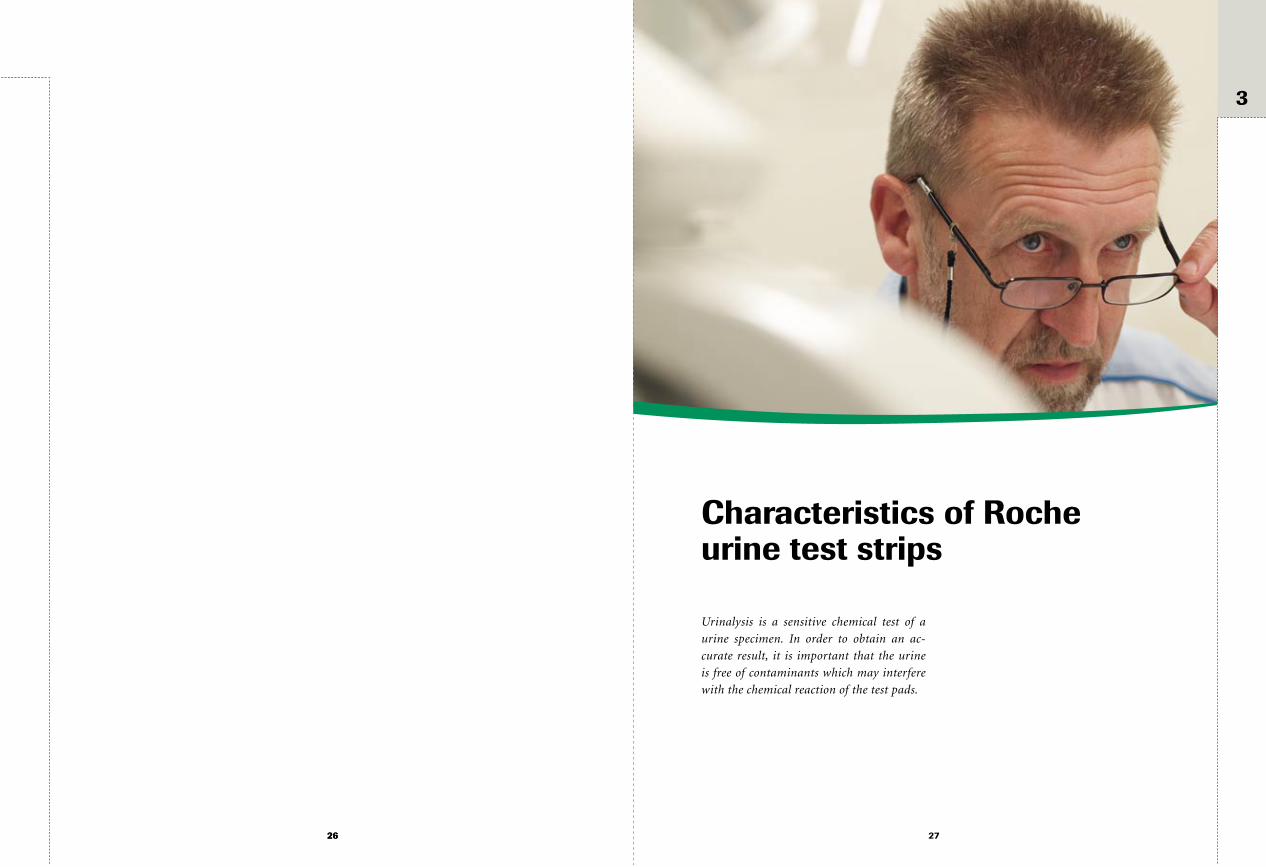
Characteristics of Roche urine test strips
Urinalysis is a sensitive chemical test of a
urine specimen. In order to obtain an ac-
curate result, it is important that the urine
is free of contaminants which may interfere
with the chemical reaction of the test pads.
3

2828
Contaminants in the urine may lead to false-positive or false-negative results. Results can also be influenced by many externaland internal factors, which may then lead to a missed or false diagnosis.
External factors may include contaminati-on by preservatives or cleaning substances which enter the urine during or after sample collection.
The main internal factor which may interfere with the result is the presence of ascorbic acid.
Nylon mesh technologyTo prevent external and internal interfe-rence, Roche has developed an unique test strip technology using a nylon mesh layer on each strip. This net sealing technology compromises a reagent and underlying ab-sorbent paper which are fixed to a plastic carrier foil by a thin nylon mesh. The strip therefore comprises several layers hold to-gether without glue, which might interfere with the result.
General problems:•Contaminationofthereagentpadbyexter-
nalinfluencesandascorbicacid•Interferences from water-repellent glue
components and possible falsification of the color by glue
Solution:•Iodated components protect blood and
glucose detection even with a high level of ascorbic acid (up to 750 mg/L)
•Reagent paper and underlying absorbentpaper are fixed to the plastic carrier foil by a thin nylon mesh without the interventions of a glue component
•Plastic carrier foil provides extra strengthand absorbent paper layers prevent splash-ing of urine
Long storage lifeA drying agent in the cap of the plastic tube protects the sensitive test strips from atmos-pheric humidity. The test strips are stable up to the expiry date specified on the package when stored and used in accordance with the directions.
Composition and benefits of test strips
Fig. 6: Structure of Roche test strip
Nylon mesh
Reagent paper
Absorber paper
Carrier foil
29
Characteristics of Roche urine test stripsComposition and benefits of test strips
Ascorbic acidVitamin C (ascorbic acid) inhibits the oxida-tion reactions for blood and glucose in the test area and can therefore lead to false-ne-gative results in the presence of hematuria and glucosuria.
Risks of ascorbic acid interferenceAn examination of over 4,000 routine urina-lysis specimens provided positive results for ascorbic acid in 22.8%. The average ascorbic acid concentration is 370 mg/L, with a range of 70-3,400 mg/L. It was shown that an in-take of 250 mg/day could produce a mean ascorbic acid level of 310 mg/L.10 The risk of false-negative results increases particu-larly sharply in theflu season,when somepeople consume large quantities of vitamin supplements, affecting the diagnosis of the following clinical pictures:
Blood: Glomerulonephritis, pyelonephritis, lithiasis, tumors
Glucose: Diabetes mellitus, glucosuria caused by kidney damage
General problems:•VitaminCisaddedtomanyfoodsandbever-
ages due to its antioxidant and preservative properties(e.g.flour,cakes,vegetables,fruit)
•Inaddition,manypeopletakeprophylacti-cally pure vitamin C in the form of vitamin tablets.
Fig. 7: Oranges: an important source of vitamin C

3030
Solution:•Test strip protection by an iodated nylon
mesh, so that ascorbic acid is eliminated by oxidation
Some urine test strips which are not pro-tected against vitamin C offer an additional test pad for ascorbic acid instead. However, it is only possible to reveal an excessive vi-tamin C concentration in the patient’s urine this way. The examination should therefore be repeated at a later time to avoid potenti-al false-negatives in the case of blood and glucose. Roche Combur-Test® strips remain stable even in the presence of high concen-trations of vitamin C, and false-negative re-actions to blood and glucose are hardly ever observed.
Did you know?A study by Brigden et al. showed that an oral
dose of as little as 100 mg of vitamin C per
day, or even a single glass of fruit juice, may
produce ascorbic acid concentrations of 10
mg/dL in the urine. With conventional urine
test strips, these concentrations may be high
enough to cause interference.10
31
Characteristics of Roche urine test stripsComposition and benefits of test strips

3232 33
Parameters of urine strips
A urine test strip can consist of different combinations of specific reagents (chemi-cal test pads). Each pad indicates a certain substance in the urine (Fig. 8). Special co-lorfast printing on the vial label allows easy and reliable evaluation of the results.
Competitive advantage•Allpadscanberead60secondsafterdip-
ping•Thestripisreadabletip-down,keepingthe
fingers clear of the specimen•Thestripisalsoreadableparalleltothevial,
competing products are often difficult to read because they are read perpendicular to the vial
Roche test strips satisfy all requirements for effective screening:•Theresultisobtainedquickly•Thetestiseasyandinexpensive•Highdiagnosticsensitivity•Highdiagnosticspecificity
Benefits•Appliednylonmeshensuresuniformcolor
development through uniform penetration of the urine
•Separate diazonium salt mesh improvesreagent stability and color differentiation in the leukocytes test
•Ascorbic acid protection avoids retestingand prevents false-negative results even with high levels of ascorbic acid
•Earlydetectionofpotentialdisease
Fig. 8: Roche Combur10 Test® strip parameters
Specific gravity
pH
Nitrate
Glucose
Urobilinogen
Blood
Leucocytes
Protein
Ketones
Bilirubin
Characteristics of Roche urine test stripsParameters of urine test strips

353534
Specific gravityWhy is it important?Specific gravity is significant in the analysis of urine for narcotics or prescribed drugs, particularly in athletes to manipulate their specimen.
Test principleThe test determines the ion concentrations in urine by reacting with a complex former and detecting the released protons.
Reference rangeValues below 1,010 g/mL are of analytical significance, since erythrocytes and leuko-cytes undergo rapid lysis in specimens with low specific gravity. This may explain negati-ve sediment results with a positive test strip reaction.
Diagnostic value•Monitoringoffluid intake inpatientswith
bladder stones•Explains differences between microscopy
and test strip results: leukocytes and eryth-rocytes might be lysed in low concentrated urine
•Interpretation of borderline results of teststrip parameters: dilution or concentration of the urine can confirm or invalidate the pathological significance
Limitations•Thetestdoesnotindicatethecontribution
of non-ionic urinary constituents, such as urea, creatinine or glucose, to the specific gravity value
•InthecaseofurinewithapHvaluegreaterthan 7.0, the specific gravity test strip read-ing may be too low and has therefore to be increased by 0.005 g/mL
•In the presence of protein between 100and 500 mg/dL or ketoacidosis, the reading tends to be elevated
•Anincreaseinurinespecificgravityduetoglucose concentrations >1,000 mg/dL (>56 mmol/L) is not determined
Influencing factorsThe specific gravity of urine depends pri-marilyontheamountoffluidsdrunkbythepatient, but factors such as heavy sweating or increased urine output provoked by di-uretics (e.g. coffee or certain drugs) also exert an influence, so that even in healthypersons the values can vary from 1,000 to 1,040 g/mL.
Characteristics of Roche urine test stripsParameters of urine test strips
Specificgravity
3636
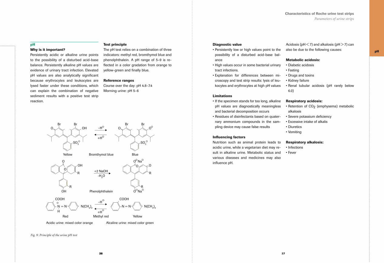
pHWhy is it important?Persistently acidic or alkaline urine points to the possibility of a disturbed acid-base balance. Persistently alkaline pH values are evidence of urinary tract infection. Elevated pH values are also analytically significant because erythrocytes and leukocytes are lysed faster under these conditions, which can explain the combination of negative sediment results with a positive test strip reaction.
Test principleThe pH test relies on a combination of three indicators: methyl red, bromthymol blue and phenolphthalein. A pH range of 5–9 is re-flectedinacolorgradationfromorangetoyellow-green and finally blue.
Reference rangesCourse over the day: pH 4.8–7.4Morning urine: pH 5–6
Fig. 9: Principle of the urine pH test
Acidic urine: mixed color orange Alcaline urine: mixed color green
O Br Br
Yellow Bromthymol blue
OH –H
+H SO3
-
+
+
+
N(CH3)2
Red Methyl red Yellow
COOH COOH
= N N = N
N
H
N(CH3)2
–H
+H
O Br Br
Blue
O
SO3
-
O
OH R R
-H2O +2 NaOH
Phenolphthalein
O OH
R
O
R
O O Na
O Na
-
-
+ -
+
+
+
37
Characteristics of Roche urine test stripsParameters of urine strips
Diagnostic value•Persistentlyloworhighvaluespointtothe
possibility of a disturbed acid-base bal-ance
•Highvaluesoccurinsomebacterialurinarytract infections.
•Explanation for differences between mi-croscopy and test strip results: lysis of leu-kocytes and erythrocytes at high pH values
Limitations•Ifthespecimenstandsfortoolong,alkaline
pH values are diagnostically meaningless and bacterial decomposition occurs
•Residuesofdisinfectantsbasedonquater-nary ammonium compounds in the sam-pling device may cause false results
Influencing factorsNutrition such as animal protein leads to acidic urine, while a vegetarian diet may re-sult in alkaline urine. Metabolic status and various diseases and medicines may also influencepH.
Acidosis (pH < 7) and alkalosis (pH > 7) can also be due to the following causes:
Metabolic acidosis:•Diabeticacidosis•Fasting•Drugsandtoxins•Kidneyfailure•Renal tubular acidosis (pH rarely below
6.0)
Respiratory acidosis:•Retention of CO2 (emphysema) metabolic
alkalosis•Severepotassiumdeficiency•Excessiveintakeofalkalis•Diuretics•Vomiting
Respiratory alkalosis:•Infections•Fever
pH

3838
LeukocytesWhy is it important?Leukocyturia is an important guide symptom for inflammatory diseases of the efferenturinary tract and kidneys, such as bacterial and abacterial infections and parasite in-festations. Abacterial leukocyturia can also constitute important evidence for the pre-sence of tuberculosis or tumors.
Test principleThe leukocytes excreted in the urine are almost exclusively granulocytes, whose es-terase activity is detected in the test strip reaction. The test zone contains an indoxyl ester, which is cleaved by the granulocyte esterase. The free indoxyl reacts with a dia-zonium salt to form a violet dye.
Reference rangeNormal < 10 leukocytes/μLBorderline 10–20 leukocytes/μLPathological > 20 leukocytes/μL
Practical detection limit10–25 leukocytes/μL
Fig. 10: Principle of the urine leukocytes test
OH + HO O NH
SO2 SO2
R1
R2
Indoxyl
Indoxyl ester Indoxyl
Diazonium salt Dye (violet)
Esterase
N
H
OH
N
H
OH
N
H
R1 R2
N
H
+ N N
O NH
O
N N
+ -
39
Characteristics of Roche urine test stripsParameters of urine strips
Diagnostic value•Leukocyturia is a cardinal symptomof in-
flammatory diseases of the lower urinarytract, mostly caused by bacteria and the kidneys
•The leukocytes excreted in the urine arealmost exclusively granulocytes, whose es-terase activity is detected in the test strip reaction
•Theteststripdetectsintactaswellaslysedcells (alkaline pH > 7 or diluted urine indi-cated by low specific gravity), which cannot be detected under the microscope
Limitations•Thetestdoesnotreacttopathogenicbac-
teria and trichomonads in urine•Proteinexcretion inexcessof500mg/dL
and glucose excretion of over 2 g/dL could lead to weaker color development and high doses of cephalexin and gentamicin
•Preservativesfalsifythetestresult(false-positive reading in the case of formalde-hyde, false-negative in case of boric acid). Medication with imipenem, meropenem and clavulanic acid could lead to falsepo-sitive results
Influencing factorsFalse-positive leukocytes:•Notcollectingcleancatchmidstream,con-
tamination by vaginal secretion or saliva•Expired,contaminatedorimproperlystored
strips•Nitrofurantoin,imipenem,meropenem,cla-
vulanic acid (antibiotics)
False-negative leukocytes:•Specimennotmixedwellorata lowtem-
perature•Proteinuria>500mg/dL•Glucosuria>2,000mg/dL•Cephalexin,gentamycin•Boricacid,sodiumazide,mercurysalts,hy-
drochloric acid
Did you know?If an inflammation is chronic or has healed,
it is not unusual to obtain a positive leuko-
cyte reaction and yet fail to find any bacte-
ria in the urine. This condition is known as
“abacterial” leukocyturia. In chronic pyelo-
nephritis leukocyturia, the only symptom is
often in the intervals between the acute epi-
sodes. The additional symptoms associated
with the acute course, such as fever, kidney
pains, proteinuria and erythrocyturia, are
absent.
Leuko-cytes

4040
NitriteWhy is it important?The presence of nitrite in the urine is one of the most important symptoms of a bacterial urinary tract infection. Women are particu-larly affected by this condition. Men suffer from these infections increasingly after the age of 60. Recognition and early treatment of urinary tract infections is of decisive im-portance, because a progressive infection may lead to chronic kidney failure, pyelone-phritic atrophic kidneys, and uremia.
Test principleThe aromatic amine sulfanilamide reacts with nitrite in the presence of an acid buf-fer to form a diazonium compound, which is coupled with 3-hydroxy-1,2,3,4- tetrahydro-benzo-(h)-quinoline to form an azo dye. Ni-trate that is present in the urine is converted by bacterial reduction into nitrite.
Fig. 11: Principle of the urine nitrite test
+ - +
H2N–SO2
H2N–SO2 H2N–SO2
H2N – SO2
Diazonium salt Azo dye (red) Coupling component
Sulfanilamide Diazonium salt Nitrite
NH3 + NO2 N + 2 H2O
N=N OH
OH N + N
N
N
H
N
H
H
H +
+
+
41
Characteristics of Roche urine test stripsParameters of urine strips
Reference rangeBacteria-free urine does not contain any nitrite.
Practical detectionlimit11 μmol/L (0.05 mg/dL)
Diagnostic value•The presence of nitrite in the urine is in-
dicative of bacterial urinary tract infections (UTIs) by nitrate-deforming bacteria e.g. E. coli, independently of the pH
•Onaverageabout50%ofbacterialUTIsaredetected with the nitrite test
•Under favorable conditions (first morningurine, high microbial count) more than 90% of bacterial UTIs are detected
•Screeningbeforeconfirmationbybacterio-logical examinations
•Leukocyturia is an important supplemen-tary finding
Limitations•Theintensityoftheredcolorisameasure
of the nitrite concentration but cannot be correlated to the severity of the infection
Influencing factorsFalse-positive nitrites•Expired,contaminatedorimproperlystored
strips, e.g. prolonged exposure to the air (nitrous gases)
•Drugsthatcolortheurinerede.g.phenazo-pyridine
•Bacterial contamination from sample col-lection
•Bacteriacanmultiplyandconvertnitratetonitrite in specimens that are more than 4 hours old
False-negative nitrites•Bacteria causingUTIsmaynot be able to
convert nitrate to nitrite•Antibiotic therapy suppresses the enzyme
metabolism and the microbial population, so that not enough nitrite is formed for the test
•Insufficientnitrateintakeortooshortreten-tion of urine in the bladder
Did you know?A single negative test does not exclude a uri-
nary tract infection, because the microbial
count and the nitrate content of the urine
can vary. Absence of color on repeated tes-
ting is also not reliable evidence for the ab-
sence of a urinary tract infection, since a pa-
thogenic microorganism that does not form
nitrite could be present. If there is clinical
suspicion of an infection, therefore, it is ad-
visable to go on to determine the microbial
species and count.
Nitrite

4242
Protein (albumin)Why is it important?The indicator reacts particularly sensitively to albumin excreted in the presence of kidney damage. Proteinuria is a frequent symptom in renal diseases, but it is also non-specific. It is not a proof of nephropathy, nor does its absence exclude nephropathy.
Test principleThe detection reaction relies on the so called protein error of pH indicators. The protein test area contains a buffer mixture and an indicator which changes color from yellow to green in the presence of protein, even though the pH is held constant.
Reference rangeBelow 10 mg/dL (for total protein)
Practical detectionlimit6 mg/dL albumin and above
Diagnostic value•Thetestispredominantlysensitiveforalbu-
min•Goodcorrelationwith albumindetermina-
tion by immunodiffusion•NoinfluencebyvaryingpHof5–9andspe-
cific gravity of the urine•Nointerferencebyquinine,quinidine,chlo-
roquine, sulfonamides•Moreconvenientandgenerallysuperiorto
precipitation tests•Medicinessuchasquinine,quinidine,chlo-
roquine, sulfonamides and penicillin have virtually no effect on the color reaction
Fig. 12: Principle of the urine protein test
Yellow 3',3",5',5"-tetrachlorphenol- 3,4,5,6-tetrabromsulfophthalein (neutral form)
Green Anion of this compound
-H
Protein
SO3 SO3
Br
Br Br
Br
Cl
O Cl Cl
Cl
OH O
Br
Br Br
Br
Cl
O Cl Cl
Cl
+ -
-
-
43
Characteristics of Roche urine test stripsParameters of urine strips
Limitations•Microalbuminuria cannot be detected be-
cause the first positive result of the test strips is 15–30 mg/dL
•The sensitivity to other proteins (e.g. g- globulins, proteses, peptones, mucopro-teins) is lower
Influencing factorsLow or false-negative protein•Proteinuriaismainlyconsistingoutofother
proteins than albumin
False-positive protein•Duringorafterinfusionofpolyvinylpyrroli-
done (blood substitute)•Stronglybasicurine(pH>9)duringthera-
py with phenazpopyridine•Residuesofdisinfectantsbasedonquater-
nary ammonium compounds or chlorhexi-dine
Did you know?Benign proteinuria
In persons with healthy kidneys, proteinuri-
as are observed predominantly up to the age
of 30, and account for up to 90% of the pro-
teinurias found in this age group. The causes
of these benign conditions include physical
stress (for example from playing sports),
emotional stress, orthostatism and lordosis.
Proteinurias associated with hypothermia,
heat, pregnancy, or the use of vasoconstric-
tively acting drugs are also usually benign.
Benign proteinuria has been observed in
20% of women during pregnancy.
Benign proteinurias occur intermittently.
While protein excretion is normal in mor-
ning urine, values reaching 500 mg/dL may
be observed in the course of the day. On the
basis of this property, benign proteinuria is
relatively easily distinguished from the pa-
thological form by repeated testing of the
first morning urine.
Extrarenal proteinuria
Protein is detected in the urine in many
mostly acute clinical pictures, such as colics,
epileptic fits, infarcts, strokes, head injuries,
and postoperative states. These proteinur-
ias disappear after the extrarenal cause has
been eliminated. Proteinurias due to fever
are usually harmless, but they do require cli-
nical supervision and course monitoring.
Renal proteinuria
An increase in the permeability of the glo-
merular capillaries due to pathological pro-
cesses leads to the development of renal pro-
teinuria.
In general the level is in excess of 25 mg/
dL, the most pronounced proteinurias being
observed in nephroses. In glomerulonephri-
tis the protein excretion is usually 200–300
mg/dL, but lower values must be recognized
within the event of glomerulonephritis asso-
ciated with few symptoms. This proteinuria
is usually accompanied by microhematuria.
Postrenal proteinuria
Postrenal proteinuria can occur following
inflammation of the bladder or prostate and
on bleeding in the urinary tract.
Protein

4444
GlucoseWhy is it important?The determination of glucose in urine has a high diagnostic value for early detection of disorders such as diabetes mellitus.
Test principleThe detection of glucose is based on a spe-cific glucose-oxidase-peroxidase reaction in which D-glucose is oxidized enzymatically by atmospheric oxygen to D-gluconolacto-ne. The hydrogen peroxide formed oxidizes the indicator TMB under peroxidase cataly-sis, to give a blue-green dye which on the yellow test paper causes a color change to green.
Reference rangeFasting morning urine <1.1 mmol/L (< 20 mg/dL)Daytime urine <1.7 mmol/L (< 30 mg/dL)Practical detection limitFor ascorbic-acid-free urine the practical detection limit is around 2.2 mmol/L (40 mg/dL), so that even slightly pathological glucosurias can be detected with high relia-bility. The upper limit of physiological gluco-suria in the first morning urine is around 0.8 mmol/L (15 mg/dL).
Fig. 13: Principle of the urine glucose test
OH
O O OH
OH
+ O2
CH2OH
3,3',5,5'-Tetramethylbenzidin
HO H OH O + H2O2
OH
CH2OH
HO
H2O + Dye (blue) POD
GOD
H2N NH2 + H2O2
β-D-Glucose Oxygen Hydrogen peroxide
45
Characteristics of Roche urine test stripsParameters of urine strips
Diagnostic value•Thesimplestandquickestwaytoscreenfor
unidentified diabetics as well as monitoring and self-testing
•Detection of renal glucosuria, e.g. duringpregnancy
•Detection of alimentary glucosuria (afterextreme carbohydrate intake)
•The enzymatically catalyzed reaction se-quence ensures that glucose is the only urinary constituent that will react and give a positive test result
•Ketonesdonotinterfere,andthepHoftheurinesimilarlydoesnotexertanyinfluenceon the test result
Limitations•Theurineglucoseconcentrationrepresents
the glucose excretion during the urine col-lection period in the bladder, and does not necessarily correlate with the actual blood glucose value
Influencing factorsLow or false-negative glucose•Metabolic products and drug metabolites
which have a reducing action
False-positive glucose•Presenceof residuesofperoxide-contain-
ing or other strongly oxidizing cleaning agents
Did you know?The absence of glucosuria does not exclude a
disturbance of the glucose metabolism, and
in particular diabetes mellitus. Glucosuria
develops when the tubular reabsorption of
glucose in the kidneys (the renal threshold)
is exceeded (Fig. 14). The renal threshold
is normally at a blood glucose level of 150–
180 mg/dL (8.3–10 mmol/L), but it is often
elevated in older people and in persons who
have had diabetes mellitus for many years.
Fig. 14: Renal threshold for glucose
Am
ount
of
gluc
ose
tran
spor
ted
[mg/
min
]
Glucose concentration in plasma [mg/100 mL]
0
200
100
400
300
500
600
0 100 200 300
Renal threshold Glomerular filtration Tubular resorption Urinary glucose excretion
Renal threshold for glucose
Glucose

4646
KetonesWhy are they important?Ketones (acetoacetic acid, ß-hydroxybutyric acid, and acetone) occur in the urine when increased fat degradation takes place in the organism owing to an insufficient supply of energy in the form of carbohydrates. De-tection of ketones in the urine (acetoacetic acid and acetone) is particularly important in checking metabolic decompensation in diabetes mellitus.
Test principleThe detection of ketones is based on the principle of Legal’s test. Acetoacetic acid and acetone react with sodium nitroprussi-de and glycine in an alkaline medium to give a violet color complex. The reaction is speci-fic for these two ketones, ß-hydroxybutyric acid does not react.
Reference rangeBelow 0.5 mmol/L (below 5 mg/dL) for ace-toacetic acid.
Practical detection limitThe test is substantially more sensitive for acetoacetic acid (detection limit 5 mg/dL = 0.5 mmol/L) than for acetone (detection li-mit about 40 mg/dL = 7 mmol/L).
Diagnostic value•Indicative of a dangerous condition for
diabetic patients called ketoacidosis which can lead to coma
•Detectionofstarvationstates•Monitoringanddetectionofdietprograms
which severely restrict intake of carbohy-drates (e.g. Atkins Diet) and zero diet
•Detectionofhyperemesisgravidarum(nau-sea during pregnancy)
Limitations•Phenylketone or phthaleine compounds
can produce red-orange to red colors on the test pad, which are different from the violet colors produced by ketone bodies
Influencing factorsFalse-negative ketones•Captopril, MESNA (2-mercapto-ethane-
sulfonic-acid sodium salt) and other sub-stances containing sulfhydryl
Fig. 15: Principle of the urine ketones test
Na2 [Fe(CN)5NO] + CH3–C–R + NaOH Na3[Fe(CN)5N=CH–C–R] + H2O
Sodium nitroprusside Ketone
O
Color complex (violet)
O
OH
47
Characteristics of Roche urine test stripsParameters of urine strips
Ketones

4848 49
Characteristics of Roche urine test stripsParameters of urine strips
UrobilinogenWhy is it important?Urobilinogen is excreted in increased amounts in the urine when, in the enterohepatic cir-culation of the bile pigments, the functional capacity of the liver is impaired or overloaded, or when the liver is bypassed.
Test principlep-methoxybenzenediazonium fluoroborate,a stable diazonium salt, forms a red azo dye with urobilinogen in an acid medium.
Reference rangeBelow 17 μmol/L (below 1 mg/dL).
Practical detection limitThe practical detection limit is around 7 mol/L (0.4 mg/dL), at which level the uro-bilinogen gives normal urine a pale light pink color. Differentiation between normal and pathological urine is possible by means of color comparison. Complete absence of urobilinogen in the urine, perhaps on com-plete obstruction of the common bile duct, cannot be detected.
Diagnostic value•Detectionofacuteandchronicliverdiseas-
es such as viral hepatitis, liver cirrhosis, and toxic hepatic damage
•Detection of hemolytic diseases such ashemolytic anemia, pernicious anemia, and intravascular hemolysis
•Elevated amounts of urobilinogen are in-dicative of compromised liver function
Limitations•The test is specific for urobilinogen and
does not react with other diazo-positive substances
•Noredcolor is formed in thepresenceofporphobilinogen, indican, paminosalicylic acid, sulfonamides, sulfonylureas and other substances occurring in the urine
Fig. 16: Principle of the urine urobilinogen test
+ -H3C–O Azo dye (red)
Diazonium salt
BF4 + UrobilinogenN N Acidmedium
Influencing factorsFalse-negative urobilinogen•Oxidationofurobilinogenifthespecimenis
left in direct sunlight for a long period.•Formaldehyde > 200 mg/dL used as pre-
servative
False-positive urobilinogen•Drugsormetaboliteswhichturnredinan
acid medium (e.g. phenazopyridine)
Possible causes of failure of urobilino-gen formation:•Complete obstruction of the common bile
duct in the absence of a bile tract infection•Complete stoppage of bile production in
the liver (very severe viral hepatitis, severe toxic liver damage)
•Absenceofintestinalflora(physiologicalinnewborn babies, rarely observed after in-tensive antibiotic therapy)
Did you know?Urobilinogen is formed by bacterial reduc-
tion from bilirubin secreted into the intes-
tine with the bile. It is then reabsorbed into
the bloodstream and is subsequently broken
down in the liver and partly excreted in the
urine.
Urobilinogen is absent in the urine in situa-
tions involving failure of bile production in
the liver cells, disturbances of bile secretion
into the intestine, and absence of bilirubin
reduction in the intestine, even though a se-
vere disease may be present. Urobili nogen

5050
BilirubinWhy is it important?In all pathological processes that increase theconcentrationofconjugatedbilirubininplasma, the excretion of bilirubin in urine canreachconsiderablehigh levels.Conju-gated bilirubin is found in the case of increa- sed intracanalicular pressure due to an ex-trahepatic or intrahepatic obstruction, and withperiportalinflammationorfibrosisandswelling or necrosis of the liver cells.
Test principleBilirubin reacts with a stable diazonium salt (2.6 - dichlorobenzenediazonium fluorobo-rate) in an acid medium of the test paper. A red-violet azo dye is formed, causing a color change to violet.
Reference rangeAdults below 3.4 mol/L (below 0.2 mg/dL).
Practical detection limitThe practical detection limit in urine free from ascorbic acid is 9 μmol/L (0.5 mg/dL). In favorable cases as little as 3–7 μmol/L (0.2–0.4 mg/dL) may give a positive reac-tion.
Diagnostic value•Increased levels of bilirubin are found in
liver disease such as icterus or obstruction ofthebileflow
Limitations•High ascorbic acid concentrations lower
the sensitivity of the bilirubin test
Influencing factorsFalse-negative bilirubin•Prolonged standing of the urine, particu-
larly in direct sunlight, leads to oxidation of the bilirubin
False-positive bilirubin•Medicines thatcolor theurine redor that
are themselves red in an acid medium, e.g. phenazopyridine
•YelloworgreenreactioncoloroftheUBGtest in the presence of high bilirubin con-centrations
Fig. 17: Principle of the urine bilirubin test
+ -
Cl
Cl
Azo dye (red-violet)
Diazonium salt
BF4 + Bilirubin N N Acid medium
51
Characteristics of Roche urine test stripsParameters of urine strips
Did you know ?As a result of conjugation (esterification)
with glucuronic acid, bilirubin becomes
water-soluble and therefore susceptible to
excretion by the renal route. The bilirubin
present in urine is always conjugated (di-
rect) bilirubin.
Diseases in which only unconjugated biliru-
bin is increased in the serum proceed with-
out bilirubinuria, because unconjugated
bilirubin is not excreted by the renal route.
The cause may be an oversupply of bilirubin
in the liver cells or a disturbance of uptake
or conjugation:
•Hemolyticjaundice
•Jaundiceofthenewborn
•Gilbert-Meulengrachtdisease
•Crigler-Najjarsyndrome
Bilirubin

5252
BloodWhy is it important?The principal causes of hematuria, (excre-tion of erythrocytes in the urine) may indi-cate UTI, kidney disease, kidney stones or tumors.
Test principleThe test is based on the peroxidative action of hemoglobin or myoglobin which cataly-zes the oxidation of the color indicator TMB by an organic hydroperoxide (2,5-dime-thylhexane-2,5-dihydroperoxide) to give a blue-green dye which on the yellow test pa-per causes a colour change to green. High sensitivity is achieved by the addition of an activator to the reagent mixture. Intact ery-throcytes are lysed on the test paper and the hemoglobin released sets off the color reaction.
Visible green spots are formed. In contrast, hemoglobin dissolved in the urine (erythro-cytes in lysed form) leads to the develop-ment of a uniform green color. Development of green spots (intact erythrocytes) or a green color (free hemoglobin/myoglobin) in the reagent area within 60 seconds indica-tes the need for further investigation.
Reference range0–5 erythrocytes/L.
Practical detection limitThe practical detection limit for intact ery-throcytes is about 5 erythrocytes/L and for hemoglobin the amount corresponding to around 10 erythrocytes/L. The practical de-tection limit of the test reaches the limit of the normal range.
Fig. 18: Principle of the urine blood test
OOH OOH
CH3 CH3
3.3', 5.5'-Tetramethylbenzidin2.5-Dimethylhexan-2.5-Dihydroperoxid
NH2 + H2N CH3 CH2 CH2 C CH3 C
OH OH
CH3 CH3
CH3 CH2 CH2 C CH3 C
+ Dye (blue-green)
CH3 CH3
CH3 CH3
Hemoglobin Myoglobin
53
Characteristics of Roche urine test stripsParameters of urine strips
Diagnostic value•Detectionofhematuria,aconcomitantsymp-
tom of renal diseases, urinary tract disorders, and extra-renal affections
•Detectionofhemoglobinuriaandmyoglobi-nuria as symptoms of hemolytic diseases, severe intoxication, extensive burns, severe muscleinjuries,heavyphysicalstrain
•Detectionofintactandlysedcells•Freehemoglobinisindicativeofintravascu-
lar hemolysis•Cellular malignancies may lead to micro-
hematuria of unknown origin because of missing symptoms
Limitations•The test is specific for hemoglobin and
myoglobin. Other cellular constituents of the urine, such as epithelia, leukocytes, and spermatozoa, have no effect
Discrepancy between test andmicroscopy•Oldspecimens,redbloodcells(RBCs)lyzed
in urine upon sitting, and non-intact RBCs are not detected under microscope
•Urinenot swirled,RBCs settle to thebot-tom, pad at the end of strip being dipped in a concentrated area
•Over-centrifugationcancausedestructionof RBCs
Influencing factorsFalse-positive blood•Expired, contaminated or improperly stored
strips. Residues from strong oxidizing reagents in urine containers or cleansing tissues
•Menstrual contamination, not collectingclean catch midstream
False-negative blood•Formalin(usedasapreservative)•Nitrite (in excess of 10mg/dL) delays the
reaction
EvaluationErythrocytesThe observation of individually separated to closely set green dots on the test paper points to the presence of intact erythrocytes. At higher concentrations the dots may be so close together that the test area appears almost uniformly green. Dilution of the urine 1:10 or 1:100 with 0.9% (physiological) sa-line and repetition of the test with another strip will make it possible to decide whether intact erythrocytes or free hemoglobin are present. Blood

5454
A finding of 5–10 erythrocytes/μL requires repeated checks on the urine, and if it is ob-tained again must be clinically clarified.
HemoglobinA homogeneous green test area points to the presence of free hemoglobin, lysed ery-throcytes or myoglobin. A weaker green color, as a first sign of a positive reaction, requires a repetition of the test with a fresh urine specimen. This second test may re-veal, among other things, intact erythrocy-tes which at the time of the first test had already become hemolyzed. Persistence of the finding requires clinical clarification in all cases. In the event of a weakly positive hemoglobin reaction, the cause may also simply be heavy physical exertion. This can be easily excluded from the medical history.
Partial hemolysisPartial hemolysis of erythrocytes present in the urine leads to the appearance on the test area of individual green dots against a diffuse green background. An exact assign-ment of the comparison color is then impos-sible, because the degree of hemolysis can be very variable as a function of age, con-centration, and pH of the urine.
Did you know?In contrast to hematuria, in which intact
erythrocytes are excreted, in hemoglobinu-
ria the urine contains free hemoglobin. This
appears in the urine when erythrocytes have
been broken down within the vascular sys-
tem. Following intravasal hemolysis, the he-
moglobin passes into the urine as soon as the
haptoglobin-binding capacity of the plasma
and the tubular reabsorption capacity for
hemoglobin have been exceeded. This usu-
ally occurs with plasma hemoglobin concen-
trations of around 60 μmol/L (100 mg/dL).
Myoglobinuria is generally due to muscular
injury or muscular necrosis, when the myo-
globin level in plasma exceeds 9–12 μmol/L
(15–20 μmg/dL).
55
Characteristics of Roche urine test stripsParameters of urine strips

5656
Microalbuminuria and its importancePatients with diabetes mellitus, cardiovascu-lar disease or hypertension often suffer from a nephropathy as a late complication. Dia-betes is the leading cause of kidney failure, accounting for 44% of new cases. Worldwi-de 50% of people with diabetes are unaware of their condition and are not treated.11
Both diabetic and hypertensive patients usually undergo a regular Micral-Test, since they are already at high risk of terminal kid-ney failure and to damage their cardiovas-cular system.
Diagnostic valueOne factor in the early recognition of neph-ropathy is microalbuminuria, defined as al-bumin concentrations between 20 and 200 mg/L urine. Values below 20 mg/L are not critical. Early diagnosis of microalbuminuria allows an appropriate therapeutic approach in order to avoid renal failure.
The potential indications include metabolic optimization, early institution of antihyper-tensive therapy (preferably with ACE inhibi-tors), and a low-protein diet in the case of diabetics. In the case of hypertension, ge-neral measures and an effective drug the-rapy are indicated in order to lower blood pressure.
Test principleThe Micral-Test allows specific detection of human albumin in the urine by a combina-tion of immunological and chromatographic processes. When the test strip is dipped into asample,urinepassesviaawickfleeceintoalayerofconjugatefleece.Thengold-labe-led antibodies bind albumin and the resul-ting antigenantibody complex migrates into the visualization field.
Excessantibody-goldconjugateisboundbyimmobilized albumin in a capture zone, mean- ing the detection field is reached only by conjugate molecules filled with urinary al-bumin. Depending on the albumin concen-tration, the detection field ranges in color from white to red.
Detection of microalbuminuria with Micral-Test
Fig. 19: Micral-Test®, an Accu-Chek® product
57
Characteristics of Roche urine test stripsDetection of microalbuminuria with Micral-Test
Test performanceNOTE: Since the reaction of this test strip is based on chromatographic and immu-nological principles, pre-analytical settings slightly differ from the procedure for con-ventional test strips.
1. Collect first morning urine from a mid-stream, so that albumin concentration is not falsified by physical activity or fluidintake
2. Dip the test strip in the urine until the area between the two black lines is cov-ered and hold it for five seconds. The test strip must not touch the container wall during this procedure, due to the possi-bility of interference effects during chro-matography
3. Place the test strip on a nonabsorbent horizontal substrate or on the urine con-tainer
4. After one minute, compare the reaction color against the colors on the label
5. Repeat the test on three days in a week
Specificity and sensitivityWith a positive detection limit of 20 mg/L for microalbuminuria, the test obtains a sensiti-vity of 95% and a specificity of 80%. On the basis of the immunological reaction, Micral-Test measures human albumin specifically. Cross-reactions with other human proteins such as IgG, IgA, leukocytes, and erythrocy-tes are below 0.5%.
EvaluationThe result is considered as medically rele-vant if at least two of the three morning uri-ne samples have been positive.
LimitationsMistakes during the handling process may be due to a number of factors: the strip may be immersed too far, or for too short a time, or the reading may be taken too soon, or there may be contact between the test strip and the wet container wall.
The following findings restrict the infor-mation value of microalbuminuria:•Acutediseasesandinfectionsoftheurinary
tract•Positive urine findings for protein, nitrite,
leukocytes or blood•Pregnancy•Severemetabolicdysregulation, forexam-
ple in diabetics•Physicalexertionatthetimeofurinecollection
in the bladder (physiological albuminuria)•Albuminofpostrenalorigin
Influence of drugsInterference due to medicinal drugs has not been observed so far, but the effects of me-dicines and/or their metabolites on Micral-Test are not all known. If there is any doubt, and if medically acceptable, the medication should be discontinued and the test repea-ted.

5858 59
Drug interference in urine
The results of urinalysis can be influenced
by drugs, leading to false-positive or false-
negative results. Roche has carried out a re-
search study to clarify the extend of possible
interference.
4

6060
The study is representative for all Roche urine strips and analyzing systems. It is ac-cepted by FDA standards representing drug and chemicals susceptible to interfere with urine test strip results. Interference factors may include levodopa, sulfamethoxazol, as-corbic acid and others; a complete list can be found in the appendix (Tab. 3, 4, 5, 6). The susceptibility to interfere with the test result is determined in at least the maximum dai-ly doses for every drug or component. If no interference has taken place the particular drug is not listed.
Specific gravityNo interferences were found.
LeukocytesFalse-positiveinfluence•Cefoxitin,curcumin,levodopa,nacetylcys-
teine, acid or tetracycline and captopril•Loweredspecificgravity
False-negativeinfluence•False-negative results can occur during
treatment with captopril. The test pad is not affected by the ingestion of ascorbic acid (up to 750 mg/dL)
NitriteFalse-positiveinfluence•Phenazopyridinefrom300mg/L
False-negativeinfluence•Medicationswith2-mercaptoethanesulpho-
nate-sodium (MESNA) and sulfonamide (trimethoprim, but only in extremely high concentrations (1080 mg/L))
The test pad is not affected by the ingestion of ascorbic acid (up to 3,000 mg/dL).
ProteinFalse-positiveinfluence•Duringtherapywithp-aminosalicyclicacid,
chloroquine, quinidine or nitrofurantoin
The test pad is not affected by the ingestion of ascorbic acid (up to 4,000 mg/dL).
GlucoseFalse-positiveinfluence•Medicationwith2-mercaptoethanesulpho-
nate-sodium (MESNA)
False-negativeinfluence•Medications with nitrofurantoin may pro-
duce false-negative readings
The test pad is not affected by the ingestion of ascorbic acid (up to 750 mg/dL).
Influencing factors
61
Drug interferences in urineInfluencing Factors
Ketones False-positiveinfluence•Captopril,curcumin,imipenemand2-mer-
captoethanesulphonate-sodium (MESNA) or other sulfhydryl-containing compounds
•Formaldehyde(stabilizer)maycausefalse-positive readings, but only in extremely high concentrations
The test pad is not affected by the ingestion of ascorbic acid (up to 4,000 mg/dL).
UrobilinogenFalse-positiveinfluence•Urinefrompatientswhoarebeingtreated
with p-aminosalicyclic acid or sulfame-thoxazol and phenazopyridine may show a false-positive reaction
•UrineswithhighpHvalues(pH>9)
False-negativeinfluence•Treatments with levodopa, cefoxitin and
sulhydril-containing compounds (n-acetyl-cysteine) cause false-negative results
The test pad is not affected by the ingestion of ascorbic acid (up to 4,000 mg/dL).
BilirubinFalse-positiveinfluence•False-positivereadingsareobtainedduring
treatments with imipenem, penicillin and p-aminosalicyclic acid or due to contami-nation of urine container with hydrochloric acid
•Highlybasicurines(pH>9)•Largeamountsofurobilinogenintheurine
affect the color change of the bilirubin test and also lead to false-positive results
False-negativeinfluence•Treatmentswith2-mercaptoethanesulpho-
nate-sodium (MESNA)
The test pad is not affected by the ingestion of ascorbic acid (up to 400 mg/dL).
Blood (erythrocytes/hemoglobin)False-positiveinfluence•Patientswithphenazopyridinemedications•High counts of leukocytes (500 LEU/μL),
highly basic urines (pH > 9) and low specif-ic gravity (< 1,005) may show false-positive test results
•Medicationswith2-mercaptoethanesulpho-nate-sodium (MESNA) can lead to false-positive or false-negative reading of reagent pad color changes
False-negativeinfluence•Whenformalinisusedtopreservetheurine
with nitrofurantoin and quinidines, false-negative results are obtained
The test pad is not affected by the ingestion of ascorbic acid (up to 1,000 mg/dL).

6262 63
Automated urinalysis
The high sensitivity and specificity of Com-
bur-Test® urine strips permit rapid and reli-
able conclusions about pathological changes
in the urine.
5

6464
It is difficult to standardize visual evaluation of urine test strips, and a number of environ-mental factors can have a negative effect on the quality of the result. These include:•Differencesinlightconditionsatthework-
place •Individual differences in color differentia-
tion•Declining concentration when examining
long series of samples•Differencesintheaccuracyofcompliance
with the specified test strip reaction time
Instrumental evaluation of urine test strips virtually eliminates the described factors and guarantees rapid, standardized measu-rement and immediate reliable documenta-tion of the result.
PhotometryUrinalysis systems evaluate test strips by re-flectance photometry using selective light-emitting diodes (LEDs) with a wavelength and measurement time tailored exactly to the chemical reaction and color develop-ment of the test field concerned. Compared to visual assessment, this produces impro-ved accuracy near the limit of detection.
Urine test strip systems
Detector
Orange
Test strip
Scheme
Green
Measuring head (sensor)
Fig. 20: Measuring head (schematic)
65
Automated urinalysisUrine test strip systems
Althoughurinalysissystemsusereflectancephotometry to evaluate the test field color changes with high precision, it is not pos-sible to completely eliminate all differences in the composition of the sample material which could have an effect on color deve-lopment. In addition, and unlike instruments for the measurement of blood glucose, uri-nalysis systems only yield semi-quantitative results. In calculating the result, a correc-tion for interference by the intrinsic color of the urine is made by measuring a blank field on the test strip (compensation field), which is illustrated in the following: •TheLED(1)emitslightofadefinedwave-
length on the surface of the test pad (2) at an optimum angle
•Thelightisreflectedfromthesurfaceandpicked up by the detector (3)
•Thephototransistorsendsananalogelec-trical signal to an A/D converter (4), which changes it to digital form
•Themicroprocessor(5)convertsthedigitalreadingtoareflectancevaluebyreferringitto a calibration standard
•Reflectancevaluesarecomparedwith thedefined range limits (reflectance valuesthat are programmed in the analyzer for each parameter)
•Outputs,semi-quantitativeresult(6)•Resultscanbeprintedoutortransferredto
the laboratory computer
Before each measurement, the optical sys-tem is tested for variations in LED bright-ness and detector sensitivity. If the strip is not in the correct position, the result is not printed out and the measurement must be repeated. The result of the specific gravity test is automatically corrected if the pH va-lue is elevated.
Fig. 21: Reflection photometry (schematic)
3 Detector
4 Analogue-digital converter
2 Test pad surface
5 Microprocessor
6 Result
1 LED
Scheme
Determination of measured values

6666
Urinalysis systems Automated urinalysis can serve many diffe-rent needs, and can be divided into three categories:
Instruments intended for single mea-surementsOne test strip is manually inserted at a time. The test strip is measured automatically, and the result is delivered after about one minute. The test strip has to be removed manually afterwards.
Semi-automatic urinalysis systemsTest strips can be inserted manually at short intervals. Transport, measurement, and dis-posal of the used test strips into an in-built container are automatic. The results are au-tomatically saved in the memory and printed out.
Fully-automatic urinalysis systemsManual dipping and insertion of the test strips is not required. The urine samples are applied from sample tubes using a rotor or rack. Sample identification, test procedure and disposal of the used test strips into an in-built container are fully automatic.
Roche urinalysis systemsRoche offers a range of automated urinaly-sis products in all three segments, meeting a variety of day-to-day needs and based on reflectance-photometricevaluation.
The Urisys line offers standardized solutions for the ward or the physician’s office as well as for low-, medium- and high-volume labo-ratories. The new cobas line enables effici-entmanagementofworkanddataflowsinmedium-volume testing sites.
The urinalysis systems can be connected to a barcode scanner for automatic sample identification. The measured results can be transferred to the laboratory computer sys-tem or PC.
Key benefits:•Consistentandreliablereading•Reliabledocumentation•Standardizedtestprocedure•Optimizedworkflow
For more detailed information on product specifications, please contact your local distributor.
67
Automated urinalysisUrine test strip systems
Urisys 2400® analyzer Fully-automatic
urinalysis systems for large-scale
laboratories
Urisys 1100® analyzer Instruments intended for single measure-ments on wards or in physicians’ offices
cobas u 411 analyzerSemi-automatic urinalysis systems for small to medium-sized laboratories
Fig. 22: System overview
A solution that satisfies all different kind of needs

6868 69
Urine microscopy indifferential diagnosis
Test strip urinalysis produces a high number
of pathological findings requiring further
diagnosis. Urine centrifugation and micros-
copy can be readily performed by any physi-
cian or medical technician.
6

7070
Many kinds of microscopes are used in the medical laboratory. All share a basic design while differing in the special functions requi-red by their fields of application. Their main components can be divided into devices for illumination, magnification, and contrast.
Illumination device Medical specimens for microscopy are ge-nerally translucent. In transmitted light mi-croscopy, usually known simply as light mi-croscopy, the light source is located at the foot of the microscope.
The collector is a lens built into the foot abo-ve the light source. It collects the light and focuses it into the condenser located under the microscope stage. The amount of incident light energy is regulated using the aperture diaphragm and brightness knob. The con-denser lens system is located directly under the microscope stage and uniformly illumina-testheobject.Usingsmallcenteringscrewsthelightfieldcanbeadjustedtothecenterofvision.Underadjustmentthelightfieldisen-larged using the light field diaphragm until it exactlycoincideswiththeobjectivelens’fieldof view. This prevents contrast-reducing light scattering(lightspottoolargefortheobjec-tive lens) and uses the maximum amount of light energy to optimize illumination of the object.
Magnification device Laboratory microscopes have an objectiveturret locatedabove theobjectplanecon-tainingobjectivelenseswithstandardfocallengths of common magnifications (10x, 40x, 100x). The tube can contain special devices, zoom magnification systems, optical chan-nels for multiviewer microscopy, an image camera, or a video processing system. At the observer end of the tube the eyepiece usually magnifies the image generated by the objective lens by 10x. Eyepieces maycontain a corrective facility to compensate for impaired eyesight and/or reticles for ta-king sample measurements.
Microscope
Microscope setup. The observer looks through the eyepiece (1) and via the tube (2) views the image magnified by the objective lens (3,4). Under the stage (5) is the condenser (6) that focusses the light ema-nating from the source in the foot (7). On the side are knobs for adjusting focus (8) and brightness (9) (courtesy Olympus, Hamburg).
12
3
4
56
7
8
9
71
Urine microscopy in differential diagnosisMicroscope
Contrast microscopy The basic light microscopy method used in medicine is bright-field microscopy, for which all laboratory microscopes are equip-ped (Fig. 1). For urine, on the other hand, phase-contrast microscopy is the more re-liable technique because highly translucent elements are less likely to be overlooked. An iris diaphragm, either separate or integrated into the condenser, forms the light to create a hollow cone, which shifts light refracted atobjectboundarysurfacesby90°relativeto the environment when it passes through the phase ring located in a special phase contrast objective. Attenuating direct lightby approximately 75% decreases brightness but considerably increases the contrast of boundary surfaces, making translucent ob-jects more readily visible (Fig. 2). In dark-field microscopy intermediate polar filters above and below the object allow light topass through in only one direction of os-cillation. If the two filter axes are offset re-lative to each other, a dark field is created because light rays can only pass through thesecondfilteriftheyhavebeendeflectedinto the plane of the second filter by a light-refractingobject.Objectsarecontrasted inwhite against a dark background (Fig. 3, 4).
Fig. 2: Phase contrast image. Drug crystal and red blood cells with accentuated border contrast (white halo, 400x).

7272 73
Urine particles and formedelements
Urine microscopy examinations can be per-
formed on centrifuged or non-centrifuged
urine. Particles examined are cells, casts,
pathogens, crystals and formed elements or
insoluble compounds accumulated in the
urine during the passage from the kidney to
the lower urinary tract.
7

7474
White blood cells There are normally very few white blood cells (also known as leukocytes) in urine sediment. They only enter the urine in the presenceofurinarytractinflammation.Re-liable detection is therefore of particular diagnostic significance. White blood cells are recruited from blood by chemotactic signals produced by the damaged tissue. They actively migrate into the interstitium through the capillary basement membrane from where they pass through the epitheli-um of the renal tubules, bladder or ureter to emerge in the urine. In contrast to dysmor-phic red blood cells, no changes in white cell shape have been described that betray a specifically renal origin.
MorphologyWhite blood cells are colorless, 10–12 μm in diameter, with a large nucleus, and granular cytoplasm (Fig. 5). Lymphocytes are a type of white blood cell with a large circular nucleus and a narrow peripheral cytoplasm; although they can be difficult to differentiate from ma-crophages in bright-field microscopy this has no diagnostic significance. Granulocytes, on the other hand, have a segmented nucleus and contain coarse granules. White blood cells are easy to differentiate from red blood cells on account of their size, nucleus, and cytoplasmic structure (Fig. 6–8). Differentia-tion from small tubular cells, which can also contain granular cytoplasm, may be more difficult (Fig. 7–12). White blood cells tend to adhere to each other or to other sediment constituents (Fig. 9–11).
Blood cells
Fig. 5: In bright field microscopy white blood cells are approx. 10 μm in size with a prominent nucleus and granular cytoplasm (1,000x, bright field micros-copy).
75
Urine particles and formed elementsBlood cells
Diagnostic significanceAlthough white blood cells in urine sedi-ment are always a sign of urinary tract in-flammation, their provenance can only beidentified by studying the accompanying sediment constituents and case history. Isolated leukocyturia in which urine cultu-res remain negative is termed sterile leu-kocyturia.Itsuggestsinflammatorydiseasedue to pathogens that cannot be identified under the microscope and are difficult to culture. In particular it suggests chlamydial infection and renal tuberculosis. Because trichomonads are readily confused with white blood cells, a fresh urine sample should be specifically tested in such cases for motile trichomonads. In prostatitis, leu-kocyturia may be the only positive finding in the sediment. Sterile leukocyturia in the presence of failing renal function points to interstitial nephritis. Post-transplant lym-phocyturiasuggestrejection.
The presence of bacteria (see Bacteria) in addition to white blood cells in fresh urine is diagnostic of urinary tract infection in a dysuric patient. Concomitant eumorphic erythrocyturia suggests hemorrhagic cys-titis or a bacterially colonized concretion (secondary renal calculus). Leukocyturia plus eumorphic erythrocyturia in the ab-sence of bacteria suggests a toxic mucosal lesion (toxic hemorrhagic cystitis), renal tuberculosis, or interstitial nephritis. The sediment should be examined for casts. Mycobacterial cultures may be indicated. White blood cells plus dysmorphic red blood cells or acanthocytes (see Red blood cells – Morphology) suggest glomerulone-phritis with concomitant interstitial nephri-tis or glomerulonephritis with concomitant urinary tract infection. White blood cells plus trichomonads are diagnostic of tricho-moniasis. White blood cells plus crystallu-ria suggest crystal nephropathy.
Blood cells

7676
Red blood cells The commonest reason for investigating a patient for hematuria is a positive test strip result. If the patient is a young adult and otherwise asymptomatic, the likeli-hood of identifying a clinically relevant cause of bleeding is <2%.12 Investigati-on should only be considered after ru-ling out contamination, e.g. by menstrual blood. However, since hematuria can be due to very different diseases, ranging from benign hereditary hematuria and chronic glomerulonephritis to renal cell carcinoma, a rational diagnostic strate-gy is essential. The cause may be renal or postrenal. Invasive tests targeting the kidney, in particular biopsy, should only be performed in confirmed kidney cases. Similarly, invasive urological investiga-tion, such as cystoscopy, is warranted only in patients with lower urinary tract disease. Evaluation of urine sediment can make a major contribution in thiscontext.
MorphologyNot all red blood cells in urine have the normal biconcave form (Fig. 13–15). Eumorphic cells are often mixed with dysmorphic cells (Fig. 16, 17), including: acanthocytes, annular red blood cells with hemoglobin-containing membrane bulges that point either toward the cen-ter of the annulus or outward, resembling “Mickey Mouse ears” (Fig. 18); echinocy-tes (burr cells), with small spiny membrane projectionsthateitherruncircumferentiallyaround the biconcave annulus or spread evenly across the surface of spherical cells (Fig. 19); codocytes (target cells), stomatocy-tes (fish-mouth cells), and knizocytes, all of which have lost their biconcave shape and resemble spheres with one or more reces-ses that produce their specific dysmorphism (Fig. 20–22); schistocytes, crescent-shaped sickle cells with split edges and erythrocyte ghosts (ghost cells), empty membrane enve-lopes of red blood cells that have released hemoglobin into the urine through cracks in the membrane (Fig. 20, 23, 24).
Diagnostic significanceThe list of potential causes of hematuria is so long that investigation has to be syste-matic. Initially a distinction is made between glomerular and non-glomerular causes because this considerably narrows the dif-ferential diagnostic spectrum. If the cause is not apparent from the case history or concomitant symptoms, the urine sediment must be screened for further evidence of a renal cause. Tell-tale evidence of renal he-maturia consists of red blood cell casts, re-nal epithelia containing red blood cell casts
Fig. 16: Scanning electron micrograph showing the biconcave shape of eumorphic red blood cells (courtesy Dr. E. Wandel, Nephrology, University of Mainz).
77
Urine particles and formed elementsBlood cells
(Fig. 25, 26), red cell phages (Fig. 27), lipidu-ria (Fig. 28), and granular or yellow-brown casts (Fig. 29) occurring in large quantities. However, certain characteristics are only found in approximately 20% of patients. Moreover, coexistent renal and postrenal disease must be considered. Urinary tract infections, ureteric calculi, urothelial and renal cell carcinomas and drug toxicity (he-morrhagic cystitis after cyclophosphamide) also occur in lupus nephritis and chronic glomerulonephritis. If, however, no further characteristic sediment constituents are in evidence, glomerular and non-glomerular hematuria can be differentiated on the ba-sis of erythrocyte shape. In short, urinary red cell morphology can be regarded as a reliable, simple and inexpensive diagnostic method.13 But how do red cell shapes arise in the urine and which shapes indicate renal bleeding? The size and shape of urinary red blood cells largely depend on three factors: urine osmolarity, concomitant hemolysis, and passage through the glomerular base-ment membrane (GBM).
OsmolarityIf the cause of bleeding is extrarenal and the urine is iso-osmolar with blood, red blood cells in the urine are the same size and sha-pe as those in blood vessels. In hyperosmo-lar (morning) urine, red blood cells shrink and assume a typical thorn-apple crystal shape. Conversely, in hypoosmolar urine (water diuresis, diuretic therapy, and diabe-tes insipidus), red blood cells can swell fol-lowing alcohol consumption.14 Cell lysis gi-ving rise to erythrocyte ghosts is increased in hyposmolar urine. Size differences due
to osmotic influences have no diagnosticrelevance and are not reliably recorded on microscopy (Fig. 30, 31).

7878
Squamous epithelial cells Squamous epithelial cells are a common constituent of urine sediment, in particular in women. There is often a large pellet af-ter sedimentation, mostly made up of squa-mous epithelial cells. Only a small proportion stem from the urethra but their presence is evidence of mixture with vaginal secretion. Detailed instructions on the correct way to collect a midstream urine sample (swab, spread the labia etc) reduce the epithelial component considerably. In men squamous epithelium is found rarely and only in rela-tively small amounts.
MorphologySquamous epithelial cells are large translu-centflat cellswitha small central nucleus(Fig. 32). In bright field microscopy they are often barely visible but visualization can be improved substantially by phase contrast microscopy (Fig. 33, 34). They are more or less square-shaped and their thin margins are usually folded, facilitating differentiation from other urine cells (Fig. 35). In many ca-ses, contrasting small cytoplasmic granules are easy to pick out. These large cells can
befoundusuallyflatbetweentheslideandthe cover slip, or sometimes folded into an irregular sickle shape (Fig. 36).
Diagnostic significanceNone in connection with renal disease. However, massive presence of squamous epithelial cells indicates contamination with vaginal secretion (Fig. 37, 38). When found in conjunctionwith fungi,bacteria,andwhiteblood cells, the commonent diagnosis is va-ginitis, with cystitis and other urinary tract infection being less likely (Fig. 39). If there are urethral symptoms and the midstream urine sample has been correctly collected, the most likely diagnosis is urethritis.
Epithelial cells
Fig. 32: Vaginal squamous epithelium is characterized by large cells in a small central nucleus and a relatively homogeneous cytoplasm. Multinucleate cells are also observed (400x, phase contrast microscopy).
79
Urine particles and formed elementsEpithelial cells
Renal tubular cells The normal regeneration process means that renal tubular cells become detached from the tubular basement membrane and are eliminated in the urine in small amounts. Morphological classification is based on the fact that cells found in epithelial cell casts are different from round epithelia of the lo-wer urinary tract. Fluorescence-labeled an-tibodies against Tamm-Horsfall protein have confirmed the origin of these cells based on their coating with this tubular secretion pro-tein as opposed to transitional epithelia.15
MorphologyTubular cells are small round epithelial cells with a large central nucleus (Fig. 40). Diffe-rentiation from white blood cells and uro-thelium is not always easy (Fig. 41). Tubu-lar cells are much smaller than transitional epithelial cells but larger than white blood cells (Fig. 42–45). They also differ from white blood cells in that they have small unlobed nuclei and a relatively homogeneous cyto-plasm that contains occasional inclusions such as fat globules, red blood cells, hemo-globin, melanin, and bilirubin granules (Fig.
46). Renal tubular cells loaded with fatty particles correspond to the oval fat bodies found in the nephrotic syndrome (Fig. 47). After lengthy exposure to alkaline urine ty-pical deformations occur, such as folding of the cell borders or condensation of the cyto-plasm, with the result that cap-like attached membrane envelopes can be confused with fatty particles (Fig. 48, 49).
Diagnostic significanceIn acute renal failure and other tubuloin-terstitial disease many tubular cells are di-scharged into the tubular lumen and elimi-nated in the urine.
Fig. 41: Tubular epithelium (right) differs from white blood cells (left and below) and red blood cells in size and cytoplasmic structure (1,000x, bright field mi-croscopy).
Epithelial cells

8080
Transitional epithelial cells Urine flows from the renal parenchymathrough the renal pelvis and ureter into the bladder. Desquamated urothelial cells are thus also found in the urine and must be differentiated from renal tubular cells whe-never possible.
MorphologyUrothelium comprises superficial and deep layers. Cells in the superficial layer are lar-ge and circular with a small central nucleus (Fig. 50). Measuring up to 40 μm they re-semble squamous epithelial cells but the difference is that they do not have folded cytoplasmic borders. Their circular shape also differentiates them from squamous epi-thelium (Fig. 51). Cells in the deep urothelial layer are smaller and often have neuron-like outgrowths (Fig. 52–54). Multinuclear cells or interconnected cell aggregates occasio-nally occur (Fig. 55, 56).
Diagnostic significanceTransitional epithelial cells are also found in small numbers in the urine of healthy sub-jects.Theycanbeplentiful inurinary tractinfection but the number is not a reliable di-agnosticcriterion.Ininflammatoryormalig-nant disease of the lower urinary tract there tend to be more cells from the deep urothe-lial layer and large cell aggregates. If there is a clinical suspicion or otherwise unexp-lained eumorphic erythrocyturia, a specialist cytological opinion should be obtained.
Fig. 50: The difference between white blood cells and urothelial cells is their size (1,000x, bright field mi-croscopy).
81
Urine particles and formed elementsEpithelial cells
Atypical cells Urine microscopy is useful in the early detec-tion of renal and lower urinary tract cancer because dysplastic mucosal cells or renal carcinoma cells advancing into the renal pelvis are discharged in the urine. Urine cytology has proven a reliable screening procedure for urothelial cancer in occupa-tional risk groups (industrial workers expo-sed to aromatic nitrosamines).16,17 If clinically indicated, since cell abnormalities cannot be detected reliably by conventional bright field microscopy, targeted cytology is indicated, using cytocentrifugation and Papanicolaou staining.
MorphologySuspect cells include multinucleated cells with prominent nucleoli and clearly vacuo-lated cells with an eccentric chromatin-rich nucleus. However, the exact cytological evalu-ation should be performed by an experienced cytopathologist.
Diagnostic significanceConventional bright field urine microscopy is unsuitable as a general screening method for malignancy of the lower urinary tract because cell structure cannot be evaluated adequate-ly. Also, given the low prevalence of urothelial carcinomas, many false-positive results are obtained and selective cytological tests are too labor-intensive and expensive. A chance finding of atypical cells or tumor cell aggre-gates is a clear indication for further cyto-logical and urological investigation. In renal cell carcinoma the sensitivity and specificity of urine cytology are even worse. Only 27% of 436 urine samples from 31 patients with confirmed renal cell carcinoma had tumor cells in their urine.18

8282
Hyaline casts Hyaline casts are formed in the renal tubules due to precipitation of the 95 kd Tamm-Hors-fall glycoprotein, which is produced only by the tubular cells in the ascending limb of the loop of Henle and in the distal tubule and is secreted into the urine (Fig. 57).19 In contrast to filtered albumin that is almost completely reabsorbed in the proximal tubule, secreted Tamm-Horsfall protein accounts for the ma-jorpartofphysiologicalproteinuria.Itisanimportant inhibitor of calcium oxalate and calcium phosphate crystallization in the uri-ne.20 It is highly likely that Tamm-Horsfall protein is of pathophysiological significance as an antigen in chronic tubulointerstiti-al renal disease.21 Precipitation of Tamm-Horsfall protein fibrils to form hyaline casts iselevatedinlowtubularflow(dehydrationby fever, sport), presence of X-ray contrast agents, Bence-Jones proteinuria (myeloma cast nephropathy), and acid urine, whereas they dissolve quickly in alkaline urine (e.g. due to bacteria). This explains why hyaline casts can also be detected in urine speci-mens of healthy persons.
MorphologyHyaline casts are sharply delineated, co-lorless, and translucent. As a result, even relatively large amounts can sometimes be missed in bright field microscopy (Fig. 58). In phase contrast microscopy, however, they are easily recognized even by an inexpe-rienced observer due to their birefringent border delineation (Fig. 59). If it is not pos-sible to perform phase contrast microscopy, hyaline casts become more readily visible if the condenser below the stage is moved to its uppermost position (Fig. 60). Since the light beam is smaller, the cast borders are less overexposed and the depth of focus is improved. Hyaline casts can be compact, fi-brous, or coiled, due to the varying density of the protein fiber network (Fig. 61–63).22,
23, 24 Their width is a function of the inner diameter of the tubular lumen in which they formed. Wide casts are common in chronic renal insufficiency because the damaged nephrons have a dilated lumen due to tu-bular cell atrophy. Often the tip of the cast contains granular inclusions that turn into a pure hyaline cast and thus represent a tran-sitional form of granular casts (Fig. 64). The situation is the same with isolated cellular inclusions which, like pure red blood cell casts or white blood cell casts, constitute evidence of a renal cell origin (Fig. 65–68). These must be differentiated from cellular urine constituents that have only become attached to hyaline casts extrarenally or during centrifugation (Fig. 69). However, by adjustingthemicrometerscrewitisusuallypossible to differentiate attached cells from cells enclosed in the cast.
Casts
Fig. 59: The same cast is visualized clearly in phase contrast microscopy (400x, phase contrast micros-copy).
83
Urine particles and formed elementsCasts
Diagnostic significanceThe detection of pure hyaline casts is of no significance in the diagnosis of renal di-seases. They are a consequence of a changed tubular environment, as occurs physiologically in healthy subjects and pa-thophysiologically after extrarenal changes in hemodynamics and hydration status.25, 26
Physiological Tamm-Horsfall proteinuria be-comes visible by precipitation in the form of hyaline casts. Renal diseases, however, involve precipitation-inducing factors, with the result that hyaline casts can be detected to an increased extent in glomerular and tu-bulointerstitial diseases.27 Beyond diagnosti-cally significant microscopy findings (white blood cell casts, acanthocyturia), evidence of hyaline casts does not provide further useful clinical information. It should be noted that in a renal manifestation of light chain disease (multiple myeloma kidney, myeloma cast ne-phropathy) only hyaline-like casts are occa-sionally found in urine sediment, consisting of a precipitate mixture of light chains and Tamm-Horsfall protein. Morphologically they cannot be reliably differentiated from hyali-ne casts. If cellular inclusions are detected in the hyaline cast matrix, diagnostic signifi-cance is higher if these cells can be shown to be of renal origin. However, a hyaline cast with a single embedded red blood cell does not provide the same diagnostic certainty of glomerular disease as the detection of mul-tiple red blood cell casts. Physiologically, red blood cells migrating through the glomerular basement membrane can be embedded in hyaline casts when passing through the ne-phron.28 Such findings should prompt close
inspection of several samples because in re-nal disease either a number of similar casts or distinct red blood cell casts can be expec-ted. Diagnostic certainty increases with the number of cellular inclusions. In short, hyali-ne casts are commonly encountered formed elements that are only significant in the dia-gnosis of renal disease if there is evidence of cellular inclusions.
Casts

8484
Granular casts As with hyaline casts, the matrix of granu-lar casts consists of the tubular secretion of Tamm-Horsfall protein. If there are relatively small cell constituents, cell degradation pro-ducts, or protein precipitates in the tubular lumen during precipitation of the glycopro-tein, they are fixed in the forming matrix and create a cast of the corresponding tu-bular segment. Inclusions of granular casts in the urine examined under a microscope can have various origins.29 In glomerular or proximal tubular damage they are precipi-tates of glomerular filtered serum proteins or degradation products of renal cells that are already fractionated by the loop of Hen-le when they enter the distal tubule, where together with Tamm-Horsfall precipitates they form granular casts (Fig. 70, 71).30 In tubulointerstitial disease they are disintegra-ted tubular cell constituents that are elimi-nated in the form of granular casts (Fig. 72, 73). However, since further degradation of cellular products also takes place in the cast matrix, even intact, enclosed tubular cells or white blood cells take the form of granular casts in time (Fig. 74).22, 23, 24 Cell borders or nuclei are then no longer apparent. During passage through the nephron granular casts can be transformed into waxy casts. Elect-ron microscopy, immunohistochemistry, and immunofluorescencewithantibodiesagainstserum proteins and tubular cell antigens have all been used to establish the varying composition of granular casts, enabling dif-ferential diagnosis between acute glomerular disease and tubular necrosis, for example.29,23 However, these methods are not used in rou-tine diagnosis.
MorphologyGranular casts are sharply delineated and contain finely to coarsely granular inclu-sions, the cell borders of which cannot be visualized reliably (Fig. 75–77). They are readily visible even at low levels of magni-fication (10 x 10) (Fig. 78). The surface is rougher than in hyaline casts because they mostly form in damaged tubules (Fig. 79). If the contrast is particularly high, pigmenturia etc. must be considered (see Yellow-brown casts) (Fig. 80). The casts can be differen-tiated from hyaline casts covered by a bac-terial lawn on the basis not only of the high number of bacteria but also their intrinsic motility at higher magnification (also at 10 x 100 under oil immersion microscopy) (Fig. 81).22, 23, 24 Amorphous crystals can also cover hyaline casts or, when clustered in groups as pseudocasts, can be confused with gra-nular casts (Fig. 82). This can be observed at low magnification as patchy sediment typical of amorphous crystals and, with the 40xobjective,bytheabsenceofsharpcastdelineation.
Fig. 77: Granular casts are easy to recognize in bright field microscopy (400x, bright field microscopy).
85
Urine particles and formed elementsCasts
Diagnostic significanceA certain amount of tubular cell degradati-on is physiological. Whether the degradati-on products appear in the form of granular casts mainly depends on the precipitation of Tamm-Horsfall protein. Consequently, a small number of granular casts can be found in the urine of healthy persons. That also explains the granular inclusions in the matrix of hyali-ne casts. However, an increased discharge of granular casts is found in many glomerular and tubulointerstitial diseases (Fig. 83).18 In erythrocyturia or leukocyturia, without con-firmed cast evidence, the increased dischar-ge of granular casts can be a strong sign of renal disease (Fig. 84). In addition, granular casts are also often found in acute renal fai-lure due to tubular necrosis, rhabdomyolysis, or toxic renal damage (Fig. 85).
Pigmented casts The renal discharge of pigmented serum constituents such as hemoglobin, myoglo-bin, and bilirubin can lead to the formation of yellow-brown casts. The relatively large amounts of hemoglobin released in hemo-lysis are transported by haptoglobin to the liver where conversion to bilirubin allows clearance via the bile. If intravascular hemo-lysis is substantial, however, the transport capacity of haptoglobin is exceeded with the result that hemoglobin is freely filtered in the kidney. In hemochromatosis, or two to three days after intravascular hemolysis, hemosi-derin granules can also be detected in the urine. Myoglobin released following skeletal muscle damage (molecular weight approx. 40,000 daltons) is always filtered and thus found in the urine.30 Rising serum creatinine
values and a positive urine dipstick test for blood are an indication for urine microscopy which will show massive amounts of yellow-brown myoglobin casts instead of intact red bloodcells. Inobstructive jaundice,filteredconjugated bilirubin stains the sedimentconstituents yellow. Consequently, granular casts that were otherwise previously color-less can appear yellow.
MorphologyEven low magnification shows a patchy brown „soiled“ sediment with a large num-ber of yellow-brown granular casts, some of which are short (Fig. 86). Higher magnifica-tion shows medium to coarse-grain granu-les with a yellow-brownish color embedded in a hyaline matrix (Fig. 87–90). Since myo-globinuria can lead to acute renal failure, epithelial cell casts can be a common sign of tubular necrosis.
Fig. 90: Stained granular casts can also be reliably differentiated from confounders (400x, bright field microscopy)

8686
Diagnostic significanceCast color alone is not a reliable guide to the type of pigment. A urine test strip can help in classification based on the results of he-moglobin/erythrocyte and urobilinogen test pads. Patients with bilirubinuria are always jaundiced.Hemoglobinuriacanbedifferen-tiated from myoglobinuria by examining the patient serum. Free hemoglobin stains the serum reddish.
Waxy casts Waxy casts are rarer than hyaline casts and never found in subjects with healthy kid-neys. Their width suggests that they form in ectaticnephronswithminimalflow.
MorphologyWaxy casts are wide and translucent with a high refractive index, which is why they are more visible than hyaline casts under bright field microscopy. Their borders are sharply delineated and the corners perpendicular (Fig. 91). Lateral “slits” are an indication of the compressed spiral structure, as is occa-sionally well visualized in an unfolded form (Fig. 92–94). It is assumed that proximal cast structures formed in non-ectatic tubular
segments fold up in a corkscrew-like man-ner in distal ectatic tubules. Sometimes this spiral structure is particularly evident due to inclusions of relatively small granules in the nucleus of the waxy cast (Fig. 95, 96). In rare cases fine granular casts are also surroun-ded by a „waxy coat“ and thus represent a mixed form with granular casts (Fig. 97).
Diagnostic significanceWide waxy casts are only found in advanced renal diseases with considerable morpho-logical tubulointerstitial changes (Fig. 98). Occasionally, waxy casts can be detected inacuteorchronicrejectionofrenaltrans-plants as well as in the polyuric phase fol-lowing anuric renal failure.
Fig. 91: Waxy casts are wide homogeneous casts with a sharp border and rounded corners (1,000x, bright field microscopy).
87
Urine particles and formed elementsCasts
Red blood cell casts Red blood cell casts form due to fixation of red blood cells located in the tubular lumen in the precipitated matrix of Tamm-Horsfall protein or occasionally without a protein matrix due to pure compression of a large number of red blood cells in the tubules (Fig. 99).21,22,23 They are of high diagnostic significance in clarifying unexplained he-maturia. However, even if the cause of renal bleeding is identified, they can often only be found in small numbers. If clinically war-ranted, systematic inspection of the entire specimen, and possible further specimens, is advisable. For this purpose the edge of the cover glass should first be examined forsuspiciouscastswiththe10xobjective,which can then be observed in detail with the40xobjective.Sensitivitycanbeincrea-sed by prefiltering large amounts of urine.31
Morphology Red blood cell casts can consist of a hyaline cast with the inclusion of single red blood cells or numerous red blood cells in a hyali-ne matrix (Fig. 100–102). Although dysmor-phic red blood cells are regarded as being
typical of glomerular bleeding, the red blood cells enclosed in casts are usually eumor-phic (Fig. 103). They can be differentiated from other cell casts or granular casts by their yellowish color, the partially retained double border structure of the red blood cells, and the absence of nuclei or nucleus-like structures. However, in interstitial renal disease, mixed-cell casts are often found with the inclusion of red blood cells, white blood cells, and renal tubular cells (Fig. 104–108). In the case of pure red blood cell casts, the formed elements are only several millimeters long (Fig. 101). If they stay in the nephron for any length of time, red blood cells enclosed in casts can degenerate to the stage at which their cell borders can no longer be recognized (Fig. 109). Emerging hemoglobin stains the cast matrix in such a way that it is no longer possible to dif-ferentiate them from yellow-brown casts occurring in rhabdomyolysis or hemolysis. However, clinical symptoms and the often concomitant hematuria with acanthocyturia generally facilitate differentiation.
Diagnostic significanceRed blood cell casts are evidence of a renal ori-gin to hematuria because red blood cells can only pass from the bloodstream to the tubular lumen through the glomerular or tubular ba-sal membrane. However, the sensitivity of red blood cell casts as a marker of renal hematuria isonlyjustabove50%.35, 36 Glomerulonephritis and systemic inflammatory disease involvingthe kidney are the commonest cause of red blood cell casts, especially in the presence of concomitant proteinuria. The number of casts provides information about the inflammatory
Fig. 99: Red blood cell casts developing in a Tamm-Horsfall matrix in renal tubules (hematoxylin-eosin, 1,000x, courtesy Dr. Weis, Pathological Institute, University of Munich).

8888
activity of the disease.37 If proteinuria is absent or only minimal, this suggests IgA nephritis and the thin basement membrane syndrome (benign familial hematuria).38, 39 Isolated prote-inuria is characteristic of diabetic nephropathy and further sediment findings tend to suggest an additional renal disease. Nevertheless, in patients with diabetic nephropathy and glo-merular hematuria with red blood cell casts a different glomerulopathy is only found by his-tological analysis in some cases.29 Traumatic renal damage and embolic renal infarctions are further causes of glomerular hematuria with the formation of red blood cell casts.30 Temporary discharge of red blood cell casts can be found in subjects with healthy kid-neys following heavy physical exercise such as marathon running.31 In renal tuberculosis all types of sediment constituents can be found. In addition to the characteristic findings of sterile leukocyturia, glomerular hematuria with casts can also occur occasionally.32
White blood cell casts In inflammatory renal diseases, infiltratingwhite blood cells are released into the tubular lumen. They can either be enclosed in a matrix of precipitated Tamm-Horsfall protein or can conglomerate directly to form a matrix-free cast. Although the white blood cells have to overcome the tubular basal membrane, dys-morphic forms do not occur as with glomeru-lar red blood cells. This is due to the ability of white blood cells to lyse cell to cell contacts and basal membranes in diapedesis and ac-tively migrate through them.
MorphologyIsolated white blood cells can be enclosed in hyaline casts. (Fig. 110–112) However, crow-ded clumps of white blood cells within or wit-hout a hyaline matrix are regarded as genuine white blood cell casts (Fig. 113). This has to be differentiated with white blood cell pseudo-casts which may occur in severe leukocyturia due to adhesion of cells following centrifuga-tion forming aggregates (Fig. 114). Renal tu-bular cell casts can occasionally be mistaken for white blood cell casts if nuclear structures can no longer be reliably delineated due to cell degradation (Fig. 115).
Diagnostic significanceWhite blood cell casts represent evidence of inflammatoryinfiltrationoftherenalintersti-tium, as in pyelonephritis, interstitial neph-ritis,renaltuberculosis,transplantrejection,and interstitial coinvolvement in glomerulo-nephritis.
Epithelial cell casts If tubular basement membrane sheds sever-al tubular cells into the lumen, epithelial cell casts form due to mechanical compression or embedding in the hyaline matrix of preci-pitated Tamm-Horsfall protein.
Fig. 111: In this cast with a granular matrix there are several white blood cell inclusions (1,000x, bright field microscopy).
89
Urine particles and formed elementsCasts
MorphologyEpithelial cells originating from a tubular segment often accumulate as a typical doub-le row of roundish cells to form an epithelial cell cast, whereas white blood cell casts con-sist of a disorganized, compressed amount of small cells (Fig. 116). But epithelial cell casts can also occur in a disorganized manner if the tubular cells originate from different tu-bular segments (Fig. 117–119). Nevertheless, unstained they can usually be differentiated from white blood cell casts by their smal-ler cell size, often polymorphic nuclei, and absent nucleoli (Fig. 120). Only in isolated cases can a Giemsa stain be performed to differentiate lymphocytic white blood cell casts from epithelial cell casts or to produce reliable evidence of mixed cell casts. Often only single epithelial cells are enclosed in an otherwise hyaline cast (Fig. 121–123).
Diagnostic significanceEpithelial cell casts are found in interstitial renal disease. They confirm the renal origin of other isolated round epithelia.
Fatty casts Serum lipids bound to lipoproteins are not normally filtered by the glomerular mem-brane. Albumin-bound or free fatty acids that pass through the glomerular basement membrane are completely reabsorbed in the proximal tubule and are thus not normal constituents of urine. However, in renal and extrarenal disease with a disorder of the fil-tration barrier they tend to enter the tubular lumen and can be enclosed in hyaline casts when passing through the nephron.
MorphologyFat globules can be enclosed singly in hy-aline casts in the form of small translucent spheres or they can fill them completely (fatty casts) (Fig. 124). Lipid globules are round, translucent, and of variable size. Fat-ty particles consisting of pure cholesterol esters are smaller, have a higher refractive index, and shine brightly in polarized light with a characteristic Maltese cross (Fig. 125, 126).
Fig. 116: In acute tubular necrosis there are typi-cally double rows of tubular epithelium in cast form (1,000x, bright field microscopy).
Fig. 124: In this small fatty cast the fatty particles are crowded close to each other. The small fatty particles have a much higher refractive index and a darker color than red blood cells (400x, bright field microscopy).

9090
Diagnostic significanceFatty casts as a sign of lipiduria are a typi-cal sediment finding in patients with sub-stantial proteinuria or nephrotic syndrome. Apart from glomerulonephritis, fatty casts can also occur in non-glomerular renal di-sease, extrarenal disease, and lipid storage diseases.33, 34, 35
Cylindroids The shape of cylindroids would appear to be somewhere between casts and mucosal strands. However, since casts do not form in the reproductive organs, the thin portion of cylindroids has nothing to do with mucus. On the contrary, it is assumed that it at least partially corresponds to the thin lumen of the loop of Henle.
MorphologyIn sediment they are characterized by a narrow, longish, spindle-like structure that occasionally can be confused with hyaline or waxy casts (Fig. 127). They also have a sharply delineated border but at one end they lack the blunt end typical of casts, which is drawn out like a tail instead. Con-fusion with mucosal strands is especially
possible under bright field microscopy if the formed hyaline portion of the cylindroid is overlooked. In phase contrast microscopy, however, differentiation is easy. Deposits of granules, red blood cells, white blood cells, and fatty particles are comparable with pure casts (Fig. 128–132).
Diagnostic significanceFogazzi reports that cylindroids in 85 of 90 sediments were associated with cell-containing casts. In this respect cylindroids are comparable with casts in terms of diag-nostic significance.21
Rare casts Bacterial castsEasily overlooked in bright field microscopy, bacteria-containing hyaline casts can rarely be found in phase contrast microscopy. It is easy to confuse them with hyaline casts coated by bacterial lawn (Fig. 133, 134). Genuine bacterial casts are almost always a sign of bacterial pyelonephritis.22, 23, 24 Cer-tainty can only be obtained by considering the case history and evaluating a urine sam-ple immediately after collection.
Yeast castsYeast deposits in hyaline casts are found in Candida sepsis with renal involvement.47 They are easily confused with granular or red blood cell casts (Fig. 135, 136).
Platelet castsCasts of agglomerated platelets with disse-minated intravascular coagulation are extre-mely rare in sepsis.
Fig. 129: Granular cylindroid with a wide head and a thin tail segment (400x, phase contrast microscopy).
91
Urine particles and formed elementsCasts
Crystal castsCrystals often form a cast shape (Fig. 137, 138). It is difficult to distinguish whether these structures originated in the kidney or only formed in the bladder. Concomitant hematuria suggests a renal origin, which is confirmed if small crystals are enclosed in a large hyaline cast (Fig. 139, 140). However, crystal deposits can look deceptively similar (Fig. 141). At all events the evaluation can only be performed in conjunction with thepatient‘s case history and clinical picture.
Pseudo casts If the urine sediment contains primarily crystals, they are usually densely packed ag-gregates that can occasionally be mistaken for granular casts (Fig. 142). Also in eryth-rocyturia or leukocyturia aggregates caused by centrifugation can give rise to cast-like structures (Fig. 143, 144). Certain artifacts can also resemble casts (Fig. 145–148).
MorphologyPseudocasts due to aggregations are usu-ally short. Even by adjusting the microme-ter screw it is not possible to detect a sharp cast border. Due to the blurred outline of these structures, they may sometimes be
confused with hyaline casts with inclusions of bacteria, cells, or crystals.
Diagnostic significanceNone.
Fig. 142: This pseudo cast is formed by calcium phos-phate crystals (1,000x, polarized light).

9292
Trichomonads Trichomonas vaginalis is a unicellular patho-gen responsible for anorectal and urogenital infections. It is transmitted by sexual inter-course or contact infection.
MorphologyTrichomonas vaginalis has a central nucleus and at approx. 15 μm it is 1.5 to 2 times larger than a white blood cell. Thus in terms of size it can tend to be confused with small round epi-thelia (Fig.149). Itsaxial rod,whichprojectsbeyond the cranial pole, is rarely distinct. The 3to5flagellaofthecaudalpoleareconstantlyin motion, with the result that in fresh urine microscopy trichomonads can easily be dif-ferentiated by their motility from white blood cells and epithelial cells. A lateral membrane emerging from theflagellarpolealso servesmotility and is the reason for the often blurred border contour at one end (Fig. 150). By ad-justingthemicrometerscrewitcanbeseenasa wavy line slightly above or below the plane in which the border contour can be sharply visualized.
Diagnostic significanceTrichomonads can cause vaginitis or urethritis, or merely represent fecal contamination. Tri-chomonas hominis is an apathogenic bowel commensal, which is why the isolated finding of trichomonads in asymptomatic patients can be meaningless. In symptomatic trichomonia-sis white blood cells are found in large num-bers, making it easy to overlook trichomonads (Fig. 151). Consequently, in a patient with urethral symptoms and isolated leukocyturia, trichomonads should be sought in a freshly processed urine sample as the cause of tri-chomonas urethritis. If there is evidence of contamination with vaginal secretion (squa-mous epithelial cells, mucosal strands), this would tend to suggest trichomonas vaginitis.
Pathogens
Fig. 149: Trichomonads are either round or oval and can easily be confused with white blood cells or epi-thelial cells (1,000x, bright field microscopy).
93
Urine particles and formed elementsPathogens
Fungi The fungi frequently found in urine are usually Candida. More accurate differentiation is not usually required because all fungi are sensiti-ve to common antimycotics.
MorphologyYeasts such as Candida albicans are 5–7 μm in size, colorless, and have round to oval bodies that can easily be confused with red blood cells or certain uric acid crystals (Fig. 152, 153). Thanks to the double forms or short chains occurring during budding differenti-ation can nevertheless be reliable, although there can be a certain similarity with protru-sions of acanthocytes (Fig. 154). However, if 5% acetic acid is added, fungi fail to dissolve, as opposed to red blood cells. Vegetative fun-gal forms (hyphae) are almost always found in addition, or tubular chains of different lengths subdivided by short septa (Fig. 155–157). En-tire networks of hyphae with roundish blasto-cytic sprouting form a mycelium (Fig. 158, 159). Other forms of yeasts are found rarely (Fig. 160).
Diagnostic significanceFungal spores are found physiologically on the skin and in the air. Pure contaminati-on of midstream urine is possible but rare. Candida albicans is a frequent pathogen of vaginitis: squamous epithelia and muco-sal strands in the urine sediment are clear evidence that the fungi are of vaginal ori-gin. Contamination-free urine collection is essential. If there is no such indication in women or if fungi are found in the urine of men, white blood cells in the urine and a po-sitive test strip result for glucose can con-firm a urinary tract infection with Candida in a diabetic, usually following instrumental intervention. In rare cases spores in the uri-ne of severely immunosuppressed patients are evidence of systemic Candida infection, having been filtered and eliminated in the urine (Fig. 161). Fungal casts forming in this way are a rarity but they represent evidence of fungemia.
Fig. 152: At low magnification yeasts cannot be dif-ferentiated from red blood cells (100x, bright field microscopy).
Patho-gens

9494
Bacteria Although urine is normally sterile, bacteria can very often be detected in urine sedi-ment.
Morphology95% of all bacteria responsible for urinary tract infections are Gram-negative rods (E. coli, Proteus mirabilis, Klebsiella pneumo-niae), present singularly or in short chains, usually in large numbers (Fig. 162, 163).37 In hospitalized patients Gram-positive coc-ci (enterococci) are of significance. Among these, staphylococci can be differentiated from amorphous urates or phosphates on account of their characteristic intrinsic mo-tility (Fig. 164). Bacteria are much smaller than red blood cells and even after lengthy examination they are still carried through the microscopic field by small currents. Their tendency toward adherence can lead to confusion with formed sediment constitu-ents because epithelial cells, hyaline casts, and white blood cells can be completely co-vered by a bacterial lawn (Fig. 165).
Diagnostic significanceTwo questions are relevant in the detection of bacteria:
1. Is bacteriuria present? If bacteria are found in urine sediment, bacteriuria is not nec-essarily present. Contamination by vagi-nal secretions, from the collection vessel, or due to bacterial growth resulting from lengthy storage are important factors that make it difficult to interpret the bacterial evidence as bacteriuria. With a doubling rate of as short as 20 minutes for the most frequently detected microorganism in urine, Escherichia coli, it is therefore only understandable that bacteriuria can only be confirmed if the urine has been collect-ed correctly and processed immediately. Presence of squamous epithelial cells sug-gests contamination with vaginal secre-tions. In the event of clinical relevance it is therefore necessary to carry out an im-mediate urine processing or select a more reliable method of urine collection (mid-stream method/bladder puncture).
Fig. 162: Rod-shaped bacteria can best be seen in bright field microscopy with the condenser in a high position. Red blood cells and white blood cells sug-gest a hemorrhagic urinary tract infection (1,000x, bright field microscopy).
95
Urine particles and formed elementsPathogens
2. Is a urinary tract infection present? Urinary tract infection is the most frequent bacte-rial infection in humans. Three factors are of significance in its diagnosis:
a) Pyuria is a crucial sediment finding for a urinary tract infection (Fig. 166). If squamous epithelial cells are also found, vaginitis must also be considered. Howev-er, if bacteriuria is significant, in only 50% of patients is it possible to detect more than 5 white blood cells per microscopic field by conventional urine microscopy (10 x 40, high-power field).49 In the event of ap-propriate clinical suspicion a white blood cell count must be performed in a Rosen-thal chamber. With this technique 96% of symptomatic patients have more than 10 white blood cells/mm3, as opposed to less than 1% of asymptomatic patients.50
b) If symptoms are typical, bacteria in the sedi-ment are an important sign. However, as-ymptomatic urinary tract infections must also be reliably diagnosed and treated, e.g. in the case of diabetics or neurological cases.
c) Due to inevitable bacterial contamination in the collection of urine the term significant
bacteriuria was created. At least for Gram-negative microorganisms, colony-forming units of more than 105 determined in dip culture plate tests are significant.51 Mixed cultures suggest contamination. Nitrite strip tests are an indirect sign of signifi-cant bacteriuria. Together with a positive white blood cell stick test the predicted value for a urinary tract infection is 97%.52 Streptococci and staphylococci are not typical pathogens of urinary tract infec-tions. Their detection in the urine either suggests contamination or is a very se-rious sign of septic spread, e.g. endocar-ditis with filtration of circulating bacteria into the urine.53 The latter is highly likely in the case of fever accompanied by pyuria and an alarming diagnosis (Fig. 167). Ge-nuine bacterial casts are only to be found in ascending urinary tract infections (Fig. 168).

9696
Crystals in urine sediment are particularly fascinating for the examiner because they occur with a large diversity of shapes and colors. Even if only a few are diagnostically relevant, one feels the compulsion to classify the crystals. Since a single chemical com-pound can occasionally assume different crystal forms depending on temperature, pH, and other factors or form various crys-tals similar to substances, reliable differenti-ation is sometimes only possible with know-ledge of urine pH and polarization behavior. Since commercial urine test strips permit approximate orientation using pH, correct determination is usually possible without having to conduct further solubility tests. Apart from drug crystals the diagnostically relevant crystals are only found in slightly acidic urine (leucine, tyrosine, cystine, cho-lesterol). Crystal behavior in polarized light is valuable. Most crystals appear in different colors when exposed to singly polarized light (birefringence). The various forms of uric acid crystals are especially easy to con-firm this way. In the case of calcium oxalate the diamond-shaped dihydrates differ from polymorphic monohydrates on account of different behavior in polarized light as the latter appear in all colors. Leucine and ty-rosine spheres can be differentiated as only leucine crystals exhibit a color reaction. In alkaline urine only calcium phosphates re-main colorless in polarized light.
Calcium oxalate Oxalic acid is taken in with food and is an intermediate metabolic product. Dissolved calcium oxalate crystallizes out at a urine pH that is normal or slightly acid to slightly alkaline so the crystals are a frequent con-comitant finding of otherwise unremarkable urine samples. Unlike red blood cells they are insoluble in 5% acetic acid but dissolve in hydrochloric acid.
MorphologyCalcium oxalate crystals in a regular octa-hedral form are monohydrates (Fig. 169). They are clear and translucent so when the micrometerscrewisadjusted,firstthefrontedges and then the rear edges can be visua-lized in the focal plane. The respective edge outside the focal plane shows through as a black or white line so calcium oxalates were described as having the shape of an envelo-pe. However, due to their strong light refrac-tion they are more like sparkling diamonds. Calcium oxalate crystals are usually smal-ler or approximately the same size as red blood cells but they can also be of gigantic
Crystals
Fig. 173: Intermingled calcium oxalate dihydrates and monohydrates. The monohydrates shimmer in colors in bright field microscopy (1,000x, bright field microscopy).
97
Main disease indicationsCrystals
size (100 μm) (Fig. 170). Occasionally, round, oval, or hourglass-shaped calcium oxalate crystals are also seen (Fig. 171–178). At low magnification round forms can be confused with red blood cells (Fig. 179).
Diagnostic significanceNone, because oxalate and calcium are physiological constituents of urine. If they are repeatedly found in fresh urine in large amounts, quantitative tests for hyperoxalu-ria must be performed.
Uric acid Uric acid is a soluble metabolite of urine metabolism. A high purine diet, congenital enzyme defects, and rapid cell degradation can all cause large amounts of uric acid to be secreted in the tubules and eliminated in the urine. If the solubility product is excee-ded, crystals can precipitate in the kidney where concretions can form. The solubility of uric acid depends on the pH. If urine pH is acid, physiological uric acid concentra-tions precipitate as crystals. Vice versa, they can be dissolved immediately by alkali. After calcium oxalates they are the next most fre-quently found urine crystals.
MorphologyUric acid crystals occur in a wide varie-ty of shapes and sizes. They are clear and transparent to dense brown in color. Most frequently they have the shape of rhombic plates or whetstones that rarely exceed the size of red blood cells (Fig. 180). When ag-glomerated, they form rosettes or cover cel-lular sediment constituents. Contaminants such as hairs or fibers serve as seed crys-tals, which can be covered by a whole layer of crystals. Barrel-shaped, rosette-shaped, rod-shaped, or hexagonal uric acid crystals are seen in rare cases. They polarize in in-terference light (Fig. 181–186).
Diagnostic significanceNone because uric acid is a physiological constituent of urine. They merely confirm an acidic urine pH. However, large aggregates, if detected repeatedly, can suggest primary or secondary hyperuricosuria.
Fig. 180: In bright field microscopy one can recognize uric acid crystals on account of their highly refrin-gent properties. The forms, on the other hand, can vary considerably (1,000x, bright field microscopy).
Crystals

9898
Amorphous urate If urine pH is neutral or slightly acid, uric acid is physiologically present as a urate due to the detachment of a proton.
MorphologyIn urine, amorphous urates do not take the formofneedlesas ina jointbiopsyspeci-men but that of small dark grains. Clusters easily form which, even in an overview, give the entire sediment a soiled appearance (Fig. 187). Urates in urine cover all other sediment constituents, which gives hyaline casts a granular exterior (Fig. 188). Never-theless, urates that have precipitated in tubules in urate nephropathy can occlude the tubular lumen as urate casts (Fig. 189). In terms of form, amorphous urates are no different from phosphates. When exposed to polarized light, urates show a colored con-trast.
Diagnostic significanceApart from urate nephropathy, in which lar-ge amounts of urate casts are discharged, the detection of amorphous urates is of no diagnostic value.
Amorphous phosphate If urine pH is slightly alkaline, amorphous phosphates are often detected.
MorphologyIn bright field microscopy they cannot be differentiated from amorphous urates (Fig. 190). However, they do not form a contrast in polarized light.
Diagnostic significanceNone. Amorphous phosphate is present in alkaline urine and can only be identified as such without chemical analysis if the pH is known.
Fig. 187: Amorphous urates are small dark grains that give sediment a soiled appearance. In between there are calcium oxalate dihydrate crystals (100x, bright field microscopy).
Fig. 190: A calcium phosphate plate is deposited on small amorphous phosphates (400x, bright field mi-croscopy).
99
Main disease indicationsCrystals
Calcium phosphate In urine, calcium phosphate is normally found dissolved in solution but it can also precipitate to form crystals in an alkaline environment.
MorphologyThe highly birefringent crystals are colorless and can take on very different forms (Fig. 191, 192). They usually form scaly crystals that can agglomerate to form entire groups (Fig. 193). Grayish granular plates or small prisms also occur (Fig. 194, 195).
Diagnostic significanceNone.
Calcium carbonate Calcium carbonate is a normal constituent of urine.
MorphologyCarbonate crystals can occur in amorphous form, as small dumbbells, or as small sphe-res (Fig. 196). Usually a large number of amorphous crystals are found which give the sediment a soiled appearance (Fig. 197). Following centrifugation the pellet is white or pink-colored.
Diagnostic significanceNone.
Magnesium ammonium phosphate Ammonium magnesium phosphate crystallizes out in alkaline to neutral urine. It dissolves when acetic acid or hydrochloric acid is added.
MorphologyThe colorless crystals are highly refringent so they can shine in all colors when exposed to interference light (Fig. 198). Due to the typical prismatic shape they are also termed coffin lid crystals or envelope crystals (Fig. 199–202). Small to very large crystals can occur alongside each other.
Diagnostic significanceLike amorphous phosphate, magnesium ammonium phosphate is a sign of alkaline urine pH.
Fig. 200: The symmetry and colors of magnesium ammonium phosphates are particularly attractive (400x, polarized light).
Fig. 191: Highly refringent calcium phosphate crystal (400x, phase contrast microscopy).
Fig. 196: Calcium carbonate crystals can also be present in small dumbbell forms (400x, phase con-trast microscopy).

100100
Dicalcium phosphate Dicalcium phosphate crystals can occasi-onally be seen in alkaline or slightly acid urine. They dissolve when acetic acid is ad-ded.
MorphologyDicalcium phosphates are colorless crystals that are druse or fan-shaped and take the form of single wedge-shaped rods (Fig. 203, 204).
Diagnostic significanceNone.
Dimagnesium phosphate In alkaline urine dimagnesium phosphates sometimes precipitate and dissolve again in acetic acid.
MorphologyThecolorlessflatrhombicplateshavesharpirregular edges and occur in various sizes that look like shattered pieces of broken glass (Fig. 205–209). They are highly refrin-gent but remain colorless in polarized light.
Diagnostic significanceNone.
Rare crystals
Fig. 204: Dicalcium phosphate and amorphous phos-phate (400x, bright field microscopy).
Fig. 205: Alkaline urine occasionally contains large, translucent dimagnesium phosphate plates (400x, bright field microscopy).
101
Main disease indicationsRare crystals
Tyrosine Tyrosine crystals only precipitate in acid uri-ne. They do not dissolve in ethanol or acetic acid.
MorphologyTyrosine precipitates in the form of long needles. Usually it is typical rosettes that form, which can easily be confused with other crystals (Fig. 210).
Diagnostic significanceTyrosine crystals are occasionally detected in patients with hepatic insufficiency.
LeucineLeucine crystals also only precipitate in acid urine and dissolve in hot acetic acid.
MorphologyThe morphology of leucine crystals is cha-racteristic. Leucine has small circular crys-tals with concentric rings (Fig. 211–213). In polarized light it appears colored.
Diagnostic significanceLike tyrosine crystals leucine spheres are only found in patients with advanced hepa-tic insufficiency.
Hippuric acid These rare crystals are found after increa-sed consumption of food containing ben-zoic acid.
MorphologyVarious forms such as needles, prisms, or rhombi can occur.
Diagnostic significanceNone.
Fig. 210: Tyrosine crystals are small needles that usually form rosettes. Here they are accompanied by erythrocyturia (400x, phase contrast microscopy).
Fig. 211: Typical leucine crystal and leukocyturia (1,000x, polarized light).

102102
Cystine Even if considerable amounts of cystine are eliminated in the urine, cystine crystals are only found in acidified urine from pH 4.
MorphologyCystine crystals have a regular hexagonal shape.Theyareflatandcolorless(Fig.214).In rare cases the crystals cluster to form ro-settes (Fig. 215).
Diagnostic significanceIn patients with calculus-induced ureteral obstruction the urine sediment should be acidified, after routine examination, with ace-tic acid to establish to a pH <4 to search for cystine crystals. The crystals are diagnostic of cystinuria, a rare genetic defect causing urolithiasis in homozygous individuals.
Fig. 214: Cystine crystals usually occur as flat hex-agonal plates that are colorless (courtesy Dr. E. Wan-del, Nephrology, University of Mainz, 400x., phase contrast microscopy).
Ammonium biurate Ammonium biurate forms after the splitting of urea by urease-forming bacteria. Crystals are only found at an alkaline urine pH.
MorphologyAmmonium biurate crystals are clearly iden-tified by their dark brown color and round shape (Fig. 216).
Diagnostic significanceNone. Even in urinary tract infections ammo-nium biurate spheres are rarer than ammoni-um magnesium phosphate crystals.
Fig. 216: Ammonium biurate spheres can scarcely be confused (400x, bright field microscopy).
103
Main disease indicationsRare crystals

104 105104
Crystal formation from drug metabolites is heavily dependent on solubility, the admi-nistered dose, and urine pH. Initial reports go back to the use of early sulfonamides (acetylsulfadiazine, actylsulfamethoxazole), which are hardly used nowadays. Neverthe-less, drug crystals can even now be found originating from a number of common me-dicinal products. Due to the use of high doses of cotrimoxazole for the treatment of opportunistic infections in the presence of AIDS, both crystalluria and complications (hematuria and acute renal failure) have been reported (Fig. 217, 218).54, 55
Withother antibiotics aswell, suchasflu-oroquinolone, amoxicillin, but also aciclovir, dose-dependent crystalluria occurs particu-larly at an acid urine pH (Fig. 219).32, 56, 57 In the case of antiretroviral protease inhibitors that are used for the treatment of HIV in-fection drug crystalluria is an important side effect (Fig. 220–222).58 Unilateral or bilateral urinary tract obstructions or even acute re-nal failure have been described. On account of these substances symptomatic crystallu-ria, which became rarer after the introduc-tion of newer sulfonamides, has returned to the differential diagnosis of hematuria and acute urinary tract obstructions.
Drug crystals
Fig. 222: When exposed to polarized light indinavir crystals shine in all colors (400x, polarized light).
Main disease indicationsDrug crystals

106106
Fat globules and cholesterol crystals Apart from fatty casts lipiduria in urine se-diment can be recognized by individual fat globules, oval fat bodies, lipid granules in epithelial cells, and cholesterol crystals.
MorphologyLipids can be present in urine in the form of globules or crystals. Lipid globules are round, translucent, and of various sizes (Fig. 223). Fused into small clusters they are de-scribed as oval fat bodies (Fig. 224, 225). Tubular cells or macrophages can contain relatively large numbers of lipid granules absorbed from the lumen by endocytosis (Fig. 226). If they consist of pure choleste-rol esters, they shine brightly with a cha-racteristic Maltese cross in polarized light. Cholesterol crystals are colorless rhombic plates with angled recesses that result in cube formations when lying on top of each other (Fig. 227, 228). They are only noticed in phase contrast microscopy. Fat globules and cholesterol crystals are instantly solub-le in ether but not in acid nor alkali. Sudan stains lipid granules and globules deep red.
Diagnostic significanceSee fatty casts.
Spermatozoa Spermatozoa or sperm cells can occasio-nally be found in both male and female uri-ne samples.
MorphologyOn account of the arrow tip-like to oval head and the long tail sperms cannot be confused even by an inexperienced investigator (Fig. 229). However, they are only sufficiently vi-sible at a high magnification (400x). In ma-turation disorders (following chemotherapy) various types can occur (Fig. 230). In a fresh urine sample it is occasionally possible to observe the intrinsic motility of the sperma-tozoa.
Diagnostic significanceIn women, spermatozoa suggest contami-nation with vaginal secretion.
Other constituents
Fig. 223: Single large fat globules are recognized by a shimmering Maltese cross. Alongside there is an oval fat body consisting of numerous small lipid particles (1,000x, bright field microscopy).
Fig. 229: Sperms can occasionally be found in the urine of healthy subjects (400x, bright field micros-copy).
107
Main disease indicationsOther constituents
ContaminationUrine microscopy aims to help recognize diseases of the kidneys and lower urinary tract. For this purpose it is important that ar-tifacts and urine sample contaminations are detected reliably in order to prevent confu-sion with genuine urine constituents. Conta-minations can enter the urine sample from patients themselves (skin, feces, clothing) and during processing in the laboratory.
Vaginal secretionThe most frequent urine contamination ori-ginates from vaginal secretion. Relatively large amount of mucus can be found in bright field microscopy and additionally provide good contrast in phase contrast mi-croscopy (Fig. 231, 232). Other solid sedi-ment constituents easily adhere to the long fibrous mucosal strands (Fig. 233). Usually squamous epithelial cells are also usually found in large numbers. Spermatozoa in wo-men also suggest contamination with vagi-nal secretion. If white blood cells and yeasts or bacteria are found, an increased level of vaginal secretion due to vaginitis must be considered. In diagnostically relevant cases women must be briefed about how to per-form the midstream method properly before another urine sample is examined under the microscope (Fig. 234).
Skin Desquamated skin scale and hairs can be found in centrifuged urine. Hairs can easily be differentiated from other structures on account of their shape, smooth surface, and split ends (Fig. 235, 236). Skin care products are also found in urine in the form of fat
globules or talcum particles (Fig. 237–239). When examined by dark field microscopy, talcum particles manifest a distorted Mal-tese cross, as opposed to fat globules (Fig. 240).
FecesLack of physical hygiene or fistulas can lead to contamination of the urine sample with fecal matter. Under the microscope this is only noticed when constituents that are nor-mally only found in feces are found in se-diment. This particularly applies to bacterial clusters, meat fibers, or worm eggs elimina-ted enterally. (Fig. 241)
ClothingMaterial fibers can enter the urine sample directly from clothing of the patient, of the assisting nursing staff or laboratory assis-tant, or through the air. They are characte-rized by their length and highly refringent properties. White blood cells can become attached to the fibers secondarily and cause confusion with casts (Fig. 242). In many ca-ses, fibers are covered with crystals (Fig. 243). This proves that many of the crystals found in urine only crystallize out after re-moval in the sample vessel.

108108
Sample vesselEven disposable vessels can already contain fibers, dust, pollen, or production residues before the urine sample is taken, which are discovered by urine microscopy following centrifugation.
AirContamination of urine with matter suspen-ded in the air occurs in every laboratory. Es-pecially in spring and summer the dispersi-on of pollen leads to regular contamination with flower pollen, which can occasionallycause uncertainty (Fig. 244). Some rare crystals such as ammonium urates can ea-sily be confused with pollen particles (Fig. 245). However, contamination is never re-stricted to a single sample. Air bubbles enter the urine following centrifugation when the sediment is agitated rigorously, or when the cover glass has not been applied properly (Fig. 246). They are clearly evident due to sharp delineation. They do not refract light so they can be clearly differentiated from li-pid globules in dark field microscopy.
Latex glovesTalcum or latex particles, which can easily be confused with amorphous crystals or fat globules, enter the urine from laboratory gloves (Fig. 247, 248). In polarized light or dark field microscopy these particles are represented by a distorted Maltese cross, which allows distinct differentiation from fat globules (Fig. 249, 250).
Glass splintersOccasionally, tiny glass splinters, which re-semble certain crystals, enter the urine from glass materials being used such as cover glasses or centrifuge tubes. However, dif-ferentiation is usually possible due to the irregular shape of the splinters. Also it is not possible to dissolve them by acidification or alkalinization.
109
Main disease indicationsOther constituents

110110 111
Diagnostics
A microscopy examinations yield important
and detailed information in the detection
and evaluation of renal and urinary tract
disorders, infections as well as other systemic
diseases.
8

112112
Glomerulonephritis In glomerulonephritis or inflammatory sys-temic disease involving the kidney, „active nephritic sediment“ is regarded as the de-tection of glomerular hematuria with acan-thocyturia and red blood cell casts, as well as more or less severe proteinuria (Fig. 251, 252). White blood cells, hyaline casts, and granular casts are usually also present. However, urine sediment analysis does not result in characteristics which would diffe-rentiate the type of glomerulonephritis.
Acute pyelonephritis If signs of a lower urinary tract infection with dysuria, frequency, bacteriuria, and leuko-cyturiaareaccompaniedbyflankpains,fe-ver, or shaking chills, an ascending infection with involvement of the renal pelvis is likely. In terms of histopathology the medulla is in-filtrated with neutrophilic granulocytes. The glomeruli are not affected. The urine sedi-ment analysis corresponds to symptomatic infection of the lower urinary tract with se-vere leukocyturia (Fig. 253). Usually a high number of renal epithelia are also found as a sign of tubular structure involvement. Alt-hough anatomically the disease is intersti-tial nephritis, granular, epithelial and white blood cell casts are only seen rarely because the renal sediment proportion is low due to concomitant infection of the lower urinary tract (Fig. 254).
Endocarditis In endocarditis the kidneys can be affected by various complications, some of which can be differentiated by urine sediment analysis. The most obvious complication is embolism of bacteria-containing valvular deposits in renal arteries. In urine sediment eumorphic hematuria and bacteriuria are in evidence, and possibly leukocyturia as well. Cell-con-taining casts and renal epithelia are not ty-pical but they can be found. If staphylococci or streptococci are found, diseases such as osteomyelitis, dental root abscess, catheter infection, or introduction of other foreign matter must be considered as the source of septic focal nephritis. Dysmorphic red blood cells, red blood cell casts, and proteinuria tend to suggest concomitant glomerulone-phritis, as occurs not only in rheumatic fever but also in other types of endocarditis in the form of peri-infectious (endocapillary pro-liferative) glomerulonephritis. Occasionally, changes in renal function, leukocyturia with white blood cell casts, granular casts, eo-sinophiluria, and few eumorphic red blood cells are only found in the course of and after the commencement of antibiotic treat-ment. In such cases acute interstitial nephri-tis must be suspected as an allergic or toxic side effect of any of the antibiotics used.
Renal diseases
113
DiagnosticsRenal diseases
Nephrotic syndrome The nephrotic syndrome is clinically cha-racterized by severe proteinuria (>3.5 g/24 h), hypercholesterolemia, hypoalbuminemia, and edema. The cause is always a disorder of the glomerular filter. A large number of serum proteins enter the primary urine and exceed the capacity of the proximal tubular cells for reabsorption. Purely interstitial re-nal diseases or diseases of the lower urinary tract cannot be the cause of a nephrotic syn-drome. Diagnosis of a nephrotic syndrome is made clinically and on the basis of appro-priate laboratory parameters. Quantification of proteinuria is performed by the chemical test methods. Proteinuria is not directly evi-dent in microscopy. Hyaline casts are preci-pitates of physiological Tamm-Horsfall pro-tein and not of serum proteins. Only by the discharge of cholesterol particles or the tu-bular cells damaged secondarily by protein-uria is it possible to infer severe proteinuria from sediment (Fig. 255, 256). Tubular cells that have become detached from the tubu-lar basement membrane due to maximum reabsorption of proteins are discharged in urine as vacuolated round epithelial cells. In the case of substantial hyperlipidemia fat-ty particles also accumulate in tubular cells which, when shed into the tubular lumen, appear in the sediment as oval fat bodies. If cellular portions are enclosed in the lumen in a Tamm-Horsfall matrix, they appear in the urine as fatty casts (Fig. 257, 258). However, only chemically determined protein detec-tion in the urine is used in monitoring.
Acute tubular necrosis Renal failure due to acute tubulointerstitial damage leads to necrotic shedding of the tubular epithelium into the tubular lumen. There are many different causes of acute tubular necrosis. At most, urine microscopy can help to support any existing suspicion. Many small round epithelial cells are always observed. The cytoplasm is often densely filled with vacuoles. Tubular cell casts and granular casts are characteristic of the si-multaneous shedding of entire tubular cell aggregates in (Fig. 259–262). Eumorphic red blood cells, red blood cell casts, and white blood cell casts are often found in addition (Fig. 263). If myoglobinuria or hemoglobinu-ria exists as the cause of acute renal failure, large numbers of yellow-brown casts are formed (Fig. 264). Differentiation is perfor-med with the aid of serum parameters.
Fig. 260: This sediment contains granular casts, amorphous phosphates, and drug crystals as a sign of toxic renal failure (400x, phase contrast micros-copy).

114114
Bacterial cystitis In a typical case, bacterial cystitis can be reliably diagnosed with a urine strip test and sediment microscopy, without further diagnostic measures. White blood cells and bacteria are found in fresh midstream uri-ne. As concomitant findings urothelia and red blood cells (hemorrhagic cystitis) can be observed occasionally (Fig. 265–267). Even though white blood cells are often found on a massive scale, the diagnosis is no less likely at lower white cell counts. On the other hand, a count of less than 10 white blood cells/mm3 in the Rosenthal chamber is insufficient to diagnose a urinary tract infec-tion. Significant bacteriuria is supported by a positive nitrite test strip result. Squamous epithelial cells, hairs and foreign matter in the urine, however, suggest contamination.
Vaginitis/urethritis/balanitis If white blood cells and pathogens are found in the urine, it does not necessarily indica-teacaseofbladderinflammation.Bacterialgrowth in the urine, urethral inflammation,or an infection of the vagina can also be the cause (Fig. 268). Since vaginitis, for example, would be adequately treated by local thera-py (vaginal suppositories), differentiation is also of clinical significance. Pyelonephritis, cystitis, urethritis, and vaginitis can naturally be sufficiently distinguished by careful study of case history. However, squamous epithe-lial cells, mucosal strands and hairs suggest contamination with vaginal secretion (Fig. 269). Fungi are a very rare cause of cystitis and nephritis but they account for the most frequent pathogen group in vaginitis (Fig. 270, 271). If white blood cells and squamous
epithelial cells are found in the urine of men with dysuria, it is most likely to be a case of urethritis or balanitis. If no bacteria are found in the urine, arrangements should be made for a chlamydia smear test.
Urogenital diseases
115
DiagnosticsUrogenital diseases

116116 117
Appendix
9

118118
1 International Society of Nephrology (2010). Kidney Diseases, Silent Killers; online: http://api.ning.com/files/AldaUIBuvvgB0kEaQESMdNMx1CcqzN5qP6nKdC1Is5k_/WKDme-diabackground.pdf
2 National Kidney and Urologic Disease Information (2009). Urinary Tract Infections in Adults; online: http://kidney.niddk.nih.gov/Kudiseases/pubs/utiadult/
3 World Health Organization (2010). Diabetes Program; online: http://www.who.int/diabe-tes/actionnow/Lifethreatening/en/
4 World Health Organization (2010). Key facts; online: http://www.who.int/mediacentre/factsheets/fs317/en/index.html
5 National Kidney Foundation. (2002). K/DOQI clinical practice guidelines for chronic kid-ney disease: evaluation, classification, and stratification. Am J Kidney Dis.;392 suppl 1:S1–266.
6 Winzer, C., Pohanka, E. (2006). Sinn und Unsinn der Kreatinin-Bestimmung. Nephro Skript. 9. 6-10
7 Lab Test (2009). Kidney and Urinary Tract Function, Disorders, and Diseases; online: http://www.labtestsonline.org/understanding/conditions/kidney-2.html
8 World Heath Organization (2009). Diabetes; online: http://www.who.int/mediacentre/factsheets/fs312/en/
9 National Kidney and Urologic Disease Information (2009). Urinary Tract Infections in Adults; online: http://kidney.niddk.nih.gov/kudiseases/pubs/utiadult/
10 Brigden ML, Edgell D, McPherson M, Leadbeater A, Hoag G. High incidence of signifi-cant urinary ascorbic acid concentrations in a west coast population--implications for routine urinalysis. Clin Chem 1992; 38:426-431.
11 World Kidney foundation (2009). Prevalence of disease; online: http://www.worldkidney-day.org/page/prevalence-of-disease
12 Froom, P. (1984), et al. Significance of microhaematuria in young adults. Br Med J, 288, 20.
13 Fasset, R.G. (1982). et al. Detection of glomerular bleeding by phase contrast microsco-py. Lancet, I, 1432.
14 Schütz, E., et al. (1985). Effect of diuresis on urinary erythrocyte morphology in glomeru-lonephritis. Klin Wochenschr, 63, 575.
15 Yin, P.D., et al. (1991). Urinary Tamm-Horsfall protein coating of free cells and its clinical diagnostic significance. Chung Hua Nei, 30, 76.
16 Beyer-Boon, M.E., et al. (1978). The efficacy of urinary cytology in the detection of urothe-lial tumors. Urol Res, 6, 3.
17 Trott, P.A. (1976). Cytological screening for cancer of the bladder. Proc R Sc Med, 69, 496.18 Piscoli, F., et al. (1985). Urine cytology in the detection of renal adenocarcinoma. Cancer,
56, 2251.19 Hoyer, JR, et al. (1979). Pathophysiology of Tamm- Horsfall protein. Kidney Int, 12, 279.
References
119
20 Worchester, E.M., et al. (1988). Crystal adsorbtion and growth slowing by nephrocalcin, albumin, and Tamm-Horsfall protein. Am J Physiol, 255, F1197.
21 Mayrer, A.R., et al. (1982). Tubulointerstitial nephritis and immunologic response to Tamm- Horsfall protein. J Immunol, 128, 2634.
22 Lindner, L.E., et al. (1983). Identification and composition of types of granular urinary casts. Am J Clin Pathol, 80, 353.
23 Lindner, L.E., et al. (1983). Hyaline casts in the urine, mechanism of formation and morpho-logic transformation. Am J Clin Pathol, 80, 347.
24 Lindner, L.E., et al. (1980). A specific urinary cast in acute pyelonephritis. Am J Clin Pathol, 73, 809.
25 Imhof, P.R., et al. (1972). Excretion of urinary casts after administration of diuretics. BMJ, 2, 199.
26 Haber, M.H., et al. (1979). Urinary casts after stress. Lab Med, 10, 351.27Fairley,J.K.,etal.(1983).Proteincompositionofurinarycastsfromhealthysubjectsand
patients with glomerulonephritis. Br Med J, 287, 1838.28 Schurek H.J. (1994). Mechanisms of glomerular proteinuria and hematuria. Kidney Int, 46
(suppl 47), S12.29Orita,Y.,etal.(1977I).Immunofluorescentstudiesofurinarycasts.Nephron,(19,19.30 Honda, et al. (1983). Acute renal failure and rhabdomyolysis. Kidney Int, 23, 888.31 Rutecki, G.J., et al. (1949). Characterisation of proteins in urinary casts. N Engl J Med
(1971, 284, .32 Fogazzi, G.B., et al. (1996). Microscopic hematuria diagnosis and management. Nephron,
72, 125.33 Fogazzi, G.B., et al. (1996). Microscopic hematuria diagnosis and management. Nephron,
72, 125.34 Fogazzi, G.B. (1996). The erythrocyte cast. Nephrol Dial Transplant, 11, 1649.35 Rizzoni, G., et al. (1983Evaluation of glomerular and non-glomerular hematuria by phase-
contrast microscopy. J Pediatr, 103, 370.36 Rasoulpour, M., et al. (1996). Inability of communitybased laboratories to identify patho-
logical casts in urine samples. Arch Pediatr Adolesc Med, 150, 1201.37 Hebert, L.A., et al. (1995). Relationship between appearance of urinary red blood cell/
white blood cell casts and the onset of renal relapse in systemic lupus erythematosus. Am J Kidney Dis, 26, 432.
38 Rogers, P.W., et al. (1973). Familial benign essential hematuria. Arch Intern Med, 131, 257.39 Tiebosch, A.T., et al. (1989). Thin-basement membrane nephropathy in adults with persis-
tent hematuria. N Engl J Med, 320, 14.40 O’Neill, W.M., et al. (1983). Hematuria and red cell casts in typical diabetic nephropathy.
Am J Med, 74, 389.
AppendixReferences

120120 121
41 Blakely, P., et al. (1994). Nephritic urinary sediment in embolic renal infarction. Clin Neph-rol, 42, 401.
42 Reid, R.I., et al. (1987). Haematuria following marathon run, source and significance. Br J Urol, 59, 133.
43 Wechsler, H., et al. (1969). The earliest signs and symptoms in male patients with genito-urinary tuberculosis. J Urol, 83, 801.
44 Braden, G.L., et al. (1988). Urinary doubly refractile lipid bodies in nonglomerular renal diseases. Am J Kidney Dis, 11, 332.
45 Desnik, R.J., et al. (1971). Diagnosis of glycosphingolipidoses by urinary sediment analysis. N Engl J Med, 284, 739.
46 Duncan, K.A., et al. (1985). Urinary lipid bodies in polycystic kidney diseases. Am J Kidney Dis, 49, 49.
47 Gregory, M.C., et al. (1984). The clinical significance of candidal casts. Am J Kidney Dis, 4, 179.
48 Rubin, R.H., et al. (1995). Infections of the urinary tract. In, Scientific American Medicine, Dale DC, Federmann Hsg.
49 McGucking, M., et al. (1978). Significance of pyuria in the urinary sediment. J Urol., 120, 452.
50 Stamm, W.E. (1983). Measurement of pyuria and ist relation to bacteriuria. Am J Med, 75, 53.
51 Jackson, G.G. (1958). Urinary findings diagnostic of pyelonephritis. JAMA, 166, 14.52 Lachs, M.S., et al. (1992). Spectrum bias in the evaluation of diagnostic tests, Lessons from
the rapid dipstic test for urinary tract infection. Ann Intern Med, 117, 135. 53 Cattell, W.R.. (1998). Lower and upper Urinary tract infection in the adult. In, Oxford text-
book of clinical nephrology. Davidson AM, et al. Hsg, 1241.54 Simon, D.I., et al. (1990). Sulfadiazine cristalleria revisited. The treatment of toxoplasma
encephalitis in patients with acquired immunodeficiency syndrome. Arch Intern Med, 150, 2379.
55 Oster, S., et al. (1990). Resolution of renal failure in toxoplasmic encephalitis despite con-tinuance of sulfadiazine. Rev Infect Dis, 12, 618.
56Wolfson,J.S.,etal.(1991).Overviewoffluoroquinolonesafety.AmJMed,91,153S.57 Moesch, C., et al. (1990). Differents types de cristalluries dàmoxicilline. Ann Biol Clin Paris,
48, 331.58 Kopp, J.B., et al. (1997). Crystalluria and urinary tract abnormalities associated with Indi-
navir. Ann Intern Med, 127, 119.
AppendixReferences

122122
Storage conditions, influencing factors and interference factors
Tab. 1: Storage conditions, influencing and interference factors
Parametermeasured
Stability in urine Influencingfactors
Interferencefactors
Remarks
4–8° C 20–25° CSpecificgravity
Fluid intake,diuretics
pH > 7 Precipitation changesthe specific gravity
pH Unstable Unstable Diet (meat ↓,
vegetarian ↑)
Rise on formationof ammonia
Leukocytes 1–4 h 1–4 h Vaginal secretion Strong color ofurine ↑High glucose andprotein values ↓Certainantibiotics ↑ or ↓
Fast lysis atspecific gravity < 1.010and pH > 7Mix urine specimenwell
Nitrite 8 h 4 h Bacterial count Strong color ofurine ↑Ascorbic acid ↓Phenazopyridine ↑
Antibiotics inhibitnitrite formation
Protein(albumin)
7 days 1 day Physical activitypregnancy
Ejaculate↑Preservatives ↑
Glucose 8 h 2 h Pregnancy,fever, old age
Bacteria ↓
Ketones 6 h 2 h Starvation,fasting,fever
Phenylketones ↑Phthaleins ↑SH compounds ↑
Test is more sensitiveto acetoacetic acidthan to acetone
Urobilinogen 2 h Light ↓Strong color ofurine ↑Phenazopyridine ↑
Oxidation in air
Bilirubin 2 h Light ↓Ascorbic acid ↓Phenazopyridine ↑
Oxidation in air
Blood(erythrocytes)
1–4 h 1–4 h Menstruation,strong physicalactivity
Oxidizing cleaningagents ↑
Fast lysis atspecific gravity < 1.010and pH > 7Mix urine specimenwell
123
Tab. 1 (Continued): Storage conditions, influencing and interference factors
AppendixStorage conditions, influencing and interference factors
Parametermeasured
Stability in urine Influencingfactors
Interferencefactors
Remarks
4–8°C 20–25° CIn sediment:BacteriaCastsEpithelial cellsErythrocytesLeukocytes
24 h1–4 h
1–4 h1–4 h
Unstablehours1–4 h1–4 h
Urinary pH Cells are lysed independence on pH andosmolality.Osmolality < 300 mmol/Lreduces storage stability
Urine culture Low pH,antibiotics,infections outsidethe bladder(kidney stones,prostate), fastidiousmicroorganisms
Indwelling catheter,collection technique(children, oldpersons),delayedworking-up
Results too low orfalse-negative results
Results too high orfalse-positive results

124124
Tab. 2: Color changes in urine
Color changes in urine
Col
or /
ap
pear
ance
En
doge
nous
cau
ses
S
uspi
cion
of
Ex
ogen
ous
caus
es
Dru
g
Food
s
Into
xica
tion
s /
infe
ctio
ns
Col
orle
ssPo
lyur
iaD
iabe
tes
mel
litus
Yello
wB
iliru
bin
Bili
rubi
naem
ia
Qui
nine
Phen
olph
thal
ein
Met
hyld
opa
Nitr
ofur
anto
in
Ant
hron
e (r
huba
rb)
Car
oten
e Vi
tam
in B
2
Bro
wn-
red
Hae
mog
lobi
nM
yogl
obin
Hae
mog
lobi
nuria
Myo
glob
inur
iaPh
enyt
oin
Sulfa
met
hoxa
zol
Red
Porp
hobi
linPo
rphy
rins
(dar
keni
ng)
Porp
hyria
Def
erox
amin
ePh
enaz
opyr
idin
e(o
rang
e)
Bet
anin
(be
etro
ots)
R
hoda
min
e B
(ice
cre
am)
Gre
enB
ileA
mitr
ipty
line
Pseu
dom
onas
Res
orci
nol
Blu
eEv
ans
blue
Met
hyle
ne b
lue
Bla
ck
Hae
mog
lobi
n(d
arke
ning
) M
elan
inH
omog
entis
ate
Mas
sive
hae
mol
ysis
in
mal
aria
Mel
anom
aA
lkap
tonu
ria
Levo
dopa
(dar
keni
ng)
Met
roni
dazo
le(d
arke
ning
)
Phen
ols
125
AppendixColor changes in urine

126126
Tab. 3: Interference factors
Interference factor
Acetaminophen
N-Acetyl-Cysteine
Salicyluric acid
Ascorbic acid
Calcium dobesilate
Cefoxitin
Gentamycine sulfate
Ibuprofen
Levodopa
Methyldopa
Phenazopyridine
Ofloxacine
Tetracycline
Drug interference factors
127
Tab. 4: Interference factors from package inserts of Combur10Test®M/UX / Chemstrip® 10 UA / Chemstrip 10 A
Interference factor Agent
Ascorbic acid
Gentamycine sulfate
Phenazopyridine
Nitrofurantoin
Cephalexin
Imipenem = Primaxin® Imipenen + Cilastin
Meropenem = Meronem® Meropenem
Clavulanic acid
Polyvinylpyrrolidone Crospovidon®
Phenylketone
Phthalein compounds
2-Mercaptoethanesulfonate-
sodium (MESNA)
Formaldehyde
Quarternary ammonium groups Mecetronium etilsulfat
Chlorhexidine
Oxidizing cleaning agent Hypochlorite
Bacteria
Trichomonads
Epithelia cells
Erythrocytes
Leukocytes
Protein
Nitrite
Glucose
Ketones
Urobilinogen
pH
Specific gravity
AppendixDrug interfernce factors

128128
Tab. 5: Interference factors from the Compendium
Interference factor Agent
Sulhydril-containing compounds N-Acetyl-Cysteine
Sulfonamide Trimethoprim
Captopril
Quinine
Quinidine
Chloroquine
Penicillin
Sulfonylureas Glybenclamide
p-Aminosalicyc acid
Indican
Porphobilinogen
Boric acid
Spermatozoa
Urea
Stercobilinogen
Interference factor
Sulfamethoxazol
Curcumin
Trimethoprim
Hydrochloric acid
i-Propanol (70%)
129
AppendixDrug interfernce factors
Tab. 6: Additional interference factors

130130
Figures
Fig. 1: Bright field image. Drug crystal with substantial cen-tral summation effects (400x).
Fig. 3: Dark field image. The yellow drug crystal as in Fig. 3 viewed in dark field microscopy. Strong contrast with fewer superimposition artifacts (400x).
Fig. 4: Dark field microscopy. Visibility in dark field micros-copy describes the refringent properties of an object, a fatty particle in this case.
Fig. 5: In bright field microscopy white blood cells are ap-prox. 10 μm in size with a prominent nucleus and granular cytoplasm (1,000x, bright field microscopy).
Fig. 6: At low magnification white blood cells are seen on a massive scale in a patient with bacterial cystitis (100x, bright field microscopy).
Fig. 2: Phase contrast image. Drug crystal and red blood cells with accentuated border contrast (white halo, 400x).
131
Microscopic urine sedimentFigures
Fig. 7: At higher magnification there are not only granular white blood cells but also relatively small red blood cells rep-resenting a sign of hemorrhagic cystitis. Below: three round epithelia (400x, bright field microscopy).
Fig. 8: The lobed nuclei and the granular cytoplasm of gran-ulocytes becomes easy to recognize at even higher magnifi-cation (1,000x, bright field microscopy).
Fig. 9: In the presence of severe leukocyturia single cells be-come attached to each other to form relatively large clusters (1,000x, bright field microscopy).
Fig. 10: Attachment of white blood cells to hyaline casts or fibers can be confused with white blood cell casts, exhibit-ing a sign of renal leukocyturia (400x, bright field micros-copy).
Fig. 11: In urine white blood cells quickly become attached to other sediment constituents (1,000x, bright field micros-copy).
Fig. 12: White blood cells are easily confused with round epithelia. The various cells differ due to typical outgrowths of epithelial cells and the different peripheral cytoplasm (1,000x, bright field microscopy).

132132
Fig. 13: Even at low magnification red blood cells can be dif-ferentiated from white blood cells because they are seen as small yellowish dots (100x, bright field microscopy).
Fig. 15: Red blood cells that have lost their biconcave shape due to osmotic forces appear spherical in phase contrast mi-croscopy (400x, phase contrast microscopy).
Fig. 16: The biconcave shape of eumorphic red blood cells is particularly well visualized when viewed under a scanning electron microscope (courtesy Dr. E. Wandel, Nephrology, University of Mainz).
Fig. 17: Echinocyte in the middle of several biconcave red blood cells (courtesy Dr. E. Wandel, Nephrology, University of Mainz).
Fig. 18: Glomerular hematuria with several acanthocytes and anulocytes (courtesy Dr. E. Wandel, Nephrology, Uni-versity of Mainz).
Fig. 14: Due to their biconcave shape eumorphic red blood cells appear as sharply delineated ring structures (400x, phase contrast microscopy).
133
Microscopic urine sedimentFigures
Fig. 19: Due to osmotic water emergence red blood cells take on the thorn apple crystal shape of echinocytes (1,000x, bright field microscopy).
Fig. 21: Dysmorphic red blood cells are codocytes and stom-atocytes (1,000x, bright field microscopy).
Fig. 22: Red blood cells. Codocytes and stomatocytes are a sign of glomerular hematuria (1,000x, bright field micros-copy).
Fig. 23: Eumorphic red blood cell surrounded by “ghost cells”, erythrocyte ghosts (1,000x, bright field microscopy).
Fig. 24: In the presence of glomerular hematuria there are acanthocytes (center) and erythrocyte ghosts (“ghost cells”) (1,000x, bright field microscopy).
Fig. 20: The dysmorphic form changes of red blood cells include hemoglobin-free “ghosts” and shrunken knizocytes as an extreme form of echinocytes (1,000x, bright field mi-croscopy).

134134
Fig. 25: Red blood cell casts only occur in hematuria with renal causes (1,000x, bright field microscopy).
Fig. 27: By ingestion of several red blood cells a macrophage has become a giant cell, a red cell phage. This is a rare sign of renal hematuria (1,000x, bright field microscopy).
Fig. 28: This granular cast contains multiple fatty particles, as only occur in severe glomerular proteinuria. Concomitant hematuria is most likely to be of renal origin. The Maltese cross, which is visible in particularly large fatty particles even in bright field microscopy, is characteristic (1,000x, bright field microscopy).
Fig. 29: Multiple granular casts suggest a renal origin of erythrocyturia (400x, bright field microscopy).
Fig. 30: Noticeably, red blood cell casts usually contain eu-morphic red blood cells (1,000x, bright field microscopy).
Fig. 26: Red blood cell casts in glomerulonephritis (400x, phase contrast microscopy).
135
Microscopic urine sedimentFigures
Fig. 31: In IgA nephropathy, the most frequent form of glom-erulonephritis, mixed eumorphic/dysmorphic erythrocytu-ria is usually found (400, phase contrast microscopy).
Fig. 33: In bright field microscopy squamous epithelia can easily be overlooked due to their homogeneous translucent structure (400x, bright field microscopy).
Fig. 34: In phase contrast microscopy, on the other hand, the border structures of squamous epithelial cells are clearly visualized (400x, phase contrast microscopy).
Fig. 35: The borders of squamous epithelia are often folded (400x, phase contrast microscopy).
Fig. 36: Heavily folded squamous epithelia can take on a sickle shape. The concomitant mucosal strands also suggest contamination of the urine with vaginal secretion (400x, phase contrast microscopy).
Fig. 32: Vaginal squamous epithelium is characterized by large cells in a small central nucleus and a relatively ho-mogeneous cytoplasm. Multinucleate cells are also observed (400x, phase contrast microscopy).

136136
Fig. 37: Distinct contamination of the urine by vaginal se-cretion with a large number of squamous epithelia (400x, bright field microscopy).
Fig. 39: Fungal hyphae and squamous epithelium in urine as a sign of sign of colpitis (400x, phase contrast micros-copy).
Fig. 40: Round epithelia of tubular origin are small circular cells with a medium-size central nucleus (400x, phase con-trast microscopy).
Fig. 41: Tubular epithelium (right) differs from white blood cells (left and below) and red blood cells on account of size and different cytoplasmic structure (1,000x, bright field microscopy).
Fig. 42: Although tubular cells (center) can vary in size, differentiation between white blood cells (top) and transi-tional epithelium (left) is usually possible (1,000x, bright field microscopy).
Fig. 38: Apart from the squamous epithelia, oil globules suggest contamination of the urine by vaginal secretion and skin care products (400x, phase contrast microscopy).
137
Microscopic urine sedimentFigures
Fig. 43: Tubular cells (right) are much smaller than tran-sitional epithelium (top right) and squamous epithelium (bottom) (1,000x, bright field microscopy).
Fig. 45: In tubulointerstitial diseases a larger amount of granule-rich round epithelium is found in the urine. Con-comitant renal hematuria is also present (1,000x, bright field microscopy).
Fig. 46: The prominent inclusion bodies of the large round epithelial cell suggest primary or secondary tubulointersti-tial damage (1,000x, bright field microscopy).
Fig. 47: Tubular cells with many large fatty particles inclu-sions are termed oval fat bodies and are a sign of severe pro-teinuria (1,000x, bright field microscopy).
Fig. 48: Degenerative changes in round epithelium are found if the urine is allowed to stand for a lengthy period prior to analysis (1,000x, bright field microscopy).
Fig. 44: The nucleus size of squamous epithelium (top) and tubular epithelium (center) is approximately the same but the cell types differ due to the size of the cytoplasm (400x, phase contrast microscopy).

138138
Fig. 49: The folded borders of this round epithelial cell are a sign of excessively long storage of the urine prior to evalua-tion (1,000x, bright field microscopy).
Fig. 51: Compared to squamous epithelial cells the transi-tional epithelium of the lower urinary tract and the bladder have a smoothly delineated cell border (1,000x, bright field microscopy).
Fig. 52: With their cytoplasmic branches transitional epi-thelial cells become firmly attached to each other and to the basement membrane (400x, bright field microscopy).
Fig. 53: Transitional epithelial cells with long thin branches can look roughly like flagella but do not have intrinsic mo-tility (400x, bright field microscopy).
Fig. 54 Some transitional epithelial cells of the deeper layers have longer branches and can easily be confused with cylin-droids (1,000x, bright field microscopy).
Fig. 50: The difference between white blood cells and urothe-lial cells is their size (1,000x, bright field microscopy).
139
Microscopic urine sedimentFigures
Fig. 55: Transitional epithelia are large circular cells that originate from the lower urinary tract. In the urine they are often eliminated as a cell aggregate (400x bright field microscopy).
Fig. 57: Hyaline casts form as a result of precipitation of Tamm-Horsfall protein in the distal tubules of the kidney (400x, hematoxylin-eosin, courtesy Dr. Weis, Pathological Institute, University of Munich).
Fig. 58: Hyaline casts can scarcely be recognized in bright field microscopy (400x, bright field microscopy).
Fig. 59: The same cast is visualized clearly in phase contrast microscopy (400x, phase contrast microscopy).
Fig. 60: By moving the condenser to its uppermost position hyaline casts can also be visualized in bright field micros-copy.
Fig. 56: Transitional epithelial cells are eliminated in the urine in a very wide range of forms (400x, phase contrast microscopy).

140140
Fig. 61: Precipitated Tamm-Horsfall protein does not al-ways have to be homogeneous in hyaline casts (400x, bright field microscopy).
Fig. 63: At the head end of this long hyaline cast there are two granulocytes. The tail has a coiled segment and an ex-tended segment (400x, phase contrast microscopy).
Fig. 64: This short hyaline cast has cell inclusions at the tip (400x, phase contrast microscopy).
Fig. 65: In the center segment of this hyaline cast a cell inclu-sion is found (400x, phase contrast microscopy).
Fig. 66: At higher magnification one can see a fat body cell embedded in the cast as a sign of tubulointerstitial damage (1,000x, bright field microscopy).
Fig. 62: At higher magnification the different texture at the tip of the hyaline cast is particularly clear (1,000x, bright field microscopy).
141
Microscopic urine sedimentFigures
Fig. 67: Whether this cast should be described as a hyaline cast with red blood cell inclusions or as a red blood cell cast is not clearly defined. At all events it is a sign of renal hema-turia (1,000x, bright field microscopy).
Fig. 69: On closer examination this image does not depict erythrocytic inclusions but deposits on a hyaline cast (400x, bright field microscopy).
Fig. 70: In this renal biopsy one can see glomerular damage due to the emergence of serum protein from the capillary loops into the ext-racapillary space of the glomerulus. From there serum protein enters the tubular lumen, where under the influence of pH and osmolar forces it can precipitate deformed casts (400x, hematoxylin-eosin, courtesy Dr. Weis, Pathological Institute, University of Munich).
Fig. 71: The renal tubules are surrounded by an inflamma-tory reaction, which causes damage to the tubular cells. Any granular cast found in the urine can be a sign of such dam-age (400x, hematoxylin-eosin, courtesy Dr. Weis, Patho-logical Institute, University of Munich).
Fig. 72: Bright field microscopy depicts granular casts as blurred or sharply delineated with an erratic internal struc-ture (400x, bright field microscopy).
Fig. 68: This hyaline cast with erythrocytic inclusions sug-gests a renal origin of otherwise eumorphic erythrocyturia (400x, phase contrast microscopy).

142142
Fig. 73: In granular casts glomerular or tubular cell detritus is encapsulated in a Tamm-Horsfall matrix (1,000x, bright field microscopy).
Fig. 75: The various portions of the casts can have very dif-ferent sizes of granule, depending on the ratio of Tamm-Horsfall matrix and inclusions (400x, phase contrast mi-croscopy).
Fig. 76: Even relatively long granular casts can be coiled in the rear segment (400x, phase contrast microscopy).
Fig. 77: Granular casts are easy to recognize in bright field microscopy (400x, bright field microscopy).
Fig. 78: Granular casts are noticeable even in an overview, which is why this magnification is more suitable for general orientation regarding the density of casts (100x, bright field microscopy).
Fig. 74: Shed tubular cells and cytoplasmic cell constitu-ents can be found inside granular casts (1,000x, bright field microscopy).
143
Microscopic urine sedimentFigures
Fig. 79: The different surface texture of hyaline casts and granular casts becomes clear under a scanning electron mi-croscope (courtesy Dr. E. Wandel, Nephrology, University of Mainz).
Fig. 81: This is not a granular cast. On closer examination one can see that a hyaline cast is covered by a bacterial lawn (1,000x, bright field microscopy).
Fig. 82: Dark granular casts in the presence of amorphous crystals are usually pseudo casts due to clustering or coated hyaline casts (400x, bright field microscopy).
Fig. 83: Granular casts together with round epithelia sug-gest tubulointerstitial damage (400x, phase contrast mi-croscopy).
Fig. 84: This cast is likely to be a fatty cast, as is already evident from the hint of a Maltese cross in bright field mi-croscopy (400x, bright field microscopy).
Fig. 80: High-contrast shimmering granular casts are a sign of pigmenturia. In this case chemical analysis or case histo-ry must be considered (400x, phase contrast microscopy).

144144
Fig. 85: A calcium oxalate crystal has been deposited on this granular cast from a patient with acute renal failure. It pro-vides no additional diagnostic information (1,000x, bright field microscopy).
Fig. 87: In a granular cast with its pigmented color no red blood cells are enclosed in the matrix. Nor were any other signs of erythrocyturia found (1,000x, bright field micros-copy).
Fig. 88: The head segment of this yellow-brown granular cast contains plenty of yellow pigment but no red blood cells (1,000x, bright field microscopy).
Fig. 89: The amorphous crystals next to the yellow-brown cast are not associated with the cause of acute renal failure (400x, phase contrast microscopy).
Fig. 90: Stained granular casts can also be reliably differ-entiated from contaminations (400x, bright field micros-copy).
Fig. 86: Even in an overview one can recognize multiple pigmented casts and relatively small pigments in a patient with rhabdomyolysis and acute renal failure (soiled sedi-ment) (100x, bright field microscopy).
145
Microscopic urine sedimentFigures
Fig. 91: Waxy casts are wide homogeneous casts with a sharp border and rounded corners (1,000x, bright field mi-croscopy).
Fig. 93: Waxy casts occasionally have a spiral structure in the tail segment (400x, phase contrast microscopy).
Fig. 94: This waxy cast is slightly inhomogeneous and has a spiral tail. In the transitional segment between the body and tail one can see how the lateral notches form due to the compressed spiral structure (400x, bright field microscopy).
Fig. 95: Occasionally, waxy casts form from a strand of granular matrix and a strand of hyaline matrix (400x, bright field microscopy).
Fig. 96: Spirals in the fragment of a waxy cast (400x, bright field microscopy).
Fig. 92: Lateral notches are typical of waxy casts (400x, phase contrast microscopy).

146146
Fig. 97: A granular cast is surrounded by a wax layer (1,000x, bright field microscopy).
Fig. 99: Red blood cell casts develop in a Tamm-Horsfall matrix in renal tubules (hematoxylin-eosin, 1,000x, cour-tesy Dr. Weis, Pathological Institute at the University of Munich).
Fig. 100: With minimal glomerular bleeding there are sometimes only isolated red cell inclusions in hyaline casts (1,000x, bright field microscopy).
Fig. 101: In this long thin cast one can see how several red blood cells are enclosed in a hyaline matrix (1,000x, bright field microscopy).
Fig. 102: These short red blood cell casts only appear to consist of compressed red blood cells (1,000x, bright field microscopy).
Fig. 98: A fragment of a waxy cast that has a completely homogeneous texture at one end and a granular texture at the other (400x, bright field microscopy).
147
Microscopic urine sedimentFigures
Fig. 103: In the upper section of the image one can see an acanthocyte. Within the cast, however, almost only eu-morphic red blood cells can be found (1,000x, bright field microscopy).
Fig. 105: This mixed cell cast also consists of red blood cells, epithelial cells, and white blood cells (1,000x, bright field microscopy).
Fig. 106: At the yellowish colored head end of this wide granular cast one can also recognize individual red blood cell contours (1,000x, bright field microscopy).
Fig. 107: This red blood cell cast contains two tubular cells (1,000x, bright field microscopy).
Fig. 108: In a patient with acute renal failure this epithe-lial cell cast was found along with single red cell inclusions (1,000x, bright field microscopy).
Fig. 104: In addition to a granular structure this cast con-tains red blood cells, white blood cells, and a still recogniz-able epithelial cell. The yellowish color is hemoglobin that has emerged (1,000x, bright field microscopy).

148148
Fig. 109: In this red blood cell/hemoglobin cast one can see how burst red blood cells release hemoglobin so cell contours can no longer be recognized with certainty (1,000x, bright field microscopy).
Fig. 111: In this cast with a granular matrix there are sev-eral white blood cell inclusions (1,000x, bright field micros-copy).
Fig. 112: This long hyaline cast also contains single white blood cells as a sign of inflammatory renal disease (400x, bright field microscopy).
Fig. 113: In a typical white blood cell cast the cells are close to each other in a hyaline matrix (1,000x, bright field mi-croscopy).
Fig. 114: Pseudo cast. In severe leukocyturia clusters of white blood cells can be confused with white blood cell casts (1,000x, bright field microscopy).
Fig. 110: This mixed cell cast contains single white blood cells and tubular cells. There is a tubular cell alongside (1,000x, bright field microscopy).
149
Microscopic urine sedimentFigures
Fig. 115: In this mixed cell cast the borders of white blood cells and tubular epithelium can scarcely be delineated (1,000x, bright field microscopy).
Fig. 117: In this pure epithelial cell cast the cell borders can no longer be clearly delineated (1,000x, bright field micros-copy).
Fig. 118: Epithelial cell cast and a finely granular cast in a patient with shock kidneys (400x, phase contrast micros-copy).
Fig. 119: Erythrocyturia and an epithelial cell cast (400x, bright field microscopy).
Fig. 120: The typical cell structure of tubular cells is still easy to recognize in this epithelial cell cast (1,000x, bright field microscopy).
Fig. 116: In acute tubular necrosis there are typically double rows of tubular epithelium in cast form (1,000x, bright field microscopy).

150150
Fig. 121: Sometimes single tubular cells are enclosed in a hyaline cast, even in subjects with healthy kidneys (400x, phase contrast microscopy).
Fig. 123: In the head segment of this hyaline casts there are several epithelial cell inclusions (1,000x, bright field mi-croscopy).
Fig. 124: In this small fatty cast the fatty particles are crowded close to each other. The small fatty particles have a much higher refractive index and a darker color than red blood cells (400x, bright field microscopy).
Fig. 125: In dark field microscopy one can recognize the shining fat bodies and the typical Maltese cross (400x, dark field microscopy).
Fig. 126: In this fatty cast there are also relatively large fatty particles (1,000x, dark field microscopy).
Fig. 122: In this cast as well with a hyaline matrix one can see enclosed tubular cells (1,000x, bright field microscopy).
151
Microscopic urine sedimentFigures
Fig. 127: In a patient with eumorphic erythrocyturia this long hyaline cylindroid was first noticed in phase contrast microscopy (400x, phase contrast microscopy).
Fig. 129: Granular cylindroid with a wide head and a thin tail segment (400x, phase contrast microscopy).
Fig. 130: In a patient with glomerulonephritis there was not only dysmorphic erythrocyturia but also a cylindroid with a hemoglobin-containing head as a sign of renal hematuria (400x, phase contrast microscopy).
Fig. 131: Granular cylindroid in acute tubular necrosis (400x, phase contrast microscopy).
Fig. 132: Granular cylindroid (400x, bright field micros-copy).
Fig. 128: Hyaline cylindroid with an epithelial cell inclu-sion as a sign of origin in the kidney (400x, phase contrast microscopy).

152152
Fig. 133: Staphylococcal cast in endocarditis with septic fo-cal nephritis (1,000x, bright field microscopy).
Fig. 135: Yeast cells have been enclosed in the tubules of the kidney in a hyaline matrix. Leukocyturia also supports the finding: fungal cast (1,000x, bright field microscopy).
Fig. 136: This yeast cell cast surrounded by white blood cells was found in the urine of a patient with fungemia (1,000x, bright field microscopy).
Fig. 137: This calcium oxalate cast in a patient with symp-tomatic hypercalciuria is not unlike a red blood cell cast (1,000x, bright field microscopy).
Fig. 138: This cast consists entirely of calcium oxalate dihy-drate. It does not shine in polarized light. Relatively small crystals can be seen inside (1,000x, polarized light).
Fig. 134: In this bacterial cast it is difficult to differentiate between bacterial inclusions and deposits. The squamous epithelia suggest contamination with genital secretion (1,000x, bright field microscopy).
153
Microscopic urine sedimentFigures
Fig. 139: Urate nephropathy. Small uric acid crystals are also enclosed in this white blood cell cast, which documents a renal origin (1,000x, polarized light).
Fig. 141: Small uric acid crystals become attached to the out-side of a mucosal strand (400x, bright field microscopy).
Fig. 142: This pseudo cast is formed by calcium phosphate crystals (1,000x, polarized light).
Fig.143: Pseudo casts are often found in the urine sediment of patients with eumorphic macroscopic hematuria. Hya-line border delineation is not even possible in phase contrast microscopy (400x, bright field microscopy).
Fig. 144: In this case a cast is evident from a layer of crystals and red blood cells. Pseudo cast in a patient with bladder carcinoma (400x, phase contrast microscopy).
Fig. 140: Uric acid crystals and an epithelial cell in the head of a hyaline cast. In addition there are yeast cells and white blood cells on a massive scale (1,000x, polarized light).

154154
Fig. 145: Hair and air bubble. The highly birefringent edge of the hair and the split ends allow certain delineation of a hyaline cast (400x, bright field microscopy).
Fig. 147: At higher magnification one can see the crystal de-posits (1,000x, bright field microscopy).
Fig. 148: Foreign matter as a pseudo cast (400x, bright field microscopy).
Fig. 149: Trichomonads are either round or oval and can easily be confused with white blood cells or epithelial cells (1,000x, bright field microscopy).
Fig. 150: Squamous epithelia suggest contamination with vaginal secretion in a patient with trichomonas colpitis (1,000x, bright field microscopy).
Fig. 146: This hair served as a seed for uric acid crystals (400x, bright field microscopy).
155
Microscopic urine sedimentFigures
Fig. 151: A trichomonas infection can initially be falsely di-agnosed as sterile leukocyturia. Intrinsic motility in fresh urine is significant (400x, bright field microscopy).
Fig. 153: Only at higher magnification is it possible to recog-nize the typical shape of the yeasts with their classic budding (1,000x, bright field microscopy).
Fig. 154: If candiduria is accompanied by leukocyturia, an infection exists that requires therapy (400x, bright field mi-croscopy).
Fig. 155: Vegetative yeast forms can be recognized from their glassy interior with a central nucleolus (1,000x, bright field microscopy).
Fig. 156: Budding yeasts, on the other hand, have a central bright spot. They are accompanied by erythrocyturia, “ghost cells”, and leukocyturia (1,000x, bright field microscopy).
Fig. 152: At low magnification yeasts cannot be differenti-ated from red blood cells (100x, bright field microscopy).

156156
Fig. 157: Contamination of the urine with squamous epi-thelium and budding yeasts (400x, phase contrast micros-copy).
Fig. 159: Squamous epithelial cells and long fungal hyphae were found in this contaminated urine sample (100x, bright field microscopy).
Fig. 160: In rare cases relatively unusual yeast forms are also detected in the urine (1,000x, bright field microscopy).
Fig. 161: Short fungal cast in a patient with fungemia. Con-comitant leukocyturia and bacteriuria (1,000x, bright field microscopy).
Fig. 162: Rod-shaped bacteria can best be seen in bright field microscopy with the condenser in a high position. Red blood cells and white blood cells suggest a hemorrhagic urinary tract infection (1,000x, bright field microscopy).
Fig. 158: Yeast mycelium with proliferous blastocytic sprouting (1,000x, bright field microscopy).
157
Microscopic urine sedimentFigures
Fig. 163: Extensive bacterial growth is found especially in urine samples that were not fresh when tested (400x, bright field microscopy).
Fig. 165: Bacteria become attached to or grow over other sediment constituents such as casts and epithelial cells (400x, bright field microscopy).
Fig. 166: Pyuria, bacteriuria, and epithelial cells are signs of contamination with vaginal secretion in colpitis. Naturally a urinary tract infection can also be present (1,000x, bright field microscopy).
Fig. 167: In the urine of a patient with endocarditis lenta there were tubular epithelia, white blood cells, and cocci. Diagnosis: septic focal nephritis (1,000x, bright field mi-croscopy).
Fig. 168: Such bacterial casts can be found in ascending uri-nary tract infections (1,000x, bright field microscopy).
Fig. 164: Cocci can also be recognized in bright field mi-croscopy at high magnification (1,000x, bright field micros-copy).

158158
Fig. 169: No matter whether they are small or large, calcium oxalate crystals resemble sparkling diamonds. Here they are accompanied by some epithelial cells. At the center there is a circular urate crystal (400x, phase contrast microscopy).
Fig. 171: Longish forms of calcium oxalate are found more rarely (400x, bright field microscopy).
Fig. 172: Calcium oxalate occurs in small round crystals or hourglass forms of various sizes. They shine in colors in po-larized light. At the center there is a granular cast with an enclosed epithelial cell (400x, polarized light).
Fig. 173: In this section calcium oxalate dihydrates and monohydrates are next to each other. The monohydrates shimmer in colors in bright field microscopy (1,000x, bright field microscopy).
Fig. 174: In dark field microscopy the refringent property of calcium oxalate monohydrates is particularly well visual-ized (1,000x, dark field microscopy).
Fig. 170: Single calcium oxalate crystals can become very large. Here there is a symmetric tetramer (400x, bright field microscopy).
159
Microscopic urine sedimentFigures
Fig. 175: Calcium oxalate monohydrates in an hourglass form. Accompanied by leukocyturia (1,000x, bright field microscopy).
Fig. 177: Various shapes and sizes of calcium oxalate mono-hydrate crystals (400x, bright field microscopy).
Fig. 178: In an overview one can see non-refringent calcium oxalate dihydrates and small oval monohydrates that al-ready shine in colors in bright field microscopy (100x, bright field microscopy).
Fig. 179: Small circular calcium oxalate monohydrates can easily be confused with red blood cells (400x, phase contrast microscopy).
Fig. 180: In bright field microscopy one can recognize uric acid crystals on account of their highly refringent proper-ties. The forms, on the other hand, can vary considerably (1,000x, bright field microscopy).
Fig. 176: Oval calcium oxalate monohydrate crystals shine in colors when exposed to polarized light (400x, polarized light).

160160
Fig. 181: Shining in colors when exposed to polarized light is characteristic (1,000x, polarized light).
Fig. 183: Rhombic uric acid crystals, which can also be very large, are typical (400x, polarized light).
Fig. 184: Small circular uric acid crystals can easily be con-fused with red blood cells (1,000x, bright field microscopy).
Fig. 185: In this air bubble there are six biconcave uric acid crystals. In polarized light it is easy to differentiate them from red blood cells (1,000x, polarized light).
Fig. 186: In this contaminated urine sample sediment con-stituents provide the seed for the formation of uric acid crys-tals in various shapes (400x, phase contrast microscopy).
Fig. 182: Barrel-shaped uric acid crystals (1,000x, bright field microscopy).
161
Microscopic urine sedimentFigures
Fig. 187: Amorphous urates are small dark grains that give sediment a soiled appearance. In between there are calcium oxalate dihydrate crystals (100x, bright field microscopy).
Fig. 189: In urate nephropathy urate casts and amorphous urate are found (1,000x, bright field microscopy).
Fig. 190: A calcium phosphate plate is deposited on small amorphous phosphates (400x, bright field microscopy).
Fig. 191: Highly refringent calcium phosphate crystal (400x, phase contrast microscopy).
Fig. 192: This fragmented calcium phosphate crystal resem-bles glass particles (400x, bright field microscopy).
Fig. 188: Amorphous urate darkens other sediment con-stituents (400x, bright field microscopy).

162162
Fig. 193: Scaly calcium phosphates agglomerate to create larger formations (400x, phase contrast microscopy).
Fig. 195: Fragmented finely granular calcium phosphate plate (400x, bright field microscopy).
Fig. 196: Calcium carbonate crystals can also be present in small dumbbell forms (400x, phase contrast microscopy).
Fig. 197: Amorphous calcium carbonate makes it difficult to evaluate diagnostically important structures (100x, bright field microscopy).
Fig. 198: Erythrocyturia and colored magnesium ammo-nium phosphate crystals (100x, polarized light).
Fig. 194: Finely granular grayish calcium phosphate plate (400x, phase contrast microscopy).
163
Microscopic urine sedimentFigures
Fig. 199: The trapezoidal shape of magnesium ammonium phosphates causes the envelope form due to translucent edges (1,000x, bright field microscopy).
Fig. 201: In alkaline urine magnesium ammonium phos-phate crystals and amorphous phosphate are found (400x, bright field microscopy).
Fig. 202: Magnesium ammonium phosphate can occasion-ally also form long rod crystals. The crystals are surrounded by amorphous phosphate. Both are a sign of alkaline urine pH (400x, phase contrast microscopy).
Fig. 203: Dicalcium phosphate crystals usually take the form of druses (1,000x, bright field microscopy).
Fig. 204: Dicalcium phosphate and amorphous phosphate (400x, bright field microscopy).
Fig. 200: The symmetry and colors of magnesium ammo-nium phosphates are particularly attractive (400x, polar-ized light).

164164
Fig. 205: Alkaline urine occasionally contains large, trans-lucent dimagnesium phosphate plates (400x, bright field microscopy).
Fig. 207: Dimagnesium phosphates are sometimes quite flat (400x, bright field microscopy).
Fig. 208: Some dimagnesium phosphates are slim and long (400x, bright field microscopy).
Fig. 209: Slim dimagnesium phosphates can be confused with needle-shaped drug crystals (400x, phase contrast mi-croscopy).
Fig. 210: Tyrosine crystals are small needles that usually form rosettes. Here they are accompanied by erythrocyturia (400x, phase contrast microscopy).
Fig. 206: The sharp-edged dimagnesium phosphates usu-ally have a thick border and a thin border, which is why they resemble glass splinters (1,000x, bright field microscopy).
165
Microscopic urine sedimentFigures
Fig. 211: Typical leucine crystal and leukocyturia (1,000x, polarized light).
Fig. 213: In dark field microscopy the highly refringent prop-erty of leucine crystals is particularly evident (1,000x, dark field microscopy).
Fig. 214: Cystine crystals usually occur as flat hexagonal plates that are colorless (courtesy Dr. E. Wandel, Nephrolo-gy, University of Mainz, 400x, phase contrast microscopy).
Fig. 215: More rarely one can see small rosettes of cystine crystals (400x, phase contrast microscopy).
Fig. 216: Ammonium biurate spheres can scarcely be con-fused (400x, bright field microscopy).
Fig. 212: Leucine crystals are occasionally colored even in bright field microscopy (1,000x, bright field microscopy).

166166
Fig. 217: In a patient with chronic polyarthritis this symp-tomatic crystalluria was present as a complication of sul-fasalazine therapy (1,000x, bright field microscopy).
Fig. 219: Crystalluria is rare in the presence of vancomycin (400x, polarized light).
Fig. 220: In the case of proteinase inhibitors for the treat-ment of HIV infection crystalluria particularly occurs in the presence of indinavir. Concomitant erythrocyturia existed in a patient with postrenal kidney failure with bilateral ureteral occlusion by crystal concretions (100x, bright field microscopy).
Fig. 221: Indinavir crystals at high magnification (400x, bright field microscopy).
Fig. 222: When exposed to polarized light indinavir crystals shine in all colors (400x, polarized light).
Fig. 218: For the treatment of pneumocystis carinii pneu-monia high doses of sulfasalazine-trimetoprim are used. Crystalluria can also occur (400x, bright field microscopy).
167
Microscopic urine sedimentFigures
Fig. 223: Single large fat globules are recognized by a shim-mering Maltese cross. Alongside there is an oval fat body consisting of numerous small lipid particles (1,000x, bright field microscopy).
Fig. 225: In a patient with nephrotic syndrome there were small free fat globules, but also numerous oval fat bodies (400x, phase contrast microscopy).
Fig. 226: Tubular cell with vacuolar cytoplasm in a patient with severe proteinuria in membranous glomerulonephritis (400x, phase contrast microscopy).
Fig. 227: This small crystal and dark crystal in a patient with minimal change glomerulonephritis are cholesterol plates. Erythrocyturia is present also (400x, phase contrast microscopy).
Fig. 228: Cholesterol crystal in nephrotic syndrome and “active nephritic” sediment (400x, phase contrast micros-copy).
Fig. 224: Oval fat body with numerous small and large fat globules (1,000x, bright field microscopy).

168168
Fig. 229: Sperms can occasionally be found in the urine of healthy subjects (400x, bright field microscopy).
Fig. 231: In bright field microscopy the mucosal strands of vaginal secretions are not clearly visible (1,000x, bright field microscopy).
Fig. 232: In phase contrast microscopy, on the other hand, mucosal strands are visualized very clearly (400x, phase contrast microscopy).
Fig. 233: Small calcium carbonate crystals become attached to sticky vaginal mucus (400x, phase contrast microscopy).
Fig. 234: However, in contaminated urine samples a red blood cell cast suggests a renal cause of hematuria (400x, phase contrast microscopy).
Fig. 230: Maturation disorders of spermatozoa are seen fol-lowing chemotherapy (1,000x, bright field microscopy).
169
Microscopic urine sedimentFigures
Fig. 235: Hairs in urine are easy to recognize from their substantial refraction and frayed ends (400x, bright field microscopy).
Fig. 237: Large oil particles usually originate from skin care products (400x, phase contrast microscopy).
Fig. 238: At low magnification talcum particles can easily be confused with cellular structures (100x, bright field mi-croscopy).
Fig. 239: Even at high magnification the differentiation of uric acid crystals, fat globules, and red blood cells is not al-ways easy (1,000x, bright field microscopy).
Fig. 240: In dark field microscopy the distorted Maltese cross of talcum particles is characteristic though (1,000x, dark field microscopy).
Fig. 236: Hairs can scarcely be confused with hyaline casts (400x, phase contrast microscopy).

170170
Fig. 241: Meat fibers prove fecal contamination of the urine (100x, bright field microscopy).
Fig. 243: Fiber particles form the seed for magnesium am-monium phosphate (100x, polarized light).
Fig. 244: Pollen particles can be found in many urine sam-ples in early summer (1,000x, bright field microscopy).
Fig. 245: Pollen particles can be confused with ammonium biurate spheres (400x, bright field microscopy).
Fig. 246: Air bubble and other foreign matter (400x, phase contrast microscopy).
Fig. 242: White blood cells become attached to material fi-bers (1,000x, bright field microscopy).
171
Microscopic urine sedimentFigures
Fig. 247: Talcum particles can initially be confused with amorphous material or fatty particles (1,000x, bright field microscopy).
Fig. 249: However, the distorted Maltese cross becomes vis-ible in polarized light (1,000x, polarized light).
Fig. 250: In talcum particles the distorted cross shines in dark field microscopy (1,000x, dark field microscopy).
Fig. 251: Active sediment with dysmorphic red blood cells and acanthocytes (1,000x, bright field microscopy).
Fig. 252: Erythrocyturia and red blood cell casts in acute glomerulonephritis (1,000x, bright field microscopy).
Fig. 248: Even in phase contrast microscopy talcum par-ticles resemble crystals (400x, phase contrast microscopy).

172172
Fig. 253: White blood cells and bacteria on a massive scale are important signs of a urinary tract infection (1,000x, bright field microscopy).
Fig. 255: This urine sample originates from a female patient with membranous glomerulonephritis. One can see hyaline casts, erythrocyturia, and a cholesterol crystal as a sign of severe proteinuria (400x, phase contrast microscopy).
Fig. 256: Dysmorphic erythrocyturia and a highly vacu-olated tubular epithelial cell in the urine of a patient with focal sclerosing glomerulopathy (1,000x, bright field mi-croscopy).
Fig. 257: Fatty casts in IgA nephropathy with severe protei-nuria (1,000x, bright field microscopy).
Fig. 258: The same cast in dark field microscopy with the typical sign of fatty particles (1,000x, dark field micros-copy).
Fig. 254: If white blood cells casts and epithelial cell casts are also found, clinical suspicion of pyelonephritis can be confirmed (1,000x, bright field microscopy).
173
Microscopic urine sedimentFigures
Fig. 259: Epithelial cell casts are typical of tubulointerstitial renal diseases (1,000x, bright field microscopy).
Fig. 261: Mixed cell casts are particularly found in tubu-lointerstitial renal diseases (1,000x, bright field microsco-py).
Fig. 262: Granular casts and erythrocyturia in a patient with shock kidneys (1,000x, bright field microscopy).
Fig. 263: Mixed cell cast in acute tubular necrosis (1,000x, bright field microscopy).
Fig. 264: Yellow-brown cast in myoglobinuria (1,000x, bright field microscopy).
Fig. 260: This sediment contains granular casts, amorphous phosphates, and drug crystals as a sign of toxic renal failure (400x, phase contrast microscopy).

174174
Fig. 265: Bacteria and white blood cells in bacterial cystitis (400x, bright field microscopy).
Fig. 267: Cocci and white blood cells suggest contamination or hematogenic spread (1,000x, bright field microscopy).
Fig. 268: Squamous epithelial cells and long chains of rod-shaped bacteria as a sign of contamination (400x, bright field microscopy).
Fig. 269: Squamous epithelial cells, bacteria, and white blood cells suggest colpitis (400x, bright field microscopy).
Fig. 270: White blood cells and yeasts without squamous epithelium, from a diabetic in this case, mean that urinary tract infection is likely (1,000x, bright field microscopy).
Fig. 266: White blood cells and bacteria on a massive scale as a sign of urinary tract infection (1,000x, bright field mi-croscopy).
175
Microscopic urine sedimentFigures
Fig. 271: The short fungal hypha is barely visible between the white blood cells (1,000x, urinary tract infection).

176
Notes