Comparison of the PTS1- and Rab8b-binding properties of Pex5p and Pex5Rp/TRIP8b
10
Comparison of the PTS1- and Rab8b-binding properties of Pex5p and Pex5Rp/TRIP8b Marc Fransen a, ⁎ , 1 , Leen Amery a, 1, 2 , Andreas Hartig b , Chantal Brees a , Anja Rabijns c , Guy P. Mannaerts a , Paul P. Van Veldhoven a, ⁎ a Katholieke Universiteit Leuven, Campus Gasthuisberg (O&N 1), Departement Moleculaire Celbiologie, LIPIT, Herestraat 49 bus 601, B-3000 Leuven, Belgium b Max F. Perutz Laboratories, Department of Biochemistry, Vienna Biocenter, Dr. Bohrgasse 9, A-1030 Wien, Austria c Katholieke Universiteit Leuven, Campus Gasthuisberg (O&N 2), Departement Farmaceutische Wetenschappen, Laboratorium voor Biokristallografie, Herestraat 49 bus 822, B-3000 Leuven, Belgium article info abstract Article history: Received 16 August 2007 Received in revised form 11 February 2008 Accepted 13 February 2008 Available online 29 February 2008 Tetratricopeptide (TPR)-domain proteins are involved in various cellular processes. The TPR domain is known to be responsible for interaction with other proteins commonly recognizing sequence motifs at the C-termini. One such TPR-protein, TRIP8b, was originally identified in rat as an interaction partner of Rab8b, and its human orthologue as a protein related to the peroxisomal targeting signal 1 (PTS1) receptor Pex5p (Pex5Rp). Somewhat later, the mouse orthologue was reported to bind the hyperpolarization-activated, cyclic nucleotide-regulated HCN channels, and, very recently, the rat orthologue was shown to interact with latrophilin 1, the calcium-independent receptor of α-latrotoxin. Here we employed various methodological approaches to investigate and compare the binding specificities of the human PTS1 receptor Pex5p and the related protein Pex5Rp/TRIP8b towards a subset of targets, including Rab8b and various C-termini resembling PTS1. The results show that the TPR domains of Pex5p and Pex5Rp/TRIP8b have distinct but overlapping substrate specificities. This suggests that selectivity in the recognition of substrates by the TPR domains of Pex5p and Pex5Rp/TRIP8b is a matter of considerable complexity, and that no single determinant appears to be sufficient in unambiguously defining a binding target for either protein. This idea is further corroborated by our observations that changes in the surrounding residues or the conformational state of one of the binding partners can profoundly alter their binding activities. The implications of these findings for the possible peroxisome-related functions of Pex5Rp/TRIP8b are discussed. © 2008 Elsevier B.V. All rights reserved. Keywords: TPR proteins Pex5p Pex5Rp/TRIP8b Rab8b PTS1 Peroxisomes 1. Introduction Proteins are composed of structural domains, and each domain contributes a specific function to the overall role of a particular protein. Among these structural modules the tetratricopeptide repeat (TPR)- domain [1] is known to be responsible for interaction with other pro- teins [2], commonly recognizing sequence motifs at the C-termini [3,4]. Although the sequence conservation among the TPRs is very low, these motifs fold into a very similar compact conformation of two short antiparallel alpha-helices termed A and B separated by a turn [5]. The interaction between the adaptor protein Hop and the C-terminal end- ings of the two molecular chaperones Hsp70 and Hsp90 consisting of the amino acid sequence – EEVD is one well-characterized example of a protein-protein interaction involving a TPR domain [3], and APC3 and APC7 of the mammalian anaphase-promoting complex (APC) recogniz- ing the C-terminal dipeptide motif – IR is another one [6]. The best studied example of a TPR protein interacting with C-terminal endings is Pex5p, a protein binding peroxisomal targeting signals (PTS1) at the C-terminus of peroxisomal matrix proteins [7–11]. According to the crystal structure [7,9] the PTS1 is locked in a groove with many interactions towards side chain residues of the TPR domain. Proteins carrying a PTS1 are recognized in the cytosol by Pex5p, and are subse- quently imported into peroxisomes by the concerted action of a whole range of peroxins in a manner not yet elucidated [12]. Currently it is generally accepted that Pex5p functions as the cycling receptor for PTS1- type matrix proteins [13]. In addition, evidence was presented that after docking at the peroxisomal membrane, Pex5p remains bound to its substrate and accompanies its cargo – at least partially – through the membrane and perhaps even into the matrix [14,15]. Finally, the recep- tor-cargo complexes are dissociated and the receptor molecules are transported back into the cytosol to allow for another round of transport. In an attempt to discover factors involved in the release of PTS1 proteins from Pex5p during or after peroxisomal import, we identified a Pex5p-related protein (Pex5Rp) in man and mouse [16]. Analysis of the Pex5p-like protein revealed the presence of a tetratricopeptide repeat (TPR) domain in its C-terminal half consisting of seven TPR motifs. This Biochimica et Biophysica Acta 1783 (2008) 864–873 ⁎ Corresponding authors. M. Fransen is to be contacted at tel.: +32 16 330114; fax: +32 16 345699. P. P. Van Veldhoven, tel.: +32 16 345801; fax: +32 16 345699. E-mail addresses: [email protected] (M. Fransen), [email protected] (P.P. Van Veldhoven). 1 M.F. and L.A. contributed equally to this work. 2 Current address: Katholieke Hogeschool Leuven, Departement Rega, Sint-Maar- tensstraat 55d, B-3000 Leuven, Belgium. 0167-4889/$ – see front matter © 2008 Elsevier B.V. All rights reserved. doi:10.1016/j.bbamcr.2008.02.013 Contents lists available at ScienceDirect Biochimica et Biophysica Acta journal homepage: www.elsevier.com/locate/bbamcr
-
Upload
marc-fransen -
Category
Documents
-
view
212 -
download
0
Transcript of Comparison of the PTS1- and Rab8b-binding properties of Pex5p and Pex5Rp/TRIP8b

Biochimica et Biophysica Acta 1783 (2008) 864–873
Contents lists available at ScienceDirect
Biochimica et Biophysica Acta
j ourna l homepage: www.e lsev ie r.com/ locate /bbamcr
Comparison of the PTS1- and Rab8b-binding properties of Pex5p and Pex5Rp/TRIP8b
Marc Fransen a, ⁎, 1, Leen Amery a, 1, 2, Andreas Hartig b, Chantal Brees a, Anja Rabijns c,Guy P. Mannaerts a, Paul P. Van Veldhoven a, ⁎a Katholieke Universiteit Leuven, Campus Gasthuisberg (O&N 1), Departement Moleculaire Celbiologie, LIPIT, Herestraat 49 bus 601, B-3000 Leuven, Belgiumb Max F. Perutz Laboratories, Department of Biochemistry, Vienna Biocenter, Dr. Bohrgasse 9, A-1030 Wien, Austriac Katholieke Universiteit Leuven, Campus Gasthuisberg (O&N 2), Departement Farmaceutische Wetenschappen, Laboratorium voor Biokristallografie,Herestraat 49 bus 822, B-3000 Leuven, Belgium
a r t i c l e i n f o
⁎ Corresponding authors. M. Fransen is to be contacted16 345699. P. P. Van Veldhoven, tel.: +32 16 345801; fax
E-mail addresses: [email protected] ([email protected] (P.P. Van Veldhov
1 M.F. and L.A. contributed equally to this work.2 Current address: Katholieke Hogeschool Leuven, D
tensstraat 55d, B-3000 Leuven, Belgium.
0167-4889/$ – see front matter © 2008 Elsevier B.V. Aldoi:10.1016/j.bbamcr.2008.02.013
a b s t r a c t
Article history:Received 16 August 2007Received in revised form 11 February 2008Accepted 13 February 2008Available online 29 February 2008
Tetratricopeptide (TPR)-domain proteins are involved in various cellular processes. The TPR domain is knownto be responsible for interaction with other proteins commonly recognizing sequence motifs at the C-termini.One such TPR-protein, TRIP8b, was originally identified in rat as an interaction partner of Rab8b, and itshuman orthologue as a protein related to the peroxisomal targeting signal 1 (PTS1) receptor Pex5p (Pex5Rp).Somewhat later, the mouse orthologue was reported to bind the hyperpolarization-activated, cyclicnucleotide-regulated HCN channels, and, very recently, the rat orthologue was shown to interact withlatrophilin 1, the calcium-independent receptor of α-latrotoxin. Here we employed various methodologicalapproaches to investigate and compare the binding specificities of the human PTS1 receptor Pex5p and therelated protein Pex5Rp/TRIP8b towards a subset of targets, including Rab8b and various C-termini resemblingPTS1. The results show that the TPR domains of Pex5p and Pex5Rp/TRIP8b have distinct but overlappingsubstrate specificities. This suggests that selectivity in the recognition of substrates by the TPR domains ofPex5p and Pex5Rp/TRIP8b is a matter of considerable complexity, and that no single determinant appears tobe sufficient in unambiguously defining a binding target for either protein. This idea is further corroborated byour observations that changes in the surrounding residues or the conformational state of one of the bindingpartners can profoundly alter their binding activities. The implications of these findings for the possibleperoxisome-related functions of Pex5Rp/TRIP8b are discussed.
© 2008 Elsevier B.V. All rights reserved.
Keywords:TPR proteinsPex5pPex5Rp/TRIP8bRab8bPTS1Peroxisomes
1. Introduction
Proteins are composed of structural domains, and each domaincontributes a specific function to the overall role of a particular protein.Among these structural modules the tetratricopeptide repeat (TPR)-domain [1] is known to be responsible for interaction with other pro-teins [2], commonly recognizing sequence motifs at the C-termini [3,4].Although the sequence conservation among the TPRs is very low, thesemotifs fold into a very similar compact conformation of two shortantiparallel alpha-helices termed A and B separated by a turn [5]. Theinteraction between the adaptor protein Hop and the C-terminal end-ings of the twomolecular chaperonesHsp70andHsp90 consistingof theamino acid sequence – EEVD is one well-characterized example of aprotein-protein interaction involving a TPR domain [3], and APC3 and
at tel.: +32 16 330114; fax: +32: +32 16 345699.. Fransen),
en).
epartement Rega, Sint-Maar-
l rights reserved.
APC7 of the mammalian anaphase-promoting complex (APC) recogniz-ing the C-terminal dipeptide motif – IR is another one [6].
Thebest studiedexampleof aTPRprotein interactingwithC-terminalendings is Pex5p, a protein binding peroxisomal targeting signals (PTS1)at the C-terminus of peroxisomal matrix proteins [7–11]. According tothe crystal structure [7,9] the PTS1 is locked in a groove with manyinteractions towards side chain residues of the TPR domain. Proteinscarrying a PTS1 are recognized in the cytosol by Pex5p, and are subse-quently imported into peroxisomes by the concerted action of a wholerange of peroxins in a manner not yet elucidated [12]. Currently it isgenerally accepted that Pex5p functions as the cycling receptor for PTS1-type matrix proteins [13]. In addition, evidence was presented that afterdocking at the peroxisomal membrane, Pex5p remains bound to itssubstrate and accompanies its cargo – at least partially – through themembrane and perhaps even into the matrix [14,15]. Finally, the recep-tor-cargo complexes are dissociated and the receptor molecules aretransported back into the cytosol to allow for another roundof transport.
In an attempt to discover factors involved in the release of PTS1proteins from Pex5p during or after peroxisomal import, we identified aPex5p-related protein (Pex5Rp) in man and mouse [16]. Analysis of thePex5p-like protein revealed the presence of a tetratricopeptide repeat(TPR) domain in its C-terminal half consisting of seven TPR motifs. This
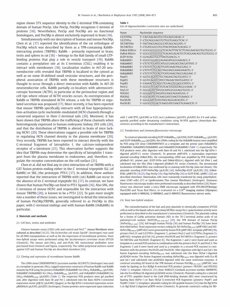
Table 1List of oligonucleotides (restriction sites are underlined)
Name Nucleotide sequence
5.E379Tfw 5′-CACCAACACGTCCCTGCAGCGACAG-3′5.E379Trv 5′-CTGCAGGGACGTGTTGGTGAAGCTCAC-3′5H.T407Efw 5′-TACTAACGAGGGCCATCAGCAGGATG-3′5H.T407Erv 5′-CTGATGGCCCTCGTTAGTATAACTCACAGC-3′DsRed-KSKLrv 5′-GGGGGCGGCCGCTCACAGTTTACTCTTCAGGAACAGGTGGTGGCG-3′DsRed-RSLLrv 5′-GGGGGCGGCCGCTCACAGGAGACTCCTCAGGAACAGGTGGTGGCG-3′EGFP-N1fw 5′-GCAGAGCTGGTTTAGTGAACCG-3′HsRab8bF1 5′-GGGCCGGATCCAGAAGATGGCGAAGACG-3′HsRab8bF2 5′-CTCTCTGGATCCATGGCGAAGACGTACGATTATC-3′HsRab8bR1 5′-CTCTCTCTGCAGTTCATCAAAGTAGCGAGC-3′HsRab8bR2 5′-CTCTCTCTGCAGCTAGCAACGAAAGAAACTG-3′HsRab8bR3 5′-CTCTCTCTGCAGTTCATCAAAGTAGCGAGGAAC-3′PeptF1 5′-GGTCCAGATCTTCCTAGAACTAGTGGATCC-3′PeptF2 5′-GGTCCAGATCTATCCTAGAACTAGTGGATCC-3′PeptR1 5′-CAAAACTGCAGCCTCGAGGTCGACGGTATCG-3′Pex5.2 5′-GCCCGTCGACCTGTCACTGGGGCAGGCCAAAC -3′Pex5.31 5′-GGGAGATCTATGGCAATGCGGGAGCTGGTG-3′PEX5H1 5′-GGGAATTCAAGATGCAAGCAGAATGGG-3′PEX5H2 5′-GGGAGATCTTCAAGGATCCAAGTTGAAAGC-3′Pex5H.F8 5′-GGGGAATTCATGTACCAGGGACACATG-3′Pex5H.R1 5′-GGGGGTCGACTCAAGGATCCAAGTTGAAAGCTC-3′
865M. Fransen et al. / Biochimica et Biophysica Acta 1783 (2008) 864–873
region shows 57% sequence identity to the C-terminal TPR-containingdomain of human Pex5p. Like Pex5p, Pex5Rp recognizes C-termini ofproteins [16]. Nevertheless, Pex5p and Pex5Rp are no functionalhomologues, and Pex5Rp is almost exclusively expressed in brain [16].
Simultaneously with our description of human andmouse Pex5Rp,Chen et al. [17] reported the identification of the rat orthologue ofPex5Rp which was described by them as a TPR-containing Rab8b-interacting protein (TRIP8b). Rab8b – primarily expressed in brain,testis and spleen in rat [18] – belongs to the Rab family of small GTP-binding proteins that play a role in vesicle transport [19]. Rab8bcontains a prenylation site at its C-terminus (CSLL) enabling it tointeract with membranes [18]. Localization studies in AtT-20 neu-roendocrine cells revealed that TRIP8b is localized in the cytosol aswell as on some ill-defined small vesicular structures, and the peri-pheral association of TRIP8b with these membrane structures isthought to occur through a direct interaction with Rab8b. In AtT-20neuroendocrine cells, Rab8b partially co-localizes with adrenocorti-cotropic hormone (ACTH), in particular at the perinuclear region andcell tips where release of ACTH vesicles occurs. As overexpression ofRab8b or TRIP8b stimulated ACTH release, a role for TRIP8b in regu-lated secretion was proposed [17]. More recently, it has been reportedthat mouse TRIP8b specifically interacts with all four hyperpolariza-tion-activation cyclic nucleotide-modulated (HCN) channels through aconserved sequence in their C-terminal tails [20]. Moreover, it hasbeen shown that TRIP8b alters the trafficking of these channels whenheterologously expressed in human embryonic kidney 293 cells [20],and that the distribution of TRIP8b is altered in brain of mice lack-ing HCN1 [20]. These observations suggest a possible role for TRIP8bin regulating HCN channel density in the plasma membrane [20].Very recently, TRIP8b has been reported to interact directly with theC-terminal fragment of latrophilin 1, the calcium-independentreceptor of α-latrotoxin [21]. This observation further supports theidea that TRIP8b may determine the specificity of latrophilin 1 trans-port from the plasma membrane to endosomes, and, therefore, re-gulate the receptor concentration on the cell surface [21].
Chen et al. did not find any in vitro interaction of rat TRIP8b with aGST fusion protein ending either in the C terminal tripeptide SLL (ofRab8b) or SKL (the prototypic PTS1) [17]. In addition, these authorsreported that the interaction of TRIP8b with (rat) Rab8b can occur inthe absence of its C-terminal tripeptide SLL [17]. In contrast, we haveshown that human Pex5Rp can bind to PTS1 ligands [16]. Also SNL, theC-terminus of mouse HCN1 and responsible for the interaction withmouse TRIP8b [20], is known to be a PTS1 [22]. To gain more insightinto a number of these issues, we investigated in detail the interactionof human Pex5Rp/TRIP8b, generally referred to as Pex5Rp in thispaper, with C-terminal endings and with human Rab8b (HsRab8b) inparticular.
2. Materials and methods
2.1. Cell lines, strains and antibodies
Chinese hamster ovary (CHO) cells and control and Pex5−/− mouse fibroblasts werecultured as described [16,23]. The Escherichia coli strain Top10F′ (Invitrogen) was usedfor all DNA manipulations as well as for the expression of recombinant proteins. Yeasttwo-hybrid assays were performed using the Saccharomyces cerevisiae strain SFY526(Clontech). The mouse anti-(His)6 and anti-FLAG M2 monoclonal antibodies werepurchased from Clontech and Sigma, respectively. The rabbit polyclonal antisera raisedagainst GST and human Pex14p are described elsewhere [24].
2.2. Cloning and expression of recombinant human Rab8b
The cDNA clone CS0DD009YM15 (accession number AL530175) (Invitrogen)was usedas a template to generate (His)6-tagged expression constructs of human Rab8b and Rab8bmutants byPCRusing theprimersHsRab8bF1/HsRab8bR1 for (His)6-HsRab8bWT (pLA190),HsRab8bF1/HsRab8bR2 for (His)6-HsRab8bΔSLL (pLA193), and HsRab8bF1/HsRab8bR3 for(His)6-HsRab8bC204S (pLA205) (Table 1). The respective PCR products were gel purifiedand, after digestion with Bam HI and Pst I, subcloned into the Bam HI/Pst I-restrictedexpression vector pQE32 (pLA190) (Qiagen) or the Bgl II/Pst I-restricted expression vectorpBADHisA (pLA193, pLA205) (Invitrogen). Fusion proteinswere expressed upon induction
with 1 mM IPTG (pLA190) or 0.2% (w/v) arabinose (pLA193, pLA205) for 4 h and subse-quently purified under denaturing conditions using Ni-NTA agarose (Amersham Bio-sciences) according to the manufacturer's instructions.
2.3. Transfections and (immuno)fluorescence microscopy
ToconstructplasmidsencodingEGFP-HsRab8bWT (pLA194), EGFP-HsRab8bΔSLL (pLA195)and EGFP-HsRab8bC204S (pLA206), the cDNAs coding for the HsRab8b fusionswere amplifiedby PCR using EST clone CS0DD009YM15 as a template and the primer pairs HsRab8bF2/HsRab8bR1, HsRab8bF2/HsRab8bR2 and HsRab8bF2/HsRab8bR3 (Table 1), respectively. ThePCR products were, after digestion with Bam HI and Pst I, subcloned into the Bgl II/Pst I-digested pEGFP-C1 vector (Clontech). To generate pMF1479, a mammalian expressionplasmid encoding DsRed-RSLL, the corresponding cDNA was amplified by PCR (template:pDsRed1-N1; primer pair: EGFP-N1fw and DsRed-RSLLrv), digested with Sal I/Not I, andsubcloned into the Xho I/Not I-digested pDsRed1-N1 vector (Clontech). The peroxisomallocalization of DsRed-RSLL was confirmed by co-stainingwith Pex14p (data not shown). Theplasmids encoding EGFP-HsPex5Rp (pLA118) [16], HsPex5Rp-EGFP (pLA120) [16], DsRed-KSKL (pMF578) [16,25], Flag-Pex5p [16], Flag-HsPex5Rp [16] or EGFP-KSKL (pMB1) [26] aredescribed elsewhere. Mammalian cells were transiently transfected by using polyethyleni-mine (CHO cells) [27] or Lipofectamine Plus (mouse fibroblasts) (Invitrogen). (Immuno)fluorescencemicroscopy studieswere performed essentially as described before [28]. Fluore-scence was observed under a Leica DMR microscope equipped with FITC/RSGFP/Bodipy/Fluo3/DIO and Texas Red filters, or evaluated on a CellM imaging station (Olympus)equipped with U-MNUA2, U-MNIBA3, and U-MWIY2 fluorescence mirror units.
2.4. Yeast two-hybrid analyses
The cotransformation of the bait and prey plasmids in chemically-competent SFY526cells aswell as the liquid culture andcolony liftfilter assays forβ-galactosidase activitywereperformed as described in themanufacturer's instructions (Clontech). The plasmids codingfor a fusion of Gal4p activation domain (AD) to the 70 C-terminal amino acids of ratpalmitoyl-CoA oxidase (RnPCOX592–658-SKL) [29], the TPR domain of human Pex5p(HsPex5p221–602) [29], full-length human Pex5p [30] and human Pex5Rp [16] have beendescribed before. Yeast expression vectors coding for AD-HsPex5pE379T (pMF1382) andAD-HsPex5RpT407E (pMF1422)were generatedby fusionPCR (pMF1382: templatepMF106 [30],primers Pex5.31 and 5.E379Trv (fragment 1), primers Pex5.2 and 5.E379Tfw (fragment 2);pMF1422: template pLA134 [16], primers Pex5H.F8 and 5H.T407Erv (fragment 3), primers5H.T407Efw and Pex5H.R1 (fragment 4)). The fragments 1 and 2 were fused and used astemplate in a secondPCR reaction in combinationwith the primers Pex5.31 andPex5.2. Thefragments 3 and 4 were fused and used as a template in a second PCR reaction in com-binationwith the primers Pex5H.F8 and Pex5H.R1. After digestionwith Bgl II and Sal I, thefusion fragment encoding HsPex5pE379T was subcloned into the Bam HI/Sal I-digestedpGAD424 vector. The fusion fragment encoding HsPex5RpT407E was digested with Eco RIand Sal I and subcloned into pGAD424 digested with the same restriction enzymes. Aconstruct encoding AD fused to the TPR domain of human Pex5Rp(321–624) (pMF341) wasmade by ligating an Eco RI/Bgl II-restricted PCR product (primers: PEX5H1 and PEX5H2(Table 1); template: I.M.A.G.E. [31] clone 1640223 (Genbank accession number AI090876)into the Eco RI/BamHI-digested pGAD424 vector (Clontech). Plasmids coding for a selectedset of PTS1 peptides fused to the Gal4 DNA-binding domain (BD) were constructed byligating Bam HI/Pst I (or Bam HI/Sal I)-restricted PCR fragments (primers: PeptF1 andPeptR1 (Table 1); templates: plasmids coding for AD-peptide fusions [32]) into theBgl II/PstI (or Bgl II/Sal I)-digested pGBT9 vector (Clontech). To generate constructs coding for BD-

Table 2Pex5p and Pex5Rp display different affinities for C-terminal endings in the yeast two-hybrid system
Peptide Sequence β-Galactosidase activity
HsPex5p(221–602) HsPex5p HsPex5Rp(321–624) HsPex5Rp
Sc01 CERSKL 775±351 265±89.1 24.9±2.7 28.0±13.0Sc02 GSHRRMDATKRRESKL 447±322 66.6±44.8 2.1±0.9 b1Sc17 WDSAVAWVPRKRVCHL b1 b1 18.5±6.6 1.9±1.2Sc23 HQLLEWFPVYNRSTKL b1 b1 3.1±1.7 2.0±2.1Sc26 KRVWRRQWTGRKLKL b1 b1 b1 b1Sc27 WYGPGPGCCRRRDLKL b1 b1 b1 b1Hs04 VVYSLLALAVQCSKL 6.8±3.0 1.1±0.4 3.8±1.3 2.3±0.9Hs13 YRFNICDAIFQGGSRL 446±115 165±42.4 69.6±23.1 46.5±43.0Hs19 GETMPPNNIGCTASRL 79.5±50.1 53.8±38.0 8.6±6.2 7.5±4.1Hs31 WRIVSPYTGGSLSCKL 394±93.2 52.1±37.0 48.9±0.9 35.0±32.8Hs35 SKVFVSGWGAGGMSRM 424±93.5 117±81.9 b1 b1Hs50 SDTIAMNCVQVKSQL 1093±127 442±275 65.0±16.5 62.8±21.1Hs51 MKHSWARYENIMSSQL 8.4±6.7 1.8±1.6 59.3±7.6 59.6±50.0Hs54 SEVGTTMRGDTLTSLL 6.4±2.5 3.5±1.8 584±126 266±112Hs55 EGGRRGMGFPAVRSLL 1824±318 583±219 1338±194 558±362Hs56 CRSGLPCLQSLL 835±111 121±62.9 1053±160 506±154Hs57 PEYSRAGQGTMWRSLL 535±151 64.1±51.3 569±68.2 221±121Hs61 TEEKVVGVGGCVKSYL 165±40.9 55.7±52.2 16.8±7.7 10.0±6.0Hs62 DGLWLGNFGVSLCSSL 12.0±2.1 3.0±1.5 178±67.0 217±130Hs63 QGNNNGQVRDWCRPSL 21.9±19.4 9.8±7.7 124±79.6 146±57.3Hs64 AGGRIASNCNLVSAL b1 b1 12.5±6.4 4.4±4.0Hs65 MKLRMVVRMNPLKCVL 12.2±7.6 b1 352±36.2 145±97.8
A select set of C-terminal endings, fused to the Gal4p DNA-binding domain, were tested for interaction with HsPex5p, HsPex5p(221–602), HsPex5Rp, or HsPex5Rp(321–624), fused tothe Gal4p activation domain, in the yeast two-hybrid system. Double transformants expressing one of the bait and one of the prey fusion proteins were selected and assayed forβ-galactosidase activity using o-nitrophenyl-β-D-galactopyranoside as the substrate. The optical densitiesweremeasured at 420 nm, normalized for culture densities (optical densitiesat 600 nm=10) and time (24 h), and corrected for the blank (‘empty’ plasmids only). The values given aremeans (±the standard deviation) for at least threemeasurements performedon cultures derived from independent colonies. Pex5p (or Pex5Rp) interactions yielding a strong β-galactosidase activity (N50 normalized units) and showing a N10-fold difference inactivity (for both the full-length and the truncated forms of the protein) relative to Pex5Rp (or Pex5p) are underlined.
866 M. Fransen et al. / Biochimica et Biophysica Acta 1783 (2008) 864–873
fusions to HsRab8bWT (pLA188), HsRab8bΔSLL (pLA189), or HsRab8bC204S (pLA203), therespective cDNAswere amplifiedby PCRusingESTcloneCS0DD009YM15as a template andthe same primer pairs as described for the cloning of the (His)6-tagged fusion proteins. PCRproducts were, after digestion with Bam HI and Pst I, subcloned into the Bam HI/Pst I-digested pGBT9 vector. To generate constructs coding for BD-fusions to DsRed-KSKL(pMF1428) or DsRed-RSLL (pMF1429), the respective cDNAs were amplified by PCR using
Fig. 1. Pex5p and Pex5Rp display different specificities for GST-PTS1 fusion proteins in afar Western assay. (A) Five µg of purified bacterially expressed GST (-), GST-GSHRRMDATKRRESKL (Sc02; SKL), GST-SKVFVSGWGAGGMSRM (Hs35; SRM), GST-SEVGTTMRGDTLTSLL (Hs54; SLL), or GST-DGLWLGNFGVSLCSSL (Hs62; SSL) wassubjected to SDS-PAGE and blotted onto nitrocellulose membranes. After blocking, theblots were probed with anti-GST antibodies (α-GST), biotinylated-Pex5p(221–602) (b-5/TPR) or biotinylated-Pex5Rp(321–624) (b-5R/TPR). Reactive complexes were identifiedwith goat anti-rabbit-alkaline phosphatase (AP) or streptavidin-AP conjugates. Forclarity, only the relevant portions of the blot are shown. (B) Comparison of the expres-sion levels of b-5/TPR and b-5R/TPR. Equal amounts of the respective bacterial lysateswere subjected to SDS-PAGE and blotted onto a nitrocellulosemembrane. After blocking,the blot was incubated with streptavidin-AP. The migration of relevant molecular massmarkers is shown on the left.
pDsRed-N1 as a template in combinationwith the primer pairs EGFP-N1fw/DsRed-KSKLrvand EGFP-N1fw/DsRed-RSLLrv, respectively. The PCR products were, after digestion withBgl II and Not I, subcloned into Bam HI/Not I-restricted pMF812, a pGBT9-derivative [30].
2.5. Pex5p/Pex5Rp binding experiments
In vitro interactions of recombinant (His)6-tagged Rab8b (and Rab8b variants) withrecombinant GST-tagged Pex5p and Pex5Rp fusion proteins were analyzed in a blot-overlay assay, essentially as described before [16,33]. In vitro interactions of recombinantGST-tagged PTS1peptideswere analyzed in a similarway, using biotinylated fusions to theTPRdomains of Pex5p [33] or Pex5Rp [16]. TheGST-taggedPTS1 peptides Sc02, Hs35,Hs54and Hs62 were constructed by ligating Bgl II/Sal I-restricted PCR fragments (oligonucleo-tides: PeptF2 and PeptR1 (Table 1); templates: plasmids coding for the respective AD-peptide fusions [32]) into the Bam HI/Sal I-digested pGEX-4T-1 vector (AmershamBiosciences). GSTandGST-peptide fusionswere expressedupon inductionwith1mMIPTGfor 4 h and subsequently purifiedusing glutathione Sepharose 4B (AmershamBiosciences)according to the manufacturer's instructions. For competition experiments, 1 μg of (His)6-Rab8bwas coated inwells of a 96-wellmicrotiter plate and incubatedwith thebiotinylatedPex5p or Pex5Rp fusion protein as described previously [33].
Table 3Pex5p, Pex5pE379T, Pex5Rp and Pex5RpT407E display different affinities for a select set oftarget proteins in the yeast two-hybrid system
BD ↓ AD → β-Galactosidase activity
HsPex5p HsPex5pE379T HsPex5Rp HsPex5RpT407E
HsRab8b b1 19.0±8.8 86.9±36.0 26.2±6.3HsRab8bΔSLL b1 b1 b1 b1HsRab8bC204S b1 9.8±3.5 42.2±7.3 33.5±3.6RnPCOX592–658-SKL 403.6±52.0 153.9±20.0 185.7±10.3 222.7±21.8DsRed-KSKL 314,1±64.7 154.7±43.9 171.5±19.6 110.5±46.6DsRed-RSLL 262.8±31.3 255.7±52.4 283.6±58.9 147.8±10.9
Yeast transformants expressing one of the bait (BD) and one of the prey (AD) fusionproteins were selected and assayed for β-galactosidase activity using o-nitrophenyl-β-D-galactopyranoside as the substrate. The optical densities were measured at 420 nm,normalized for culture densities (optical densities at 600 nm=10) and time (24 h),and corrected for the blank ('empty' plasmids only). The values given are means (±thestandard deviation) for at least threemeasurements performed on cultures derived fromindependent colonies.

Fig. 2. Pex5p and Pex5Rp display similar binding specificities for HsRab8b, HsRab8bΔSLL,and HsRab8bC204S in a far Western assay. Purified bacterially expressed (His)6-HsRab8b(WT), (His)6-HsRab8bΔSLL (ΔSLL), and (His)6-HsRab8bC204S (C204S) were subjected toSDS-PAGE and blotted onto nitrocellulose membranes. After blocking, the blots were(A) incubated with a mouse 6xHis monoclonal antibody followed by anti-mouse IgGcoupled to alkaline phosphatase, or (B) probed with the supernatant of bacterial lysatescontaining GST, GST-Pex5Rp (GST-5R) or Pex5p (GST-5). GST-containing proteincomplexes were identified with a rabbit anti-GST antiserum and an anti-rabbit-APconjugate. The migration of the 30 kDamolecular mass marker is shown on the left. Theslightly larger size of the (His)6-HsRab8bΔSLL and (His)6-HsRab8bC204S proteins is due tothe presence of 28 extraneous amino acids behind the (His)6-tag (see ‘Materials andmethods’ section).
867M. Fransen et al. / Biochimica et Biophysica Acta 1783 (2008) 864–873
2.6. Sequencing, computer analysis and modelling
DNA sequencing was done by using an ALF sequencer (Amersham Biosciences) andvector- and gene-specific primers or by sending the samples to Agowa (Germany).Automated knowledge-based proteinmodelling was performedwith the SWISSMODELprogram at the ExPaSy proteomics server [34].
3. Results
3.1. Pex5p and Pex5Rp display different affinities for C-terminal endings
Applying the two-hybrid system, we compared the binding affinitiesofHsPex5pandHsPex5Rp towards a setof C-terminal endingspreviouslyidentified as ligands of either the human or the yeast PTS1 receptorPex5p among themultitude of peptides of a randomlygenerated peptidelibrary [32]. Since both fusion proteins consisting of the Gal4p DNA-binding domain (BD) and the full-length Pex5p [30] or Pex5Rp (data notshown) autoactivate the LacZ reporter gene, the interaction assays werecarried out by employing the Gal4p DNA-binding domain extended byshort C-terminal endings and the Gal4p activation domain (AD) fused to
Fig. 3.HsRab8b and the peptide pSKL compete for binding to Pex5p and Pex5Rp in vitro.Microtiter wells were coated with 1 µg of purified bacterially expressed (His)6-HsRab8band incubated with the supernatant of a bacterial lysate containing biotinylatedHsPex5p(221–602) (squares) or biotinylated Pex5Rp(321–624) (triangles) in the presence ofincreasing amounts of the peptides pSKL (CSYHKHLKPLQSKL [29]) (open symbols) orpΔSKL (CVHESYHKHLKPLQ [29]) (closed symbols). After extensive washing, boundPex5p and Pex5Rp were detected photometrically with streptavidin-alkaline phospha-tase using p-nitrophenyl phosphate as a substrate.
either the full-length proteins or the respective TPR domains. This set-upis different to the previously used one [32] and might explain quanti-tative differences when compared.
The results of these experiments showed that AD-Pex5Rp fusionproteins can interact with a distinct set of C-terminal endings, and thebinding specificities differ between Pex5p and Pex5Rp fused to the ADof Gal4p (Table 2). AD-Pex5Rp showed highest affinities towardspeptides ending in SLL, SSL, and CVL, and little or no binding topeptides ending in SKL or SRM, which represent together with SLL thepreferred binding partners of AD-Pex5p. In general, the valuesobtained with the TPR containing fragments were higher than thoseobtained with the full-length proteins, which may represent avariation in the expression levels or stabilities of the correspondingAD-fusion proteins. However, the conclusion that Pex5p and Pex5Rpdisplay different affinities for C-terminal endings in the employedassay system remains.
Fig. 4. Molecular modelling of the Pex5Rp interaction (A) Overlay of the backbonestructures of the TPR domains of Pex5p (green) and Pex5Rp (red). Amino acids of Pex5pinteracting with the PTS1 peptide YQSKL (blue) as described in [7] are indicated. Thoseamino acids that differ between Pex5p and Pex5Rp are boxed. (B) Interactions betweenthe PTS1 peptide YQSKL (blue) and the amino acids of Pex5p (white and green) andPex5Rp (white and red). Those amino acids involved in the PTS1 interaction that don'tdiffer between Pex5p and Pex5Rp are indicated in white. Different amino acids arerepresented in green (Pex5p) and red (Pex5Rp): Lys (Pex5p) and Arg (Pex5Rp) on theleft, Glu and Ser (Pex5p) and Thr and Gly (Pex5Rp) on the right.

868 M. Fransen et al. / Biochimica et Biophysica Acta 1783 (2008) 864–873
In previous experiments [16] employing blot-overlay assays andrat liver proteins, Pex5p and Pex5Rp seemed to recognize the sameset of PTS1-containing proteins. To resolve this issue, the PTS1peptides Sc02, Hs35, Hs54 and Hs62 (see Table 2) were fused to GST,
Fig. 5. SLL, the C-terminal tripeptide of Rab8b, can function as a peroxisomal targeting signaRab8b variants, were processed for (in)direct fluorescence microscopy 24 h after transfectio(the exposure time for DAPI was 12 ms). (A–C) Distinct fluorescent staining patterns obseexposure time) a portion of the protein localizes to peroxisomes, as illustrated by its colocaltripeptide SLL of Rab8b (ΔSLL) abolishes peroxisomal targeting. (E) An almost exclusive peroof the outlined regions. Bars: 20 µm.
and binding to the TPR domains of Pex5p and Pex5Rp wasinvestigated. As shown in Fig. 1A, peptides Hs35 (ending in SRM)and Hs62 (ending in SSL) gave rise to an interaction with Pex5p orPex5Rp only, respectively, matching the two-hybrid results. However,
l in CHO cells. CHO cells, transiently transfected with plasmids encoding the indicatedn. Images of each channel were acquired separately with the indicated exposure timesrved for EGFP-Rab8b. Note that in cells expressing high levels of EGFP-Rab8b (= shortization with the peroxisomal membrane protein Pex14p. (D) Deletion of the C-terminalxisomal localization is observed with EGFP-Rab8bC204S. The insets show an enlargement

869M. Fransen et al. / Biochimica et Biophysica Acta 1783 (2008) 864–873
while Pex5Rp showed no affinity for peptide Sc02 (ending in SKL) andPex5p showed very low affinity for peptide Hs54 (ending in SLL) in theyeast two-hybrid system, both proteins did efficiently interact withthe corresponding GST-peptide fusion proteins in a blot-overlay assay(Fig. 1A). A plausible explanation for this discrepancy is that the(native) BD-PTS1 and (renatured) GST-PTS1 fusion proteins displayconformational differences.
3.2. Pex5p and Pex5Rp bind the C-terminus of human Rab8b in vitro
As the C-terminus of human Rab8b consists of SLL which might berecognized by Pex5p and Pex5Rp (Table 2, Fig. 1A, and [32]), theinteraction of Rab8b with Pex5p and Pex5Rp was analyzed in moredetail using yeast two-hybrid (Table 3), blot overlay (Fig. 2), andpeptide competition (Fig. 3) assays. In the yeast two-hybrid assay aninteraction was found only between Pex5Rp and HsRab8b, and thisinteraction depended on the presence of SLL (Table 3). In contrast, inthe blot overlay and peptide competition assays, bacterially expressed(His)6-tagged HsRab8b was recognized by both Pex5p and Pex5Rp.Both interactions depended on the presence of SLL (Fig. 2) and couldbe competitively inhibited by a peptide ending in SKL, but not by arelated peptide lacking this C-terminal tripeptide (Fig. 3). Summar-ized, these results suggest that bacterially expressed HsRab8b con-tains a C terminal end, which is recognized by TPR domains from bothPex5p and Pex5Rp. In contrast, in yeast cells only one interaction wasvisualized in a transcription-based assay, indicating structural diffe-rences between bacterially produced interaction partners and pro-teins expressed in vivo. As (i) CSLL, the C-terminal tetrapeptide ofRab8b represents a CAAX prenylationmotif [35], and (ii) prenylation ofthe cysteine residue in the CAAX box may induce structural changesand lead to proteolyis of the three C-terminal (AAX) amino acids invivo [35,36], we also investigated whether HsRab8bC204S, a prenyla-tion-defective variant of Rab8b, was able to interact with Pex5p andPex5Rp. Pex5p interactedwith HsRab8bC204S and its wild-type versionin a blot-overlay assay (Fig. 2), but not in the yeast two-hybrid assay
Fig. 6. Peroxisomal import of EGFP-Rab8bC204S is dependent upon the PTS1-receptor Pex5p. Ccontrol (5+/+) and Pex5p-deficient (5−/−) mouse fibroblasts, and processed for direct fluoresc
(Table 3). Pex5Rp recognized HsRab8bC204S in both assays (Table 3),ruling out that the two-hybrid interaction might be compromised byin vivo modification.
3.3. Comparative protein modelling of Pex5p and Pex5Rp
Since the interaction between Pex5p or Pex5Rp and the C-terminiof their partners ismediated through its TPR-containing domain [4,16],and the crystal structure of the TPR domain of Pex5p in complexwith aPTS1 peptide [7] or a substrate protein [9,10] has already been charac-terized, the distinct PTS1-binding affinities of Pex5p and Pex5Rp couldpossibly be explained by comparative protein modelling. The C-ter-minal regions of Pex5p and Pex5Rp contain seven TPR motifs each.Crystal structure analysis of this domain of Pex5p in complex witha PTS1 peptide has revealed the presence of two clusters of threeTPRs separated by a hinge region representing the fourth repeat [7].Modelling of the TPR domain of Pex5Rp using SWISS MODEL [34]revealed an identical fold of the main chain (Fig. 4A), consistent withthe observed interactions. However, different amino acid side chainsmight account for differences between Pex5p and Pex5Rp in theirability to interactwith C-termini (Fig. 4B). The asparagine residues thatbind the peptide backbone and the C-terminal carboxylate (Asn378,Asn489, Asn497, and Asn524 in the short isoform of Pex5p [29]) [7] areconserved, as well as Arg520 of Pex5p which is engaged in a water-mediated interaction with the carboxylate ion. The amino acid Lys490of Pex5p, which is also involved in the latter interaction, is replaced byanother basic amino acid, arginine, the influence of which is not clear.Almost all residues responsible for the recognition of the C-terminaltripeptide Ser-Lys-Leu (Glu348, Val374, Thr377, Asn378, Asn489,Ala493, Tyr508, Asn524, and Ser528 in Pex5p; [7]) are conserved,except glutamate at position 379 of Pex5p is replaced by a threonine(at position 407 of Pex5Rp). This probably results in a change of thepocket formed by the acidic residues (Glu348 and Glu379 in Pex5p) towhich usually the positively charged lysine of the C-terminal tripep-tide SKL is bound via a water-mediated interaction. Indeed, peptides
onstructs encoding EGFP-PTS1 and EGFP-HsRab8bC204S were transiently transfected intoence microscopy 24 h after transfection. Bars: 20 µm.

870 M. Fransen et al. / Biochimica et Biophysica Acta 1783 (2008) 864–873
with R or K at the penultimate position of the PTS1 do bind weakly toPex5Rp (Table 2). Also, a Pex5p variant in which the glutamate atposition 379 was replaced by a threonine generally displayed com-pared to thewild-type protein amarkedly lower and higher affinity forproteins ending in -SKL and -SLL, respectively (Table 3). Interestingly, aPex5Rp variant inwhich the threonine at position 407was replaced byglutamate displayed a lower affinity for proteins ending in -SLL, but nosignificant increase in affinity for proteins ending in -SKL (Table 3).Although these observations are in line with the hypothesis thatthe glutamate at position 379 of Pex5p (or the threonine at position407 of Pex5Rp) is important for recognition of proteins with positi-vely charged (or non-charged) residues at the penultimate position of
Fig. 7. Localization of Rab8bvariants and Pex5Rp or Pex5p upon co-expression in CHO cells. CHand (A) Flag-Pex5Rp or (B) Flag-Pex5p. After 24 h, the cells were fixed and processed for fluo
C-terminal endings, they also indicate that other amino acids involvedin interactions with residues upstream of the ultimate tripeptide maymodulate binding. For instance, the serine at position 380 of Pex5p,which is thought to form hydrogen bonds with the fourth last aminoacid, is replaced by a glycine in Pex5Rp, a substitution which is alsofound in S. cerevisiae Pex5p.
3.4. The C-terminus of human Rab8b can function as a PTS1 in vivo
In order to investigatewhether the C-terminal tripeptide of HsRab8bcan function as a PTS1 in vivo, CHO cells were transfectedwith plasmidscoding for EGFP-HsRab8b or EGFP-HsRab8bΔSLL. Cells expressing these
Ocellswere transiently transfectedwith plasmids encoding the indicatedRab8bvariantsrescence analysis. The insets show an enlargement of the outlined regions. Bars: 20 µm.

871M. Fransen et al. / Biochimica et Biophysica Acta 1783 (2008) 864–873
proteins exhibited distinctivefluorescent stainingpatterns ranging fromapunctate and/or tubular cytoplasmic to perinuclear staining (Fig. 5A–C,left column). Interestingly, in cells which express high levels of EGFP-HsRab8b (image data corresponding to short exposure times) some ofthe punctate structures observed were peroxisomes, as illustrated bytheir colocalization with the peroxisomal marker protein Pex14p(inserts Fig. 5C). As this peroxisomal staining pattern was never seenwith EGFP-HsRab8bΔSLL (inserts Fig. 5D), these observations show thatthe C-terminus of human Rab8b can function as a PTS1 in vivo. In orderto investigatewhether the inefficient targeting of Rab8b to peroxisomeswas the result of a CAAX box-mediated processing step, we also exa-mined the subcellular localization of EGFP-HsRab8bC204S. Interestingly,we found that this protein localized almost exclusively toperoxisomes inwild-type CHO cells (Fig. 5E). Similarly, in mouse fibroblasts a punctatestaining representative of peroxisomal localization was observed,whereas in Pex5p-deficient mouse fibroblasts EGFP-HsRab8bC204Sgave rise to a diffuse cytosolic staining pattern (Fig. 6). These obser-vations provide additional evidence that a single amino acid changeoutside the C-terminal tripeptide has the potential to strongly influence,either directly or indirectly, recognition by Pex5p.
3.5. Rab8b can act as a membrane-recruitment factor for Pex5Rp, but notPex5p
Pex5Rp was observed to be recruited to membrane structuresthrough binding to Rab8b [17]. Therefore, we investigated whether this
Fig. 8. Effect of overexpression of Pex5Rp on the import efficiency of DsRed-KSKL and DsRDsRed-KSKL or DsRed-RSLL and EGFP-HsPex5Rp (⁎5R) or HsPex5Rp-EGFP (5R⁎). After 24 h,localization of DsRed-fusion proteins was determined by its punctate (peroxisomal) or diffuseresults of a representative experiment using HsPex5Rp-EGFP (5R⁎) are shown.
small GTPase could also function as a membrane-recruitment factor forPex5p. CHO cells were co-transfected with constructs coding for EGFP-Rab8b or EGFP-Rab8bΔSLL and Flag-Pex5p or Flag-Pex5Rp, and the sub-cellular distribution pattern of the ectopically expressed fusion proteinswas analyzed by fluorescence microscopy. While Rab8b was able torecruit Pex5Rp to punctate and/or tubular cytoplasmic structures asexpected (Fig. 7A, upper panels), this protein failed to recruit Pex5p tosuch structures (Fig. 7B, upper panels). Interestingly, membrane recruit-ment of Pex5Rp seemed to be controlled by the C-terminus of Rab8b(Fig. 7A, middle panels). This observation is in agreement with theresults presented in Fig. 2 and Table 3, and suggests that (non-pre-nylated) Rab8b is a substrate for Pex5Rp, but not Pex5p, in vivo. Incombinationwith the results presented in Table 2 (e.g. compare Sc01 toHs04 for the ending –SKL), one may speculate that a cysteine imme-diately upstreamof C-terminal tripeptidyl endings is quite unfavourablefor Pex5p, but not Pex5Rp. This hypothesis is in linewith the observationthat Rab8bC204S is a substrate for Pex5p inwild-type cells (Fig. 5E, Fig. 6)and can be retained in the cytosol upon overexpression of Pex5p (Fig. 7B,lower panels). Note that also Pex5Rp is able to retain Rab8bC204S in thecytosol (Fig. 7A, lower panels).
3.6. Effect of overexpression of Pex5Rp on PTS1 protein import
As the results presented above suggest that Pex5p and Pex5Rp maycompete for binding to PTS1-like termini of proteins in vivo, we co-expressed EGFP-tagged Pex5Rp fusion proteins with either DsRed-KSKL
ed-RSLL in CHO cells. CHO cells were transiently transfected with plasmids coding forthe cells were fixed and processed for direct fluorescence analysis. (A) The subcellular(cytosolic) staining pattern in at least 100 cells, and the results were quantified. (B) The

872 M. Fransen et al. / Biochimica et Biophysica Acta 1783 (2008) 864–873
or DsRed-RSLL in CHO cells, and studied the effect of overexpression ofthe green fluorescent proteins on the subcellular localization of the redfluorescent reporter proteins (Fig. 8). When expressed alone DsRed-KSKL or DsRed-RSLL were associated with peroxisomes in almost allcells. Simultaneous overexpression of EGFP-tagged Pex5Rp had onlylittle effect on the subcellular localization of DsRed-KSKL, but resulted ina diffuse cytosolic staining pattern of DsRed-RSLL in approximately halfof the cells. These observations provide additional support for thehypothesis that Pex5Rp may interfere with PTS1-dependent import ofproteins in vivo, especially with those not ending in the canonical -SKL.
4. Discussion
Human Pex5Rp was shown to bind to peroxisomal proteins and toC-termini ending in the prototypical PTS1 tripeptide –SKL [16],although the corresponding rat orthologue did not [17]. The aim ofthis work was to study the interaction of Pex5Rp with C-termini inmore detail and to compare which ones the human Pex5p and Pex5Rpcan bind. By applying blot overlay, yeast two-hybrid, and in vivo co-expression assays, we showed that the binding specificities of Pex5Rpand Pex5p are different. In addition, we demonstrated that the gluta-mate at position 379 of Pex5p (or the threonine at position 407 ofPex5Rp) is important for recognition of proteins with positively char-ged (or non-charged) residues at the penultimate position of PTS1-likeC-terminal endings. With respect to the employed binding assays, itshould be noted that the results obtained by far Western analysis donot always corroborate those obtained from ‘in vivo’ experiments. Thismay not be so surprising taking into account that the context of the C-terminus exerts a great impact on its ability to interact with Pex5Rp orPex5p ([32] and this work), and the conformation of C-termini ofnative proteins can be expected to differ from those of the corres-ponding denatured proteins. Why Chen et al. [17] did not observe anyinteraction between Pex5Rp and GST-SKL remains unclear. The mostlikely explanation is that the amino acids immediately upstream ofSKL prevent binding. However, as this experimental detail is lacking,this can not be verified.
The rat orthologue of Pex5Rp has been described as a TPR-con-taining Rab8b-interacting protein (TRIP8b) [17]. Rab8b contains atypical C-terminal CaaX prenylation motif, CSLL. As the C-terminaltripeptide SLL can act as a PTS1 [32], we investigated whether expres-sion of EGFP-HsRab8b in CHO cells resulted in a peroxisomal loca-lization. In most cells, this protein was predominantly localized to as-yet-unidentified cytoplasmic membrane structures largely mimickingthe pattern reminiscent of that reported for myc-tagged rat Rab8b inrat basophilic leukaemia RBL.2H3, neuroblastoma PC12, and AtT-20neuroendocrine cells [17,18]. However, in a minority of cells EGFP-Rab8b was also partially associated with peroxisomes. Likely, someportion of the (overexpressed) wild-type HsRab8b escaped prenyla-tion and was mistargeted to peroxisomes in a Pex5p-dependent man-ner. This observation suggests that, if prenylation and proteolysis areprevented, the C-terminal tripeptide of Rab8b can act as a PTS1. Thishypothesis is in line with our observation that HsRab8bC204S, a pre-nylation deficient variant, is almost exclusively associated with pero-xisomes. Whether a portion of HsRab8b is targeted to peroxisomesunder physiological conditions remains to be investigated. If theamount of unprenylated Ras and Ras-related proteins is negligible invivo, the majority of Rab8b will not be localized to peroxisomes. Inaddition, it is very likely that the cysteine immediately upstream of theC-terminal tripeptide SLL plays a crucial role in favouring an inter-action with Pex5Rp compared to Pex5p. Moreover, other recognitioneventsmay contribute to the binding specificity in vivo. For example, itmay be that the interactions studied here are modulated by Rabin 8,another Rab8b binding protein. Rabin 8 is a Rab8-specific GDP/GTPexchange factor involved in actin remodelling and polarized mem-brane transport [37]. In addition, Rabin 8 was found to recruit a largeprotein complex, the BBSome, to the centrosome and to activate Rab8
leading to docking and fusion of vesicles near the ciliary membranes[38].
Related to the HCN1 and TRIP8b interaction, it is interesting to notethat this binding is dependent on SNL, the C-terminal tripeptide ofmouseHCN1 [20]. Previously, we have shown that this C-terminal tripeptide –
being also present in the human peroxisomal enzyme D-aspartate oxi-dase – can function as a PTS1, and that its peroxisome targeting efficiencybecomes higher when preceded by a positively charged residue [22]. Assuch a positively charged residue is absent in HCN1 (which ends in ASNL)and this transporter is most likely transported to the plasma membranevia the secretorypathway [39],HCN1 isunlikely tobe recognizedbyPex5pin vivo. On the other hand, this illustrates again the similarities in thebinding folds of the TPR domains of Pex5Rp/TRIP8b and Pex5p.
The absence of a well-defined gene-related phenotype (e.g. a patientcell line, a mouse model, or a deletion strain of the yeast orthologue)makes it difficult to define a functional role for Pex5Rp. However, theobservations that (i) endogenous Pex5Rp/TRIP8b and HCN1 colocalizewithin dendritic arbors of hippocampal CA1 and neocortical layer Vpyramidal neurons, (ii) the labeling of Pex5Rp/TRIP8b largely disappearsin HCN1 knockoutmice, and (iii) Pex5Rp/TRIP8b drastically decreases thesurface expression of HCN protein, provide strong evidence that Pex5Rp/TRIP8b is involved in controlling HCN channel density in the plasmamembrane [20]. In addition, the recent observation that Pex5Rp/TRIP8bcan interactwith latrophilin, amemberof the secretin familyofG-protein-coupled receptors, provides support for the hypothesis that Pex5Rp/TRIP8b may play a more general role in the trafficking pathway from theplasma membrane to endosomes for a select set of membrane proteins[21]. Thesefindings, togetherwith ourobservations that Pex5Rp/TRIP8b is(almost exclusively) expressed in brain and does not interact with otherperoxins [16], make it unlikely that Pex5Rp/TRIP8b possesses a peroxi-some-related function. Nevertheless, our observations that this proteincan compete with Pex5p for PTS1-binding in vitro and is able to retain aperoxisomal reporter protein in the cytosol upon overexpression in CHOcells, suggest that the PTS1-protein import efficiency in a cell may beaffected by altered Pex5Rp protein expression levels.
Acknowledgements
Wethank J.VanLooy,M.Brams, andV.Brys for theirexcellent technicalsupport, and Dr. M. Baes (Laboratorium voor Cellulair Metabolisme, K.U.Leuven) for the control and Pex5−/− mouse fibroblasts. We also thank thereviewers for their constructive feedback to help improve the quality ofthe manuscript. This work was supported by grants from the FlemishGovernment (Geconcerteerde Onderzoeksacties GOA2004/08), the Fondsvoor Wetenschappelijk Onderzoek – Vlaanderen (OnderzoeksprojectG.0237.04), and the FP6 European Union Project ‘Peroxisome’ (LSHG-CT-2004-512018). L.A. was supported by a Junior post-doctoral fellowshipfrom the K.U. Leuven.
References
[1] R.S. Sikorski, M.S. Boguski, M. Goebl, P. Hieter, A repeating amino acid motif inCDC23 defines a family of proteins and a new relationship among genes requiredfor mitosis and RNA synthesis, Cell 60 (1990) 307–317.
[2] L.D. D'Andrea, L. Regan, TPR proteins: the versatile helix, Trends Biochem. Sci. 28(2003) 655–662.
[3] C. Scheufler, A. Brinker, G. Bourenkov, S. Pegoraro, L.Moroder, H. Bartunik, F.U. Hartl,I. Moarefi, Structure of TPR domain-peptide complexes: critical elements in theassembly of the Hsp70-Hsp90 multichaperone machine, Cell 101 (2000) 199–210.
[4] C. Brocard, F. Kragler, M.M. Simon, T. Schuster, A. Hartig, The tetratricopeptiderepeat-domain of the PAS10 protein of Saccharomyces cerevisiae is essential forbinding the peroxisomal targeting signal SKL, Biochem. Biophys. Res. Commun.204 (1994) 1016–1022.
[5] A.K. Das, P.W. Cohen, D. Barford, The structure of the tetratricopeptide repeats ofprotein phosphatase 5: implications for TPR-mediated protein-protein interac-tions, EMBO J. 17 (1998) 1192–1199.
[6] H.C. Vodermaier, C. Gieffers, S. Maurer-Stroh, F. Eisenhaber, J.M. Peters, TPRsubunits of the anaphase-promoting complex mediate binding to the activatorprotein CDH1, Curr. Biol. 13 (2003) 1459–1468.
[7] G.J. Gatto, B.V. Geisbrecht, S.J. Gould, J.M. Berg, Peroxisomal targeting signal-1recognitionby the TPRdomains ofhumanPEX5,Nat. Struct. Biol. 7 (2000)1091–1095.

873M. Fransen et al. / Biochimica et Biophysica Acta 1783 (2008) 864–873
[8] C. Brocard, A. Hartig, Peroxisome targeting signal 1: is it really a simple tripeptide?Biochim. Biophys. Acta 1763 (2006) 1565–1573.
[9] W.A. Stanley, F.V. Filipp, P. Kursula, N. Schuller, R. Erdmann, W. Schliebs, M. Sattler,M.Wilmanns, Recognition of a functional peroxisome type 1 target by the dynamicimport receptor Pex5p, Mol. Cell 24 (2006) 653–663.
[10] W. Stanley, M. Wilmanns, Dynamic architecture of the peroxisomal importreceptor Pex5p, Biochim. Biophys. Acta 1763 (2006) 1592–1598.
[11] E.L.Maynard, J.M. Berg, Quantitative analysis of peroxisomal targeting signal type-1binding to wild-type and pathogenic mutants of Pex5p supports an affinitythreshold for peroxisomal protein targeting, J. Mol. Biol. 368 (2007) 259–1266.
[12] I. Heiland, R. Erdmann, Biogenesis of peroxisomes. Topogenesis of the peroxisomalmembrane and matrix proteins, FEBS J. 272 (2005) 2362–2372.
[13] G. Dodt, S.J. Gould, Multiple PEX genes are required for proper subcellulardistribution and stability of Pex5p, the PTS1 receptor: evidence that PTS1 proteinimport is mediated by a cycling receptor, J. Cell Biol. 135 (1996) 1763–1774.
[14] V. Dammai, S. Subramani, The human peroxisomal targeting signal receptor,Pex5p, is translocated into the peroxisomal matrix and recycled to the cytosol, Cell105 (2001) 187–196.
[15] A.M. Gouveia, C.P. Guimaraes, M.E. Oliveira, C. Reguenga, C. Sa-Miranda, J.E.Azevedo, Characterization of the peroxisomal cycling receptor, Pex5p, using a cell-free in vitro import system, J. Biol. Chem. 278 (2003) 226–232.
[16] L. Amery, H. Sano, G.P. Mannaerts, J. Snider, J. Van Looy, M. Fransen, P.P. VanVeldhoven, Identification of Pex5p-related novel peroxisome-targeting signal 1(PTS1)-binding proteins in mammals, Biochem. J. 357 (2001) 635–646.
[17] S. Chen, M.C. Liang, J.N. Chia, J.K. Ngsee, A.E. Ting, Rab8b and its interacting partnerTRIP8b are involved in regulated secretion in AtT-20 cells, J. Biol. Chem. 276 (2001)13209–13216.
[18] J. Armstrong, N. Thompson, J.H. Squire, J. Smith, B. Hayes, R. Solari, Identification ofa novel member of the Rab8 family from the rat basophilic leukaemia cell line,RBL.2H3, J. Cell Sci. 109 (1996) 1265–1274.
[19] F. Schimmöller, I. Simon, S.R. Pfeffer, Rab GTPases, directors of vesicle docking,J. Biol. Chem. 273 (1998) 22161–22164.
[20] B. Santoro, B.J. Wainger, S.A. Siegelbaum, Regulation of HCN channel surface expressionby a novel C-terminal protein-protein Interaction, J. Neurosci. 24 (2004) 10750–10762.
[21] N.V. Popova, A. Plotnikov, I.E. Deev, A.G. Petrenko, Interaction of calcium-independent latrotoxin receptor with intracellular adaptor protein TRIP8b, Dokl.Biochem. Biophys. 414 (2007) 149–151.
[22] L. Amery, C. Brees, M. Baes, C. Setoyama, R. Miura, G.P. Mannaerts, P.P. VanVeldhoven, C-terminal tripeptide Ser-Asn-Leu (SNL) of human D-aspartate oxidaseis a functional peroxisome-targeting signal, Biochem. J. 336 (1998) 367–371.
[23] M. Baes, P. Gressens, E. Baumgart, P. Carmeliet, M. Casteels, M. Fransen, P. Evrard, D.Fahimi, P.E. Declercq, D. Collen, P.P. Van Veldhoven, G.P. Mannaerts, A mousemodel for Zellweger syndrome, Nat. Genet. 17 (1997) 49–57.
[24] L. Amery, M. Fransen, K. De Nys, G.P. Mannaerts, P.P. Van Veldhoven, Mitochondrialand peroxisomal targeting of 2-methylacyl-CoA racemase in humans, J. Lipid Res.41 (2000) 1752–1759.
[25] M. Fransen, T. Wylin, C. Brees, G.P. Mannaerts, P.P. Van Veldhoven, Human Pex19pbinds peroxisomal integral membrane proteins at regions distinct from theirsorting sequences, Mol. Cell. Biol. 21 (2001) 4413–4424.
[26] I.M. Vastiau, E.A. Anthonio, M. Brams, C. Brees, S.G. Young, S. Van de Velde, R.J.Wanders, G.P. Mannaerts, M. Baes, P.P. Van Veldhoven, M. Fransen, Farnesylation ofPex19p is not essential for peroxisome biogenesis in yeast and mammalian cells,Cell. Mol. Life Sci. 63 (2006) 1686–1699.
[27] O. Boussif, F. Lezoualc'h, M.A. Zanta, M.D. Mergny, D. Scherman, B. Demeneix, J.P.Behr, A versatile vector for gene and oligonucleotide transfer into cells in cultureand in vivo: polyethylenimine, Proc. Natl. Acad. Sci. U. S. A. 92 (1995) 7297–7301.
[28] L. Amery, C. Brees, M. Baes, C. Setoyama, R. Miura, G.P. Mannaerts, P.P. VanVeldhoven, C-terminal tripeptide Ser-Asn-Leu (SNL) of human D-aspartate oxidaseis a functional peroxisome-targeting signal, Biochem. J. 336 (1998) 367–371.
[29] M. Fransen, C. Brees, E. Baumgart, J.C. Vanhooren, M. Baes, G.P. Mannaerts, P.P. VanVeldhoven, Identification and characterization of the putative human peroxisomalC-terminal targeting signal import receptor, J. Biol. Chem. 270 (1995) 7731–7736.
[30] M. Fransen, C. Brees, K. Ghys, L. Amery, G.P. Mannaerts, D. Ladant, P.P. VanVeldhoven, Analysis of mammalian peroxin interactions using a non-transcrip-tion-based bacterial two-hybrid assay, Mol. Cell. Prot. 1 (2002) 243–252.
[31] G. Lennon, C. Auffray, M. Polymeropoulos, M.B. Soares, The I.M.A.G.E. Consortium:an integrated molecular analysis of genomes and their expression, Genomics 33(1996) 151–152.
[32] G. Lametschwandtner, C. Brocard, M. Fransen, P.P. Van Veldhoven, J. Berger, A. Hartig,Thedifference in recognition of terminal tripeptides asperoxisomal targeting signal 1between yeast and human is due to different affinities of their receptor Pex5p to thecognate signal and to residues adjacent to it J. Biol. Chem. 273 (1998) 33635–33643.
[33] M. Fransen, C. Brees, P.P. Van Veldhoven, G.P. Mannaerts, The visualization ofperoxisomal proteins containing a C-terminal targeting sequence on Western blotby using the biotinylated PTS1-receptor, Anal. Biochem. 242 (1996) 26–30.
[34] N. Guex, M.C. Peitsch, SWISS-MODEL and the Swiss-Pdb viewer: an environmentfor comparative protein modelling, Electrophoresis 18 (1997) 2714–2723.
[35] F.L. Zhang, P.J. Casey, Protein prenylation: molecular mechanisms and functionalconsequences, Annu. Rev. Biochem. 65 (1996) 241–269.
[36] M. Zerial, H. McBride, Rab proteins as membrane organizers, Nat. Rev. Mol. Cell.Biol. 2 (2001) 107–117.
[37] K. Hattula, J. Furuhjelm, A. Arffman, J. Peränen, A Rab8-specific GDP/GTP exchangefactor is involved in actin remodeling and polarized membrane transport, Mol.Biol. Cell 13 (2002) 3268–3280.
[38] M.V. Nachury, A.V. Loktev, Q. Zhang, C.J. Westlake, J. Peränen, A. Merdes, D.C.Slusarski, R.H. Scheller, J.F. Bazan, V.C. Sheffield, P.K. Jackson, A core complex of BBSproteins cooperates with the GTPase Rab8 to promote ciliary membranebiogenesis, Cell 129 (2007) 1201–1213.
[39] B. Much, C. Wahl-Schott, X. Zong, A. Schneider, L. Baumann, S. Moosmang, A.Ludwig, M. Biel, Role of subunit heteromerization and N-linked glycosylation inthe formation of functional hyperpolarization-activated cyclic nucleotide-gatedchannels, J. Biol. Chem. 278 (2003) 43781–43786.