Comparison of exercise versus sport participation motives among ...

RESEARCH ARTICLE Open Access
Comparison of the effects of exercise withchondroitin sulfate on knee osteoarthritis inrabbitsNing Ma†, Tingting Wang†, Lianyu Bie, Yang Zhao, Lidong Zhao, Shai Zhang, Li Gao and Jianhua Xiao*
Abstract
Background: The aim of the study is to compare the effects of exercise therapy with chondroitin sulfate (CS)therapy in an experimental model of osteoarthritis (OA).
Methods: Twenty-one New Zealand rabbits were randomly divided into four groups: normal group (N group, n = 3);OA control group (C group, n = 6); OA plus medication group (CS group, n = 6); and OA plus exercise group (E group,n = 6). Four weeks after modeling, the rabbits were subjected to exercise (artificial, 30 min/time, 4 times/week) ormedicated with CS (2% CS, 0.3 ml/time, once/week) for 4 weeks. Histopathological changes in treated joints wereexamined after staining. X-ray and scanning electron microscopy was used to evaluate the different therapies byexamining the surfaces and joint spaces of the articular cartilage. RT-qPCR was used to assess chondrogenic geneexpression including Col2, Col10, mmp-13, il-1β, adamats-5, and acan in the experimental groups.
Results: Histology showed both treatment groups resulted in cartilage that was in good condition, with increasednumbers of chondrocytes, and the results of X-ray and scanning electron microscopy showed the therapeutic effectof exercise therapy is equivalent to CS therapy, surface articular cartilage was flat, and the of cartilage layer wasthinning. All treated groups induced the expression of Col10 and Col2 and decreased expression of mmp-13, il-1β,and adamats-5 compared with the control groups. The expression of acan was upregulated in the E group anddownregulated in the CS group. Furthermore, expression of Col10 was higher and il-1β was lower in the exercisegroup compared to that of the CS group.
Conclusion: These results indicate that exercise has a positive effect on OA compare with CS, and it also suppliesreference for the movement mode to improve function.
Keywords: Osteoarthritis, Exercise, Chondroitin sulfate, Rabbit
BackgroundOsteoarthritis (OA), one of the most common chronicconditions, is a chronic degenerative joint disease char-acterized by cartilage degeneration and erosion, fibrosis,and osteophyte formation, leading to pain and disabilityin the worst cases [1].The early changes in articular cartilage in OA is char-
acterized by the loss of proteoglycan and a reduction inthe expression of collagen 2 (Col2) gene without anychanges to the regularity of the structure the articular
tissue [2]. In contrast, the expression of PCNA, Ki67,and matrix metalloproteinase 13 (mmp-13) are signifi-cantly increased [3–5], in addition to observations ofchondrocyte changes similar to the stages of cell hyper-trophy [6, 7]. Severe OA mainly manifests as cartilagesurface cracks or fibrosis and defects in the cartilagewith the banded structure in disorder [8]. There arecracks in the fibrotic area, extending to the calcifiedzone accompanied by severe loss of proteoglycan anddegradation of Col2 [9], and upregulation of the expres-sion of mmp-13 and mmp-2 [10]. The expression ofgenes related to the terminal differentiation of chondro-cytes has been found in cell clusters around the cracks,such as Col2, collagen 10 (Col10), and TGF-β3 [11].
* Correspondence: [email protected]†Equal contributorsKey Laboratory of the Provincial Education Department of Heilongjiang forCommon Animal Disease Prevention and Treatment, College of VeterinaryMedicine, Northeast Agricultural University, Harbin 150030, China
© The Author(s). 2018 Open Access This article is distributed under the terms of the Creative Commons Attribution 4.0International License (http://creativecommons.org/licenses/by/4.0/), which permits unrestricted use, distribution, andreproduction in any medium, provided you give appropriate credit to the original author(s) and the source, provide a link tothe Creative Commons license, and indicate if changes were made. The Creative Commons Public Domain Dedication waiver(http://creativecommons.org/publicdomain/zero/1.0/) applies to the data made available in this article, unless otherwise stated.
Ma et al. Journal of Orthopaedic Surgery and Research (2018) 13:16 DOI 10.1186/s13018-018-0722-4

There are many ways to treat OA including slimmingin overweight patients, medication, non-pharmacologicalinterventions, and surgery. During the last 15 years, thefocus of treatment has begun to shift to non-medicinetreatment. It is generally believed that exercise can re-duce joint pain and improve joint function. Latham andLiu suggest exercise can strengthen the quadriceps [12],which effectively reduces lower limb pain and improvejoint function [13], and Bennell et al. believe the key tothe treatment of OA is restoring joint function [14, 15].Iijima et al. studied moderate exercise can also preventOA from causing joint tissue damage [16]. Chondroitinsulfate (CS), a slow-acting drug for the treatment ofarthritis, has an anti-inflammatory effect by reducing thesynthesis of proteolytic and inflammation-related en-zymes, and proinflammatory cytokines [17, 18]. In 2010,the International Society of OA pointed out that CS notonly reduces the symptoms of OA but also modifies thestructure of the cartilage after prolonged injection, sothat it is more advantageous in the therapy of OA [19].This paper studies whether exercise has some stimula-
tion on cartilage excluding the effect of muscularchanges, weight, joint loading, and so on; leads to somechanges in the surface of cartilage such as collagen, poly-saccharide, and chondrocytes; and ultimately achievesthe purpose of treatment of arthritis. CS appears to actas a protective agent of cartilage in the treatment of OA,so it can be used as a positive control group, to studythe effect of exercise on the treatment of arthritis usingclinical, histological, and genetic techniques.
MethodsAnimals and surgeryNew Zealand rabbits (n = 21) with a body weight in therange 2.5 ± 0.5 kg were used in the study. The rabbitsfasted for 12 h before surgery and were then anesthe-tized with ether. They were randomly divided into fourgroups. Except for the normal group (N, n = 3), rabbitswere made osteoarthritic as per the Hulth-Telhag model[20]; as described before, the medial side of the jointcapsule was opened without cutting the articular surfaceand the anterior cruciate ligament was cut and a portionof the meniscus removed, under sterile conditions. Thenormal group had a sham operation. Penicillin andstreptomycin (Yocon Biotechnology Co., Ltd., China)were administered twice daily for 3 days post-operativelyto avoid surgery-related infection. After 4 weeks, therabbits were randomly assigned to the various groups.
TreatmentRabbits were randomly divided into three groups afterthe arthritis models were successfully established.Rabbits in the 2% CS group (CS, n = 6, 30 min/time)were injected with 0.3 ml CS (Biosharp, China) once per
week. In the exercise group (E, n = 6), put the electricanimal treadmill upside down and the rabbits were lyingon the board, make it passively exercised for 30 min,four times a week. The control group (C, n = 6, saline)animals served as a control and were maintained as anosteoarthritic control and not given any treatment.
HistologyAfter 34 days of treatment, the articular cartilage fromthe middle part of the load-bearing region was shavedoff with a scalpel. Sections were frozen and stored in li-quid nitrogen if scheduling so required. Some pieceswere fixed in 10% formaldehyde solution then embeddedin paraffin, then finally stained with hematoxylin eosin(HE), toluidine blue, and alcan blue and prepared forlight microscopy according to standard procedures.
X-ray assessmentAnterioposterior and left-lateral images were performedon the knee joints of animals after 34 days of treatment,using X-rays and direct digital radiography (DR), to esti-mate the degree of cartilage degradation and joint spacereduction under anesthesia. Exposure to X-ray was con-ducted at 300 MA, 50 kV with a 0.03-s exposure time,and a 100-cm tube to film distance for AP projection.
Scanning electron microscopyAfter sacrifice, the knees of the hind legs of the animalswere dissected in sterile conditions. Medial femur tissuemeasuring 0.4 mm × 0.4 mm × 0.2 mm was harvested,submerged in a sterile solution of 0.9% NaCl at roomtemperature to prevent oxidation. All three groups werefixed in 4% paraformaldehyde for 2 days then submergedin OsO4 for 2 h. The samples were transferred to a crit-ical point dryer then coated with gold to generate an 18-nm-thick coating. Changes in the microstructure weredetermined using scanning electron microscopy (SEM).
RT-qPCR analysisTotal RNA was extracted with Trizol® Reagent (Trans-Gen Biotech, Beijing, China) according to the manufac-turer’s instructions. Reverse transcription was performedusing PrimeScriptTM RT Master Mix (Takara) accordingto the manufacturer’s instructions. The resultant cDNAwas used to amplify Col2, Col10, acan, mmp-13,interleukin-1β (IL-1β), and ADAM metallopeptidasewith thrombospondin type 5 motif (admats-5) transcrip-tion. Primers were synthesized by Comate BioscienceCompany Limited (Beijing, China) and are shown inTable 1. Quantitative reverse transcription polymerasechain reaction (RT-qPCR) amplification was carried outusing SYBR green master mix (Toyobo) in a LightCycler2.0 (Toyobo) using the following conditions: pre-incubation at 95 °C for 30 s; 45 cycles of amplification
Ma et al. Journal of Orthopaedic Surgery and Research (2018) 13:16 Page 2 of 9

with denaturation at 95 °C for 5 s, annealing at 57 °C for20s, and extension at 72 °C for 20 s; and 1 cycle of meltcurves at 95 °C for 0 s, 65 °C for 15 s. Finally, a coolingstep was performed at 40 °C for 30 s. All samples wereperformed in triplicate. After each reaction, the numberof cycles required for the threshold (Ct) value to beexceeded was recorded, reflecting exponential kineticmeasurements.
Data analysisThe 2-ΔΔCt method was adopted to calculate relativemRNA concentrations against glyceraldehyde-3-phosphatedehydrogenase (GAPDH) as the reference gene [21]. Alldata were statistically analyzed using Graph Pad Prism v7.0software (Graph Pad Software, Inc., La Jolla, CA, USA).Data are expressed as mean ± standard deviation (SD).Analysis of significance was calculated using two-way ana-lysis of variance (ANOVA). Differences having p < 0.01were regarded as very significant, and results were consid-ered statistically significant in all analyses at p < 0.05.
ResultsTwo rabbits were randomly selected from the experi-mental groups for X-ray photography using the sameconditions. The articular cavity was observed narrowingin comparison with normal knees, proving that theexperimental model of OA was successful.
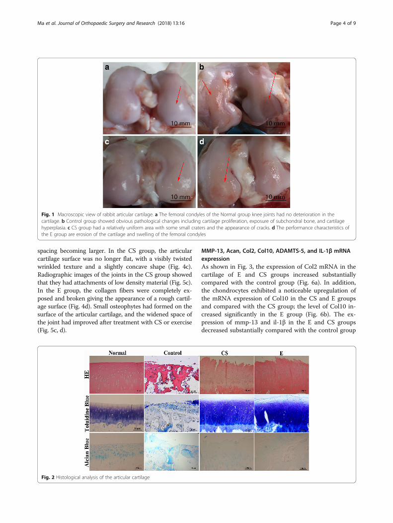
Assessment of tissue morphology and histologyIn the healthy rabbit knees, all the joints containedhealthy and smooth articular cartilage using a visiblecheck by naked eye. Obvious morphological differenceswere observed in the CS group and exercise group com-pared to the controls. Compared with the N group, ar-ticular cartilage hyperplasia, cartilage surface roughness,and obvious cracks were observed in all the experimen-tal groups, in addition to a thinned cartilage layer (Fig. 1),resulting in an alteration to the histology, consistentwith these morphological changes. Normal chondrocytessecrete cartilage matrix that principally comprises colla-gen II fibers and proteoglycans. In the event of chondro-cyte degeneration or necrosis, they either secrete
abnormal proteoglycan or no longer secrete any at all, asevidenced by loss of toluidine blue staining or unevendyeing [22, 23]. HE, alcian blue and toluidine blue stain-ing were conducted to observe tissue histomorphology(Fig. 2). The normal cartilage matrix was consistentlycolored, chondrocytes were arranged in order, and thetidal lines were intact. The cartilage in the control groupwas markedly thinned and the subchondral bone slightlyenlarged. It can be seen from the toluidine blue stainingthat the cartilage surface was smooth in the CS and Egroups with partial destaining and tidal lines that wereclear compared with those of the control group. Thenumber of cartilage cells in the E and CS groups wassignificantly larger than in the C group, as was the thick-ness of cartilage layer. HE staining showed that the sur-face of the cartilage in the exercise and CS groups wasirregular, and the chondrocytes were disordered andshowed evidence of mitosis and proliferation.The severity of histological changes in the tissue was
evaluated using Mankin scores [24]. According to thescoring, differences between the CS and E groups werenot clear. Mean scores for the control, CS, exercise, andnormal groups were 12.5 ± 2.8, 6.0 ± 0.8, 7.5 ± 1.5, and0.0 ± 0.0 at 34 days, respectively (Fig. 3), indicating thatsevere-to-mild changes representing OA were observed,consistent with the histological changes.
Scanning electron microscopy and radiography of thesurface of articular cartilageThe macroscopical and microscopical assessment of theknee joints showed multiple states. Normal articular car-tilage exhibited a uniform area without splits, lacunae,or cartilage proliferation and was investigated usingscanning electron microscopy (Fig. 4a). According to X-ray analysis, the surface was smooth and continuous(Fig. 5a). Those in the control group were significantlydeteriorated with a rough appearance where the surfacehad fissures that had widened and collagen fibers thatwere exposed, loose, broken, and turned upwards (Fig. 4b).As shown in Fig. 5b, formation of cartilage proliferationand cartilage surface defects and thinning of the cartilagelayer can be seen indirectly from the radiograph, with
Table 1 Transcripts and sequences of each primer used in RT-qPCR
Gene NCBI number Forward Primer, 5′–3′ Reverse Primer, 5′–3′
GAPDH KJ_875954.1 CCCTCAATGACCACTTTGTG GGTTTGAGGGCTCTTACT CCT
Col2 S_83370 GCACCCATGGACATTGGAGG AGCCCCGCACGGTCTTGCTT
Col10 XM_002714724 GAAAACCAGGCTATGGAACC GCTCCTGTAAGTCCCTGTTGTC
MMP-13 NM_1082037 TTCGAGTCATGCCACAAAT TAAGCTTTGCCCTGAAACCT
IL-1β NM_001082201 TGACGGCCTGAGAACTTTCT CATACGTGCCAGACAACACC
ADAMTS-5 AF_247708 CAGTGTTCTCGCTCTTGTGG CTGGGTGCAGGGTTATTGC
Aggrecan L_38480 ATGGCTTCCACCAGTGCG CGGATGCCGTAGGTTCTCA
Col2 collagen 2, mmp-13 matrix metalloproteinase 13, Col10 collagen 10, IL-1β interleukin-1β, ADAMTS-5 ADAM metallopeptidase with thrombospondin type 5 motif
Ma et al. Journal of Orthopaedic Surgery and Research (2018) 13:16 Page 3 of 9
spacing becoming larger. In the CS group, the articularcartilage surface was no longer flat, with a visibly twistedwrinkled texture and a slightly concave shape (Fig. 4c).Radiographic images of the joints in the CS group showedthat they had attachments of low density material (Fig. 5c).In the E group, the collagen fibers were completely ex-posed and broken giving the appearance of a rough cartil-age surface (Fig. 4d). Small osteophytes had formed on thesurface of the articular cartilage, and the widened space ofthe joint had improved after treatment with CS or exercise(Fig. 5c, d).
MMP-13, Acan, Col2, Col10, ADAMTS-5, and IL-1β mRNAexpressionAs shown in Fig. 3, the expression of Col2 mRNA in thecartilage of E and CS groups increased substantiallycompared with the control group (Fig. 6a). In addition,the chondrocytes exhibited a noticeable upregulation ofthe mRNA expression of Col10 in the CS and E groupsand compared with the CS group; the level of Col10 in-creased significantly in the E group (Fig. 6b). The ex-pression of mmp-13 and il-1β in the E and CS groupsdecreased substantially compared with the control group
Fig. 1 Macroscopic view of rabbit articular cartilage. a The femoral condyles of the Normal group knee joints had no deterioration in thecartilage. b Control group showed obvious pathological changes including cartilage proliferation, exposure of subchondral bone, and cartilagehyperplasia. c CS group had a relatively uniform area with some small craters and the appearance of cracks. d The performance characteristics ofthe E group are erosion of the cartilage and swelling of the femoral condyles
Fig. 2 Histological analysis of the articular cartilage
Ma et al. Journal of Orthopaedic Surgery and Research (2018) 13:16 Page 4 of 9

(Figs. 6c, d). Similarly, the expression of adamts-5 in theCS group decreased significantly and lowered in the Egroup (Fig. 6e). However, treatment of exercise observ-ably increased the mRNA of acan in contrast to the CSgroup (Fig. 6f ).
DiscussionOA is one of the most common joint diseases affectinghuman health, ultimately a degenerative disease of thejoints, without racial and regional differences [25]. Ourfindings verified that in the control group. the surface ofthe cartilage was not smooth and was accompanied by
destruction of the articular cartilage, cartilage fibrosis,osteophyte formation, synovial proliferation, and loss ofjoint function leading to pain and potential disability (Fig.1).In recent years, CS has been listed as a daily-use drug
to improve the symptoms of OA. The European Leagueagainst Rheumatism has suggested that CS is an effectivedrug for the treatment of knee OA [4]. As there are noblood vessels within articular cartilage, relying on syn-ovial fluid to provide nutrition, CS can be used as a de-livery pipeline, transporting nutrients to the joints,allowing an increase in joint metabolism and inhibitionof the pain of OA [26–29]. The cartilage surface in theCS group was smooth but with a visible distortion in thetexture and a slight breakage of collagenous and elasticfibers compared with the control group (Fig. 4c). Jr. etal. suggest CS allows direct mechanical and frictionalforces to be buffered and traps water molecules withinits chains [30]. Our results showed that the drug groupalleviated the pathological changes to OA under the ac-tion of CS. In this experiment, intumescentia of the ar-ticular surface is too small to be seen with the naked eyecompared with the control group (Fig. 1c). The articularcartilage was slightly discolored in the CS group, with asurface roughness that was not clearly obvious.Histology revealed that there were biological growths onthe cartilage surface in addition to a significant increasein the number of chondrocytes and quantity of matrix,constituting a significant increase in the number of eachcartilage unit (Fig. 2). CS is an important component ofarticular cartilage. Mice that are knockout for the N-
Fig. 3 Mankin scale for joint destruction (a–d). Data are means ± SD ofthree rabbits for each group. *p < 0.05 and **p < 0.01 versus untreatedcontrol, n.s. not significant
Fig. 4 Scanning electron microscopy view of surface of fresh cartilage. a Normal group was uniform without splits or lacunae. b Control grouphad deep splits on the surface. c Cartilage from rabbits where chondroitin sulfate (CS) had been administered were relatively uniform, with avisibly twisted wrinkled texture in other areas. d Rabbits that had exercised (E group) had a relatively rough surface
Ma et al. Journal of Orthopaedic Surgery and Research (2018) 13:16 Page 5 of 9

acetylgalactosyltransferase-1 gene, an important enzymeused in the synthesis of CS, have some characteristicssimilar to those suffering from OA, including a reduc-tion in proteoglycan molecules, the rapid catabolism ofacan and disordered Col2 fibers [31]. Compared with
the control group, the expression of Col2 (p < 0.01) andCol10 (p < 0.05) mRNA increased, while acan decreased(P < 0.05). CS exerts its anti-inflammatory effectsthrough the inhibition of reactive oxygen species activityor the activation of protein kinases, reducing the
Fig. 5 Lateral and craniocaudal radiographs from the joints of rabbits
Fig. 6 Gene Expression. RT-qPCR was used to examine the mRNA expression of a Col2, b Col10, c mmp-13, d il-1, e adamts-5, and f acan genesin each group. The results showed no evident differences between the CS and E groups for the genes a Col2 and c mmp-13. The expression of cmmp-13, d il-1, and e adamts-5 genes in the treated groups were decreased compared to that of the control group. The expression of f acanand a Col2 genes in the E group increased, while the expression of f acan decreased in the CS group. *p < 0.05, **p < 0.01, ***p < 0.001 indicatesignificant differences between groups. n.s. not significant. The 2-ΔΔCt method was adopted with GAPDH as the reference gene
Ma et al. Journal of Orthopaedic Surgery and Research (2018) 13:16 Page 6 of 9

synthesis of proteolytic enzymes (mmp-3, mmp-9, andmmp-13), inflammation-related enzymes (pla2, cox-2,nos-2), and proinflammatory factors (il-1β and tnf-α) [6,7]. In this study, administration of CS reduced the ex-pression of mRNA for mmp-13, il-1, and adamts-5 (p <0.01), which concurs with previous [32] studies.At present, exercise as a convenient and cheap treat-
ment of OA has been investigated in recent years. Theprevious study revealed that exercise had an effect onbone stabilization of the OA joint [33]. Exercise inter-vention for strength training does not provide superioroutcomes in pain or disability [34]; exercise therapy doesnot slow down the symptoms of arthritis by enhancingmuscle strength or reducing weight on the joint. In thisexperiment, after the success of arthritis modeling, therabbits were subjected to exercise therapy for a period of4 weeks, once every 2 days. Compared with the controlgroup, the joint swelling was not apparent (Fig. 1d). Andthe X-ray structures revealed that thickness of articularcartilage has been improved compared with the controlgroup (Fig. 5d), which was consistent with the researchof Kiviranta et al. [35]. Chondrocytes are the key tomaintaining healthy cartilage extracellular matrix [36]; inFig. 5 the number of cells in each cartilage unit compris-ing cartilage and chondrocytes increased significantly.The exercise stimulates the proliferation of chondro-cytes, and then achieves the purpose of restoration.The level of matrix collagenase in OA was increased,
and the expression of MMPs and adamts-5 were higherin the deteriorated matrix, while expression of Col2 andacan were exhibited [35]. There was a high concentra-tion of Col2 in the cartilage matrix amongst collagenfibers. The dynamic balance of Col2 and proteoglycanproduction and consumption is the direct cause of theloss of biomechanical properties in articular cartilage. Inthe case of inflammatory infiltration in articular cartil-age, chondrocytes lose their normal phenotype and con-vert into mast cell to release Col10. In the exercisegroup, the relative expression of Col2 and Col10 genes in-creased compared to that in the control group (p < 0.01),and the relative expression of Col10 mRNA increased(p < 0.01) in the CS group (Fig. 6a, b). The MMP andADAMTS families have been shown to be the twomajor types of enzymes that remove cartilage extracel-lular matrix, which is composed of proteoglycan andcollagen. MMP-13 acts mainly on cartilage collagen, soits inhibition would inhibit the degradation of Col2,preserving the cartilage extracellular matrix structure[37]. Similarly, in Fig. 6c, the mRNA levels of mmp-13decreased significantly (p < 0.05) in the exercise groupcompared with those in the control group; it would re-duce morphological changes in the cartilage and slowdown the process of OA. However, there was no signifi-cant difference in the expression of mmp-13 between
the exercise and CS groups. ADAMTS-5 mainly de-composes acan, the degradation of which is preventedafter knockout of the adamts-5 gene, reducing thepathological process of OA [38, 39]. In the CS group,the relative expression of the acan gene increased com-pared with the control group (p < 0.01), while the mRNAlevels of adamts-5 decreased significantly (p < 0.05). IL-1βcan affect the activity of chondrocytes through the synthe-sis of matrix metalloproteinases and plays a key role inblocking the synthesis of extracellular matrix Col2 andproteoglycans in chondrocytes [40]. Exercise training ef-fectively regulates the inflammatory process caused byknee OA [41]. We observed a decrease in the il-1β mRNAlevels (p < 0.05) in the exercise group (Fig. 6d). This studywas consistent with the conclusion by Yang Y et al., andthey corroborated that moderate treadmill exercise can al-leviate the severity of cartilage lesions in experimental OAthrough its anti-inflammatory activity of LXA4 and theNF-κB pathway [42]. The surface of the articular cartilagehad small osteophyte formations in the exercise group butnot the CS group (Fig. 5c, d). The cartilage in the CSgroup was cracked and the number of cartilage units haddecreased. Nevertheless, the crevices in the exercise groupcartilage were smaller and the number of cartilage cellssignificantly increased.Our findings suggest that exercise therapy can improve
the metabolism of chondrocytes by promoting the perme-ation and diffusion of synovial fluid to the articular cartil-age due to joint movement and promote regeneration ofcartilage tissue by increasing its nutrition metabolism. Inaccelerated repair of the injured joint and tissue surround-ing the articular cartilage, an exercising joint can stimulatethe proliferation of chondrocytes, which is conducive tothe transformation of undifferentiated stem cells intochondrocytes to fulfil the purpose of repairing articularcartilage and provide relief of injury of the joint. The resultof this experiment implies a possibility that exercise notonly avoids drug metabolism but also improves the symp-toms of OA. Using exercise, CS can be replaced in thetreatment of OA in some respects.
ConclusionsIn conclusion, we have demonstrated that exercise has agood therapeutic effect on OA equate to CS. The limita-tions of this study should focus on changing the way ofmovement to have an effect on the cartilage and notonly changing gait biomechanics to reduce pain and im-prove physical function.
AbbreviationsADAMTS-5: ADAM metallopeptidase with thrombospondin type 5 motif;Col10: Collagen 10; Col2: Collagen 2; CS: Chondroitin sulfate; Ct: Cyclesrequired for the threshold; DR: Direct digital radiography;GAPDH: Glyceraldehyde-3-phosphate dehydrogenase; HE: Hematoxylin andeosin stain; IL-1β: Interleukin-1β; mmp-13: Matrix metalloproteinase 13;OA: Osteoarthritis; RT-qPCR: Reverse transcription and quantitative
Ma et al. Journal of Orthopaedic Surgery and Research (2018) 13:16 Page 7 of 9
polymerase chain reaction; SD: Mean ± standard deviation; SEM: Scanningelectron microscopy
AcknowledgmentsThanks are due to Bing Sun for the help with the X-rays and to Zaisi Liu forthe excellent technical assistance.
FundingThis study was supported by grants from the National Key Research andDevelopment Program of China (2016YFD0501106) in the Key Laboratory ofthe Provincial Education Department of Heilongjiang for Common AnimalDisease Prevention and Treatment.
Availability of data and materialsPlease contact author for data requests.
Authors’ contributionsAll authors were involved in the conception and design of the study, or inthe acquisition analysis and interpretation of data, and in revising it criticallyfor important intellectual content. The experiments were designed by NM,TW, and JX. The experiments were performed by NM, TW, LB, YZ, SZ, LZ, andLG. The data were analyzed by NM and TW. The paper was written by NM.All authors read and approved the final manuscript.
Ethics approvalAnimal experiments were performed according to a protocol approved bythe Animal Experimentation Committee of Northeast Agricultural University,Harbin, China. Rabbits were euthanized by venous air embolism afterinjection of ether. All efforts were made to minimize suffering.
Consent for publicationNot applicable
Competing interestsThe authors declare that they have no competing interests.
Publisher’s NoteSpringer Nature remains neutral with regard to jurisdictional claims inpublished maps and institutional affiliations.
Received: 10 October 2017 Accepted: 10 January 2018
References1. Abramson SB, Attur M. Developments in the scientific understanding of
osteoarthritis. Arthritis Res Ther. 2009;11:227.2. Xu L, Flahiff CM, Waldman BA, Wu D, Olsen BR, Setton LA, et al.
Osteoarthritis-like changes and decreased mechanical function of articularcartilage in the joints of mice with the chondrodysplasia gene (cho).Arthritis Rheum. 2003;48:2509–18.
3. Johnston SA. Osteoarthritis. Joint anatomy, physiology, and pathobiology.Vet Clin N Am Small Anim Pract. 1997;27:699.
4. Tchetina EV, Kobayashi M, Yasuda T, Meijers T, Pidoux I, Poole AR.Chondrocyte hypertrophy can be induced by a cryptic sequence of type IIcollagen and is accompanied by the induction of MMP-13 and collagenaseactivity: implications for development and arthritis. Matrix Biol.2007;26:247–58.
5. Lorenz H, Wenz W, Ivancic M, Steck E, Richter W. Early and stableupregulation of collagen type II, collagen type I and YKL40 expression levelsin cartilage during early experimental osteoarthritis occurs independent ofjoint location and histological grading. Arthritis Res Ther. 2004;7:R156.
6. Appleton CTG, Mcerlain DD, Pitelka V, Schwartz N, Bernier SM, Henry JL, etal. Correction: forced mobilization accelerates pathogenesis: characterizationof a preclinical surgical model of osteoarthritis. Arthritis Res Ther. 2008;10:407.
7. Matyas JR, Huang D, Chung M, Adams ME. Regional quantification ofcartilage type II collagen and aggrecan messenger RNA in joints with earlyexperimental osteoarthritis. Arthritis Rheum. 2002;46:1536–43.
8. Pfander D, Swoboda B, Kirsch T. Expression of early and late differentiationmarkers (proliferating cell nuclear antigen, syndecan-3, annexin VI, andalkaline phosphatase) by human osteoarthritic chondrocytes. Am J Pathol.2001;159:1777–83.
9. Hollander AP, Pidoux I, Reiner A, Rorabeck C, Bourne R, Poole AR. Damageto type II collagen in aging and osteoarthritis starts at the articular surface,originates around chondrocytes, and extends into the cartilage withprogressive degeneration. J Clin Investig. 1995;96:2859–69.
10. Tetlow LC, Adlam DJ, Woolley DE. Matrix metalloproteinase andproinflammatory cytokine production by chondrocytes of humanosteoarthritic cartilage: associations with degenerative changes. ArthritisRheum. 2001;44:585.
11. Veje K, Hyllestedwinge JL, Ostergaard K. Topographic and zonal distributionof tenascin in human articular cartilage from femoral heads: normal versusmild and severe osteoarthritis. Osteoarthr Cartil. 2003;11:217–27.
12. Latham N, Liu CJ. Strength training in older adults: the benefits forosteoarthritis. Clin Geriatr Med. 2010;26:445.
13. Erdemir A, Bennetts C, Davis S, Reddy A, Sibole S. Multiscale cartilagebiomechanics: technical challenges in realizing a high-throughputmodelling and simulation workflow. Interface Focus. 2015;5:20140081.
14. Bennell KL, Buchbinder R, Hinman RS. Physical therapies in themanagement of osteoarthritis: current state of the evidence. Curr OpinRheumatol. 2015;27:304–11.
15. Bijlsma JW, Berenbaum F, Lafeber FP. Osteoarthritis: an update withrelevance for clinical practice. Lancet. 2011;377:2115.
16. Iijima H, Aoyama T, Ito A, Tajino J, Yamaguchi S, Nagai M, et al. Exerciseintervention increases expression of bone morphogenetic proteins andprevents the progression of cartilage-subchondral bone lesions in a post-traumatic rat knee model. Osteoarthr Cart. 2016;24:1092–102.
17. Du SP, García AG, Vergés J, Montell E. Immunomodulatory and anti-inflammatory effects of chondroitin sulphate. J Cell Mol Med. 2009;13:1451–63.
18. Volpi N. Advances in chondroitin sulfate analysis: application in physiologicaland pathological states of connective tissue and during pharmacologicaltreatment of osteoarthritis. Curr Pharm Des. 2006;12:639–58.
19. Rogart JN, Barrach HJ, Chichester CO. Articular collagen degradation in theHulth-Telhag model of osteoarthritis. Osteoarthr Cartil. 1999;7:539–47.
20. Linti G, Bühler M, Monakhov KY, Zessin T. Effect of chondroitin sulphate insymptomatic knee osteoarthritis: a multicentre, randomised, double-blind,placebo-controlled study. Ann Rheum Dis. 2007;66:639–45.
21. Livak KJ, Schmittgen TD. Analysis of relative gene expression data usingreal-time quantitative PCR and the 2(−Delta Delta C(T)) method. Methods.2012;25:402–8.
22. Geyer G, Linss W. Toluidine blue staining of cartilage proteoglycan subunits1. Acta Histochem. 1978;61:127–34.
23. Mitchell N, Shepard N, Harrod J. The use of brominated toluidine blue O inX-ray microanalysis for proteoglycan. Histochemistry. 1980;68:245–51.
24. Liu Y, Wu D, Song F, Zhu C, Hui Y, Zhu Q, et al. Activation of α7 nicotinicacetylcholine receptors prevents monosodium iodoacetate-inducedosteoarthritis in rats. Cell Physiol Biochem. 2015;35:1026–32.
25. Goldring MB. Articular cartilage degradation in osteoarthritis. HSS J. 2012;8:7–9.26. Monfort J, Martelpelletier J, Pelletier JP. Chondroitin sulphate for
symptomatic osteoarthritis: critical appraisal of meta-analyses. Curr Med ResOpin. 2008;24:1303.
27. Hochberg MC, Zhan M, Langenberg P. The rate of decline of joint spacewidth in patients with osteoarthritis of the knee: a systematic review andmeta-analysis of randomized placebo-controlled trials of chondroitin sulfate.Curr Med Res Opin. 2008;24:3029.
28. Hochberg M, Chevalier X, Henrotin Y, Hunter DJ, Uebelhart D. Symptomand structure modification in osteoarthritis with pharmaceutical-gradechondroitin sulfate: what's the evidence? Curr Med Res Opin. 2013;29:259–67.
29. Bjordal JM, Johnson MI, Lopes-Martins RA, Bogen B, Chow R, Ljunggren AE.Short-term efficacy of physical interventions in osteoarthritic knee pain. Asystematic review and meta-analysis of randomised placebo-controlledtrials. BMC Musculoskelet Disord. 2007;8:51.
30. Jr VC, Spiker W, Erickson J. A review of evidence-based medicine for glucosamineand chondroitin sulfate use in knee osteoarthritis. Arthroscopy. 2009;25:86.
31. Watanabe Y, Takeuchi K, Higa OS, Sato M, Tsujita M, Abe M, et al.Chondroitin sulfate N-acetylgalactosaminyltransferase-1 is required fornormal cartilage development. Biochem J. 2010;432:47.
32. Allen J, Imbert I, Havelin J, Henderson T, Stevenson G, Liaw L, et al. Effectsof treadmill exercise on advanced osteoarthritis pain in rats. Arthritis Rheum.2017;69:1407–17.
33. Brosseau L, Taki J, Desjardins B, Thevenot O, Fransen M, Wells GA, et al. TheOttawa panel clinical practice guidelines for the management of knee
Ma et al. Journal of Orthopaedic Surgery and Research (2018) 13:16 Page 8 of 9

osteoarthritis. Part one: introduction, and mind-body exercise programs. ClinRehabil. 2017;31:582–95.
34. Bartholdy C, Juhl C, Christensen R, Lund H, Zhang W, Henriksen M. The roleof muscle strengthening in exercise therapy for knee osteoarthritis: Asystematic review and meta-regression analysis of randomized trials. SemArthritis Rheum. 2017;47:9-21.
35. Kiviranta I, Tammi M, Jurvelin J, Säämänen AM, Helminen HJ. Moderaterunning exercise augments glycosaminoglycans and thickness of articularcartilage in the knee joint of young beagle dogs. J Orthop Res. 1988;6:188–95.
36. Cheng T, Zhang L, Fu X, Wang W, Xu H, Song H, et al. The potentialprotective effects of calcitonin involved in coordinating chondrocyteresponse, extracellular matrix, and Subchondral Trabecular bone inexperimental osteoarthritis. Connect Tissue Res. 2013;54:139.
37. Li NG, Shi ZH, Tang YP, Wang ZJ, Song SL, Qian LH, et al. New hope for thetreatment of osteoarthritis through selective inhibition of MMP-13. CurrMed Chem. 2011;18:977–1001.
38. Echtermeyer F, Bertrand J, Dreier R, Meinecke I, Neugebauer K, Fuerst M, etal. Syndecan-4 regulates ADAMTS-5 activation and cartilage breakdown inosteoarthritis. Nat Med. 2009;15:1072–6.
39. Glasson SS, Askew R, Sheppard B, Carito B, Blanchet T, Ma HL, et al. Deletionof active ADAMTS5 prevents cartilage degradation in a murine model ofosteoarthritis. Nature. 2005;434:644–8.
40. Mastbergen SC, Bijlsma JW, Lafeber FP. Synthesis and release of humancartilage matrix proteoglycans are differently regulated by nitric oxide andprostaglandin-E2. Ann Rheum Dis. 2008;67:52–8.
41. Rojas-Ortega M, Cruz R, Vega-López MA, Cabrera-González M, Hernández-Hernández JM, Lavalle-Montalvo C, et al. Exercise modulates the expressionof IL-1β and IL-10 in the articular cartilage of normal and osteoarthritis-induced rats. Pathol Res Pract. 2015;211:435.
42. Yang Y, Wang Y, Kong Y, Zhang X, Bai L. The effects of different frequencytreadmill exercise on lipoxin A4 and articular cartilage degeneration in anexperimental model of monosodium iodoacetate-induced osteoarthritis inrats. PLoS One. 2017;12:e0179162.
• We accept pre-submission inquiries
• Our selector tool helps you to find the most relevant journal
• We provide round the clock customer support
• Convenient online submission
• Thorough peer review
• Inclusion in PubMed and all major indexing services
• Maximum visibility for your research
Submit your manuscript atwww.biomedcentral.com/submit
Submit your next manuscript to BioMed Central and we will help you at every step:
Ma et al. Journal of Orthopaedic Surgery and Research (2018) 13:16 Page 9 of 9