COMPARISON OF THE AUTOMATED OSCILLOMETRIC BLOOD …€¦ · Objective: The Doppler ABI test may be...
62
FACULTY OF MEDICINE AND HEALTH SCIENCES Academic Year 2013 - 2014 COMPARISON OF THE AUTOMATED OSCILLOMETRIC BLOOD PRESSURE METHOD WITH THE DOPPLER ULTRASOUND METHOD FOR MEASUREMENT OF THE ANKLE-BRACHIAL INDEX Jonas DELFORCHE Promoter: Prof. Dr. T. De Backer Dissertation presented in the 2 nd Master year in the programme of Master of Medicine in Medicine
Transcript of COMPARISON OF THE AUTOMATED OSCILLOMETRIC BLOOD …€¦ · Objective: The Doppler ABI test may be...

FACULTY OF MEDICINE AND HEALTH SCIENCES
Academic Year 2013 - 2014
COMPARISON OF THE AUTOMATED OSCILLOMETRIC
BLOOD PRESSURE METHOD WITH THE DOPPLER
ULTRASOUND METHOD FOR MEASUREMENT OF THE
ANKLE-BRACHIAL INDEX
Jonas DELFORCHE
Promoter: Prof. Dr. T. De Backer
Dissertation presented in the 2nd Master year in the programme of
Master of Medicine in Medicine

“The author and the promoter give the permission to use this thesis for consultation and to copy
parts of it for personal use. Every other use is subject to the copyright laws, more specifically the
source must be extensively specified when using results from this thesis.”
Date
Signature (student) (promoter)
Name (student) (promoter)

Preface – word of thanks
First of all, I would like to thank my promoter, Prof. Dr. Tine De Backer, for her continuous
accompaniment and support, for the evening sessions where constructive discussions arose and
for the astounding availability she expressed throughout the 2 years we’ve spent working on this
experimental study.
Secondly, I would like to say thank you to Dr. Laurence Campens, who recruited subjects in the
years 2010-2011, was always available for meetings, responded comprehensively to any question
concerning the methods used and played an important role in the realization of this thesis.
I wish to thank my fellow students, responding immediately to my request of contributing to my
thesis by volunteering for the inter-observer study.
I also would like to thank Prof. Vermassen, who allowed me to take measurements at the
Department of Thoracic and Vascular Surgery at the Ghent University Hospital.
A big thank you goes out to the personnel at the Department of Cardiology at the Ghent
University Hospital where I’ve spent several weeks collecting data. A special thanks for Tamara
Leune, head nurse of the Cardiology ward, who always treated me with respect and indulgence
and aided me in approaching patients and other personnel.
I wish to thank Rik Vuylsteke, vascular technician, for introducing me to the Doppler technique
and ABI determination with clarity.
I would like to thank Prof. George Van Maele, who was a big help in the statistical analysis and
interpretation.
Last but not least, I would like to thank my parents and brothers, who I can always count on, for
their patience, confidence and support, and for allowing me to enrich myself in 7 years of
education.

Table of contents
Abstract ................................................................................................................... 1
List of acronyms and abbreviations ......................................................................... 3
1. Introduction ......................................................................................................... 4
1.1 Peripheral artery disease (PAD) ..................................................................................... 4
1.1.1 Etiopathogenesis of PAD ........................................................................................ 4
1.1.2 Epidemiology ........................................................................................................... 4
1.1.3 Clinical presentation and symptomatology ............................................................. 4
1.1.4 Consequences of PAD ............................................................................................. 6
1.1.5 Risk factors for developing PAD ............................................................................. 7
1.1.6 Treatment of PAD .................................................................................................... 7
1.1.7 Detection of PAD with the ankle-brachial index .................................................... 8
1.2 Indications for ABI measurement .................................................................................. 8
1.3 How is the ABI measured? ............................................................................................. 9
1.4 How is the ABI interpreted? ......................................................................................... 11
1.5 Objective ...................................................................................................................... 12
2. Methods ............................................................................................................ 13
2.1 Introduction .................................................................................................................. 13
2.2 Literature study ............................................................................................................ 13
2.3 Ethical committee ......................................................................................................... 13
2.4 Participant recruitment ................................................................................................. 13
2.5 ABI measurement ......................................................................................................... 14
2.5.1 The Doppler ultrasound method (golden standard) .............................................. 14
2.5.2 The automated oscillometric blood pressure method ........................................... 15
2.6 Questionnaire ............................................................................................................... 15

2.7 Statistical methods ........................................................................................................ 15
3. Results .............................................................................................................. 17
3.1 Study Population .......................................................................................................... 17
3.2 Cardiovascular risk factors in PAD .............................................................................. 18
3.3 Correlation of measurements of both methods ............................................................ 19
3.2.1 Bland Altman plot .................................................................................................. 20
3.2.2 Intraclass correlation coefficient (ICC) ................................................................ 22
3.4 Agreement of both methods on PAD diagnosis ........................................................... 23
3.5 Agreement between different observers (Inter-observer study) ................................... 25
3.6 Comparison of time required for Doppler and oscillometric method .......................... 26
3.7 Conclusion .................................................................................................................... 29
4. Discussion ......................................................................................................... 30
4.1 Limitations of the ABI ................................................................................................. 31
4.2 Alternative tests for detecting PAD non-invasively ..................................................... 33
4.3 Limitations with the Doppler method .......................................................................... 34
4.4 Limitations with the automated oscillometric method ................................................. 34
4.5 Study limitations .......................................................................................................... 34
4.6 Alternative calculations of the ABI .............................................................................. 35
4.7 Future prospective ........................................................................................................ 36
5. References ......................................................................................................... 37
6. Appendix ............................................................................................................. I

Nederlandstalige samenvatting
Titel: Vergelijking van de automatische bloeddrukmeting met de doppler bloeddrukmeting
voor het bepalen van de enkel-arm index.
Achtergrond: Omdat perifeer arterieel vaatlijden (peripheral arterial disease, PAD) frequent
voorkomt, ernstige gevolgen kan hebben (hoog risico op myocardinfarct, cerebrovasculair
accident en sterfte, risico op amputatie), grote kosten met zich meebrengt en vaak aanwezig is bij
asymptomatische patiënten, is het sterk aangeraden om te screenen voor PAD. De enkel-arm
index (ankle-brachial index, ABI) is een eenvoudige, betrouwbare manier om PAD op te sporen.
PAD wordt vermoed als de ABI ≤ 0.90.
Objectieven: De Doppler ABI test kan tijdrovend zijn en opleiding in het correct gebruik van de
Doppler techniek is een vereiste. Daarom wordt de automatisch oscillometrische
bloeddrukmeting voorgesteld voor het meten van de ABI. Op deze manier kan zowel het
technische aspect als de benodigde tijd voor het meten van de ABI sterk verminderd worden.
Deze studie onderzoekt de overeenkomst van metingen enerzijds via de automatisch
oscillometrische methode (ABIosc) en anderzijds via de gouden standaard Doppler methode
(ABIdop) en vergelijkt de benodigde tijd voor het uitvoeren van beide methodes.
Design: Observationeel cross-sectioneel onderzoek.
Participanten: Honderd achtennegentig (198) deelnemers werden gerekruteerd op de diensten
cardiologie en thoracovasculaire chirurgie in het Universitair Ziekenhuis van Gent tussen januari
2010 en november 2013. Bij 39 deelnemers werd PAD gedetecteerd door de Doppler methode
(ABIdop≤0.90). Bij 2 deelnemers kon de ABI niet gemeten worden door onsamendrukbare
arteriën.
Resultaten: De twee ABI metingen (ABIdop en ABIosc) bleken goed te correleren (Spearman
correlatie coëfficiënt (rs) van 0.604). Intra-class correlation coëfficient (ICC) werd berekend op
0.665 en een Bland Altman plot toonde weinig verschil en geen opmerkelijke systematische bias
tussen metingen van beide methodes. Desondanks had de automatisch oscillometrische methode
wel de neiging om de ABI te onderschatten of overschatten wanneer de ABI gemeten door
Doppler respectievelijk hoog of laag was. De gemiddelde benodigde tijd voor het meten van de

ABI met de Doppler methode was 12 minuten 22 seconden, de gemiddelde benodigde tijd met de
oscillometrische methode was slechts 4 minuten 59 seconden.
Conclusie: Uit deze gegevens kan de automatisch oscillometrische methode beschouwd worden
als vrij goed overeenkomend met de Doppler ABI methode wat betreft de gemeten ABI waarden
en als een eenvoudigere en snellere manier voor het meten van de ABI voor
screeningsdoeleinden. Verder grootschalig onderzoek dient hierover een meer definitief oordeel
te vormen.
Keywords: Bloeddrukmeting, enkel-arm index, oscillometrie, Doppler, perifeer arterieel
vaatlijden,…

1
Abstract
Title: Comparison of the automated oscillometric blood pressure method with the Doppler
ultrasound method for measurement of the ankle-brachial index.
Background: Because peripheral artery disease (PAD) is highly prevalent, has serious
consequences (high risk of myocardial infarction, stroke and death, risk of amputation),
implicates high economic costs and, more worrisome, is often present in absence of symptoms, it
is strongly advised to screen for PAD. The ankle-brachial index (ABI) represents a simple,
reliable method for detecting PAD. PAD is suspected when the ABI ≤ 0.90.
Objective: The Doppler ABI test may be time-consuming and technical instruction on accurate
use of the Doppler technique is necessary. This is why the use of automated oscillometric blood
pressure measurement is proposed to measure the ABI. In this way, the technical aspect and time
for measurement could be reduced greatly. This study investigates the agreement between the
automated oscillometric ABI determination (ABIosc) and the golden standard Doppler ABI
determination (ABIdop) while also comparing time needed to perform each measurement
method.
Design: Observational cross-sectional study.
Study population: Hundred ninety-eight (198) participants were recruited at the Departments of
Cardiovascular Diseases and Thoracovascular Surgery at the Ghent University Hospital between
January 2010 and November 2013. In 39 subjects (19.7%), PAD was detected by Doppler
method (ABIdop ≤0.9). In 2 subjects the ABI could not be determined due to incompressible
arteries.
Results: The two ABI measurements (ABIdop and ABIosc) were found to be correlating well
(Spearman’s correlation coefficient (rs) of 0.604). Intra-class correlation coefficient (ICC) was
calculated at 0.665 and a Bland Altman plot showed little difference and no obvious systematic
bias between measurements of the two methods. However, the automated oscillometric method
was likely to underestimate or overestimate the ABI when the ABI measured by Doppler method
was either high or low respectively. The average time to perform the Doppler method was
2
12 minutes, 22 seconds compared to only 4 minutes, 59 seconds needed to perform the oscillometric method.
Conclusion: From these data, the automated oscillometric method for measuring the ABI for
screening purposes can be considered as having an acceptable agreement on its values with the
Doppler ABI method and as being more efficient in terms of easiness and time consumption.
Further large population based studies could give a more definite view on this subject.
Keywords: Blood pressure measurement, ankle-brachial index, Doppler, oscillometry, peripheral
arterial disease…

3
List of acronyms and abbreviations
ABI
ABIdop
ABIosc
Ankle-Brachial (Arm) Index
ABI measured by Doppler
ABI measured by automated oscillometric device
CI 95% confidence interval
CLI Critical limb ischemia
CT Computed tomography
CVD Cardiovascular disease
ICC Intra class correlation coefficient
ABIdop1 ABIdop measured by first observer
ABIdop2 ABIdop measured by second observer
LEAD Lower extremity artery disease
LR- Likelihood ratio for a negative result
LR+ Likelihood ratio for a positive result
MeanABI The mean of ABIdop and ABIosc
MR Magnetic resonance imaging
NPV Negative predictive value
PAD
PADdop
PADosc
Peripheral arterial (artery) disease
PAD diagnosed by Doppler method
PAD diagnosed by automated oscillometric method
PPV Positive predictive value
PVR Pulse volume recording
SD Standard Deviation
κ Kappa coefficient

4
1. Introduction
“Cardiovascular disease (CVD) is the leading cause of death and disability in Europe, posing a
great social and economic burden” stated the European Society for Cardiology in their latest
guidelines for peripheral artery diseases.(1) Because CVD can be, at least partially, prevented by
appropriate care, it is important to detect risk factors and early stages of the disease.
1.1 Peripheral artery disease (PAD)
1.1.1 Etiopathogenesis of PAD
Atherosclerosis plays an important role in the development of CVD. Atherosclerosis is a systemic
disease that is characterized by the narrowing and stiffening of blood vessels through the process
of plaque forming within these vessels throughout the body. It can lead to serious problems such
as coronary artery disease, carotid artery disease and peripheral artery disease (PAD). PAD is a
condition where arterial blood vessels to the head, organs and limbs are narrowed due to
atherosclerosis. This narrowing can reduce the blood flow. Atherosclerosis more frequently
affects the arteries of the legs compared to the arteries of the arms.(1) PAD in the legs is also
known as lower extremity artery disease (LEAD).
1.1.2 Epidemiology
PAD has shown to be prevalent. In a recent German study called the Heinz Nixdorf recall study,
4,814 subjects aged 45-75 years were screened for PAD. Overall, PAD affected 6.4% among men
and 5.1% among women and became much more prevalent in older individuals: 18.2 % in those
aged 70-75 years.(2) In an American study, the National Health and Nutrition Examination
Survey study, including 2174 participants, the prevalence of PAD in the United States among
adults aged 40 years and over was 4.3%. Among those aged 70 years or over, the prevalence of
PAD was 14.5%. Furthermore, this study found that black people are almost 3 times as much
affected (odds ratio= 2.83) as white people.(3)
1.1.3 Clinical presentation and symptomatology
PAD can lead to different manifestations depending on the site and the severity of the disease.
The symptoms are caused by a reduced blood flow in the affected arteries. Especially during
physical activity, insufficient oxygen is supplied to muscles and organs which results in ischemic

5
symptoms. Signs and symptoms include: pain, numbness, achiness or heaviness in the leg
muscles when walking or climbing stairs; weak or absent pulses in the legs or feet; sores or
wounds on the toes, feet, or legs that heal slowly, poorly, or not at all; a pale or bluish colour to
the skin; a lower temperature in one leg than the other leg; poor nail growth on the toes and
decreased hair growth on the legs.(4) A classic symptom of PAD is painful cramping of leg
muscles after walking a distance that is relieved by rest called intermittent claudication. The site
of claudication is distal to the diseased arterial segment. For example, buttock, hip, and thigh
claudication are seen with aortoiliac disease and calf claudication with femoral-popliteal disease.
With severe PAD, patients may present with signs and symptoms of critical limb ischemia (CLI).
These include rest pain, cold, pallor, paraesthesia or numbness, weak or absent pulses in the leg
or feet, with or without tissue loss (nonhealing ulcers or gangrene). The symptoms of CLI are
often referred to as “the six P’s” occurring in the affected leg: paraesthesia (altered sensation),
pain, pallor (mottled colouration), pulselessness, paralysis, poikilothermia (coolness). Rest pain
usually occurs at night (because of the horizontal position, which deprives the patient of the
effect of gravity on blood flow through the tight lesions) and improves when the legs are in a
dependent position. Rest pain is a sign of more severe or multilevel arterial occlusions.(5)
The presentation of PAD is categorized according to the Fontaine or Rutherford
classification.(Table 1)

6
Table 1. Classification of PAD: Fontaine and Rutherford classification.(6)
Fontaine classification Rutherford classification
Stage Clinical Grade Category Clinical
I Asymptomatic 0 0 Asymptomatic
IIa Mild claudication (>200m)* I 1 Mild claudication
IIb Moderate to severe claudication (<200m)*
I 2 Moderate claudication
I 3 Severe claudication
III Ischemic rest pain II 4 Ischemic rest pain
IV Ulceration or gangrene
III 5 Minor tissue loss
III 6 Major tissue loss
*: leg pain can occur after walking a longer distance (>200 meters) or after walking a relatively short
distance (<200 meters).
However, most patients do not have the typical intermittent claudication; they have atypical leg
symptoms or no symptoms at all.(7) About 60% of the PAD patients was asymptomatic in a
population-based study.(8) ‘Importantly, even with a similar extent and level of disease
progression, symptoms and their severity may vary from one patient to another.’(1)
In addition to leg symptoms, symptomatic patients often report poor quality of life related to their
limited mobility and subsequent decline in overall functional capacity.(9, 10)
1.1.4 Consequences of PAD
The prognosis for PAD patients with claudication is bad: about 30% can be expected to die
within 5 years, and 1-3% will undergo major amputation.(11) Other and more than the risk to
lose a limb, PAD is associated with a very high risk of myocardial infarction, stroke and
death.(1, 7, 11-14) The progression of PAD is predictive for cardiovascular disease morbidity and
mortality.(15) Because of the grave consequences, PAD is also a big economic burden.
Smolderen et al. calculated that the two-year hospitalization costs were highest for patients with

7
PAD (approximately 2953 euro) compared to the costs for cerebrovascular disease and coronary
artery disease. This high cost for PAD was explained mainly because of the high expenses on
peripheral revascularizations and amputations.(16)
1.1.5 Risk factors for developing PAD
Table 2. Risk factors for PAD
Risk factors for PAD
Advanced age
Male gender
Family history of premature CVD
Cigarette smoking
Diabetes mellitus
Hypertension
Hypercholesterolemia (dyslipidaemia)
Low kidney function
Risk factors for PAD (table 2) include: advanced age, (family) history of premature CVD,
cigarette smoking, diabetes mellitus, hypertension, hypercholesterolemia and low kidney
function.(3) While the risk factors for PAD are similar to those for atherosclerotic disease in
general as in coronary artery disease and cerebrovascular disease, diabetes(17) and cigarette
smoking(1, 17) have a particularly strong association with PAD. Smoking is the most important
risk factor for the progression of local disease in the legs, with an amputation rate 11 times
greater in smokers than in non-smokers.(11) Diabetes, male gender, and hypertension are also
important risk factors for progression of PAD.(11)
1.1.6 Treatment of PAD
Management of PAD includes aggressive management of atherosclerotic risk factors, a structured
exercise program, use of antiplatelet agents and, when indicated, percutaneous or surgical
revascularization.(5)

8
1.1.7 Detection of PAD with the ankle-brachial index
Because PAD is highly prevalent, has serious consequences (not only amputation, but also and
mainly myocardial infarction, stroke and death), implicates high economic costs and, more
worrisome, often can be present in absence of symptoms, it is strongly advised to screen for
PAD.
This demands for adequate diagnostic and prognostic tools. ‘Although other methods exist to
assess the peripheral vasculature more objectively, the ankle-brachial index (ABI) represents a
simple, reliable method for detecting PAD.’(18) PAD is suspected when ABI is 0.90 or lower
(≤0.90). The ABI test is inexpensive, accurate, and relatively easy to perform so that the ABI has
achieved a major role in screening patients for PAD.(19, 20)
1.2 Indications for ABI measurement
The ABI can be used to confirm the diagnosis in patients with symptoms suggestive of PAD or it
can be used to screen for asymptomatic PAD. The question is: Who should be screened for PAD?
Specific clinical information should be used to identify individuals who should undergo ABI
examination. ‘Clinical data that should guide this assessment includes: the presence of
atherosclerosis risk factors (especially age, smoking, and diabetes), clinical history (a history of
atherosclerotic coronary artery, carotid artery, or renal artery disease and lower extremity
symptoms), and an abnormal lower extremity pulse examination.’(21) Recommendations on
screening asymptomatic patients with PAD vary across current guidelines. Ferket et al.(22)
compared 8 leading guidelines of which 7 were developed in North America and 1 was
developed by an international collaboration (Europe, Japan, North America, Australia and South
Africa). Target groups in these guidelines generally comprised middle-aged persons with one or
more cardiovascular risk factors and the elderly.(22) In the most recent American College of
Cardiology–American Heart Association (ACC/AHA) Practice Guidelines, high-risk patients
who should undergo ABI testing were identified as follows (table 3).

9
Table 3. Who Should Undergo screening with Ankle-Brachial Index Testing?
Age ≥ 70 years
Age = 50-69 years with history of diabetes or smoking
Age ≤ 49 years with diabetes and one additional risk factor (smoking, diabetes, hypertension, or elevated cholesterol levels)
Individuals who present with claudication or more severe limb ischemic symptoms
Abnormal lower extremity pulse examination
Known atherosclerotic disease elsewhere (coronary, carotid or renal arteries)
Furthermore, ABI measurement is also indicated for assessment of healing potential and
evaluation of vascular compromise in patients with trauma of the lower legs. Follow-up
evaluation with ABI is advised in graft surveillance, worsening of ischemic symptoms and
assessment of revascularization therapy.(18, 23)
1.3 How is the ABI measured?
Figure 1. The ankle-brachial index test. As the blood pressure cuff
deflates, the systolic blood pressure is recorded. Reprinted with
permission, National Heart, Lung, and Blood Institute; National Institutes
of Health; U.S. Department of Health and Human Services.(24)

10
Figure 2. Illustration on how to calculate the ABI. This patient is diagnosed with PAD. Reprinted with
permission, Cleveland Clinic Center for Medical Art & Photography © 2012-2013. All Rights
Reserved.(25)
Blood pressure is measured at both arms (brachial artery) and ankles (posterior tibial artery and
dorsal pedis artery) with a Doppler probe and a sphygmomanometer. The ABI is then calculated
by dividing the systolic blood pressure at the ankle by the systolic blood pressure at the arm. The
higher of the two ankle pressures (posterior tibial artery or dorsal pedis artery) is used as the
numerator and the higher of the two arm pressures is used as the denominator for both
limbs.(Figure 2) If the index is 0.9 or less in either lower extremity, the diagnosis of PAD is put
forward. Further investigation is then advised. Recently, some alternative calculations of the ABI

11
have been proposed. These are reviewed in the discussion. Moreover, measuring the ABI after
exercise might diagnose PAD in patients with normal or borderline ABI at rest.(26, 27)
1.4 How is the ABI interpreted?
There is a U-shaped relationship between the ABI and mortality risk.(Figure 3)
Figure 3. The U-shaped relationship between ankle-brachial index and death.
This data was gained from 4,393 patients in the Strong Heart Study. Patients
were followed for a mean of 8.3 years. All-cause (white bar) and cardiovascular-
disease-related (black bar) mortality rates were lowest in people with values of
1.0 to 1.4. Adapted from the Strong Heart Study.(28)
With a patient at rest, results ranging from 0.91 to 1.30-1.40 are normal. Readings below 0.91
indicate PAD and are associated with an increased risk of cardiovascular morbidity and
mortality.(1, 7, 11-14). An ABI < 0.50 indicates a high risk of amputation.(1) On the other hand,
readings above 1.30-1.40 are suggestive of stiffened and thus incompressible arteries in the
ankles. This is often seen in elderly patients, patients with diabetes, or patients with end-stage
renal failure requiring dialysis.(18) These patients also have an increased cardiovascular and
overall mortality.(28) ‘Importantly, a substantial proportion of patients with an elevated ABI
12
actually do have occlusive artery disease. Alternative tests such as measurement of the toe
systolic pressures and Doppler waveform analysis are useful to unmask LEAD’.(1) These
alternative tests are covered in the discussion.
Another important question is: “What is known about the sensitivity and specificity of the ABI?”
Suggested PAD diagnosis by ABI can be verified objectively by non-invasive imaging
techniques such as Duplex-, computed tomography- (CT-) or magnetic resonance- (MR-)
angiography. A standard contrast angiogram, the golden standard for imaging PAD, is an
invasive intravascular perfusion study that uses X-rays. Nowadays, it is only performed when
surgical revascularization is considered.(29) Specificity and sensitivity of the ABI to detect PAD
have been estimated on 441 patients, respectively at 96% and 79% (obtaining the best results
when defining PAD as an ABI ≤0.9).(19) A more recent review by Dachun et al., comprising
2043 patients, reported a high specificity (83.3–99.0%) and accuracy (72.1–89.2%), but a
sensitivity that varied greatly (15–79%). Sensitivity was low, especially in elderly individuals and
patients with diabetes.(20) Here, falsely elevated ABIs can occur due to stiffening of the arteries
at the ankles, thereby lowering the sensitivity of the ABI test for PAD.
1.5 Objective
While the disease is relatively easy to detect, PAD is still underdiagnosed in primary care. This
may be a barrier to effective prevention and treatment of the high ischemic cardiovascular risk
associated with PAD.(7) Preferably, a screening test should be cheap, broadly applicable, quick
and easy to execute, sensitive and specific. The ABI test is cheap, broadly applicable, quite
sensitive (aside from patients with stiffened arteries) and specific. The ABI test may be, however,
time-consuming and technical instruction on accurate use of the Doppler technique is necessary.
This is why the use of automated (oscillometric) blood pressure measurement is proposed to
measure the ABI. In this way, the technical aspect and time for measurement could be reduced
greatly. This paper investigates the agreement between the automated oscillometric ABI
determination and the golden standard Doppler ABI determination while also comparing time
needed to perform each measurement method.
13
2. Methods
2.1 Introduction
To identify the agreement between the automated oscillometric measurement of the ABI and the
golden standard Doppler ultrasound measurement of the ABI, both measurements were
performed consecutively on the same patient in a randomized order. Patients were examined in
supine position after resting quietly for 5 to 10 minutes in a room with ambient temperature. A
questionnaire was taken to evaluate cardiovascular risk profile. Time needed to perform each
method of measuring ABI was recorded.
2.2 Literature study
First, a literature study was performed to gain insight in the pathology of PAD and the diagnostic
approach with Doppler device and automated oscillometric device. The search engine ‘Pubmed’
was searched by using MeSH terms ‘Ankle-brachial index’ (subheadings: methods,
instrumentation),‘Peripheral arterial disease’, ‘Oscillometry’ and others. Hundred and seven
(107) articles were found in this manner. After reading the abstracts, 24 articles were selected.
Snowball method (following references of selected articles) was used to collect 19 more articles
of interest. Endnote X7 was used to collect, adjust and insert references into Word.
2.3 Ethical committee
Before starting the experimental phase of this study, permission was granted by the ethical
committee of the Ghent University Hospital on 26 October 2012, Project number EC
2009|S90.(Attachment 1)
2.4 Participant recruitment
Participants were recruited at the Departments of Cardiovascular Diseases and Thoracovascular
Surgery at the Ghent University Hospital between January 2010 and November 2013. An
informed consent (attachment 2) was signed prior to the tests. A total of 198 subjects with various
cardiovascular risk profiles (table 5) were recruited. All subjects were included, however, missing
values are present in some cases of which in 2, ABI could not be determined because of
incompressible arteries.

14
2.5 ABI measurement
The ankle-brachial index is measured in two ways: firstly using a handheld Doppler probe
(Hadeco Bidop ES-100V3, Inc., Kawasaki, Japan) and sphygmomanometer, and secondly using a
validated automated blood pressure device (Datascope Acutorr Plus, Paramus, NJ, USA), both
with an appropriately sized blood-pressure cuff.
Measurements were performed by 2 operators, one a doctor already acquainted with Doppler
method and I, who received a short training in Doppler handling and ABI measurement by an
experienced vascular technician at the Ghent University Hospital
2.5.1 The Doppler ultrasound method (golden standard)
The blood-pressure cuff is placed on the patient’s upper arm and gel is applied at the level where
the brachial pulse is palpated. A Doppler auditory signal is obtained by placing the Doppler probe
(Hadeco Bidop ES-100V3, Inc., Kawasaki, Japan) towards the patient’s head in a 45- to 60-
degree angle with respect to the artery, against the arterial current. Next, the cuff is inflated
rapidly to 30 mm Hg above the point of cessation of brachial artery flow (silence on the Doppler
machine). Then, the blood pressure cuff is slowly deflated until, again, a Doppler signal is being
received. At this moment, the systolic pressure for the brachial artery is noted. The same
sequence is repeated on the other arm.
After measuring the systolic pressure at both arms, the cuff is placed above the ankle. The
posterior tibial artery is palpated and gel is applied on this area. Once again, the cuff is inflated
rapidly to 30 mm Hg above the level at which flow ceases, then deflated slowly until a Doppler
signal is being received and the systolic pressure for the posterior tibial artery is noted. Finally,
the dorsalis pedis artery is palpated and the same sequence is followed. The same routine is then
repeated on the other leg.
The ABI is calculated by dividing the systolic blood pressure at the ankle by the systolic blood
pressure at the arm.(Figure 2) The higher of the two ankle pressures (posterior tibial or dorsalis
pedis artery systolic blood pressure) is used as the numerator and the higher of the two arm
pressures is used as the denominator for both limbs. If the ABI is 0.9 or lower in either lower
extremity, the diagnosis of peripheral artery disease (PAD) is put forward.
15
2.5.2 The automated oscillometric blood pressure method
A validated automated blood-pressure device (Datascope Acutorr Plus, Paramus, NJ, USA) was
used as an alternative method to obtain the ABI. ‘Oscillometers measure the magnitude of the
pressure oscillation in the limb as the cuff is deflated from suprasystolic pressures. As the
pressure in the cuff decreases and approaches systolic blood pressure, oscillation rapidly
increases and eventually reaches a peak after which lowering the cuff pressure causes the
oscillation to decrease. Systolic blood pressure is calculated when the oscillation increases
rapidly, the diastolic pressure is calculated when the oscillation decreases rapidly.’(30) This
measurement is a lot easier to perform than the Doppler method. The cuff is placed consecutively
on both arms and ankles, the systolic (and diastolic) blood pressures are noted, and the ABI is
calculated in the same way as the Doppler method. With the oscillometric device, diagnosis of
PAD is also put forward when ABI is ≤ 0.9.
2.6 Questionnaire
A questionnaire on cardiovascular risk factors (attachment 3) was filled in.
Significant risk factors for PAD were subsequently investigated in this study population.
2.7 Statistical methods
ABI measured by Doppler (ABIdop) and ABI measured by oscillometric device (ABIosc) were
calculated and dichotomized at the value of 0.91 to diagnose respectively PADdop and PADosc
in subjects. All ABIdop and ABIosc were set out in pairs per limb to be investigated for
correlation using the Spearman correlation coefficient and for agreement using the Bland Altman
plot and intraclass correlation coefficient (ICC). Analysis of the differences between both
methods was conducted to see whether these measures varied systematically over the range of
ABI. Diagnostic accuracy (the agreement between PADosc and PADdop per patient) was
evaluated by kappa coefficient, sensitivity, specificity, positive predictive value, negative
predictive value, positive likelihood ratio and negative likelihood ratio.
To estimate the average time that can be saved by measuring the ABI with an automated
oscillometric device instead of a Doppler device, time needed to perform each method was
compared by Wilcoxon paired test and a boxplot was used for visual representation. Finally, an
inter-observer agreement study was performed.

16
Significance or α level was set to 0.05 and all P-values were calculated 2-tailed. All analyses
were conducted using SPSS version 22.

17
3. Results
3.1 Study Population
The study population comprised 198 subjects. In 39 subjects (19.7%), PAD was detected by
Doppler method (ABIdop ≤0.9), leaving 157 where PAD was not detected (ABIdop between 0.91
and 1.39) and 2 subjects of whom the ABI could not be determined due to incompressible arteries.
In the PAD group, the mean age was 67 years (standard deviation, SD=13.8 years), maximum
age was 84 years old and minimum age was 19 years old. For the no-PAD group, subjects were
significantly younger (cfr. infra) with a mean age of 53 (SD= 18.6 years), a maximum age of 85
and a minimum age of 19.(Appendix)
Of the 39 participants suspected of having PAD, only 51.3% experienced symptoms of the
disease.(Table 4) This is in accordance with Sigvant et al., who reported 60% of PAD patients to
be asymptomatic.(8)
Table 4. Fontaine classification of participants in the PAD group versus the no PAD group.
Fontaine stage Frequency Percent
Missings I Asymptomatic 1 50.0
IIb Moderate to severe intermittent claudication occurring after a <200m
walk 1 50.0
Total 2 100.0
No PAD I Asymptomatic 145 92.4
IIa Mild claudication occurring after >200meters walk 7 4.5
IIb Moderate to severe claudication occurring after a <200m walk 5 3.2
Total 157 100.0
PAD I Asymptomatic 19 48.7
IIa Mild intermittent claudication occurring after >200meters walk 6 15.4
IIb Moderate to severe claudication occurring after <200m walk 5 12.8
III Ischemic rest pain 6 15.4
IV Trophic lesions (ulceration, gangrene...) 3 7.7
Total 39 100.0

18
3.2 Cardiovascular risk factors in PAD
Table 5 shows prevalence of some important cardiovascular risk factors in subjects with PAD
(defined by ABI measured by Doppler ≤0.9) compared to subjects without PAD. P value is also
noted, resulting from a Mann-Whitney U test. Significance or α level is set to 0.05 (2-sided) with
the null hypothesis that the reported cardiovascular risk factors have equal presence among
subjects of both groups. Consequently, a significant difference between the two groups is
acknowledged when P value is < 0.05.
Table 5. Cardiovascular risk profiles for PAD and no-PAD subjects
PAD (N=39) No PAD (N=157) P value
Age (years) 67 (SD=13.8)
Min: 19
Max: 84
53 (SD=18.6)
Min: 19
Max: 85
<0.001
Men
Women
25 (64%)
14 (36%)
73 (46%)
84 (54%)
Smoking 15 (38.5%) 28 (17.8%) <0.001
Hypertension* 28 (71.8%) 53 (33.8) <0.001
Ethylism (more than 3 consumptions a day) 4 (10.3%) 1 (0.6%) 0.695
Diabetes 13 (33.3%) 31 (19.7%) 0.073
Overweight (BMI >25) 10 (25.6%) 47 (29.9%) 0.717
Obese (BMI > 30) 9 (23.1%) 24 (15.3%) 0.252
Hypercholesterolemia 24 (61.5%) 51 (32.5%) 0.001
Cardiovascular disease (CVD)** 18 (46.2%) 58 (36.9%) 0.305
Renal insufficiency 14 (35.9%) 26 (16.6%) 0.008
Family history of CVD** 10 (25.6%) 45 (28.7%) 0.946
19
*: Office systolic blood pressure >140 mmHg and/or diastolic blood pressure >90 mmHg or patients taking blood
pressure lowering medication.
**: History of angor pectoris, stroke, transient ischemic attack (TIA), acute coronary syndrome (ACS), carotid
artery stenosis, bypass or stent or dilatation of leg arteries, heart transplantation, percutaneous coronary intervention
(PCI) and coronary artery bypass grafting (CABG).
Cardiovascular risk factors, significantly (P < 0.05) more present in the PAD group versus the no
PAD group, were: advanced age, smoking, hypertension, hypercholesterolemia and renal
insufficiency. Diabetes showed a trend with a P-value of 0.073.
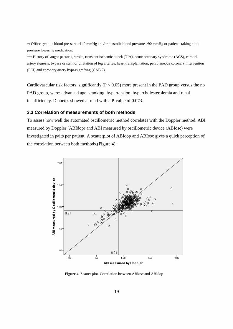
3.3 Correlation of measurements of both methods
To assess how well the automated oscillometric method correlates with the Doppler method, ABI
measured by Doppler (ABIdop) and ABI measured by oscillometric device (ABIosc) were
investigated in pairs per patient. A scatterplot of ABIdop and ABIosc gives a quick perception of
the correlation between both methods.(Figure 4).
Figure 4. Scatter plot. Correlation between ABIosc and ABIdop

20
To evaluate correlation between ABIdop and ABIosc, Spearman correlation coefficient was
determined.(Table 6) The Spearman correlation coefficient was chosen over the Pearson
correlation coefficient because variables ABIdop and ABIosc were not normally
distributed.(Appendix 3,Appendix 4)
Table 6. Spearman correlation coefficient of ABIdop and ABIosc.
ABI measured
by Doppler
ABI measured
by Oscillometric
device
Spearman's rho ABI measured by Doppler Correlation Coefficient 1 .604
P-value . .000
N 385 381
ABI measured by
oscillometric device
Correlation Coefficient .604 1
P-value .000 .
N 381 387
Note that per patient, 2 ABIdop (right ABIdop and left ABIdop) and 2 ABIosc (right ABIosc and
left ABIosc) were compared. That is why N is now theoretically 396 (the number of limbs)
instead of 198 (the number of subjects).
The Spearman correlation coefficient (rs) is 0.604 with P <0.001. However, correlation does not
give adequate information on agreement between both (linear) variables.(31, 32) Therefore,
Bland Altmanplot and ICC (intraclass correlation coefficient) were used to illustrate agreement.
3.2.1 Bland Altman plot
The Bland Altman method can generally be used to assess agreement between two methods of
clinical measurement and can also be used to determine whether these methods vary
systematically. If methods vary systematically, measurements can not be used interchangeably
and systematic adaptation of one method should be considered. An important remark is that
neither one of the methods is required to have perfect sensitivity, nor specificity in order to
compare the two methods. Although the Doppler method is referred to as the ‘golden standard’ in
measuring the ABI, the true golden standard for detecting PAD would be invasive contrast
angiography. Doppler-based ABI is thus far from perfect in diagnosing PAD and therefore in
measuring the ABI accurately. The same applies for the oscillometric-based ABI. The Bland

21
Altman plot has the ability to compare these two imperfect methods.(Figure 5) To construct a
Bland Altman plot, the mean of the two measurements (MeanABI) should be assigned to the x-
axis and the difference between the two values (ABIdop-ABIosc) to the y-axis.
Figure 5. Bland Altmanplot of the differences between both methods against
their mean showing good agreement and no obvious systematic difference
between ABIdop and ABIosc. Mean + 1.96SD and mean – 1.96SD form the
95% limits of agreement.
No obvious systematic difference in ABI between both methods is detected. This is also shown in
figure 6, showing frequencies of the differences between the two measurements distributed
around zero, relatively following the Gaussian curve. However, as mean ABI gets bigger, the
difference between ABIdop and ABIosc becomes more positive. This means that higher values of
the ABI more frequently are underestimated by the oscillometric method. As mean ABI gets
smaller, the difference between ABIdop and ABIosc becomes more negative. This means that
lower values of the ABI more frequently are overestimated by the oscillometric method. The
latter can lead to false negative results: patients with PAD would not be detected by the
automated oscillometric method.
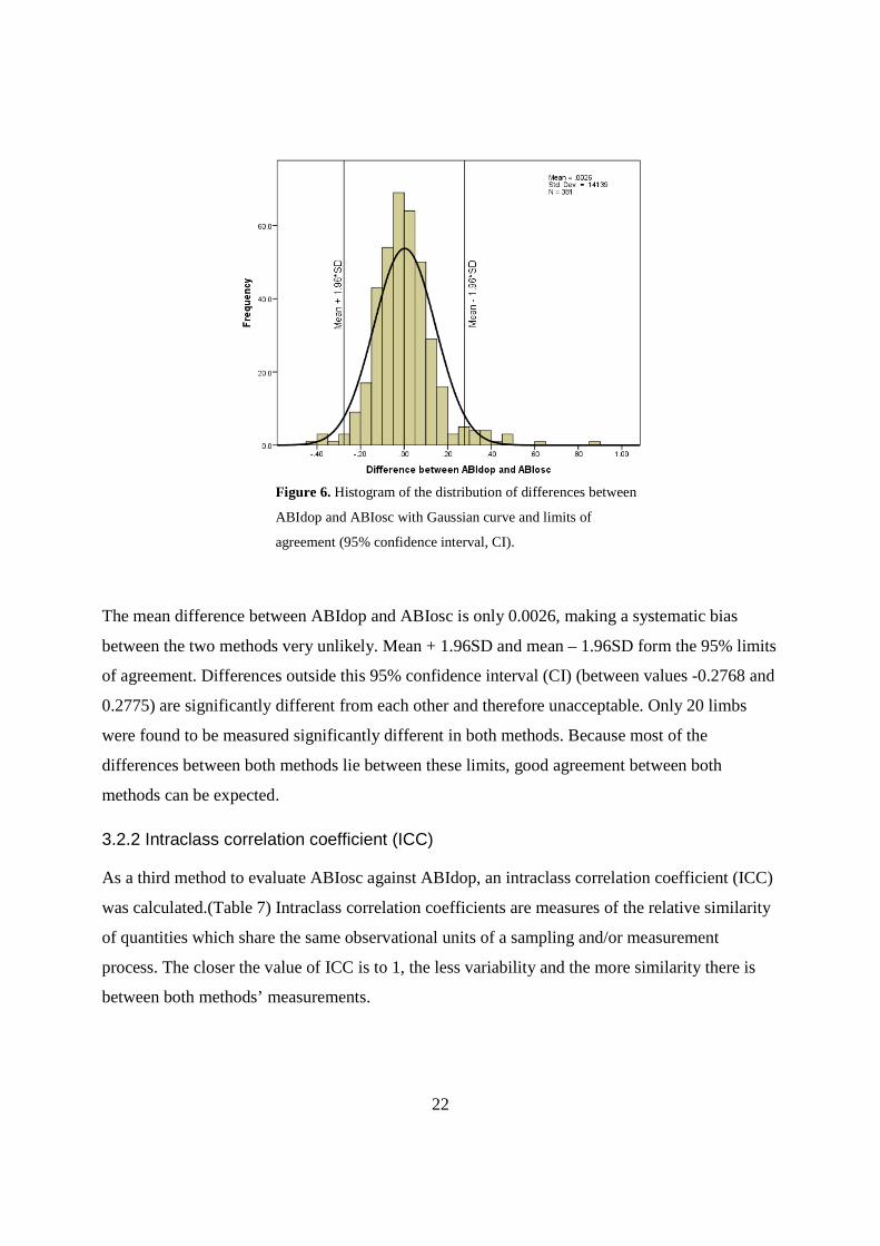
22
Figure 6. Histogram of the distribution of differences between
ABIdop and ABIosc with Gaussian curve and limits of
agreement (95% confidence interval, CI).
The mean difference between ABIdop and ABIosc is only 0.0026, making a systematic bias
between the two methods very unlikely. Mean + 1.96SD and mean – 1.96SD form the 95% limits
of agreement. Differences outside this 95% confidence interval (CI) (between values -0.2768 and
0.2775) are significantly different from each other and therefore unacceptable. Only 20 limbs
were found to be measured significantly different in both methods. Because most of the
differences between both methods lie between these limits, good agreement between both
methods can be expected.
3.2.2 Intraclass correlation coefficient (ICC)
As a third method to evaluate ABIosc against ABIdop, an intraclass correlation coefficient (ICC)
was calculated.(Table 7) Intraclass correlation coefficients are measures of the relative similarity
of quantities which share the same observational units of a sampling and/or measurement
process. The closer the value of ICC is to 1, the less variability and the more similarity there is
between both methods’ measurements.

23
Table 7. Intraclass correlation coefficient (ICC) of ABIosc and ABIdop.
Intraclass
Correlation
95% Confidence Interval
Lower Bound Upper Bound
Single Measures .665 .605 .717
Average Measures .799 .754 .835
‘Single measures ICC’ (reliability of single measures) can be used when only one measurement
of the ABI is performed. ‘Average measures ICC’ (reliability of averages of measures) can be
used when a mean of different ABI measurements is used.(33) In this case, ABI was mostly
measured only once. The single measure ICC is always lower than the average measures ICC.
The single measures ICC value is 0.665 (CI: 0.605 to 0.717) which is an adequate to good
relative reliability, according to Fleiss.(34) (Table 8)
Table 8. Relative reliability according to Fleiss.(34)
Value of ICC Reliability
> 0.75 excellent
0.40 – 0.75 adequate
< 0.40 poor
3.4 Agreement of both methods on PAD diagnosis
In order for the oscillometric method to be reliable, it should identify true patients as having PAD
and healthy subjects as having no PAD. Unfortunately, the true golden standard to which
diagnosis of PAD should be compared to, invasive contrast angiography, was not performed in
this study. The next best thing to compare it to is the suspected diagnosis by the Doppler-based
ABI. To evaluate how both methods agree on PAD diagnosis, a 2x2 cross table is shown (table 9)
and diagnostic performance (table 10) and kappa coefficient were calculated.

24
Table 9. 2x2 cross table of PAD diagnosed by oscillometric device and PAD diagnosed by Doppler.
Peripheral artery disease diagnosed
by oscillometric device
Total No PAD PAD
Peripheral artery disease
diagnosed by Doppler
No PAD 153 3 156
PAD 12 27 39
Total 165 30 195
In 15 subjects, PAD classification differed between both methods. The diagnostic performance of
the automated oscillometric device method, assuming the Doppler method to be the golden
standard was (table 10): sensitivity 69.23% (CI: 52.27% to 82.45%), specificity 98.08% (CI:
94.04% to 99.50%), accuracy 92.30% (CI: 87.70% to 94.50%), positive predictive value (PPV)
0.90 % (CI: 0.723% to 0.973%) and negative predictive value (NPV) 0.92% (CI: 0.873% to
0.960%). The likelihood ratio for a positive result (LR+) was 36.00 (CI: 11.51 to 112.6) , the
likelihood ratio for a negative result (LR-) was 0.31 (CI: 0.196 to 0.503).
Table 10. Diagnostic performance of the automated oscillometric device method.
Statistic Value 95% confidence interval Sensitivity 69.23% 52.27% to 82.45% Specificity 98.08% 94.04% to 99.50% Accuracy 92.30% 52.27% to 82.45% Positive predictive value 90 % 72.3% to 97.3% Negative predictive value 92% 87.3% to 96.0% Positive likelihood ratio 36.00 11.51 to 112,6 Negative likelihood ratio 0.31 0.196 to 0.503 Kappa coefficient 0.737 0.581 to 0.813
The Kappa coefficient (κ) is 0.737 with P < 0.001. κ=0.737 can be considered as a substantial
agreement according to Landis & Koch.(35) (Table 11)
Table 11. Agreement according to Landis & Koch.(35)
Value of Kappa Statistic Strength of Agreement
0.81 – 1.00 excellent
0.61 – 0.80 substantial
0.41 – 0.60 moderate
0.21 – 0.40 fair
0.00 – 0.20 slight
< 0.00 poor
25
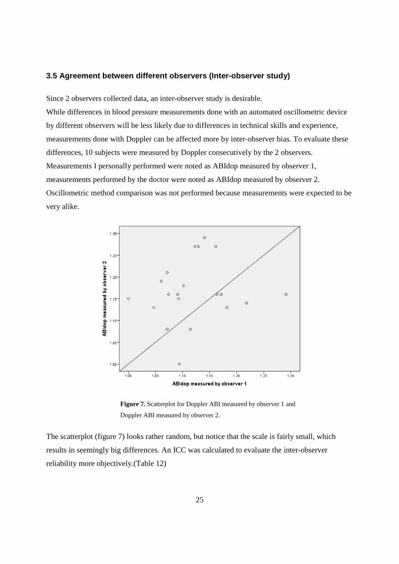
3.5 Agreement between different observers (Inter-ob server study)
Since 2 observers collected data, an inter-observer study is desirable.
While differences in blood pressure measurements done with an automated oscillometric device
by different observers will be less likely due to differences in technical skills and experience,
measurements done with Doppler can be affected more by inter-observer bias. To evaluate these
differences, 10 subjects were measured by Doppler consecutively by the 2 observers.
Measurements I personally performed were noted as ABIdop measured by observer 1,
measurements performed by the doctor were noted as ABIdop measured by observer 2.
Oscillometric method comparison was not performed because measurements were expected to be
very alike.
Figure 7. Scatterplot for Doppler ABI measured by observer 1 and
Doppler ABI measured by observer 2.
The scatterplot (figure 7) looks rather random, but notice that the scale is fairly small, which
results in seemingly big differences. An ICC was calculated to evaluate the inter-observer
reliability more objectively.(Table 12)
26
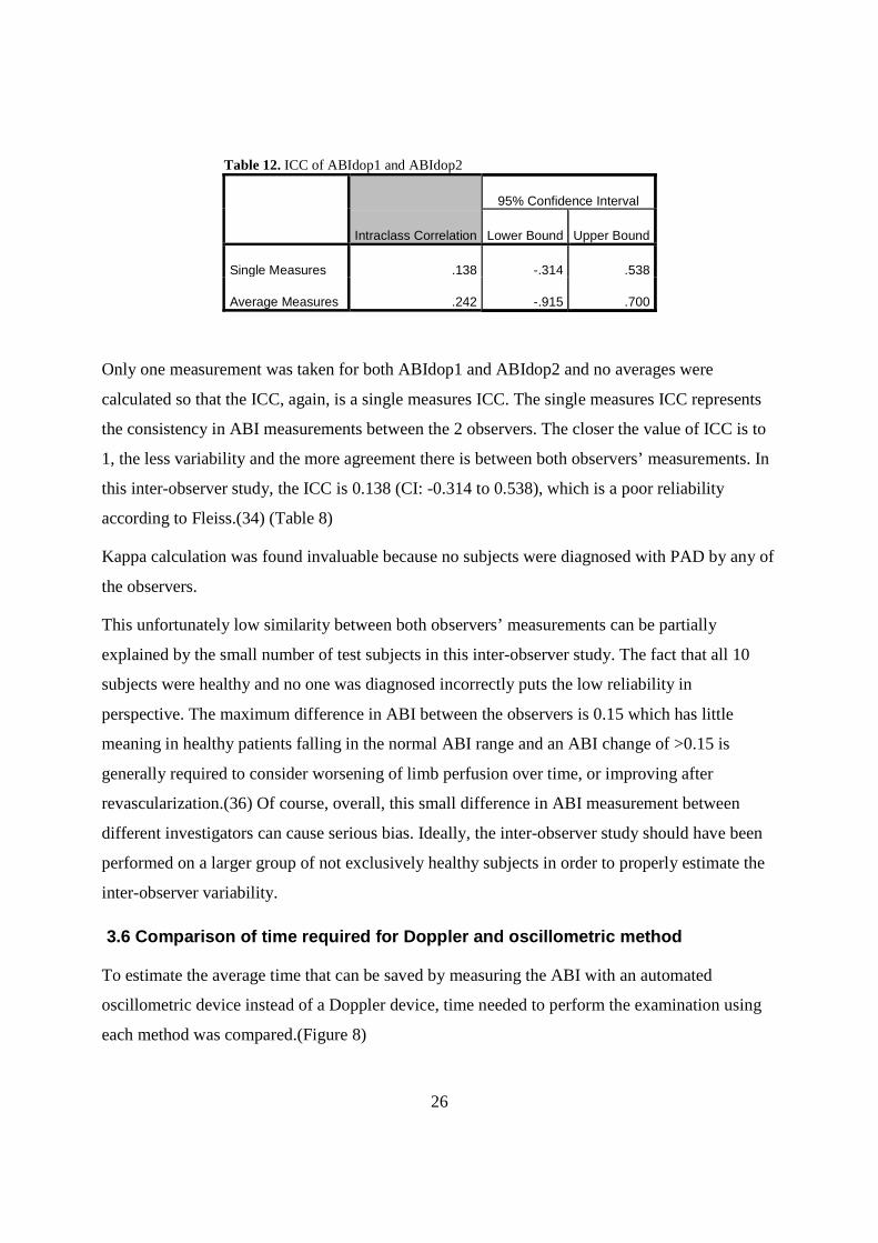
Table 12. ICC of ABIdop1 and ABIdop2
Intraclass Correlation
95% Confidence Interval
Lower Bound Upper Bound
Single Measures .138 -.314 .538
Average Measures .242 -.915 .700
Only one measurement was taken for both ABIdop1 and ABIdop2 and no averages were
calculated so that the ICC, again, is a single measures ICC. The single measures ICC represents
the consistency in ABI measurements between the 2 observers. The closer the value of ICC is to
1, the less variability and the more agreement there is between both observers’ measurements. In
this inter-observer study, the ICC is 0.138 (CI: -0.314 to 0.538), which is a poor reliability
according to Fleiss.(34) (Table 8)
Kappa calculation was found invaluable because no subjects were diagnosed with PAD by any of
the observers.
This unfortunately low similarity between both observers’ measurements can be partially
explained by the small number of test subjects in this inter-observer study. The fact that all 10
subjects were healthy and no one was diagnosed incorrectly puts the low reliability in
perspective. The maximum difference in ABI between the observers is 0.15 which has little
meaning in healthy patients falling in the normal ABI range and an ABI change of >0.15 is
generally required to consider worsening of limb perfusion over time, or improving after
revascularization.(36) Of course, overall, this small difference in ABI measurement between
different investigators can cause serious bias. Ideally, the inter-observer study should have been
performed on a larger group of not exclusively healthy subjects in order to properly estimate the
inter-observer variability.
3.6 Comparison of time required for Doppler and os cillometric method
To estimate the average time that can be saved by measuring the ABI with an automated
oscillometric device instead of a Doppler device, time needed to perform the examination using
each method was compared.(Figure 8)
27
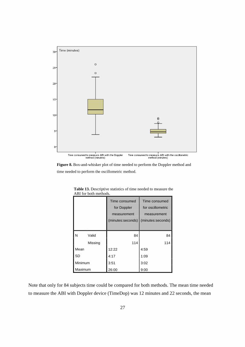
Figure 8. Box-and-whisker plot of time needed to perform the Doppler method and
time needed to perform the oscillometric method.
Table 13. Descriptive statistics of time needed to measure the ABI for both methods.
Time consumed
for Doppler
measurement
(minutes:seconds)
Time consumed
for oscillometric
measurement
(minutes:seconds)
N Valid 84 84
Missing 114 114
Mean 12:22 4:59
SD 4:17 1:09
Minimum 3:51 3:02
Maximum 26:00 9:00
Note that only for 84 subjects time could be compared for both methods. The mean time needed
to measure the ABI with Doppler device (TimeDop) was 12 minutes and 22 seconds, the mean
28
time needed to measure the ABI with an automated oscillometric device (TimeOsc) was much
less: only 4 minutes and 59 seconds.(Table 13) This means that the average time gained if
oscillometric measurement is used instead of Doppler measurement in this study population is 7.4
minutes per subject. Also, a much bigger range of time needed to measure the ABI with Doppler
can be illustrated. This can be explained by the frequent difficulties in using the Doppler
technique on some subjects, resulting in big maximum values. Problems on Doppler usage are
covered in the discussion.
A Wilcoxon paired test revealed a P value <0.001 so that the null hypothesis (TimeDop equals
TimeOsc) could be rejected. This means that there was a (very) significant difference between the
time needed for Doppler ABI measurement and time needed for oscillometric ABI measurement.
Another important observation is that there is a learning curve in handling the Doppler device
efficiently: time needed for ABI measurements decreases with practice.(Figure 9) Measurements
performed by an investigator with little experience with Doppler device will take much longer
than an investigator with ample experience. This observation was not so obvious in the
oscillometric method.(Figure 10)
Figure 9. Scatterplot of time needed to measure ABI with Doppler, progression in time.
Figure 10. Scatterplot of time needed to measure ABI with automated oscillometric device, progression in time.

29
‘Nr’ represents the sequence in which subjects were investigated. For the first 98 subjects, time
was unfortunately not recorded. So only the last 100 subjects, measured by the second observer
(myself) are shown. It is clear that, even after measuring about 200 limbs, there is still a large
range in time needed to measure the ABI in different subjects, which is also acknowledged by
experienced investigators.
3.7 Conclusion
In this paper, the automated oscillometric device method was compared to the golden standard
Doppler method to measure the ABI. Investigating 198 subjects consecutively with Doppler and
oscillometric device in a randomized order, the two ABIs (ABIdop and ABIosc) were found to be
correlating well with a Spearmans correlation coefficient (rs) of 0.604. There was also an
adequate to good ICC of 0.665 and a Bland Altman plot showed little difference and no obvious
systematic bias between measurements of the two methods. However, the automated
oscillometric method was likely to underestimate or overestimate the ABI when the ABI
measured by Doppler method was either high or low respectively.
Both methods seem to be agreeing well on PAD diagnosis. A substantial Kappa coefficient (κ) of
0.737 was calculated. Diagnostic performance of the automated oscillometric method versus the
Doppler method was good: sensitivity 69.23% (CI: 52.27% to 82.45%), specificity 98.08% (CI:
94.04% to 99.50%), accuracy 92.30% (CI: 87.70% to 94.50%), PPV 0.90 % (CI: 0.723% to
0.973%) , NPV 0.92% (CI: 0.873% to 0.960%), LR+ 36.00 (CI: 11.51 to 112.6) and LR- 0.31
(CI: 0.196 to 0.503).
Significant difference between both methods in time needed to measure the ABI was found: a
mean of 12 minutes and 22 seconds for the Doppler method versus a mean of 4 minutes and 59
seconds for the automated method. A learning curve for the Doppler method could be illustrated.
Unfortunately the inter-observer study on 10 healthy subjects revealed poor similarity (ICC=
0.138) between both investigators’ Doppler measurements. However, this should be seen in the
light of a small sample size, a relatively narrow range within normal ABI values and different
experience level between the two observers.
Taking into consideration all pros and cons, the automated oscillometric method can be suggested
for measuring the ABI for screening purposes.

30
4. Discussion
Because of its serious health consequences, high economic costs and given the fact that it is often
present in the absence of symptoms, screening for PAD is strongly advised, especially in certain
risk groups (table 3). While the disease is relatively easy to detect by the ABI test, PAD still
remains underdiagnosed in primary care. The main limitations in assessing the ABI in daily
practice are time constraints, lack of reimbursement and staff availability.(37) Also lack of
education in Doppler handling and ABI calculation and too low awareness of PAD may also play
a role. In this thesis, the Doppler method was challenged by the automated oscillometric method
for measuring the ABI because the latter method is supposed to relieve some of these above
mentioned constraints. Measurements of the automated oscillometric method were compared to
the golden standard Doppler method and reasonable advantages (in time and technique) in favour
of the automated oscillometric method were demonstrated in a group of 198 volunteers.
While Doppler-based ABI is the golden standard for measuring the ABI, invasive contrast
angiography is the golden standard in diagnosing PAD. Because invasive contrast angiography or
other imaging techniques were not performed in this setting, diagnostic performance of the
automated oscillometric method could only be assessed using the Doppler-based ABI as a
reference.
Diagnostic performance of the automated oscillometric method versus Doppler method (PADosc
compared to PADdop) found in this study population is comparable to studies on the same
subject. In this study, sensitivity was 69.23% (CI: 52.27% to 82.45%), specificity 98.08% (CI:
94.04% to 99.50%), PPV 90 % (CI:72.3% to 97.3%) and NPV 92% (CI: 87.3% to 96.0%).
Beckman et al. reported a sensitivity of 73-88%, specificity of 85-95%, PPV of 65-88% and NPV
of 88-96%, depending on which leg was compared.(30) The most recent assessment of sensitivity
and specificity of oscillometric versus Doppler method was performed by Verberk et al. in a large
meta-analysis comprising 25 studies and a total of 4186 subjects. The average sensitivity and
specificity of the oscillometric ABI estimation in PAD diagnosis found in this meta-analysis was
69% (SD 6%) and 96% (SD 1%), respectively (with Doppler-based ABI as reference).(38) This
is very much in line with our results.

31
Because the automated oscillometric device is proposed to be used for screening purposes, it is
important to have a substantial specificity in order to correctly identify healthy persons. A false
positive result could imply that the screened subject could possibly be submitted to costly
investigations, subjected to unnecessary fear or maybe even be overtreated.
Although ABIosc and ABIdop were found to agree well in this study, the automated oscillometric
method was likely to underestimate or overestimate the ABI when the ABI measured by Doppler
method was either high or low respectively. Accordingly, Verberk et al. and Jönsson et al. both
reported that in general, the oscillometric devices tend to overestimate the ankle pressure in
patients with moderate to severe PAD, giving a falsely high ABI.(38, 39) This means that the
oscillometric method would yield more false negatives and would therefore have a lower
sensitivity.
In this study population, especially in subjects with a low ABI, the oscillometric device seemed
to overestimate the ABI.(Figure 4) In the view of screening, this can be tolerated. However, for
precise assessment of the ABI in PAD patients, for e.g. making a decision whether a patient
needs invasive treatment, post-treatment evaluation, follow-up, etc., automated oscillometric
measurement might lose its advantages over the Doppler method due to its lesser agreement at
low ABI values. Automated oscillometric measurement of the ABI is thus preferably only
suggested for screening purposes.
4.1 Limitations of the ABI
The reasons that ABI is rarely used in routine clinical practice include time constraints, lack of
reimbursement and staff availability.(37) This study confirms that ABI measurement by Doppler
can indeed be time consuming. In this study, on average, a Doppler ABI measurement was
performed in 12 minutes and 22 seconds. An automated ABI measurement only took 4 minutes
and 59 seconds on average, demonstrating an important advantage for the latter method in
measuring the ABI.
For an ABI to be measured properly, training is advised on how to correctly use a Doppler device
for blood pressure measurement on peripheral blood vessels. It is important how the Doppler
device is held (preferably at a 45-60-degree angle towards the blood flow) and where it is located
(placing the probe, after applying some gel, where the pulse is felt). This sometimes can be

32
difficult, even for experienced investigators. The learning curve (figure 9) also illustrates that,
even after the observer went through quite some practice, he/she might need notably more time
for certain patients compared to others. (Figure based on my own results.) This can be explained
by the common difficulties in measuring the systolic blood pressures with a Doppler device as I
have experienced myself during the recordings: incompressible or hardly palpable arteries,
anatomical variations of arteries, a loud environment so that the Doppler signal can not be heard
on time.
Inaccurate measurements can occur. As noted in the introduction, the ABI test is a non-invasive
tool for detecting PAD with its own diagnostic accuracy. A recent review reported a high
specificity (83.3–99.0%) and accuracy (72.1–89.2%), but a sensitivity that varied greatly (15–
79%). Sensitivity was low, especially in elderly individuals and patients with diabetes.(20) Here,
falsely elevated ABIs can occur due to stiffening of the arteries in the ankles, thereby lowering
the sensitivity of the ABI test for PAD. ‘Importantly, a substantial proportion of patients with an
elevated ABI actually do have occlusive artery disease’.(1) These patients also have an increased
cardiovascular and overall mortality.(28)(Figure 3) Other diagnostic tests (e.g. toe systolic
pressures, pulse volume recording, duplex ultrasonography, MR-angiography, and CT-
angiography) are recommended for those with calcified vessels (e.g., older individuals, those
with long-standing diabetes or end-stage renal disease) suspected of having PAD and/or having a
resting ABI value of more than 1.3.
The shortcomings of the ABI test also include the potential to miss mild proximal disease of the
aorta and iliac arteries in those with well-developed collaterals. An exercise ABI (cfr. infra)
should be determined when the resting ABI value is normal and there is a high clinical suspicion
for PAD.
Some articles use different cut-off points for the ABI to suspect PAD. Sometimes PAD is
suspected when ABI <0.90, while in other articles, PAD is already suspected when ABI <0.91.
Consensus should be made to define universal cut-off points.

33
4.2 Alternative tests for detecting PAD non-invasiv ely
In subjects with an ABI > 1.3, different alternative tests can be used to diagnose PAD.
1) Toe systolic pressures. Toe systolic pressures are used instead of ankle systolic pressures
to calculate the ABI because the smaller arteries in the toes are less susceptible to
calcification.(40)
2) Pulse volume recording (PVR). Pulse volume recordings are obtained with partially
inflated segmental blood pressure cuffs that detect volume changes sequentially down a
limb. Volume changes beneath the cuffs resulting from systole and diastole cause small
pressure changes within the cuffs, which, with the use of appropriate transducers, can be
displayed as arterial waveforms. Characteristics of the arterial waveforms reveal any
occlusions within the investigated arteries.
3) Duplex ultrasonography. Duplex ultrasonography incorporates both echo (grayscale
ultrasound) to visualize the blood vessel architecture (measurement of the intima-media
thickness and tracking of atherosclerosis) and colour-Doppler ultrasound to visualize the
flow within the blood vessel.
4) MR-angiography and CT-angiography. These imaging techniques are less invasive
than contrast angiography, although CT-angiography carries risks related to ionising
radiation, and both contrast enhanced MR-angiography and CT-angiography carry risks
associated with the use of contrast agents.
The last 3 techniques have the ability to identify the location and severity of arterial narrowing
and occlusion.
In subjects with a normal resting ABI but a high clinical suspicion of PAD, an exercise ABI is
recommended.
Exercise ABI. The ABI is measured after exercise (mostly) on a treadmill. This could yield a
better sensitivity for detecting PAD.(26, 27) Moreover, it can assess the functional status of a
patient with PAD. “How long can the patient walk before ischemic symptoms occur?” And it
provides important prognostic information on long-term outcome.(27) However, other studies
report that it does not lead to more patients being detected.(41)

34
4.3 Limitations with the Doppler method
Errors in ABI measurement with the Doppler device are frequently due to an incorrect
positioning angle of the Doppler probe, a loud environment so that the Doppler signal can not be
heard on time, not applying enough gel on the spot where the Doppler probe makes skin contact,
too much pressure on the artery by the Doppler probe, …
4.4 Limitations with the automated oscillometric me thod
The main problem with this technique is that the amplitude of the oscillations depends on several
factors other than blood pressure, most importantly the stiffness of the arteries. Oscillometric
devices tend to overestimate the ABI in PAD-affected limbs.(38, 39)
While a validated oscillometric device was used, some ankle pressures could not be measured
because of unknown reasons. Kim et al.(25) indicated that the inconsistency between different
validation studies of oscillometric ABI measurement is likely because the devices were designed
for measuring blood pressure in non-obstructed arms, not in the legs, and especially not in
diseased legs. Also, simultaneous oscillometric arm-leg measurements have more accuracy in
measuring the ABI than sequential oscillometric measurements as performed in this study.(38)
4.5 Study limitations
Measurements were taken by 2 observers. Although methods on measurement were matched
beforehand and training was received, an inter-observer study revealed a substantial inter-
observer bias. However, this should be seen in the light of a small sample size, a relatively
narrow range within normal ABI values and different experience level between the two observers.
Ideally, the inter-observer study should have been performed on a larger group of not exclusively
healthy subjects in order to properly estimate the inter-observer variability.
Investigating the intra- and inter-observer variability of Doppler-based ABI, inconsistencies
between articles were encountered. On the one hand, Richart et al. concluded that Doppler
technique requires trained investigators because of an intra-observer variability of approximately
10%.(42) On the other hand, a study by Holland-Letz et al. that focused solely on intra- and inter-
observer bias in ABI measurement reported an intra- and inter-observer variability of 8% and 9%

35
respectively and a fairly high ICC of 0.423.(43) Upon these results, Holland-Letz et al. concluded
that ABI determinations are highly reproducible and reliable under routine conditions and that
ABI measurements can be performed with little training, even by people other than physicians.
However, in this particular study population, only 2 out of 108 subjects had an ABI ≤ 0.9 which
makes these observations less reliable.
The automated oscillometric method was not tested for inter-observer variability because results
were expected to be much more solid. However, it would have been interesting to see how
oscillometric measurements agreed within the 2 different observers and to have compared this
result with the Doppler inter-observer variability. The only thing that might be supposed is that
the inter-observer variability of the oscillometric method would probably be much less than the
inter-observer variability of the Doppler method.
More intense training on Doppler technique could have improved the accuracy of Doppler
measurements. By measuring blood pressures multiple times and calculating the mean, more
accurate results could have been achieved in both Doppler method and oscillometric method. It is
also generally suggested to reassess the ABI within different visitations over time, before
diagnosing peripheral artery disease and before making therapeutic decisions.(44)
In 84 participants out of 198, time needed for performing the ABI test with each method was
measured. Results show a clear advantage for the oscillometric method.
Standardized questionnaires to screen and diagnose intermittent claudication such as the
Edinburgh Claudication Questionnaire were not used. Instead, a questionnaire was created to get
a more global view on the patients’ cardiovascular risk profiles.(Attachment 3)
4.6 Alternative calculations of the ABI
The higher of the two brachial pressures is used as the denominator to account for the possibility
of subclavian artery stenosis, which can decrease the blood pressure in the upper extremity and
thus falsely elevate the ABI.
Ankle pressures used for the ABI calculation can be chosen differently from the pressures in the
classical calculation of the ABI using the highest ankle pressure of either the posterior tibial
artery or the dorsal pedis artery for each limb.

36
Various articles report a higher sensitivity in detecting PAD by using an alternate calculation of
the ABI. One method uses the mean of the pressures in the dorsal pedis and posterior tibial
arteries in each leg.(45) Others suggest that for situations in which the pressures in the two ankle
arteries are different, the lower value may be of benefit, raising the diagnostic sensitivity.(46, 47)
4.7 Future prospective
Implications of this study include that screening patients for PAD by measuring the ABI could be
performed by the automated oscillometric device instead of the Doppler device with more ease
and in a shorter time. Hopefully, measuring the ABI will become more of a routine test in
vascular investigation and cardiovascular risk assessment once oscillometric devices are fully
validated for this purpose.
The implementation of automated oscillometric devices in routine ABI assessments beyond
screening requires further demonstration of concordance with the golden standard Doppler-based
ABI. Large population based studies could give a more definite view on this subject.
This study can be considered as an asset and already demonstrates some advantages of automated
ABI determination.
.

37
5. References
1. Tendera M, Aboyans V, Bartelink ML, Baumgartner I, Clement D, Collet JP, et al. ESC Guidelines
on the diagnosis and treatment of peripheral artery diseases: Document covering atherosclerotic disease
of extracranial carotid and vertebral, mesenteric, renal, upper and lower extremity arteries: the Task
Force on the Diagnosis and Treatment of Peripheral Artery Diseases of the European Society of
Cardiology (ESC). European heart journal. 2011;32(22):2851-906.
2. Kroger K, Stang A, Kondratieva J, Moebus S, Beck E, Schmermund A, et al. Prevalence of
peripheral arterial disease - results of the Heinz Nixdorf recall study. European journal of epidemiology.
2006;21(4):279-85.
3. Selvin E, Erlinger TP. Prevalence of and risk factors for peripheral arterial disease in the United
States: results from the National Health and Nutrition Examination Survey, 1999-2000. Circulation.
2004;110(6):738-43.
4. MedlinePlus. Peripheral Arterial Disease [Internet]. U.S. National Library of Medicine [updated
2013 november 21; cited 2013 november 22]. Available from:
http://www.nlm.nih.gov/medlineplus/peripheralarterialdisease.html.
5. Mahameed AA. Peripheral Arterial Disease [Internet]. Cleveland, USA: The Cleveland Clinic
Foundation; [8/03/2014]. Available from:
http://www.clevelandclinicmeded.com/medicalpubs/diseasemanagement/cardiology/peripheral-
arterial-disease/.
6. Dormandy JA, Rutherford RB. Management of peripheral arterial disease (PAD). TASC Working
Group. TransAtlantic Inter-Society Consensus (TASC). Journal of vascular surgery. 2000;31(1 Pt 2):S1-s296.
7. Hirsch AT, Criqui MH, Treat-Jacobson D, et al. Peripheral arterial disease detection, awareness,
and treatment in primary care. JAMA. 2001;286(11):1317-24.
8. Sigvant B, Wiberg-Hedman K, Bergqvist D, Rolandsson O, Andersson B, Persson E, et al. A
population-based study of peripheral arterial disease prevalence with special focus on critical limb
ischemia and sex differences. Journal of vascular surgery. 2007;45(6):1185-91.
9. McDermott MM, Greenland P, Liu K, Guralnik JM, Criqui MH, Dolan NC, et al. Leg symptoms in
peripheral arterial disease: associated clinical characteristics and functional impairment. Jama.
2001;286(13):1599-606.
10. Priollet P. Quality of life and peripheral obliterative arteriopathy. Perspective for the future.
Drugs. 1998;56 Suppl 3:49-58.
11. Dormandy J, Heeck L, Vig S. The natural history of claudication: risk to life and limb. Seminars in
vascular surgery. 1999;12(2):123-37.
12. Zheng ZJ, Sharrett AR, Chambless LE, Rosamond WD, Nieto FJ, Sheps DS, et al. Associations of
ankle-brachial index with clinical coronary heart disease, stroke and preclinical carotid and popliteal
atherosclerosis: the Atherosclerosis Risk in Communities (ARIC) Study. Atherosclerosis. 1997;131(1):115-
25.
13. Hooi JD, Kester AD, Stoffers HE, Rinkens PE, Knottnerus JA, van Ree JW. Asymptomatic peripheral
arterial occlusive disease predicted cardiovascular morbidity and mortality in a 7-year follow-up study.
Journal of clinical epidemiology. 2004;57(3):294-300.

38
14. Criqui MH, McClelland RL, McDermott MM, Allison MA, Blumenthal RS, Aboyans V, et al. The
ankle-brachial index and incident cardiovascular events in the MESA (Multi-Ethnic Study of
Atherosclerosis). Journal of the American College of Cardiology. 2010;56(18):1506-12.
15. Criqui MH, Ninomiya JK, Wingard DL, Ji M, Fronek A. Progression of peripheral arterial disease
predicts cardiovascular disease morbidity and mortality. Journal of the American College of Cardiology.
2008;52(21):1736-42.
16. Smolderen KG, Wang K, de Pouvourville G, Bruggenjurgen B, Rother J, Zeymer U, et al. Two-year
vascular hospitalisation rates and associated costs in patients at risk of atherothrombosis in France and
Germany: highest burden for peripheral arterial disease. European journal of vascular and endovascular
surgery : the official journal of the European Society for Vascular Surgery. 2012;43(2):198-207.
17. Criqui MH. Peripheral arterial disease--epidemiological aspects. Vascular medicine (London,
England). 2001;6(3 Suppl):3-7.
18. Grenon SM, Gagnon J, Hsiang Y. Ankle–Brachial Index for Assessment of Peripheral Arterial
Disease. New England Journal of Medicine. 2009;361(19):e40.
19. Lijmer JG, Hunink MG, van den Dungen JJ, Loonstra J, Smit AJ. ROC analysis of noninvasive tests
for peripheral arterial disease. Ultrasound in medicine & biology. 1996;22(4):391-8.
20. Dachun X, Jue L, Liling Z, Yawei X, Dayi H, Pagoto SL, et al. Sensitivity and specificity of the ankle--
brachial index to diagnose peripheral artery disease: a structured review. Vascular medicine (London,
England). 2010;15(5):361-9.
21. Hirsch AT, Haskal ZJ, Hertzer NR, Bakal CW, Creager MA, Halperin JL, et al. ACC/AHA 2005
Practice Guidelines for the management of patients with peripheral arterial disease (lower extremity,
renal, mesenteric, and abdominal aortic): a collaborative report from the American Association for
Vascular Surgery/Society for Vascular Surgery, Society for Cardiovascular Angiography and Interventions,
Society for Vascular Medicine and Biology, Society of Interventional Radiology, and the ACC/AHA Task
Force on Practice Guidelines (Writing Committee to Develop Guidelines for the Management of Patients
With Peripheral Arterial Disease): endorsed by the American Association of Cardiovascular and
Pulmonary Rehabilitation; National Heart, Lung, and Blood Institute; Society for Vascular Nursing;
TransAtlantic Inter-Society Consensus; and Vascular Disease Foundation. Circulation. 2006;113(11):e463-
654.
22. Ferket BS, Spronk S, Colkesen EB, Hunink MG. Systematic review of guidelines on peripheral
artery disease screening. The American journal of medicine. 2012;125(2):198-208.e3.
23. Gerhard-Herman M, Gardin JM, Jaff M, Mohler E, Roman M, Naqvi TZ. Guidelines for noninvasive
vascular laboratory testing: a report from the American Society of Echocardiography and the Society for
Vascular Medicine and Biology. Vascular medicine (London, England). 2006;11(3):183-200.
24. National Institutes of Health DoHaHS. How Is Peripheral Arterial Disease Diagnosed? [Internet].
U.S. Department of Health & Human Services [updated 2011 April 01; cited 2013 November 24].
Available from: http://www.nhlbi.nih.gov/health/health-topics/topics/pad/diagnosis.html.
25. Kim ES, Wattanakit K, Gornik HL. Using the ankle-brachial index to diagnose peripheral artery
disease and assess cardiovascular risk. Cleveland Clinic journal of medicine. 2012;79(9):651-61.
26. Stein R, Hriljac I, Halperin JL, Gustavson SM, Teodorescu V, Olin JW. Limitation of the resting
ankle-brachial index in symptomatic patients with peripheral arterial disease. Vascular medicine (London,
England). 2006;11(1):29-33.

39
27. de L, II, Verhagen HJ, Stolker RJ, van Domburg RT, Poldermans D. The value of treadmill exercise
test parameters together in patients with known or suspected peripheral arterial disease. European
journal of preventive cardiology. 2012;19(2):192-8.
28. Resnick HE, Lindsay RS, McDermott MM, Devereux RB, Jones KL, Fabsitz RR, et al. Relationship of
high and low ankle brachial index to all-cause and cardiovascular disease mortality: the Strong Heart
Study. Circulation. 2004;109(6):733-9.
29. Begelman SM, Jaff MR. Noninvasive diagnostic strategies for peripheral arterial disease.
Cleveland Clinic journal of medicine. 2006;73 Suppl 4:S22-9.
30. Beckman JA, Higgins CO, Gerhard-Herman M. Automated oscillometric determination of the
ankle-brachial index provides accuracy necessary for office practice. Hypertension. 2006;47(1):35-8.
31. Bland JM, Altman DG. Statistical methods for assessing agreement between two methods of
clinical measurement. Lancet. 1986;1(8476):307-10.
32. Altman DG. Practical statistics for medical research: Taylor & Francis Ltd; 1990.
33. Van Maele G, Drs. Heidi Buysse, Dr. Pascal Coorevits, Dr. Ellen Deschepper. Statistische
gegevensverwerking met behulp van IBM SPSS STATISTICS 19. University Press; 2011.
34. Fleiss JL. Analysis of data from multiclinic trials. Controlled clinical trials. 1986;7(4):267-75.
35. Landis JR, Koch GG. An application of hierarchical kappa-type statistics in the assessment of
majority agreement among multiple observers. Biometrics. 1977;33(2):363-74.
36. Baker JD, Dix DE. Variability of Doppler ankle pressures with arterial occlusive disease: an
evaluation of ankle index and brachial-ankle pressure gradient. Surgery. 1981;89(1):134-7.
37. Mohler ER, 3rd, Treat-Jacobson D, Reilly MP, Cunningham KE, Miani M, Criqui MH, et al. Utility
and barriers to performance of the ankle-brachial index in primary care practice. Vascular medicine
(London, England). 2004;9(4):253-60.
38. Verberk WJ, Kollias A, Stergiou GS. Automated oscillometric determination of the ankle-brachial
index: a systematic review and meta-analysis. Hypertension research : official journal of the Japanese
Society of Hypertension. 2012;35(9):883-91.
39. Jonsson B, Lindberg LG, Skau T, Thulesius O. Is oscillometric ankle pressure reliable in leg vascular
disease? Clinical physiology (Oxford, England). 2001;21(2):155-63.
40. Brooks B, Dean R, Patel S, Wu B, Molyneaux L, Yue DK. TBI or not TBI: that is the question. Is it
better to measure toe pressure than ankle pressure in diabetic patients? Diabetic medicine : a journal of
the British Diabetic Association. 2001;18(7):528-32.
41. Gouveri E, Papanas N, Marakomichelakis G, Tzavara C, Drakopanagiotakis F, Maltezos E, et al.
Post-exercise ankle-brachial index is not an indispensable tool for the detection of peripheral arterial
disease in an epidemiological survey. A post-hoc analysis of the Athens Study. International angiology : a
journal of the International Union of Angiology. 2013;32(5):518-25.
42. Richart T, Kuznetsova T, Wizner B, Struijker-Boudier HA, Staessen JA. Validation of automated
oscillometric versus manual measurement of the ankle-brachial index. Hypertension research : official
journal of the Japanese Society of Hypertension. 2009;32(10):884-8.

40
43. Holland-Letz T, Endres HG, Biedermann S, Mahn M, Kunert J, Groh S, et al. Reproducibility and
reliability of the ankle-brachial index as assessed by vascular experts, family physicians and nurses.
Vascular medicine (London, England). 2007;12(2):105-12.
44. Weatherley BD, Chambless LE, Heiss G, Catellier DJ, Ellison CR. The reliability of the ankle-
brachial index in the Atherosclerosis Risk in Communities (ARIC) study and the NHLBI Family Heart Study
(FHS). BMC cardiovascular disorders. 2006;6:7.
45. McDermott MM, Tian L, Liu K, Guralnik JM, Ferrucci L, Tan J, et al. Prognostic value of functional
performance for mortality in patients with peripheral artery disease. Journal of the American College of
Cardiology. 2008;51(15):1482-9.
46. Schroder F, Diehm N, Kareem S, Ames M, Pira A, Zwettler U, et al. A modified calculation of
ankle-brachial pressure index is far more sensitive in the detection of peripheral arterial disease. Journal
of vascular surgery. 2006;44(3):531-6.
47. Espinola-Klein C, Rupprecht HJ, Bickel C, Lackner K, Savvidis S, Messow CM, et al. Different
calculations of ankle-brachial index and their impact on cardiovascular risk prediction. Circulation.
2008;118(9):961-7.

I
6. Appendix
Appendix 1. Summary of descriptive characteristics for continuous variable age for no PAD and PAD group.
No PAD N Valid 153
Missing 4
Mean 53,38
Median 55
SD 18,579
Range 66
Minimum 19
Maximum 85
Percentiles Q1 39,00
Q3 70,00
PAD N Valid 39
Missing 0
Mean 66,77
Median 68
SD 13,800
Range 65
Minimum 19
Maximum 84
Percentiles Q1 60,00
Q3 78,00

II
Appendix 2. Histogram of ABIdop, not
normally distributed.
Shapiro-wilk test: p < 0.001
Appendix 3. Histogram of ABIosc, not
normally distributed.
Shapiro-wilk test: p < 0.001

III
Attachment 1: Ethical Committee

IV

V
Attachment 2: Informed Consent
Informatiebrief voor de deelnemers aan experimenten
1 Titel van de studie:
Accuraatheid van automatische bloeddrukmeting bij het bepalen van de enkel/arm
index (E/A index).
2 Doel van de studie:
Men heeft u gevraagd om deel te nemen aan een studie.
Deze studie wordt uitgevoerd om te bepalen of een automatische bloeddrukmeter kan
worden gebruikt om uit te maken of er vernauwingen aanwezig zijn in de onderste
ledematen. De aanwezigheid van deze vernauwingen is een belangrijke risicofactor voor
hart- en bloedvaten. Deze alternatieve methode met automatische bloeddrukmeter zal
worden vergeleken met een reeds bestaande standaard methode (Doppler) hieronder
verder beschreven.
3 Beschrijving van de studie:
Het gaat om een éénmalig onderzoek waarbij de enkel/arm index via twee verschillende
methodes zal worden gemeten.
Er zal u worden gevraagd om eerst vijf minuten in een rustige onderzoekskamer neer te
liggen vooraleer het onderzoek met de metingen plaatsvindt. Om de twee
onderzoeksmethodes te kunnen vergelijken, zal er in een willekeurige volgorde een
bloeddrukmeter, die manueel wordt opgeblazen en een bloeddrukmeter die automatisch

VI
opblaast, worden aangelegd. De bloeddrukmeter wordt afwisselend ter hoogte van
beide armen en vervolgens ter hoogte van de beide enkels aangebracht. Bij de manuele
bloeddrukmeter wordt er met een toestelletje geluidssignalen opgevangen van de
bloedvaten en kan de bloeddruk worden bepaald (Doppler methode). De andere
methode bestaat erin om een bloeddrukmeter aan te leggen die automatisch opblaast
en automatisch de bloeddruk bepaalt ter hoogte van beide armen en enkels. Tussen de
beide onderzoeken wordt er vijf minuten gepauzeerd.
De resultaten van deze beide methodes zullen worden vergeleken om te zien of de te
testen methode evenwaardig is aan de reeds bestaande methode.
De verwachte totale duur van de studie is 30 minuten
Er zullen in totaal 60 personen aan deze studie deelnemen.
4 Wat wordt verwacht van de deelnemer?
Voor het welslagen van de studie, is het uitermate belangrijk dat u volledig meewerkt
met de onderzoeker en dat u zijn/haar instructies nauwlettend opvolgt.
5 Deelname en beëindiging:
De deelname aan deze studie vindt plaats op vrijwillige basis.
Uw deelname in de studie kan helpen om in de toekomst patiënten beter te kunnen
helpen.

VII
U kan weigeren om deel te nemen aan de studie, en u kunt zich op elk ogenblik
terugtrekken uit de studie zonder dat u hiervoor een reden moet opgeven en zonder dat
dit op enigerlei wijze een invloed zal hebben op uw verdere relatie en/of behandeling
met de onderzoeker of de behandelende arts.
Uw deelname aan deze studie zal worden beëindigd als de onderzoeker meent dat dit in
uw belang is. U kunt ook voortijdig uit de studie worden teruggetrokken als u de in deze
informatiebrief beschreven procedures niet goed opvolgt of u de beschreven items niet
respecteert.
Als u deelneemt, wordt u gevraagd het toestemmingsformulier te tekenen.
5.1 Procedures:
Om de enkel/arm index te bepalen zal volgens een willekeurige volgorde
achtereenvolgens gebruik worden gemaakt van een automatische bloeddrukmeter en
van een manuele bloeddrukmeter en een doppler toestel.
5.2 Studieverloop:
Het gaat om een éénmalige meting waarbij de enkel/arm index via de twee
verschillende methodes zal worden bepaald.
6 Risico’s en voordelen:
Er zijn geen risico’s verbonden aan dit onderzoek.
Deelname aan deze studie brengt voor u geen onmiddellijk therapeutisch voordeel.

VIII
Deze studie werd goedgekeurd door een onafhankelijke Commissie voor Medische
Ethiek verbonden aan het UZ Gent en wordt uitgevoerd volgens de richtlijnen voor de
goede klinische praktijk (ICH/GCP) en de verklaring van Helsinki opgesteld ter
bescherming van mensen deelnemend aan klinische studies. In geen geval dient u de
goedkeuring door de Commissie voor Medische Ethiek te beschouwen als een aanzet
tot deelname aan deze studie.
7 Kosten:
Uw deelname aan deze studie brengt geen extra kosten mee voor U.
8 Vergoeding:
Er wordt geen vergoeding voorzien.
9 Vertrouwelijkheid:
In overeenstemming met de Belgische wet van 8 december 1992 en de Belgische wet
van 22 augustus 2002, zal u persoonlijke levenssfeer worden gerespecteerd en zal u
toegang krijgen tot de verzamelde gegevens. Elk onjuist gegeven kan op uw verzoek
verbeterd worden.
Vertegenwoordigers van de opdrachtgever, auditoren, de Commissie voor Medische
Ethiek en de bevoegde overheden hebben rechtstreeks toegang tot Uw medische
dossiers om de procedures van de studie en/of de gegevens te controleren, zonder de
vertrouwelijkheid te schenden. Dit kan enkel binnen de grenzen die door de betreffende
wetten zijn toegestaan. Door het toestemmingsformulier, na voorafgaande uitleg, te
ondertekenen stemt U in met deze toegang.

IX
Als u akkoord gaat om aan deze studie deel te nemen, zullen uw persoonlijke en
klinische gegevens tijdens deze studie worden verzameld en gecodeerd (hierbij kan
men uw gegevens nog terug koppelen naar uw persoonlijk dossier). Verslagen waarin U
wordt geïdentificeerd, zullen niet openlijk beschikbaar zijn. Als de resultaten van de
studie worden gepubliceerd, zal uw identiteit vertrouwelijke informatie blijven.
10 Letsels ten gevolge van deelname aan de studie:
De onderzoeker voorziet in een vergoeding en/of medische behandeling in het geval
van schade en/of letsel tengevolge van deelname aan de studie. Voor dit doeleinde is
een verzekering afgesloten met foutloze aansprakelijkheid conform de wet inzake
experimenten op de menselijke persoon van 7 mei 2004. Op dat ogenblik kunnen uw
gegevens doorgegeven worden aan de verzekeraar
11 Contactpersoon:
Als er letsel optreedt tengevolge van de studie, of als U aanvullende informatie wenst
over de studie of over uw rechten en plichten, kunt U in de loop van de studie op elk
ogenblik contact opnemen met:
Dr. T. De Backer Cardioloog UZ Gent 09/332.34.71

X
Toestemmingsformulier
Ik, _________________________________________ heb het document “Informatiebrief voor de deelnemers aan experimenten” met als voettekst “Informed Consent dd. 18 oktober 2009 pagina 1 tot en met 3 gelezen en er een kopij van gekregen. Ik stem in met de inhoud van het document en stem ook in deel te nemen aan de studie. Ik heb een kopij gekregen van dit ondertekende en gedateerd formulier voor “Toestemmingsformulier”. Ik heb uitleg gekregen over de aard, het doel, de duur, en de te voorziene effecten van de studie en over wat men van mij verwacht. Ik heb uitleg gekregen over de mogelijke risico’s en voordelen van de studie. Men heeft me de gelegenheid en voldoende tijd gegeven om vragen te stellen over de studie, en ik heb op al mijn vragen een bevredigend antwoord gekregen. Ik stem ermee in om volledig samen te werken met de toeziende onderzoeker. Ik zal hem/haar op de hoogte brengen als ik onverwachte of ongebruikelijke symptomen ervaar.
Ik ben me ervan bewust dat deze studie werd goedgekeurd door een onafhankelijke
Commissie voor Medische Ethiek verbonden aan het UZ Gent en dat deze studie
zal uitgevoerd worden volgens de richtlijnen voor de goede klinische praktijk (ICH/GCP)
en de verklaring van Helsinki, opgesteld ter bescherming van mensen deelnemend aan
experimenten. Deze goedkeuring was in geen geval de aanzet om te beslissen om deel
te nemen aan deze studie.
Ik mag me op elk ogenblik uit de studie terugtrekken zonder een reden voor deze beslissing op te geven en zonder dat dit op enigerlei wijze een invloed zal hebben op mijn verdere relatie met de onderzoeker. Men heeft mij ingelicht dat zowel persoonlijke gegevens als gegevens aangaande mijn gezondheid, ras en seksuele leven worden verwerkt en bewaard gedurende minstens 30 jaar. Ik stem hiermee in en ben op de hoogte dat ik recht heb op toegang en verbetering van deze gegevens. Aangezien deze gegevens verwerkt worden in het kader van medisch-wetenschappelijke doeleinden, begrijp ik dat de toegang tot mijn gegevens kan uitgesteld worden tot na beëindiging van het onderzoek. Indien ik toegang wil tot mijn gegevens, zal ik mij richten tot de toeziende onderzoeker, die verantwoordelijk is voor de verwerking.

XI
Ik begrijp dat auditors, vertegenwoordigers van de opdrachtgever, de Commissie voor Medische Ethiek of bevoegde overheden, mijn gegevens mogelijk willen inspecteren om de verzamelde informatie te controleren. Door dit document te ondertekenen, geef ik toestemming voor deze controle. Bovendien ben ik op de hoogte dat bepaalde gegevens doorgegeven worden aan de opdrachtgever. Ik geef hiervoor mijn toestemming, zelfs indien dit betekent dat mijn gegevens doorgegeven worden aan een land buiten de Europese Unie. Ten alle tijden zal mijn privacy gerespecteerd worden. Ik ben bereid op vrijwillige basis deel te nemen aan deze studie. Naam van de vrijwilliger: _________________________________________ Datum: _________________________________________ Handtekening: Ik bevestig dat ik de aard, het doel, en de te voorziene effecten van de studie heb uitgelegd aan de bovenvermelde vrijwilliger. De vrijwilliger stemde toe om deel te nemen door zijn/haar persoonlijk gedateerde handtekening te plaatsen. Naam van de persoon die voorafgaande uitleg heeft gegeven: _________________________________________ Datum: _________________________________________ Handtekening:

XII
Attachment 3: Questionnaire
VRAGENLIJST STUDIE ENKEL ARM INDEX
Via deze vragenlijst willen we een overzicht krijgen van uw risicofactoren op het ontwikkelen
van perifeer vaatlijden.
Uw gegevens worden enkel gebruikt in het kader van deze studie en zullen niet worden gedeeld
met derden.
1) Wat is uw leeftijd?
2) Gewicht:
3) Lengte:
4) Rookt u? Zo ja, Hoeveel?:
of Heeft u ooit gerookt? Hoelang bent u gestopt?
5) Heeft u suikerziekte?
6) Heeft u teveel cholesterol of neemt u medicatie om de cholesterol te verlagen? ja/nee
7) Heeft u hypertensie of neemt u bloeddrukverlagende medicatie? Ja/nee

XIII
8) Hoeveel glazen alcohol consumeert U gemiddeld per dag (beschouwd over 1 week)?:
9) Hoeveel keer per week heeft u beweging of doet u aan sport?
10) Bent u gekend met nierinsufficiëntie?
11) Ervaart u pijn/last in de benen na een afstand stappen? Ja/nee
Indien ja:
- Gaat de pijn over indien u stilstaat?
- Na welke afstand stappen begint de pijn? </> dan 200m
- Heeft u ook pijn in rust?
12) Heeft u ooit een ingreep ondergaan thv de bloedvaten van uw benen (ballondilatatie, stent,
overbrugging)?

XIV
13) Ervaart u soms een druk op de borstkas bij inspanning?
Indien ja:
- Gaat de pijn over wanneer u rust?
- Indien u deze druk ervaart, straalt deze dan uit naar de arm, hals?
- Neemt u hiervoor medicatie, zo ja: welke?
14) Heeft u een voorgeschiedenis van hartinfarct en/of heeft u ooit een ingreep gehad van de
bloedvaten van uw hart (ballondilatatie, stent, overbrugging)?
15) Bent u gekend met een vernauwing thv de halsvaten of heeft u hiervoor reeds een ingreep
ondergaan?
16) Heeft u een voorgeschiedenis van TIA of hersentrombose?

XV
17) Zijn er mensen in uw familie die problemen hebben van het hart en/of de bloedvaten op jonge
leeftijd (< 65j) ? Zo ja, welke aandoeningen? (hartinfarct, hersentrombose, plotse dood?
hypertensie?)
18) Welke medicatie neemt u?
Dank voor uw deelname.
Jonas Delforche, student Master Geneeskunde
Dr T. De Backer – Dr L. Campens – Dr. M. De Pauw
Dienst Cardiologie
UZ Gent


![ABI Group Organization Chartabishowatech.co.in/wp-content/uploads/2017/06/20170619...2017/06/19 · ABI Group - Footprint 3 1991 –Incorporation of ABI Showatech India Limited [ABI]](https://static.fdocuments.in/doc/165x107/60047efe48a8ad05f75fc13b/abi-group-organization-20170619-abi-group-footprint-3-1991-aincorporation.jpg)