Comparison of LANCE Ultra ULTRA TR-FRET to PerkinElmer ......Introduction Protein kinases play a...
6
Introduction Protein kinases play a major role in normal cellular functions such as cell proliferation, angiogenesis, and cell adhesion. Dysregulation of kinase activity has been shown to be associated with several human diseases including cancer, diabetes and morphological disorders. This makes kinases crucial targets for the discovery and development of new drugs. Analysis of the human genome has revealed the existence of nearly 520 genes encoding kinases. The abundance of these potential therapeutic targets provides a compelling impetus for developing efficient and robust high-throughput screening (HTS) assay platforms for the discovery of kinase modulators. Time-resolved fluorescence resonance energy transfer (TR- FRET) assays are homogeneous proximity assays in which energy is transferred from a donor to an acceptor molecule. A number of TR-FRET platforms are currently available that differ principally in the nature of the donor and acceptor dyes. The LANCE ® technology, for example, uses an europium chelate (Eu) as donor dye, which offers a number of advantages, including a high quantum yield, large Stokes’ shift and a narrow-banded emission at around 615 nm. Furthermore, the lifetime of emitted light from Eu chelate dyes is exceptionally long, allowing for time-delayed measurements. The unique fluorescence properties of Eu chelates make them ideal energy donors in TR-FRET assays. In classical LANCE assays the acceptor dye is allophycocyanin (APC). APC receives the energy from irradiated Eu chelate mole- cules in close proximity, and in turn emits light at 665 nm. Although APC allows for the efficient capture and re-emission of the transferred energy, there are some disadvantages. APC is a bulky and light-sensitive protein prone to generating potential steric hindrances in some assay configurations. Comparison of LANCE Ultra TR-FRET to PerkinElmer’s Classical LANCE TR-FRET Platform for Kinase Applications A P P L I C A T I O N N O T E LANCE ULTRA TR-FRET TECHNOLOGY www.perkinelmer.com Authors Mireille Legault, Philippe Roby, Lucille Beaudet and Nathalie Rouleau PerkinElmer Life and Analytical Sciences
Transcript of Comparison of LANCE Ultra ULTRA TR-FRET to PerkinElmer ......Introduction Protein kinases play a...
Introduction
Protein kinases play a major rolein normal cellular functions suchas cell proliferation, angiogenesis,and cell adhesion. Dysregulationof kinase activity has been shownto be associated with severalhuman diseases including cancer,diabetes and morphologicaldisorders. This makes kinasescrucial targets for the discoveryand development of new drugs.Analysis of the human genomehas revealed the existence ofnearly 520 genes encodingkinases. The abundance of thesepotential therapeutic targetsprovides a compelling impetusfor developing efficient androbust high-throughput screening(HTS) assay platforms for thediscovery of kinase modulators.
Time-resolved fluorescenceresonance energy transfer (TR-FRET) assays are homogeneousproximity assays in which energyis transferred from a donor to anacceptor molecule. A number ofTR-FRET platforms are currentlyavailable that differ principally
in the nature of the donor andacceptor dyes. The LANCE®
technology, for example, uses aneuropium chelate (Eu) as donordye, which offers a number ofadvantages, including a highquantum yield, large Stokes’ shiftand a narrow-banded emission ataround 615 nm. Furthermore, thelifetime of emitted light from Euchelate dyes is exceptionallylong, allowing for time-delayedmeasurements. The uniquefluorescence properties of Euchelates make them ideal energydonors in TR-FRET assays.
In classical LANCE assays theacceptor dye is allophycocyanin(APC). APC receives the energyfrom irradiated Eu chelate mole-cules in close proximity, and inturn emits light at 665 nm.Although APC allows for theefficient capture and re-emissionof the transferred energy, thereare some disadvantages. APC is a bulky and light-sensitiveprotein prone to generatingpotential steric hindrances insome assay configurations.
Comparison of LANCE UltraTR-FRET to PerkinElmer’sClassical LANCE TR-FRETPlatform for Kinase Applications
AP
PL
IC
AT
IO
N
NO
TE
LA
NC
E U
LT
RA
TR
-FR
ET
TE
CH
NO
LO
GY
www.perkinelmer.com
Authors Mireille Legault, Philippe Roby, Lucille Beaudet and Nathalie Rouleau
PerkinElmer Life and Analytical Sciences

In addition, small molecules suchas peptides and oligonucleotidescannot be labeled directly withAPC, resulting in the need to use abiotinylated substrate combinedwith APC-streptavidin (APC-SA). To overcome these limitations,PerkinElmer has developed the newLANCE Ultra HTS platform inwhich the APC acceptor dye hasbeen replaced by a new ULight™
acceptor dye (Figure 1).
ULight is a small, light resistantacceptor dye with spectral charac-teristics similar to APC. Its red-shifted emission is less sensitive toquenching by colored compounds,which eliminates the need forratiometric data analysis.Importantly, the ULight dye’s lowmolecular weight allows directlabeling of molecules of any size. In kinase assays, the dye can becoupled directly to kinase sub-strates or to streptavidin for thecapture of biotinylated substrates.
This application note presents acomparison of LANCE Ultra withthe classical LANCE platform. The development of a Src tyrosinekinase assay measuring the phos-phorylation of a poly Glu-Tyr (poly-GT) substrate was used as a model system.
Materials and Methods
Materials
Table 1 lists the materials used for this study, including suppliersand product numbers.
2
Figure 1. Schematic representation of LANCE Ultra and LANCE for Src kinase assays. Following Src kinase reactions, labeled phospho-poly-GT is detected using three different LANCE assay formats. A) Poly-GT is labeled with the small ULight dye. B) Poly-GT is biotinylated and captured byULight-labeled streptavidin. C) Poly-GT is biotinylated and captured by APC-SA. In all three assays, the phosphorylated amino acid is recognized by an anti-phospho-tyrosine antibody (PY20) labeled with Eu. Upon excitation of the Eu-labeled antibody, energy is transferred to the acceptor dyeresulting in emission of light at 665 nm.
LANCE Ultra(ULight-poly-GT-based)
Classical LANCE(APC-streptavidin-based)
LANCE Ultra(ULight-streptavidin-based)
Acceptor:ULight
Acceptor:Allophycocyanin (APC)
Acceptor:ULight
Kinase Assays
Assay conditions were optimizedindependently for each of the threeTR-FRET platforms. All concentra-tions listed below are final concen-trations in either the kinase ordetection reaction. LANCE Ultraand classical LANCE signals weredetected using the same settings onthe EnVision™ Multilabel Reader(excitation at 320 nm and emissionat 665 nm).
LANCE Ultra (ULight-poly-GT)Optimized conditions consisted of 1 nM Src, 100 nM ULight-poly-GTand ATP at the EC50 value. Assaycomponents were diluted in Kinase Buffer and added to a whiteOptiPlate™-384 in a final volume of10 µL. Plates were incubated for 90min at room temperature (RT) andkinase reactions were stopped bythe addition of 10 mM EDTA. Fiveminutes after the addition of EDTA,2 nM of Eu-labeled PY20 antibody,diluted in detection buffer, was
added for the detection of phospho-products. Reactions were incubatedfor 1 h at RT prior to signal reading.
LANCE Ultra (ULight-Streptavidin;biotinylated poly-GT)Kinase reactions were performed asdescribed for the ULight-poly-GTassay. Five minutes after theaddition of EDTA, Eu-labeled PY20 (2 nM) antibody and ULight-Streptavidin (ULight-SA; 100 nM),diluted in Detection Buffer, wereadded for the detection of phospho-products. Reactions were incubatedfor 1 h at RT prior to signal reading.
Classical LANCE (APC-Streptavidin; biotinylated poly-GT)Kinase reactions were performed asdescribed for the ULight-poly-GTassay. Five minutes after theaddition of EDTA, Eu-labeled PY20(2 nM) antibody and APC-SA (APC-SA; 100 nM), diluted in DetectionBuffer, were added for the detectionof phospho-products. Reactionswere incubated for 1 h at RT priorto signal reading.
Results
Assay Optimization
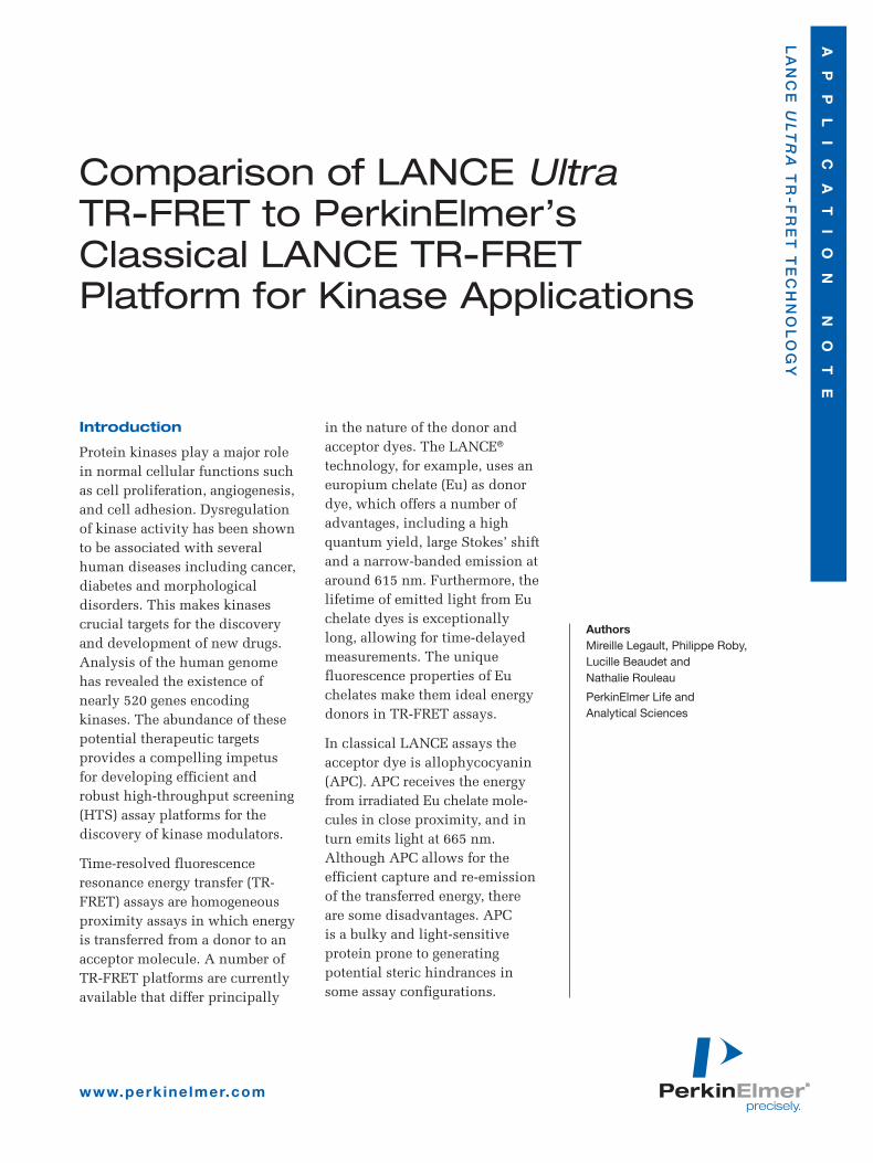
Assay optimization was performedin order to compare LANCE Ultraand classical LANCE assay formats.To determine the substrate concen-tration resulting in the best signalwindow, as well as to establish EC50
values, poly-GT was titrated. EC50
values in the low nanomolar range(0.6 nM to 4.7 nM) were obtainedfor all assay formats (Figure 2).Moreover, a specific signal could bedetected using substrate concentra-tions as low as 300 pM. Due tosaturation of the labeled strepta-vidin, a hook effect was observed at100 nM substrate concentration inboth the LANCE ULight-SA and theclassical LANCE formats. For allassays, maximal signal window wasgenerated at 100 nM poly-GT andthis substrate concentration wasused in all other experiments.
Table 1. Reagents and Consumables
Item Supplier Product No.
ULight-poly-GT PerkinElmer TRF0100-M
ULight-Streptavidin PerkinElmer TRF0102-M
SureLight™ Allophycocyanin-Streptavidin PerkinElmer CR130-100
Biotinylated-poly-GT PerkinElmer Custom product
LANCE Eu-PY20 Antibody PerkinElmer AD0066
LANCE Detection Buffer 10X PerkinElmer CR97-100
Src Kinase, active Millipore Corp. 14-326
ATP (adenosine 5'-triphosphate disodium salt) Sigma-Aldrich, Inc. A2383
Staurosporine Sigma-Aldrich, Inc. S4400
White OptiPlate-384 PerkinElmer 6007290
TopSeal-A™ PerkinElmer 6005250
EnVision Multilabel Reader PerkinElmer 2102-0010
Excitation Filter UV2(TRF) 320 nm PerkinElmer 2100-5060
Mirror LANCE/DELFIA™ Dual PerkinElmer 2100-4160
Emission Filter: Eu 615 nm PerkinElmer 2100-5090
Emission Filter: LANCE 665 nm PerkinElmer 2100-5110
Src Kinase Buffer: 50 mM Tris-HCl, pH 7.5, 10 mM MgCl2, 1 mM EGTA, 2 mM DTT and 0.01% Tween-20
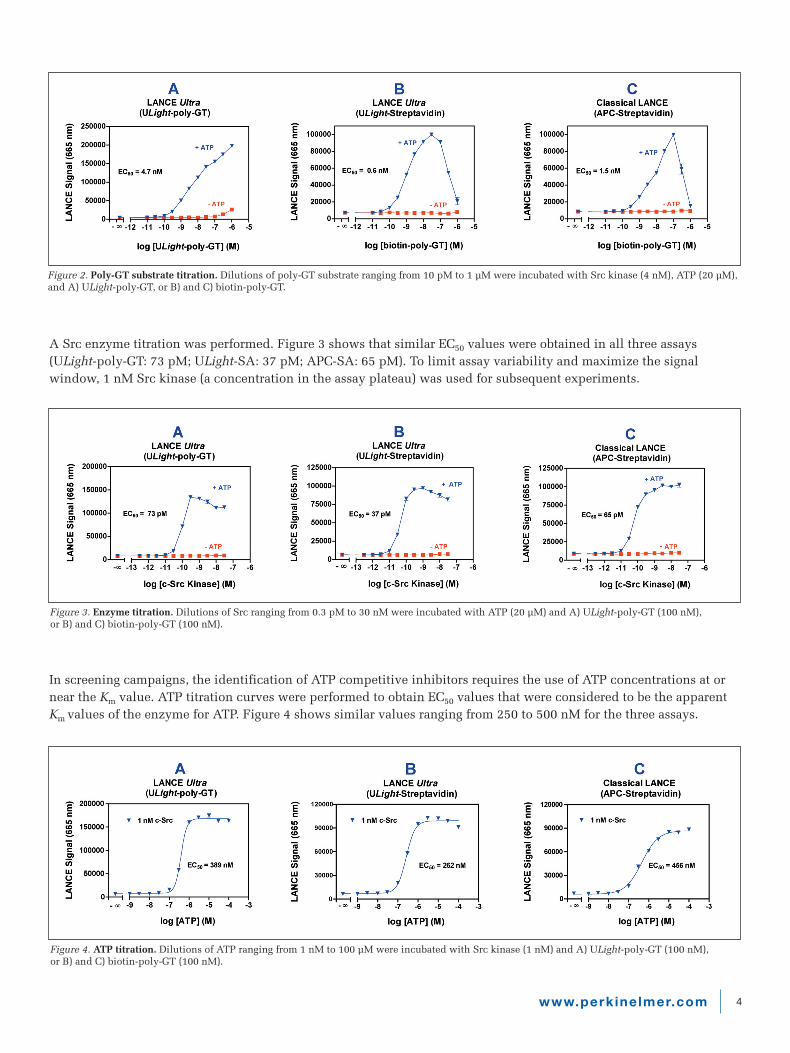
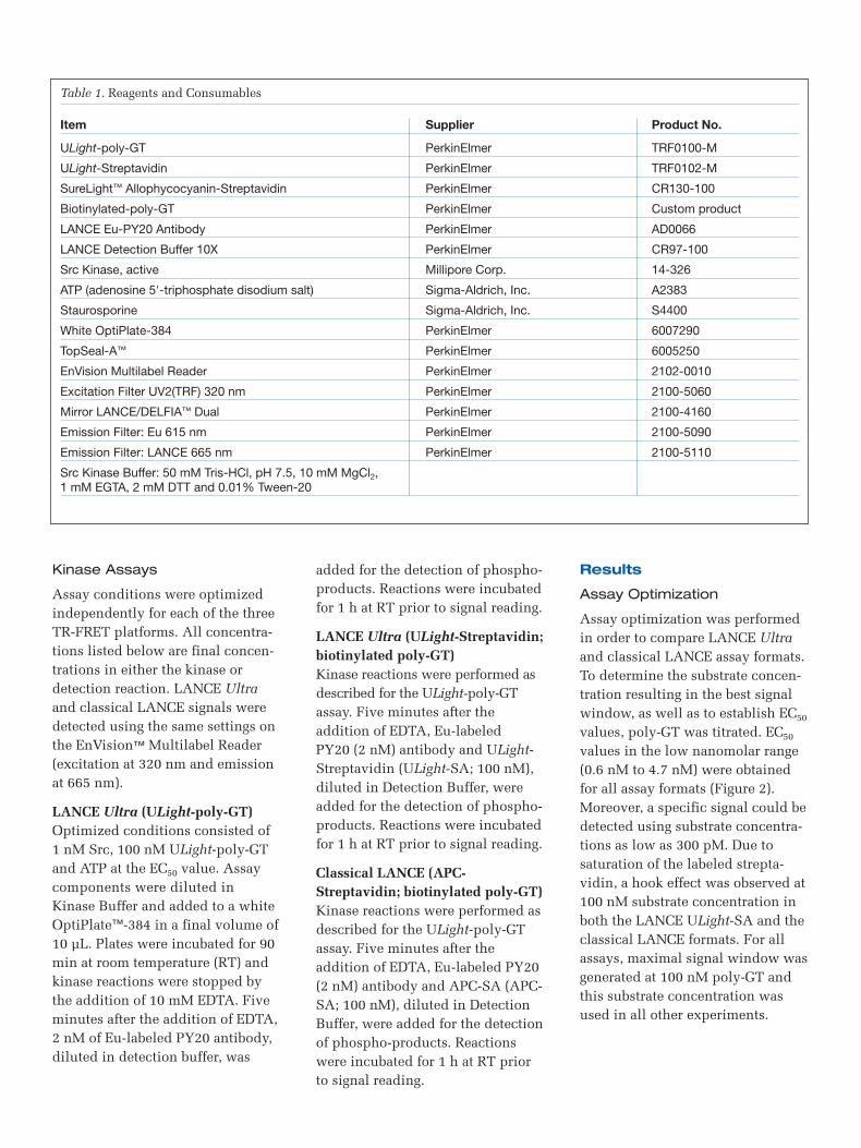
Figure 3. Enzyme titration. Dilutions of Src ranging from 0.3 pM to 30 nM were incubated with ATP (20 µM) and A) ULight-poly-GT (100 nM), or B) and C) biotin-poly-GT (100 nM).
A Src enzyme titration was performed. Figure 3 shows that similar EC50 values were obtained in all three assays (ULight-poly-GT: 73 pM; ULight-SA: 37 pM; APC-SA: 65 pM). To limit assay variability and maximize the signalwindow, 1 nM Src kinase (a concentration in the assay plateau) was used for subsequent experiments.
In screening campaigns, the identification of ATP competitive inhibitors requires the use of ATP concentrations at ornear the Km value. ATP titration curves were performed to obtain EC50 values that were considered to be the apparentKm values of the enzyme for ATP. Figure 4 shows similar values ranging from 250 to 500 nM for the three assays.
Figure 4. ATP titration. Dilutions of ATP ranging from 1 nM to 100 µM were incubated with Src kinase (1 nM) and A) ULight-poly-GT (100 nM), or B) and C) biotin-poly-GT (100 nM).
4www.perkinelmer.com
Figure 2. Poly-GT substrate titration. Dilutions of poly-GT substrate ranging from 10 pM to 1 µM were incubated with Src kinase (4 nM), ATP (20 µM),and A) ULight-poly-GT, or B) and C) biotin-poly-GT.

5www.perkinelmer.com
Evaluation of assay performance
Staurosporine is a broad spectrum kinase inhibitor competing for ATP binding. Staurosporine inhibition of the Srcenzyme was evaluated to compare the performance of the three optimized LANCE assay formats. Competition curvesperformed at the apparent Km for ATP show that the three TR-FRET platforms give virtually identical IC50 values forstaurosporine (ULight-poly-GT: 19 nM; ULight-SA: 26 nM; APC-SA: 46 nM). These values are all within one log ofpublished IC50 values for a staurosporine inhibition of Src1.
Figure 5. Staurosporine inhibition curves. Dilutions of staurosporine ranging from 100 pM to 10 µM were pre-incubated for 10 min at RT usingoptimized Src and substrate conditions. ATP concentrations at the EC50 value were used: A) 400 nM, B) 300 nM, and C) 500 nM.
Figure 6. Z'-factor determination for the Src kinase assay. Optimized enzyme and substrate concentrations were used. ATP was added at the EC50 value. Kinase reactions were performed in the absence (24 wells) or presence of 10 µM staurosporine (24 wells).
The robustness of the three assay platforms was assessed by determining the Z'-factor2 in the presence or absence of 10 µM staurosporine (Figure 6). Assays were performed using ATP concentrations at the apparent Km. Reaction mix-tures with or without staurosporine were dispensed into 24 wells of a 384-well OptiPlate. Z'-factors of 0.87 and 0.85were obtained for the LANCE Ultra ULight-poly-GT and ULight-Streptavidin containing reactions, respectively. The Z'-factor calculated for the classical LANCE assay was similar, with a value of 0.85.

For a complete listing of our global offices, visit www.perkinelmer.com/lasoffices
©2006 PerkinElmer, Inc. All rights reserved. The PerkinElmer logo and design are registered trademarks of PerkinElmer, Inc. LANCE is a registered trademark and DELFIA, EnVision,OptiPlate, SureLight, TopSeal-A and ULight are trademarks of PerkinElmer, Inc. or its subsidiaries, in the United States and other countries. ULight is covered under patent numbersUS20050202565 and US20040166515. All other trademarks not owned by PerkinElmer, Inc. or its subsidiaries that are depicted herein are the property of their respective owners.PerkinElmer reserves the right to change this document at any time without notice and disclaims liability for editorial, pictorial or typographical errors.
007755_01 Printed in USA
PerkinElmer Life and Analytical Sciences710 Bridgeport AvenueShelton, CT 06484-4794 USAPhone: (800) 762-4000 or (+1) 203-925-4602www.perkinelmer.com
Discussion
This application note describes theoptimization of an HTS Src kinaseassay using PerkinElmer’s LANCEUltra and classical LANCE plat-forms. When compared to classicalLANCE, the new LANCE Ultraassays give similar EC50 values forthe substrate, enzyme, and ATP.Additionally, the results obtainedfrom the staurosporine inhibitioncurves and Z'-factor determinationsillustrate the robust performanceand HTS suitability of both LANCE Ultra assay formats.
The power of LANCE Ultracomes from the combination ofPerkinElmer’s Eu chelate donor dyeand new ULight acceptor dye. TheULight dye offers distinct advan-tages over APC. It is light resistantand its small size allows for thedirect labeling of molecules of anysize. Moreover, the possibility ofdirectly labeling kinase substrateswith the ULight dye will greatlysimplify assay development. Forcases where a biotinylated substrateis preferred, the use of ULight-SAprovides an ideal alternative.
References
1. Sigma Aldrich. <http://www.sig-maaldrich.com/Area_of_Interest/Biochemicals/Enzyme_Explorer/Key_Resources/Protein_Kinase_Explorer/PK_Inhibitor_Specificity.html>.
2. Zhang, J.H., Chung, T.D.Y.,Oldenburg, K.R. 1999. A simplestatistical parameter for use inevaluation and validation of highthroughput screening assays. J.Biomol. Screen. 4:67-73.