Comparison of hormonal assay by ELISA , ELFA and ECL
42
Comparison of hormonal assay by ELISA , ELFA , and ECL Methods By: Rijaa A.
-
Upload
rijaa -
Category
Health & Medicine
-
view
49 -
download
5
Transcript of Comparison of hormonal assay by ELISA , ELFA and ECL
Comparison of hormonal assay
byELISA , ELFA , and ECL
MethodsBy: Rijaa A.

• Introduction• Hormone measurement is necessary for
the diagnosis of a wide range of clinical conditions and is essential for monitoring the effectiveness of treatment.• As the number of hormone requests in
the clinical field rises exponentially, it has become importance to create hormone assays accessible to researchers with a different range of devices.

• Before the introduction of radioimmunoassay most hormones were measured by bioassay and/or chemical methods. • The sensitivity of these methods was low,
and large amounts of samples were needed .• Radioimmunoassay first applied In 1959,
to measurement of insulin, after which many protein hormones were determined by this method.

• Because radioactivity poses a potential health threat, so Research had to be go on for finding a safer alternative .• The first paper on the ELISA
procedure published in 1971 as a replacement for radioimmunoassays by Sweden and Netherlands Scientists independently .

• Recently, non-isotopic immunoassay methods utilizing chemiluminescence, fluorescence and enzymes as labels are widely used. • These methods have resulted in
sensitivities adequate to replace radioimmunoassays.

Important components in immunoassay
•1-Antibody (antiserum) .•2-Antigen . •3-Labeling materials .

• 1-Antibody (antiserum)• proteins produced by the immune
system which help defend against antigens .
• The variable regions are though to be the place for recognition and binding with the antigen.
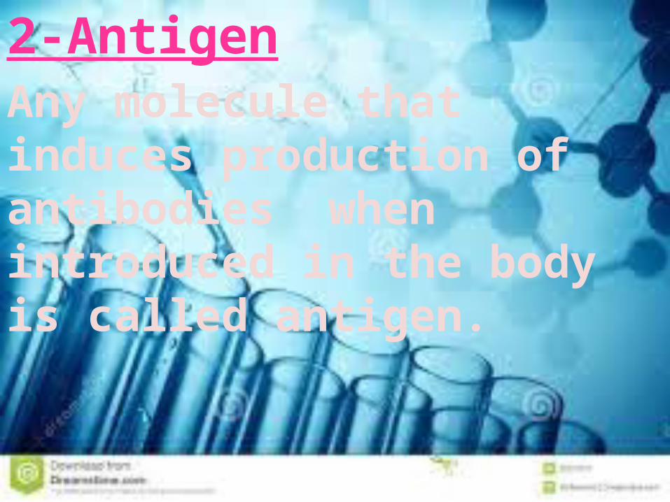
2-Antigen Any molecule that induces production of antibodies when introduced in the body is called antigen.

3-Labeling materials In immunoassay, it is necessary to use any marker to know the antigen-antibody binding. For such purpose, they label either antigen or antibody with some materials that do not interefere with the binding.

ELISA Why
known as……………?? Enzyme-Linked ImmunoSorbent
Assay

• 1. Antigen/antibody of interest is absorbed on to plastic surface (‘sorbent’).• 2. Antigen is recognised by specific
antibody (‘immuno’).• 3. This antibody is recognised by second
antibody (‘immuno’) which has enzyme attached (‘enzyme-linked’).• 4. Substrate reacts with enzyme to
produce product, usually coloured.

•ELISA• Is used to the detection of 1)Antibodies .2)Proteins .3)Peptides .4)Biomolecules .

Basic principal of ELISA• ELISAs can provide a useful
measurement of antigen or antibody concentration.
A general ELISA is a five-step procedure: 1) coat the microtiter plate wells with antigen . 2) block all unbound sites to prevent false positive results .3) add primary antibody to the wells .

4) add secondary antibody conjugated to an enzyme .5) reaction of a substrate with the enzyme to produce a colored product, thus indicating a positive reaction.
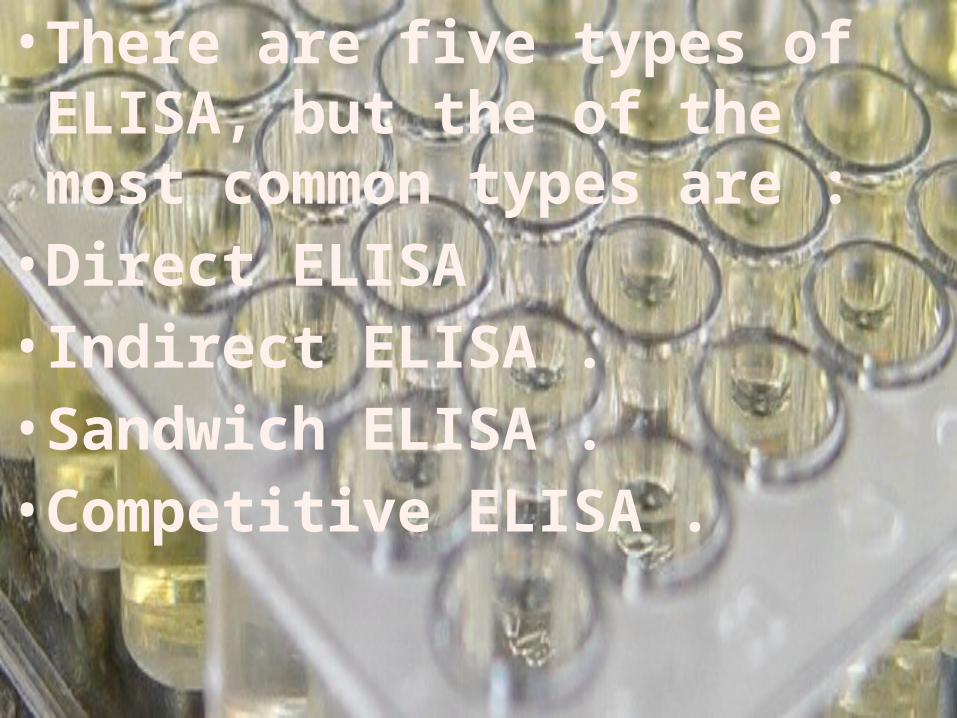
• There are five types of ELISA, but the of the most common types are :• Direct ELISA • Indirect ELISA .• Sandwich ELISA .• Competitive ELISA .


Competitive ELISA

ELFA (Enzyme Linked
Fluorescent Assay)

Technology which is based on a one-step immunoassay sandwich method and a final fluorescent detection step.• Two ready-to-use reagents .• 1- The SPR® Solid Phase Receptacle
serves as a solid phase and pipetting device. Coated with mouse monoclonal
anti-human immunoglobulins.

• 2- The reagent strip contains pre-dispensed reagents:• Conjugate: alkaline phosphatase-labelled
mouse monoclonal anti-human immunoglobulin .• Fluorescent substrate: 4-MUP (4-Methyl-
umbelliferyl phosphate) & DEA (diethanolamine).• Washing buffers .

1- Measuring Principle The reaction occurs within the interior of the SPR whereby antibodies and conjugate form a sandwich.(4-MUP) is cycled into SPR and conjugate enzyme catalyses the hydrolysis of the substrate into 4-Methyl-umbelliferone which is measured at 450nm.

The intesity of the Fluorescence is inversely proportional to the concentration of antigen present in the sample .


ECLElectrochemiluminescence

“Electro” refers to electrical stimulation. + “Chem” indicates a chemical reaction. +“Luminescence”means “produces light.” = Electrochemiluminescence (ECL)
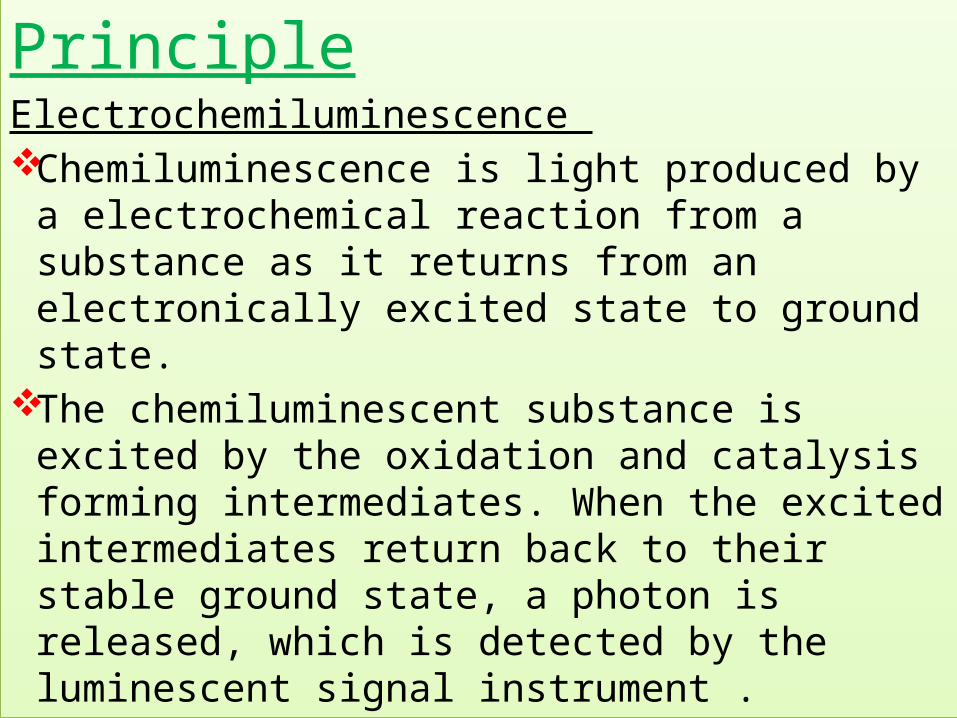
PrincipleElectrochemiluminescence Chemiluminescence is light produced by a
electrochemical reaction from a substance as it returns from an electronically excited state to ground state.
The chemiluminescent substance is excited by the oxidation and catalysis forming intermediates. When the excited intermediates return back to their stable ground state, a photon is released, which is detected by the luminescent signal instrument .

An enzyme converts a substrate to a reaction product that emits photons of light instead of developing a visible color.
ECL is a unique and highly sensitive luminescence (light) detection system that amplifies the signal (you want) to detect ultra-low concentrations of analyte. and reduces any signals you don’t want to deliver .

• Source kicks an electron of an atom out of its lowest energy “ground” state into a higher energy “excited” state .
• Electron returns the energy in the form of light so it can fall back to its “ground” state .
• [A] + [B] → [◊] → [Products] + light
• [A], [B]: reactants• [◊]: excited intermediate .

For example, if [A] is luminol and [B] is hydrogen peroxide in the presence of a suitable catalyst we have:• luminol + H2O2 →3-APA[◊] →3-APA +
light• Where:• 3-APA is 3-aminophthalate• 3-APA[◊] is the excited state producing
light as it return to a lower energy level.

• What happens in Chemiluminescence immunoassays• Monoclonal antibody coated well ↓ Test specimen (serum) ↓ HRP labelled antibody conjugate ↓ Test antigen: sandwich between solid phase Ab and enzyme labelled Ab

Incubate at 37° C↓
Remove unbound enzyme labeled Ab↓
Chemiluminescence reagent added↓
Read relative light unit with luminometer


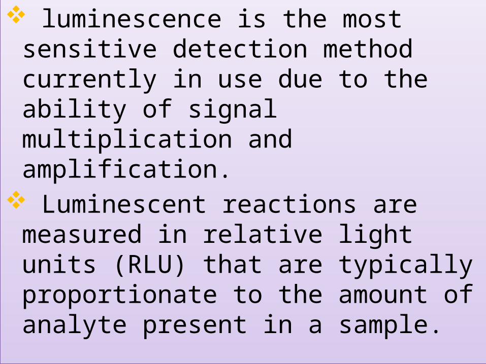
luminescence is the most sensitive detection method currently in use due to the ability of signal multiplication and amplification.
Luminescent reactions are measured in relative light units (RLU) that are typically proportionate to the amount of analyte present in a sample.

• Properties Ultra-high sensitivity (10, 000 times higher
than and the light absorption method and 1000 times higher than the fluorescence detection method) .
Show a linear relationship between luminous intensity and the concentration of measured substance .
No preincubation step .Substrate can be added several minutes prior
to detection.


• A COMPARATIVE ANALYSIS ON THREE DIFFERENT FULLY AUTOMATED ANTI-TOXOPLASMA IGM IMMUNOASSAYS

• Diagnoses acute toxoplasmosis during the first trimester of pregnancy.
• Sensitivity and specificity are calculated for each technique:• ELISA technique showed a specificity of 83%
and a sensitivity of 95%.
• ELFA technique showed a specificity of 90% and a sensitivity of 97.5%.
• ECL technique showed a specificity of 100% and a sensitivity of 100%.

Comparison Between
ChemilluminescentAssay
• No stop solution
• Higher sensitivity
• No light source(Filter)
ELISA• Stop solution
required (not in kinetic assay)
• Less sensitive
• Light source needed

THANK YOU