Comparison of Enterprise and Neuroform Stent-Assisted Coil … · 2014-03-17 · tion digital...
10
181 Copyrights © 2014 The Korean Society of Radiology INTRODUCTION With the development of new endovascular techniques and devices, endovascular treatment for patients with intracranial aneurysms is increasingly being accepted as a safe and useful method (1, 2). However, wide-neck, large, and giant aneurysms are challenges in the conventional endovascular treatment of aneurysms (1, 3-6). In these cases, stent placement is another treatment option and has been successfully used to treat intra- cranial aneurysms (1, 7). ere are two types of self-expanding intracranial stents currently available. One has an open-cell de- sign such as the Neuroform (Boston Scientific, Natick, MA, USA) and the other has a closed-cell design such as the Solitaire, Leo, and Enterprise (Cordis, Miami Lakes, FL, USA) (8). e Neuro- form is an open-cell, nitinol, self-expanding microstent. e En- terprise is a closed-cell, nitinol, self-expanding, and retractable stent (9). Stent deployment for a straight vessel is relatively simple, and there is no practical difference in using either stent. When de- ployed in a curved vessel, however, these two types of stents will Original Article pISSN 1738-2637 / eISSN 2288-2928 J Korean Soc Radiol 2014;70(3):181-190 http://dx.doi.org/10.3348/jksr.2014.70.3.181 Received September 7, 2013; Accepted September 26, 2013 Corresponding author: Seung Kug Baik, MD Department of Radiology, Pusan National University Yangsan Hospital, 20 Geumo-ro, Mulgeum-eup, Yangsan 626-770, Korea. Tel. 82-55-360-1834 Fax. 82-55-360-1848 E-mail: [email protected] This is an Open Access article distributed under the terms of the Creative Commons Attribution Non-Commercial License (http://creativecommons.org/licenses/by-nc/3.0) which permits unrestricted non-commercial use, distri- bution, and reproduction in any medium, provided the original work is properly cited. Purpose: To compare the mid-term follow-up angiographic findings in distal inter- nal carotid artery (ICA) aneurysms treated by stent-assisted coil embolization using the Enterprise or Neuroform stent. Materials and Methods: We included 68 patients with 70 aneurysms: 31 cases with Enterprise and 39 cases with Neuroform. Inclusion criteria were 1) location of the stent within the distal ICA, including the carotid siphon; 2) follow-up angio- gram after > 6 months, and 3) single use of the stent for 1 parent artery. Results: The patients’ mean age was 54.9 years (16 male and 52 female). Mean fol- low-up duration was 9.1 months. At follow-up, there were intraluminal filling de- fects of the parent artery in 19.4% of the Enterprise group and no filling defect in the Neuroform group. There was no significant in-stent stenosis in either group. Straightening of the parent artery was seen in 35.5% of the Enterprise group and 20.5% of the Neuroform group. Two Enterprise cases showed delayed migration. Conclusion: The Enterprise showed statistically significant intraluminal filling de- fects of the parent artery compared with the Neuroform. The rates of significant in- stent stenosis and straightening of the parent artery were not significantly different between the Enterprise and the Neuroform groups. Index terms Cerebral Aneurysm Stent-Assisted Coil Embolization Carotid Siphon Enterprise Neuroform Comparison of Enterprise and Neuroform Stent-Assisted Coil Embolization of Distal Internal Carotid Artery Aneurysms: Midterm Results from a Single-Center Experience 1 원위내경동맥의 동맥류에 대한 Enterprise와 Neuroform 스텐트 보조 색전술의 비교: 단일센터에서의 경험의 중간 결과 1 Won-Jin Choi, MD 1 , Seung Kug Baik, MD 1 , Jeong A Yeom, MD 1 , Young Soo Kim, MD 1 , Sang Weon Lee, MD 2 Departments of 1 Radiology, 2 Neurosurgery, Research Institute for Convergence of Biomedical Science and Technology, Pusan National University Yangsan Hospital, Pusan National University School of Medicine, Yangsan, Korea
Transcript of Comparison of Enterprise and Neuroform Stent-Assisted Coil … · 2014-03-17 · tion digital...

181Copyrights © 2014 The Korean Society of Radiology
INTRODUCTION With the development of new endovascular techniques and
devices, endovascular treatment for patients with intracranial aneurysms is increasingly being accepted as a safe and useful method (1, 2). However, wide-neck, large, and giant aneurysms are challenges in the conventional endovascular treatment of aneurysms (1, 3-6). In these cases, stent placement is another treatment option and has been successfully used to treat intra-cranial aneurysms (1, 7). There are two types of self-expanding
intracranial stents currently available. One has an open-cell de-sign such as the Neuroform (Boston Scientific, Natick, MA, USA) and the other has a closed-cell design such as the Solitaire, Leo, and Enterprise (Cordis, Miami Lakes, FL, USA) (8). The Neuro-form is an open-cell, nitinol, self-expanding microstent. The En-terprise is a closed-cell, nitinol, self-expanding, and retractable stent (9).
Stent deployment for a straight vessel is relatively simple, and there is no practical difference in using either stent. When de-ployed in a curved vessel, however, these two types of stents will
Original ArticlepISSN 1738-2637 / eISSN 2288-2928J Korean Soc Radiol 2014;70(3):181-190http://dx.doi.org/10.3348/jksr.2014.70.3.181
Received September 7, 2013; Accepted September 26, 2013Corresponding author: Seung Kug Baik, MDDepartment of Radiology, Pusan National University Yangsan Hospital, 20 Geumo-ro, Mulgeum-eup, Yangsan 626-770, Korea.Tel. 82-55-360-1834 Fax. 82-55-360-1848E-mail: [email protected]
This is an Open Access article distributed under the terms of the Creative Commons Attribution Non-Commercial License (http://creativecommons.org/licenses/by-nc/3.0) which permits unrestricted non-commercial use, distri-bution, and reproduction in any medium, provided the original work is properly cited.
Purpose: To compare the mid-term follow-up angiographic findings in distal inter-nal carotid artery (ICA) aneurysms treated by stent-assisted coil embolization using the Enterprise or Neuroform stent.Materials and Methods: We included 68 patients with 70 aneurysms: 31 cases with Enterprise and 39 cases with Neuroform. Inclusion criteria were 1) location of the stent within the distal ICA, including the carotid siphon; 2) follow-up angio-gram after > 6 months, and 3) single use of the stent for 1 parent artery.Results: The patients’ mean age was 54.9 years (16 male and 52 female). Mean fol-low-up duration was 9.1 months. At follow-up, there were intraluminal filling de-fects of the parent artery in 19.4% of the Enterprise group and no filling defect in the Neuroform group. There was no significant in-stent stenosis in either group. Straightening of the parent artery was seen in 35.5% of the Enterprise group and 20.5% of the Neuroform group. Two Enterprise cases showed delayed migration. Conclusion: The Enterprise showed statistically significant intraluminal filling de-fects of the parent artery compared with the Neuroform. The rates of significant in-stent stenosis and straightening of the parent artery were not significantly different between the Enterprise and the Neuroform groups.
Index termsCerebral AneurysmStent-Assisted Coil EmbolizationCarotid SiphonEnterpriseNeuroform
Comparison of Enterprise and Neuroform Stent-Assisted Coil Embolization of Distal Internal Carotid Artery Aneurysms: Midterm Results from a Single-Center Experience1
원위내경동맥의 동맥류에 대한 Enterprise와 Neuroform 스텐트 보조 색전술의 비교: 단일센터에서의 경험의 중간 결과1
Won-Jin Choi, MD1, Seung Kug Baik, MD1, Jeong A Yeom, MD1, Young Soo Kim, MD1, Sang Weon Lee, MD2
Departments of 1Radiology, 2Neurosurgery, Research Institute for Convergence of Biomedical Science and Technology, Pusan National University Yangsan Hospital, Pusan National University School of Medicine, Yangsan, Korea

Comparison of Enterprise and Neuroform Stent-Assisted Coil Embolization of Distal Internal Carotid Artery Aneurysms
182 jksronline.orgJ Korean Soc Radiol 2014;70(3):181-190
criteria, including 31 cases with the Enterprise stent (Enterprise group) and 39 cases with the Neuroform stent (Neuroform group).
Endovascular Procedures
All endovascular procedures were performed using a biplane angiography unit with 3-dimensional rotational angiography ca-pability (Axiom Artis Zee Biplane; Siemens, Erlangen, Germa-ny). For patients with unruptured aneurysms, dual antiplatelet drugs [300 mg/day aspirin and 75 mg/day clopidogrel (Plavix; Sanofi-Synthelabo, Seoul, Korea)] were given for at least 3 days before the procedure. For patients with ruptured aneurysms, Ti-rofiban (Aggrastat, Merck & Co, Whitehouse Station, NJ, USA) was started after the aneurysms were secured with the coil.
In our institution, since June 2012 platelet inhibition function has been performed with the Multiplate® analyzer system (Dyna-byte Medical, Munich, Germany) immediately before starting the procedure. If the patient was considered as a nonresponder [> 45 units (U) for the area under the curve of clopidogrel or > 30 U for that of aspirin], an additional anti-platelet drug was adminis-tered as soon as the procedure was finished.
All of the procedures were performed under local anesthesia. Endovascular access was obtained by a standard transfemoral approach unilaterally. After placement of the sheath, all patients received systemic heparinization: a 3000 to 5000 IU bolus of heparin (50 IU/kg body weight), followed by continuous infu-sion of 1000 to 2000 IU of heparin/h to double the baseline acti-vated clotting time. A 6 Fr shuttle guiding catheter (Shuttle-SL; Cook, Bloomington, IN, USA) was placed into the proximal ICA. A stent delivery microcatheter (Prowler Select Plus, 2.3 Fr; Cor-dis, Miami Lakes, FL, USA, or Neurorenegade Hi-Flo, 2.8 Fr; Boston Scientific, Natick, MA, USA) was first navigated and po-sitioned across the aneurysm neck. The stent was then loaded into the microcatheter and deployed by withdrawing the micro-catheter while stabilizing the delivering wire. Three stenting strategies were employed in this study: the mesh technique, the jailing technique, and the salvaging stent placement (12). At the end of coiling, final angiographic images were acquired to con-firm adequate coiling.
After the procedure, low-molecular-weight heparin (2850 IU/0.3 mL) (Fraxiparine; Sanofi-Synthelabo, Seoul, Korea) was ad-ministered subcutaneously for 2 days. All the patients were contin-
result in different appearances of the parent artery and stent it-self (10). The distal internal carotid artery (ICA), and especially the carotid siphon, is known to have a tortuous vasculature, as the name suggests. Thus stent deployment for the distal ICA in-cluding the carotid siphon involves a complicated anatomical region and might result in more complications during and after the stent deployment procedure.
Follow-up imaging after stent-assisted coil embolization (SACE) procedures has usually been focused on the presence or absence of residual filling in the aneurysm (7, 11). To our knowledge, no published studies have included an evaluation of the angiographic follow-up results focused on the distal ICA as the parent artery and comparison of the stent-deployment ef-fects between these two stents in the SACE of distal ICA aneu-rysms.
In this study, we retrospectively analyzed and compared the follow-up angiographic results of distal ICA aneurysms includ-ing the carotid siphon, treated by a SACE technique using the Enterprise or the Neuroform. We discuss the stent-associated ef-fects, focusing on the angiographic findings of the parent artery.
MATERIALS AND METHODS
Patients
This study was approved by the institutional review board at our institution. Informed consent from patients was waived due to the retrospective study design.
Imaging data of distal ICA aneurysms treated with SACE from December 2008 to September 2012 were analyzed in this retrospective study. Inclusion criteria were: 1) the location of the stent within the distal ICA, including the paraclinoid ICA, the carotid siphon; 2) the availability of immediate post-implanta-tion digital subtraction angiography (DSA) and follow-up DSA images from 6 months after the stent implantation, from Febru-ary 2009 to April 2013; 3) the use of a self-expanding nitinol in-tracranial stent: the Enterprise or the Neuroform stent (4.5 mm in diameter by 28 mm in length for the Enterprise and 4.5 mm in diameter by 20 mm in length for the Neuroform); 4) single deployment of the stent for one parent artery (two or more stent insertions for the same side of the parent artery were excluded).
We reviewed our database for cases of aneurysms treated by SACE and found 68 patients with 70 aneurysms that met these

Won-Jin Choi, et al
183jksronline.org J Korean Soc Radiol 2014;70(3):181-190
rysm without opacification of the aneurysm sac; RS 3, contrast filling of the sac of the aneurysm.
Two radiologists evaluated all the images independently and these measurements and calculations were determined by con-sensus. All imaging data were reviewed on a Picture Archiving and Communication System (PACS) workstation (Maroview; version 5.4.10.52, Marotech, Seoul, Korea).
Clinical Evaluation
The patients’ medical histories, procedural reports, and clini-cal outcomes were analyzed retrospectively by reviewing their electronic medical record charts. A complete neurologic exami-nation was performed on all patients by a neurosurgeon, at the baseline, and immediately after the procedure and at the follow-up by experienced physicians certified in stroke assessment. A modified Rankin scale (mRS) score (14) was assessed at the fol-low-up evaluations. Any adverse events, especially ischemic stroke events, were checked and recorded.
Statistical Analyses
All data were presented as mean ± standard deviation. Fisher’s exact test was used for comparisons of categorical data. Student’s t-test was used to determine statistical significance for continu-ous parameters including age, aneurysm size, stent-substented arc angle, carotid siphon angle, and significant in-stent stenosis. Significant differences were established for p < 0.05. Statistical analysis was performed by using MedCalc (version 12.7.0., MedCalc Software, Mariakerke, Belgium).
RESULTS
Patients
In total, 68 patients with 70 aneurysms were available for anal-ysis. The patients’ mean age was 54.9 ± 10.7 years (range, 26–78 years), including 16 men and 52 women. Patient demographic in-formation and aneurysm characteristics are provided in Table 1.
Follow-up DSA images were performed for all patients, rang-ing from 6 to 36 months (mean 9.1 ± 4.7 months) after the ini-tial stent-assisted coiling procedure. Most of the patients (66/68) had unilateral (right or left) stent insertion. However, 2 patients had bilateral Enterprise stent insertion for coiling of each side of distal ICA aneurysms.
ued on aspirin and clopidogrel postoperatively for 6 months to 1 year, depending on the patient’s condition.
Follow-Up and Image Analysis
All patients were followed up with DSA beyond 6 months after the initial SACE procedure. Angiograms were performed at high magnification in the working angles for the coil embolization and the stenting. Working angles were reproduced by optimizing the orientation of osseous landmarks with the stent markers and coil mass.
Follow-up DSA images were then compared with the imme-diate post-embolization angiograms with a similar projection and the same magnification ratio for the following parameters.
1) The term “intraluminal filling defect” was defined as a focal contrast filling defect at the stent-deployment area of the parent artery, between the stent filament and the vessel wall in the fol-low-up angiogram only (the “intraluminal filling defect” did not appear in the initial immediate post-embolization angiogram). And the presence of an incomplete intraluminal filling defect was recorded.
2) The stent-subtended arc angle, measured independently of the radius of the siphon curvature, was evaluated on the basis of the angle at which the stent was deployed in the parent vessel (7).
3) The carotid siphon angle was measured by the following method: first, the center was placed at the midpoint of the carot-id siphon; second, a line was drawn between the center point and upper and lower portion of the carotid siphon; finally the angle between the two lines was measured.
4) The term “straightening” was defined as more than 10% of an angle increase of the parent artery between the immediate postembolization angiograms and the follow-up angiograms, us-ing the stent-subtended arc angle and carotid siphon angle.
5) The mean diameter at the distal and proximal portion of the stent-deployment sites was measured to identify in-stent ste-nosis (or intimal hyperplasia). We defined the term “significant in-stent stenosis” as indicating more than 50% narrowing within the stent-deployment area of the parent artery.
6) Any changes in the stent itself on the follow-up angiogram, such as delayed stent migration, were also recorded.
7) Aneurysmal occlusion was graded on a 3-point Raymond scale (RS) (13): RS 1, complete obliteration of the aneurysm in-cluding the neck; RS 2, contrast filling of the neck of the aneu-
Comparison of Enterprise and Neuroform Stent-Assisted Coil Embolization of Distal Internal Carotid Artery Aneurysms
184 jksronline.orgJ Korean Soc Radiol 2014;70(3):181-190
Angiographic results are summarized in Table 2.
Aneurysm Occlusion Grade
On the immediate post-treatment angiogram, the occlusion grades were as follows for the Enterprise group: RS 1 in 23 (74.2%), RS 2 in 4 (12.9%), and RS 3 in 4 (12.9%) aneurysms; in the Neu-roform group, we observed RS 1 in 27 (69.2%), RS 2 in 8 (20.5%), and RS 3 in 4 (10.3%) aneurysms. The occlusion grades for the follow-up DSA in the Enterprise group were RS 1 in 21 (67.7%), RS 2 in 8 (25.8%), and RS 3 in 2 (6.5%); follow-up occlusion grades in the Neuroform group were RS 1 in 34 (87.2%), RS 2 in 4 (10.3%), and RS 3 in 1 (2.6%) aneurysms. Six (75%) of 8 aneu-rysms with persistent contrast filling of the sac were totally oc-
Angiographic Results of the Parent Artery
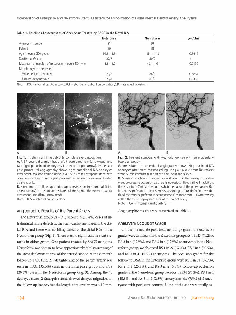
The Enterprise group (n = 31) showed 6 (19.4%) cases of in-traluminal filling defects at the stent-deployment area of the dis-tal ICA and there was no filling defect of the distal ICA in the Neuroform group (Fig. 1). There was no significant in-stent ste-nosis in either group. One patient treated by SACE using the Neuroform was shown to have approximately 40% narrowing of the stent deployment area of the carotid siphon at the 6-month follow-up DSA (Fig. 2). Straightening of the parent artery was seen in 11/31 (35.5%) cases in the Enterprise group and 8/39 (20.5%) cases in the Neuroform group (Fig. 3). Among the 70 deployed stents, 2 Enterprise stents showed delayed migration on the follow-up images, but the length of migration was < 10 mm.
Table 1. Baseline Characteristics of Aneurysms Treated by SACE in the Distal ICAEnterprise Neuroform p-Value
Aneurysm number 31 39Patient 29 39Age (mean ± SD), years 56.3 ± 9.9 54 ± 11.3 0.3445Sex (female/male) 22/7 30/9 1Maximum dimension of aneurysm (mean ± SD), mm 4.1 ± 1.7 4.6 ± 1.6 0.2189Morphology of aneurysm Wide neck/narrow neck 29/2 35/4 0.6867 Unruptured/ruptured 28/3 37/2 0.6489
Note.-ICA = internal carotid artery, SACE = stent-assisted coil embolization, SD = standard deviation
Fig. 1. Intraluminal filling defect (incomplete stent apposition).A. A 67-year-old woman has a left P-com aneurysm (arrowhead) and two right paraclinoid aneurysms (arrow and open arrow). Immediate post-procedural angiography shows right paraclinoid ICA aneurysm after stent-assisted coiling using a 4.5 × 28 mm Enterprise stent with complete occlusion and a just proximal paraclinoid aneurysm treated by stent only. B. Eight-month follow-up angiography reveals an intraluminal filling defect (arrow) at the substented area of the siphon (between proximal arrowhead and distal arrowhead).Note.-ICA = internal carotid artery
Fig. 2. In-stent stenosis. A 64-year-old woman with an incidentally found aneurysm. A. Immediate post-procedural angiography shows left paraclinoid ICA aneurysm after stent-assisted coiling using a 4.5 × 20 mm Neuroform stent. Subtle contrast filling of the aneurysm sac is seen. B. Six-month follow-up angiography shows that the aneurysm under-went progressive occlusion as there is no residual flow visible. In addition, there is mild (40%) narrowing of substented area of the parent artery. But it is not significant in-stent stenosis, according to our definition: we de-fined the term “significant in-stent stenosis” as more than 50% narrowing within the stent-deployment area of the parent artery.Note.-ICA = internal carotid artery
A AB B

Won-Jin Choi, et al
185jksronline.org J Korean Soc Radiol 2014;70(3):181-190
ic flat-panel computed tomography (CT) have shown crumpling and ovalization of these stents during use of bending elastic tube models (10, 17), there are little in vivo data on the apposition of the intracranial stent-strut to the parent vessel wall, even after over 10 years of clinical use (17, 18). The siphon curvature of the ICA in humans has similar characteristics to the bending tube model. We analyzed the comparative follow-up angiographic findings of distal ICA aneurysms treated by SACE using the En-terprise and the Neuroform stent, focusing on the stent-subtend-
cluded at the follow-up, whereas 2 (25%) showed persistent neck opacification. The frequency of aneurysms with RS 3 decreased from 8/70 (11.4%) on initial post-treatment angiogram evaluation to 3/70 (4.3%) on the follow-up angiogram, while the overall fre-quency of aneurysms with RS 1 increased slightly from 50/70 (71.4%) to 55/70 (78.6%). The initial and follow-up angiographic outcomes of aneurysms using the 3-point RS are summarized in Table 3.
Clinical Outcome
In the Enterprise group with unruptured aneurysms (n = 26), none of the patients had neurologic symptoms at the baseline or at the follow-up. In the Enterprise group with ruptured aneu-rysms (n = 3), one patient (3.4%) showed mild lower extremity numbness (mRS = 1) at the follow-up. In the Neuroform group with unruptured aneurysms (n = 37), none of the patients except two had neurologic symptoms at the baseline. One patient (2.6%), who had been previously diagnosed with poliomyelitis, had left leg weakness (mRS = 2) at the baseline. Another patient (2.6%) initial-ly had gait disturbance and weakness (mRS = 3) due to a previous cerebral infarction. At the follow-up, these two patients showed no additional neurologic symptoms. In the Neuroform group with ruptured aneurysms (n = 2), these two patients had severe head-ache and mental changes at the baseline but after treatment and at the follow-up, they showed no neurologic symptoms.
DISCUSSION
After the introduction of self-expandable flexible microstents, SACE has become a feasible option for the treatment of wide-neck aneurysms and more recently for small and medium-sized aneurysms (15, 16).
There are predominantly 2 types of self-expanding flexible mic-rostents in use–the open-cell design Neuroform and the closed-cell design Enterprise. Although in vitro studies using angiograph-
Fig. 3. Parent artery straightening. A 36-year-old woman presented with severe headache. Examination revealed subarachnoid hemor-rhage due to ruptured aneurysm. A. Immediate post-procedural angiography shows a right paraclinoid ICA aneurysm after stent-assisted coiling using a 4.5 × 28 mm Enterprise stent with complete occlusion. B. Twelve-month follow-up angiography reveals increased vascular cur-vature of the carotid siphon (“straightening”) and focal intraluminal filling defect at the substented area (arrow).Note.-ICA = internal carotid artery
A B
Table 2. Comparative Mid-Term Follow-Up Result about Morpho-logic Changes of the Parent Artery (Distal ICA)
Enterprise (n = 31)
Neuroform (n = 39) p-Value
Intraluminal filling defect 6 0 0.0056Straightening 11 8 0.1856Significant in-stent stenosis 0 0 1Delayed stent migration 2 0 0.1925
Note.-ICA = internal carotid artery
Table 3. Initial and Follow-Up Results for Aneurysm Occlusion Occlusion Grade at Immediate SACE Occlusion Grade at Follow-Up
RS 1 (%) RS 2 (%) RS 3 (%) RS 1 (%) RS 2 (%) RS 3 (%) Enterprise (n = 31) 23 (74.2) 4 (12.9) 4 (12.9) 21 (67.7) 8 (25.8) 2 (6.5)
Neuroform (n = 39) 27 (69.2) 8 (20.5) 4 (10.3) 34 (87.2) 4 (10.3) 1 (2.6)
Total (n = 70) 50 (71.4) 12 (17.1) 8 (11.4) 55 (78.6) 12 (17.1) 3 (4.3)
Note.-RS = Raymond scale, SACE = stent-assisted coil embolization

Comparison of Enterprise and Neuroform Stent-Assisted Coil Embolization of Distal Internal Carotid Artery Aneurysms
186 jksronline.orgJ Korean Soc Radiol 2014;70(3):181-190
alence of ISA in curved vessels may be a direct result of the in-herited closed-cell design of the Enterprise stent. In larger diam-eter vessels, such as the distal ICA, the Enterprise stent showed a higher tendency for ISA, which might be due to the small outer radial force of the stent needed to deploy the vessel wall suffi-ciently. When the maximal diameter of the parent vessel is > 4 mm, stent dislodgement may occur (11). Therefore, deployment of the Enterprise in curved or tortuous vessels such as the carot-id siphon with its larger diameter (> 4 mm) causes a greater in-cidence of ISA.
ISA may cause complications such as immediate flow reduc-tion, embolism, and restenosis (21). In patients with coronary stent deployment, ISA can be associated with no neointimal hy-perplasia. ISA without neointimal hyperplasia was significantly associated with the presence of thrombus at the follow-up, and may constitute a potent substrate for late stent thrombosis (22). There are several reports on the morphological changes, particu-larly conformity of the parent artery, after stent deployment (1, 7, 9, 10, 23, 24). In our study, the Enterprise group with ISA showed no additional neurologic symptoms at follow-up. To demon-strate the clinical significance of ISA in cases treated with Enter-prise devices, further studies will be needed.
Second, after stent deployment, activation and proliferation of regional smooth muscle cells develops and may lead to neointimal hyperplasia, which can cause a re-stenosis within the stent (25, 26). Endothelial cells have a critical role in the control of smooth mus-cle growth, and when regulation is disturbed, neointimal hyper-plasia causes stenosis (25, 27). In-stent stenosis is commonly en-countered within 3–6 months after stent deployment (25, 28, 29). Some histopathologic reports have demonstrated that neo-intimal hyperplasia is implicated as the primary mechanism in in-stent stenosis (25, 30, 31). In our study, there was no signifi-cant in-stent stenosis in either of the two groups. There was no statistically significant difference between the two stents in the occurrence of significant in-stent stenosis (0% and 0%, respec-tively, p = 1). In our study, no patient showed any significant neu-rologic symptoms or sequelae. According to a recent review arti-cle using a computerized database search of SACE (32), the overall rate of delayed in-stent stenosis was 5.3%, with individu-al study rates varying from 0% to 20.6%, which were favorable outcomes compared with our result.
Third, based on our literature review, significant changes in
ed area. To our knowledge, however, there are no published in vivo reports focused on the distal ICA including the carotid si-phon as the parent artery, using DSA for the follow-up evaluation.
The main findings of this comparative study are as follows: 1) an intraluminal filling defect of the parent artery in the Enter-prise group (statistically significant difference), 2) no significant in-stent stenosis (statistically insignificant difference), 3) mild geometrical changes in the vasculature (statistically insignificant difference), and 4) delayed stent migration (statistically insignif-icant difference).
First, only the Enterprise group showed intraluminal filling de-fects of the parent artery (n = 6/31, 19.4%). Our results showed a statistically significant greater occurrence of intraluminal filling defects using the Enterprise compared with using the Neuro-form (19.4% and 0%, respectively, p = 0.0056). We thought that incomplete stent apposition (ISA), defined as separation of one or more stent struts from the underlying vessel wall and obser-vation of blood speckles between the stent struts and the vessel wall (19), and foreign body reaction around the struts (20) due to ISA were the main causes for the intraluminal filling defect. ISA in distal ICA aneurysms, with the stent-subtended area at the carotid siphon, may occur because the carotid siphon has a curved segment with a relatively large radius. In a recent study, Heller et al. (17) reported that for the ISA, the crescent sign as a distinctive semi-lunar signal pattern, identified using 3T mag-netic resonance angiography (MRA), denoted flow outside the boundaries of the stent struts using the Enterprise stent. They reported that 17 of 18 cases with the crescent sign representing ISA were recognized when stents deposited in the comparative-ly tortuous ICA showed a probable link between the interaction with the architecture of the closed-cell design stent and the par-ent vessel anatomy. However, the number of recorded ISA in our study was relatively small compared with the study using 3T MRA within 3 days of the procedure. The lower incidence of ISA in our study might be due to the study design in which our results from the analysis of the DSA were taken from images obtained more than 6 months after the procedure. Because DSA is not a cross-sectional image and mainly shows intraluminal contrast fill-ing of the vascular structure, ISA would be more clearly seen by MRA or flat-panel CT. In addition, different image analysis tim-ings after the procedure may influence the detection of ISA.
A further study by Heller et al. (7) showed that the high prev-

Won-Jin Choi, et al
187jksronline.org J Korean Soc Radiol 2014;70(3):181-190
changes in 3-dimensional vascular geometry by using 3-dimen-sional angiographic datasets and to determine uniform mea-surement standards. We therefore used the same working view angle to reduce error.
In conclusion, our study is a mid-term comparative follow-up of stent-assisted coil embolization with two stents (the Enter-prise and the Neuroform) in distal ICA aneurysms including aneurysms of the carotid siphon. With regard to the parent ar-tery, the Enterprise may cause statistically significant intralumi-nal filling defects in the carotid siphon compared with the Neu-roform. The rates of significant in-stent stenosis or straightening of the parent artery were not statistically different between the Enterprise and the Neuroform.
REFERENCES
1.HuangQH,WuYF,XuY,HongB,ZhangL,LiuJM.Vascular
geometrychangebecauseofendovascularstentplace-
mentforanteriorcommunicatingarteryaneurysms.AJNR
AmJNeuroradiol2011;32:1721-1725
2.MolyneuxAJ,KerrRS,BirksJ,RamziN,YarnoldJ,Sneade
M,etal.Riskof recurrentsubarachnoidhaemorrhage,
death,ordependenceandstandardisedmortalityratios
afterclippingorcoilingofanintracranialaneurysminthe
InternationalSubarachnoidAneurysmTrial (ISAT): long-
termfollow-up.LancetNeurol2009;8:427-433
3.WakhlooAK,LinfanteI,SilvaCF,SamaniegoEA,DabusG,
EtezadiV,etal.Closed-cellstentforcoilembolizationof
intracranialaneurysms:clinicalandangiographicresults.
AJNRAmJNeuroradiol2012;33:1651-1656
4.LavineSD,LarsenDW,GiannottaSL,TeitelbaumGP.Parent
vesselGuglielmidetachablecoilherniationduringwide-
neckedaneurysmembolization:treatmentwithintracra-
nialstentplacement:twotechnicalcasereports.Neuro-
surgery2000;46:1013-1017
5.PhatourosCC,SasakiTY,HigashidaRT,MalekAM,Meyers
PM,DowdCF,etal.Stent-supportedcoilembolization:
thetreatmentoffusiformandwide-neckaneurysmsand
pseudoaneurysms.Neurosurgery2000;47:107-113;discus-
sion113-115
6.GallasS,PascoA,CottierJP,GabrillarguesJ,DrouineauJ,
CognardC,etal.Amulticenterstudyof705rupturedin-
the geometry of the native intracranial vasculature as a result of stent deployment were confirmed by several previous studies (1, 7, 23). Our study also showed alteration of the carotid siphon vas-culature, demonstrated as “straightening”, after stent implantation. Stent placement significantly changes the parent artery–aneurysm angle and also the angle between the afferent and efferent ves-sels. This may perform an important role in the alteration of the local hemodynamics, encouraging the healing of aneurysms (1). In our study, the occurrence of straightening between the Enter-prise and the Neuroform was not significantly different (35.5% and 20.5%, respectively, p = 0.1856).
Fourth, there have been reports of delayed migration related to the Enterprise stent (9, 33-35). According to the literature, possible explanations for this delayed migration include the in-herited structural characteristics of the Enterprise, the diameter difference between either side of the stent-deployed vessels, and the angle formed between the locations proximal and distal to the stent expanding after stent placement. Similarly to a previ-ous study (9), we found the phenomenon of angle expansion, defined as straightening in our study, associated with the use of not only the Enterprise stent but also the Neuroform stent. After angular expansion, the stented segment of the distal portion may develop closer in line with the proximal portion, thus de-creasing resistance of stent migration proximally. In our study, delayed migrations were seen only with the Enterprise stent.
Our initial angiographic results showed that 62/70 (88.6%) of aneurysms treated with SACE had total occlusion (RS 1) or only minimal flow into the neck (RS 2) (Table 3), and follow-up an-giograms showed 67/70 (95.7%) of aneurysms had no flow or only minimal flow into the neck of the aneurysms. Consistent with previous reports (11, 18, 36-38), initial subtotal occlusion tends to progress to total occlusion in either type of stent.
There were several limitations in this study. First, the study was retrospective in nature with the possibility of selection bias. Second, the number of patients was relatively small. Further studies are required with a larger number of cases and adequate follow-up to identify whether the long-term outcome of endo-vascular treatment is affected by vascular configuration change. Third, in this study, the measurements were acquired from se-lected 2-dimensional projection angiographic images using the PACS workstation. As mentioned by the authors of a previous study (1, 23), it is very difficult to describe the morphologic

Comparison of Enterprise and Neuroform Stent-Assisted Coil Embolization of Distal Internal Carotid Artery Aneurysms
188 jksronline.orgJ Korean Soc Radiol 2014;70(3):181-190
self-expandingstentsindistalsmallcerebralvessels.AJNR
AmJNeuroradiol2007;28:533-536
17.HellerRS,MieleWR,Do-DaiDD,MalekAM.Crescentsignon
magneticresonanceangiographyrevealingincompletestent
apposition:correlationwithdiffusion-weightedchangesin
stent-mediatedcoilembolizationofaneurysms.JNeurosurg
2011;115:624-632
18.PiotinM,BlancR,SpelleL,MounayerC,PiantinoR,Schmidt
PJ,etal.Stent-assistedcoilingofintracranialaneurysms:
clinicalandangiographicresultsin216consecutiveaneu-
rysms.Stroke2010;41:110-115
19.RathoreS,TerashimaM,HabaraM,KinoshitaY,NasuK,Ka-
tohO,etal.Incompletestentappositionaftercoronarystent
implantation:mythorreality?JIntervCardiol2009;22:341-
349
20.BennettMR.Vascularpathologyasaresultofdrug-elut-
ingstents.Heart2007;93:895-896
21.duMesnildeRochemontR,YanB,ZanellaFE,RüfenachtDA,
BerkefeldJ.Conformabilityofballoon-expandablestentsto
thecarotidsiphon:aninvitrostudy.AJNRAmJNeuroradiol
2006;27:324-326
22.OzakiY,OkumuraM,IsmailTF,NaruseH,HattoriK,KanS,
etal.Thefateofincompletestentappositionwithdrug-
elutingstents:anopticalcoherencetomography-based
naturalhistorystudy.EurHeartJ2010;31:1470-1476
23.KingRM,ChuehJY,vanderBomIM,SilvaCF,CarniatoSL,
SpilbergG,etal.Theeffectofintracranialstentimplanta-
tiononthecurvatureofthecerebrovasculature.AJNRAm
JNeuroradiol2012;33:1657-1662
24.GaoB,BaharogluMI,CohenAD,MalekAM.Stent-assisted
coilingofintracranialbifurcationaneurysmsleadstoim-
mediateanddelayedintracranialvascularangleremodel-
ing.AJNRAmJNeuroradiol2012;33:649-654
25.YoonKW,KimYJ.In-stentstenosisofstentassistedendo-
vasculartreatmentonintracranialcomplexaneurysms.J
KoreanNeurosurgSoc2010;48:485-489
26.KipshidzeN,DangasG,TsapenkoM,MosesJ,LeonMB,Ku-
trykM,etal.Roleoftheendotheliuminmodulatingneo-
intimalformation:vasculoprotectiveapproachestoattenu-
aterestenosisafterpercutaneouscoronaryinterventions.J
AmCollCardiol2004;44:733-739
27.WakhlooAK,MandellJ,GounisMJ,BrooksC,LinfanteI,
tracranialaneurysmstreatedwithGuglielmidetachable
coils.AJNRAmJNeuroradiol2005;26:1723-1731
7.HellerRS,MalekAM.Parentvessel sizeandcurvature
stronglyinfluenceriskofincompletestentappositionin
enterpriseintracranialaneurysmstentcoiling.AJNRAmJ
Neuroradiol2011;32:1714-1720
8.KrischekO,MiloslavskiE,FischerS,ShrivastavaS,Henkes
H.Acomparisonoffunctionalandphysicalpropertiesof
self-expanding intracranial stents [Neuroform3,Wing-
span,Solitaire,Leo+,Enterprise].MinimInvasiveNeuro-
surg2011;54:21-28
9.GaoB,MalekAM.Possiblemechanismsfordelayedmigra-
tionoftheclosedcell--designedenterprisestentwhen
usedintheadjunctivetreatmentofabasilararteryaneu-
rysm.AJNRAmJNeuroradiol2010;31:E85-E86
10.EbrahimiN,ClausB,LeeCY,BiondiA,BenndorfG.Stent
conformityincurvedvascularmodelswithsimulatedan-
eurysmnecksusingflat-panelCT:aninvitrostudy.AJNR
AmJNeuroradiol2007;28:823-829
11.LubiczB,FrançoisO,LevivierM,BrotchiJ,BalériauxD.Pre-
liminaryexperiencewiththeenterprisestentforendovas-
culartreatmentofcomplexintracranialaneurysms:poten-
tialadvantagesandlimitingcharacteristics.Neurosurgery
2008;62:1063-1069;discussion1069-1070
12.YangPF,LiuJM,HuangQH,ZhaoWY,HongB,XuY,etal.
Preliminaryexperienceandshort-termfollow-upresults
oftreatmentofwide-neckedorfusiformcerebralaneu-
rysmswithaself-expanding,closed-cell,retractablestent.
JClinNeurosci2010;17:837-841
13.RaymondJ,RoyD,BojanowskiM,MoumdjianR,L’Espérance
G.Endovasculartreatmentofacutelyrupturedandunrup-
turedaneurysmsofthebasilarbifurcation.JNeurosurg
1997;86:211-219
14.vanSwietenJC,KoudstaalPJ,VisserMC,SchoutenHJ,van
GijnJ.Interobserveragreementfortheassessmentofhand-
icapinstrokepatients.Stroke1988;19:604-607
15.MaldonadoIL,MachiP,CostalatV,MuraT,BonaféA.Neu-
roformstent-assistedcoilingofunrupturedintracranialan-
eurysms:short-andmidtermresultsfromasingle-center
experiencewith68patients.AJNRAmJNeuroradiol2011;
32:131-136
16.TurkAS,NiemannDB,AhmedA,Aagaard-KienitzB.Useof
Won-Jin Choi, et al
189jksronline.org J Korean Soc Radiol 2014;70(3):181-190
tionofanenterprisestent.AJNRAmJNeuroradiol2009;
30:E57
34.KellyME,TurnerRD4th,MoskowitzSI,GonuguntaV,Hus-
sainMS,FiorellaD.Delayedmigrationofaself-expanding
intracranialmicrostent.AJNRAmJNeuroradiol2008;29:
1959-1960
35.LavineSD,MeyersPM,ConnollyES,SolomonRS.Sponta-
neousdelayedproximalmigrationofenterprisestentafter
stagedtreatmentofwide-neckedbasilaraneurysm:tech-
nicalcasereport.Neurosurgery2009;64:E1012;discussion
E1012
36.IzarB,RaiA,RaghuramK,RotruckJ,CarpenterJ.Com-
parisonofdevicesusedforstent-assistedcoilingofintra-
cranialaneurysms.PLoSOne2011;6:e24875
37.SedatJ,ChauY,MondotL,VargasJ,SzapiroJ,LonjonM.
Endovascularocclusionofintracranialwide-neckedaneu-
rysmswithstenting(Neuroform)andcoiling:mid-term
andlong-termresults.Neuroradiology2009;51:401-409
38.BiondiA,JanardhanV,KatzJM,SalvaggioK,RiinaHA,
GobinYP.Neuroformstent-assistedcoilembolizationof
wide-neckintracranialaneurysms:strategiesinstentde-
ploymentandmidtermfollow-up.Neurosurgery2007;61:
460-468;discussion468-469
WinerJ,etal.Stent-assistedreconstructiveendovascular
repairofcranialfusiformatheroscleroticanddissectingan-
eurysms:long-termclinicalandangiographicfollow-up.
Stroke2008;39:3288-3296
28.MarksMP,WojakJC,Al-AliF,JayaramanM,MarcellusML,
ConnorsJJ,etal.Angioplastyforsymptomaticintracranial
stenosis:clinicaloutcome.Stroke2006;37:1016-1020
29.FiorellaD,AlbuquerqueFC,WooH,RasmussenPA,Ma-
sarykTJ,McDougallCG.Neuroformin-stentstenosis:inci-
dence,naturalhistory,andtreatmentstrategies.Neuro-
surgery2006;59:34-42;discussion34-42
30.KearneyM,PieczekA,HaleyL,LosordoDW,AndresV,Schain-
feldR,etal.Histopathologyofin-stentrestenosisinpatients
withperipheralarterydisease.Circulation1997;95:1998-
2002
31.HoffmannR,MintzGS,DussaillantGR,PopmaJJ,Pichard
AD,SatlerLF,etal.Patternsandmechanismsofin-stent
restenosis.Aserialintravascularultrasoundstudy.Circula-
tion1996;94:1247-1254
32.McLaughlinN,McArthurDL,MartinNA.Useofstent-as-
sistedcoilembolizationforthetreatmentofwide-necked
aneurysms:asystematicreview.SurgNeurolInt2013;4:43
33.RodriguezGJ,MaudA,TaylorRA.Anotherdelayedmigra-
Comparison of Enterprise and Neuroform Stent-Assisted Coil Embolization of Distal Internal Carotid Artery Aneurysms
190 jksronline.orgJ Korean Soc Radiol 2014;70(3):181-190
원위내경동맥의 동맥류에 대한 Enterprise와 Neuroform 스텐트 보조 색전술의 비교: 단일센터에서의 경험의 중간 결과1
최원진1 · 백승국1 · 염정아1 · 김영수1 · 이상원2
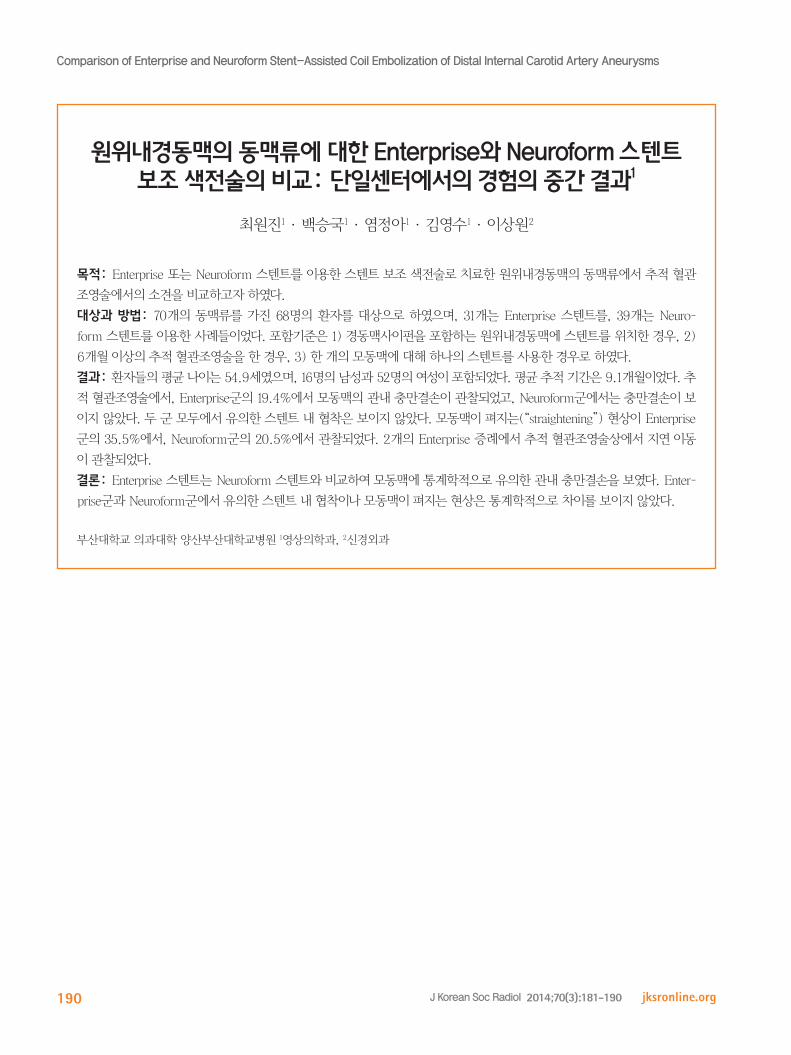
목적: Enterprise 또는 Neuroform 스텐트를 이용한 스텐트 보조 색전술로 치료한 원위내경동맥의 동맥류에서 추적 혈관
조영술에서의 소견을 비교하고자 하였다.
대상과 방법: 70개의 동맥류를 가진 68명의 환자를 대상으로 하였으며, 31개는 Enterprise 스텐트를, 39개는 Neuro-
form 스텐트를 이용한 사례들이었다. 포함기준은 1) 경동맥사이펀을 포함하는 원위내경동맥에 스텐트를 위치한 경우, 2)
6개월 이상의 추적 혈관조영술을 한 경우, 3) 한 개의 모동맥에 대해 하나의 스텐트를 사용한 경우로 하였다.
결과: 환자들의 평균 나이는 54.9세였으며, 16명의 남성과 52명의 여성이 포함되었다. 평균 추적 기간은 9.1개월이었다. 추
적 혈관조영술에서, Enterprise군의 19.4%에서 모동맥의 관내 충만결손이 관찰되었고, Neuroform군에서는 충만결손이 보
이지 않았다. 두 군 모두에서 유의한 스텐트 내 협착은 보이지 않았다. 모동맥이 펴지는(“straightening”) 현상이 Enterprise
군의 35.5%에서, Neuroform군의 20.5%에서 관찰되었다. 2개의 Enterprise 증례에서 추적 혈관조영술상에서 지연 이동
이 관찰되었다.
결론: Enterprise 스텐트는 Neuroform 스텐트와 비교하여 모동맥에 통계학적으로 유의한 관내 충만결손을 보였다. Enter-
prise군과 Neuroform군에서 유의한 스텐트 내 협착이나 모동맥이 펴지는 현상은 통계학적으로 차이를 보이지 않았다.
부산대학교 의과대학 양산부산대학교병원 1영상의학과, 2신경외과