Comparison of efficacy and safety of single versus ...
16
RESEARCH ARTICLE Comparison of efficacy and safety of single versus repeated intra-articular injection of allogeneic neonatal mesenchymal stem cells for treatment of osteoarthritis of the metacarpophalangeal/metatarsophalangeal joint in horses: A clinical pilot study Carmelo Magri ID 1☯ *, Michael Schramme 1☯ , Marine Febre 2☯ , Eddy Cauvin 3‡ , Fabrice Labadie 2‡ , Nathalie Saulnier 2‡ , Ise ´ Franc ¸ ois 1‡ , Antoine Lechartier 4‡ , David Aebischer 5‡ , Anne-Sophie Moncelet 6‡ , Ste ´ phane Maddens 2☯ 1 Cline ´ quine, VetAgro Sup, Campus Ve ´te ´ rinaire de Lyon, Marcy l’Etoile, France, 2 Vetbiobank SAS, Campus Ve ´te ´ rinaire de Lyon, Marcy l’Etoile, France, 3 Azurvet, Cagnes-sur-Mer, France, 4 Clinique e ´ quine ve ´te ´ rinaire de Meheudin, Ecouche ´ , France, 5 Clinique e ´ quine Areda, Bex, Switzerland, 6 Clinique e ´ quine de Grosbois, Marolles-en-Brie, France ☯ These authors contributed equally to this work. ‡ These authors also contributed equally to this work. * [email protected] Abstract The purpose of this prospective study was to evaluate the effects of single and repeated intra-articular administration of allogeneic, umbilical cord-derived, neonatal mesenchymal stem cells (MSC) in horses with lameness due to osteoarthritis (OA) of a metacarpophalan- geal joint (MPJ). Twenty-eight horses were included. Horses were divided into two groups. Horses in group MSC1 received an MSC injection at M0 and a placebo injection at M1 (1 month after M0). Horses in group MSC2 received MSC injections at M0 and at M1. Joint injections were performed with a blinded syringe. Clinical assessment was performed by the treating veteri- narian at M1, M2 and M6 (2 and 6 months after M0), including lameness evaluation, palpa- tion and flexion of the joint. Radiographic examination of the treated joints was performed at inclusion and repeated at M6. Radiographs were anonymized and assessed by 2 ECVDI LA associate members. Short term safety assessment was performed by owner survey. A 2- month rehabilitation program was recommended to veterinarians. There was a significant improvement of the total clinical score for horses in both groups. There was no significant difference in the total clinical score between groups MSC1 and MSC2 at any time point in the study. There was no significant difference in the total radiographic OA score, osteophyte score, joint space width score and subchondral bone score between inclusion and M6. Owner-detected adverse effects to MSC injection were recorded in 18% of the horses. Lameness caused by OA improved significantly over the 6-month duration of the study after treatment with allogeneic neonatal umbilical cord-derived MSCs combined with 8 weeks PLOS ONE | https://doi.org/10.1371/journal.pone.0221317 August 29, 2019 1 / 16 a1111111111 a1111111111 a1111111111 a1111111111 a1111111111 OPEN ACCESS Citation: Magri C, Schramme M, Febre M, Cauvin E, Labadie F, Saulnier N, et al. (2019) Comparison of efficacy and safety of single versus repeated intra-articular injection of allogeneic neonatal mesenchymal stem cells for treatment of osteoarthritis of the metacarpophalangeal/ metatarsophalangeal joint in horses: A clinical pilot study. PLoS ONE 14(8): e0221317. https://doi.org/ 10.1371/journal.pone.0221317 Editor: Paolo Fiorina, Children’s Hospital Boston, UNITED STATES Received: June 19, 2019 Accepted: August 2, 2019 Published: August 29, 2019 Peer Review History: PLOS recognizes the benefits of transparency in the peer review process; therefore, we enable the publication of all of the content of peer review and author responses alongside final, published articles. The editorial history of this article is available here: https://doi.org/10.1371/journal.pone.0221317 Copyright: © 2019 Magri et al. This is an open access article distributed under the terms of the Creative Commons Attribution License, which permits unrestricted use, distribution, and reproduction in any medium, provided the original author and source are credited.
Transcript of Comparison of efficacy and safety of single versus ...
RESEARCH ARTICLE
Comparison of efficacy and safety of single
versus repeated intra-articular injection of
allogeneic neonatal mesenchymal stem cells
for treatment of osteoarthritis of the
metacarpophalangeal/metatarsophalangeal
joint in horses: A clinical pilot study
Carmelo MagriID1☯*, Michael Schramme1☯, Marine Febre2☯, Eddy Cauvin3‡,
Fabrice Labadie2‡, Nathalie Saulnier2‡, Ise Francois1‡, Antoine Lechartier4‡,
David Aebischer5‡, Anne-Sophie Moncelet6‡, Stephane Maddens2☯
1 Clinequine, VetAgro Sup, Campus Veterinaire de Lyon, Marcy l’Etoile, France, 2 Vetbiobank SAS,
Campus Veterinaire de Lyon, Marcy l’Etoile, France, 3 Azurvet, Cagnes-sur-Mer, France, 4 Clinique equine
veterinaire de Meheudin, Ecouche, France, 5 Clinique equine Areda, Bex, Switzerland, 6 Clinique equine de
Grosbois, Marolles-en-Brie, France
☯ These authors contributed equally to this work.
‡ These authors also contributed equally to this work.
Abstract
The purpose of this prospective study was to evaluate the effects of single and repeated
intra-articular administration of allogeneic, umbilical cord-derived, neonatal mesenchymal
stem cells (MSC) in horses with lameness due to osteoarthritis (OA) of a metacarpophalan-
geal joint (MPJ).
Twenty-eight horses were included. Horses were divided into two groups. Horses in
group MSC1 received an MSC injection at M0 and a placebo injection at M1 (1 month after
M0). Horses in group MSC2 received MSC injections at M0 and at M1. Joint injections were
performed with a blinded syringe. Clinical assessment was performed by the treating veteri-
narian at M1, M2 and M6 (2 and 6 months after M0), including lameness evaluation, palpa-
tion and flexion of the joint. Radiographic examination of the treated joints was performed at
inclusion and repeated at M6. Radiographs were anonymized and assessed by 2 ECVDI LA
associate members. Short term safety assessment was performed by owner survey. A 2-
month rehabilitation program was recommended to veterinarians. There was a significant
improvement of the total clinical score for horses in both groups. There was no significant
difference in the total clinical score between groups MSC1 and MSC2 at any time point in
the study. There was no significant difference in the total radiographic OA score, osteophyte
score, joint space width score and subchondral bone score between inclusion and M6.
Owner-detected adverse effects to MSC injection were recorded in 18% of the horses.
Lameness caused by OA improved significantly over the 6-month duration of the study after
treatment with allogeneic neonatal umbilical cord-derived MSCs combined with 8 weeks
PLOS ONE | https://doi.org/10.1371/journal.pone.0221317 August 29, 2019 1 / 16
a1111111111
a1111111111
a1111111111
a1111111111
a1111111111
OPEN ACCESS
Citation: Magri C, Schramme M, Febre M, Cauvin
E, Labadie F, Saulnier N, et al. (2019) Comparison
of efficacy and safety of single versus repeated
intra-articular injection of allogeneic neonatal
mesenchymal stem cells for treatment of
osteoarthritis of the metacarpophalangeal/
metatarsophalangeal joint in horses: A clinical pilot
study. PLoS ONE 14(8): e0221317. https://doi.org/
10.1371/journal.pone.0221317
Editor: Paolo Fiorina, Children’s Hospital Boston,
UNITED STATES
Received: June 19, 2019
Accepted: August 2, 2019
Published: August 29, 2019
Peer Review History: PLOS recognizes the
benefits of transparency in the peer review
process; therefore, we enable the publication of
all of the content of peer review and author
responses alongside final, published articles. The
editorial history of this article is available here:
https://doi.org/10.1371/journal.pone.0221317
Copyright: © 2019 Magri et al. This is an open
access article distributed under the terms of the
Creative Commons Attribution License, which
permits unrestricted use, distribution, and
reproduction in any medium, provided the original
author and source are credited.
rest and rehabilitation. There is no apparent clinical benefit of repeated intra-articular admin-
istration of MSCs at a 1-month interval in horses with MPJ OA when compared to the effect
of a single injection.
Introduction
Osteoarthritis (OA) of the metacarpophalangeal/metatarsophalangeal joints (MPJs) is one of
the most common causes of lameness in Sports horses [1]. Several local and systemic treat-
ments have been described, including intra-articular viscosupplementation (hyaluronic acid,
polyacrylamide gel), anti-inflammatory biological therapeutics [platelet-rich plasma (PRP),
autologous conditioned serum (ACS or IRAP), polysulphated glycosaminoglycans (PSGAGS)
and steroidal drugs (corticosteroids, stanozolol). Such intra-articular treatments have been
shown to have predominantly symptom-modifying effects.
Mesenchymal stem cell (MSC) therapy has been developed as a means of promoting scar-
free tissue regeneration in a variety of musculoskeletal injuries in horses [2], and has been
characterized predominantly for the treatment of overstrain injuries of the superficial digital
flexor tendon [3–5]. Even if the direct regenerative properties of MSCs are now being ques-
tioned [6], their immunomodulatory effects may offer an alternative mechanism to improve
the quality of the healing process of injured tissues through disease-modifying effects.
MSCs have been used in equine joints for the treatment of osteoarthritis [7] and for surgical
resurfacing of chondral defects during arthroscopic interventions [8–10]. Although the results
of intra-articular injection of MSCs were initially disappointing in the management of OA
[11], more promising results have since been reported [8,12,13]. Preclinical data in a rabbit
model have shown that equine MSCs may be able to prevent cartilage degradation after
induced meniscal injury [14].
MSCs can be harvested from different sources, and autologous preparations derived from
bone marrow or adipose tissue were the first to be introduced in equine orthopaedics
[4,11,15]. The preparation and culture of autologous cells is time-consuming and their proper-
ties can be variable depending on the individual horse donor. Therefore the therapeutic use of
allogeneic MSCs derived from bone marrow, adipose tissue, peripheral blood, gingiva, and
other tissues from selected adult donors has also been explored [16–20]. More recently, umbil-
ical cord, umbilical cord blood or placenta have gained acceptance as sources of neonatal allo-
geneic MSCs because these cells may have greater biological potential and an immunologically
privileged state compared to adult MSCs [21]. Several studies argue for a relatively higher
immunomodulatory potential of these cells both in horses and other species, justifying their
investigation for application in joints to help improve the clinical status of the joint through
balancing the inflammatory environment [14].
As the persistence of injected MSCs in the joint is limited[14,22,23], their therapeutic effect
is likely to be temporary. A single intra-articular injection of MSCs can therefore no longer be
considered as an ‘OA treatment for life’ and repeated intra-articular dosing may have more
beneficial effects. The optimal time for a first MSC-injection has been shown to be during a
period of joint inflammation [14] but optimal times and dosages for repeat injections still need
to be defined. For a pharmacological drug, optimal time for dosing is usually derived from
drug metabolism and pharmacokinetic (DMPK) studies. However, MSCs have unconven-
tional DMPK parameters which furthermore are not completely understood [24]. As a conse-
quence, MSC dosing regimens have been derived from experience and approximation.
One or two allogeneic neonatal MSC injection for treatment of fetlock OA in horses
PLOS ONE | https://doi.org/10.1371/journal.pone.0221317 August 29, 2019 2 / 16
Data Availability Statement: All relevant data are
within the paper and its Supporting Information
files.
Funding: Azurvet provided support in the form of
salary for author EC. Vetbiobank provided support
in the form of salaries for authors (MF, FL, NS) and
contributed to the study design, data collection and
analysis, decision to publish and preparation of the
manuscript. The specific roles of these authors are
articulated in the ’Authors Contribution Section’.
Competing interests: The authors have read the
journal’s policy and the authors of this manuscript
have the following competing interests: SM is a
current and principal shareholder of Vetbiobank.
MF, FL, NS, are employees of Vetbiobank. There is
no commercial or financial connection between
Vetbiobank and the seven co-authors (CM, MS, EC,
IF, AL, DA, AM). EC is shareholder of Azurvet. The
product used in this study is developed for
commercial use in France and Europe. There are
no patents associated with this research to declare.
These commercial affiliations do not alter
adherence of all authors to all PLOS ONE policies
on sharing data and materials. MS and CM are
current employees of the National Veterinary
School Of Lyon, Vetagro Sup in France.

Repeated administration of adult MSCs has raised concerns related to allogenicity [25], but
neonatal umbilical cord MSCs may be better tolerated than adult allogenic MSCs [26].
The purpose of this study was to evaluate the effects of single and repeated intra-articular
administration of allogeneic, umbilical cord-derived, neonatal MSCs in horses with lameness
due to documented OA of an MPJ. We hypothesized that 2 intra-articular MSC injections
with a 4-week inter-injection interval would improve the clinical and radiological outcome
parameters at 6 months compared to a single intra-articular injection.
The primary objectives of this study were to determine the 1) clinical and 2) radiological
outcomes of horses with OA of an MPJ treated with intra-articular MSCs and 3) to compare
the outcomes between horses that were injected once or twice. The secondary objective was to
assess the safety of single and repeated intra-articular administration of allogeneic neonatal
MSCs in equine MP joints.
Materials and methods
Study design (Fig 1)
This was a multicenter, randomized, double-blinded, controlled, clinical pilot study, involving
veterinary surgeons from 10 different equine veterinary hospitals in France. All horses were
client-owned and all owners signed an informed consent prior to inclusion. The study was
implemented in accordance with University regulations. All horses were sedated during joint
injections and all efforts were made to minimize suffering. Horses were divided into two
groups (MSC1 and MSC2) according to the number of MSC injections received. The study
compared a single MSC injection (group MSC1) with 2 MSC injections (group MSC2). Horses
in group MSC1 received an MSC injection at M0 and a placebo injection at M1 (1 month after
M0). Horses in group MSC2 received an MSC injection at M0 and at M1. Follow-up examina-
tions were performed at M1, M2 (2 months after M0) and M6 (6 months after M0). Owners
and attending veterinarians were blinded to the selected treatment regimen for each horse dur-
ing the 6-month duration of the study. Short term safety assessment was performed by owner
survey using dedicated forms. At M6, after the final lameness evaluation, treatment group allo-
cation was unveiled to both owners and veterinarians.
Inclusion and exclusion criteria
Horses were selected for the study if they had a persistent single-limb lameness of at least 2
months’ duration that was localized to a MPJ with diagnostic analgesia, that was exacerbated
by a fetlock flexion test and if radiographs showed osteoarthritis of that MPJ.
Clinical examination included lameness evaluation, palpation and flexion of the joint.
Lameness was scored from 0 to 5 based on an adaptation of the AAEP (American Association
of Equine Practitioners) lameness grading system for trotting on a straight line only (0 sound;
1 lameness difficult to detect and inconsistent; 2 lameness difficult to detect but consistent; 3
lameness consistently observable in a straight line; 4 obvious lameness with marked head nod-
ding; 5 minimal weight-bearing in motion and/or at rest) [27]. When available, objective gait
Fig 1. Case selection and time-line of the study.
https://doi.org/10.1371/journal.pone.0221317.g001
One or two allogeneic neonatal MSC injection for treatment of fetlock OA in horses
PLOS ONE | https://doi.org/10.1371/journal.pone.0221317 August 29, 2019 3 / 16
analysis was performed with an inertial sensor-based lameness diagnosis system (Lameness
Locator ND) while horses were trotted on concrete in straight lines. The parameters used
included mean HDMax (Maximum head height difference), mean HDMin (Minimum head
height difference) and Vector Sum for fore limb lameness, and mean PDmax (Maximum pel-
vic height difference) and mean PDmin (Minimum pelvic height difference) for hindlimb
lameness.[28] Additional scores were given for the horse’s response to fetlock flexion at rest
(passive flexion 0–4 = no, slight, moderate, marked, severe pain response), for the increase in
lameness grade following a fetlock flexion test (active flexion 0–4 = no, slight, moderate,
marked, severe lameness increase) and for joint distention (0–4 = no, slight, moderate,
marked, severe distention). The four clinical scores (lameness, passive flexion response, active
flexion response and joint distention) were added to obtain a composite clinical score out of a
total of 17.
Radiographic examination consisted of 4 projections (dorso-palmar/plantar, latero-medial,
dorsolateral-palmaromedial oblique and dorsomedial-palmarolateral oblique views). Magnetic
resonance imaging (MRI), ultrasonography and diagnostic arthroscopy were also used in
some horses for identification of cartilage degeneration and osteophytes.
Previously attempted treatments were recorded as well as the time of initial diagnosis of
OA. Horses with other orthopaedic injuries and horses that received intra-articular treatment
less than 1 month prior to the MSC injection were not included. Any other intra-articular
treatment given during the 6-month duration of the study was a cause for exclusion from the
study, as was the incidence of any other orthopaedic injury that could have influenced lame-
ness evaluation of the treated limb during the follow-up period. Previous treatment with MSCs
was also a cause for exclusion. Systemic administration of anti-inflammatory medication was
accepted during the study if it occurred within 48 hours of or more than 4 weeks after MSC
injection and was reported in the follow-up information. Administration of any anti-inflam-
matory medication was not allowed less than 15 days before any of the clinical evaluations
(M0, M1, M2 and M6).
Cell product preparation
Equine umbilical cord-derived (UCd)-MSCs were provided by Vetbiobank (Marcy l’Etoile,
France). Umbilical cords were recovered following parturition from selected mares and foals
belonging to French National Studfarms (IFCE, Saumur, France), in accordance with a previ-
ously described protocol [29]. Extensive adventitious agent screening (including viruses, para-
sites and mycoplasma) was performed on mares’ blood samples before foaling and on neonatal
tissue samples afterwards. All batches of MSCs used in this study were isolated from umbilical
cord tissue, cultured, manufactured and characterised as previously described [14]. Briefly,
cells displayed a conventional phenotype for equine MSCs (i.e. CD44+; CD29+; CD90+;
MHC1+; MHC2-; CD45-; CD34-), differentiated in vitro into adipogenic and osteogenic cell
lineages and expressed chondrogenic markers upon differentiation [30]. MSCs were frozen in
10% (v/v) DMSO and stored in liquid nitrogen.
On the day prior to injection, each MSC dose was retrieved from liquid nitrogen, thawed at
37˚C and washed extensively with Dulbecco’s Phosphate Buffered Salinea (D-PBS, Pan Bio-
tech). The investigational product consisted of approximately 10 x106 viable cells (median: 11
x106; minimum: 9 x10⁶ and maximum 16 x106) at the 4th passage (minimum 2, maximum 8
passages), re-suspended after thawing in 2 mL D-PBS and transferred to a sterile 5mL ready-
to-use syringe. The placebo product consisted of syringes containing the same volume of
D-PBS to ensure a similar color and consistency of content compared to MSC-loaded syringes.
Blinding was further assured by camouflaging the syringe’s content with opaque adhesive
One or two allogeneic neonatal MSC injection for treatment of fetlock OA in horses
PLOS ONE | https://doi.org/10.1371/journal.pone.0221317 August 29, 2019 4 / 16

sterile tape applied around the barrel of the syringe. Loaded syringes were shipped overnight,
refrigerated between 2 to 12˚C in an appropriately sterile container, to equine hospitals partici-
pating in the study and used within 12 hours of reception. These conditions assure less than
10% cell death at the time of injection.
Eleven different horse donors were used to prepare equal numbers of MSC batches. It was
mandatory for horses in group MSC2 that the cell batch used for the second injection was
from a different donor horse than the one used for first injection. Major Histocompatibility
Complex (MHC) typing was not performed in this study.
Treatment (Fig 1)
Joint injections were performed at M0 (MSCs) and M1 (MSCs or placebo) under sedation
with an alpha-2-agonist (detomidineb 0,01 mg/kg IV, xylazinec 0,06 m/kg IV or romifidined
0,08 mg/kg IV) alone or in combination with butorphanole (0,01 mg/kg IV). The injection
procedure was identical for both MSC and placebo syringes. Prior to injection both MSC and
placebo syringes were gently inverted 8–10 times to bring MSCs into homogeneous suspen-
sion. The injections were performed slowly, over a minimum duration of 7–10 seconds, using
20-gauge needles to prevent cell damage from shear stress [31].
The use of systemic anti-inflammatory medication with phenylbutazonef (2,2 mg/kg IV),
flunixine meglumineg (1,1 mg/kg IV) and/or dexamethasoneh (0,1 mg/kg IV) immediately
before injection was considered acceptable to avoid potential post-injection reactions. Imme-
diately after injection a standard lower limb bandage was placed on the limb and maintained
for 24 hours.
Rehabilitation
A 2-month rehabilitation program was recommended to veterinarians (Fig 2) consisting of
stall rest with short, progressively increasing, periods of controlled hand-walking daily (from
15–30 minutes). After 2 months horses were free to resume their normal exercise routine.
Deviations from the proposed rehabilitation protocol had to be reported by the horse’s treating
veterinarian to the study monitor.
Follow-up evaluation
Clinical assessment was performed by the treating veterinarian at M1, M2 and M6 as for the
inclusion. Radiographic examination of the treated MPJ was repeated at M6. Radiographs of
both studies were anonymized and assessed by 2 ECVDI LA associate members (EC, MS). An
OA score was given to each radiographic exam (4 views) using a customized scoring system
(based on subchondral bone changes, joint space width, and periarticular new bone formation)
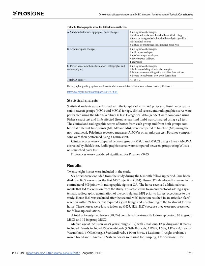
to obtain a total radiographic score out of 10 (Table 1). Owners were asked to document in
writing any post-injection reactions at the level of the injected MPJ during the first 48 hours
after each injection and to send the completed form to the stem cell laboratory (Vetbiobank).
Fig 2. Rehabilitation program. The 2-month rehabilitation program consisted of stall rest with periods of controlled
hand walking daily. After 2 months horses were free to resume their normal exercise routine.
https://doi.org/10.1371/journal.pone.0221317.g002
One or two allogeneic neonatal MSC injection for treatment of fetlock OA in horses
PLOS ONE | https://doi.org/10.1371/journal.pone.0221317 August 29, 2019 5 / 16
Statistical analysis
Statistical analysis was performed with the GraphPad Prism 6.0 programi. Baseline compari-
sons between groups (MSC1 and MSC2) for age, clinical scores, and radiographic scores were
performed using the Mann-Whitney U test. Categorical data (gender) were compared using
Fisher’s exact test and limb affected (front versus hind limb) was compared using a χ2 test.
The clinical and radiographic scores of horses from each group and from both groups com-
bined at different time points (M1, M2 and M6), were compared to baseline (M0) using the
non-parametric Friedman repeated measures ANOVA on a rank sum test. Post hoc compari-
sons were then performed using a Dunn’s test.
Clinical scores were compared between groups (MSC1 and MSC2) using a 2-way ANOVA
corrected by Sidak’s test. Radiographic scores were compared between groups using Wilcox-
on’s matched pairs test.
Differences were considered significant for P values�0.05.
Results
Twenty-eight horses were included in the study.
Six horses were excluded from the study during the 6-month follow-up period. One horse
died of colic 3 weeks after the first MSC injection (H24). Horse H28 developed lameness in the
contralateral MP joint with radiographic signs of OA. The horse received additional treat-
ments that led to exclusion from the study. This case led us to amend protocol adding a sys-
tematic radiographic examination of the contralateral MPJ prior to horses’ acceptance to the
study. Horse H23 was excluded after the second MSC injection resulted in an articular ‘flare’
reaction within 24 hours that required a joint lavage and un-blinding of the treatment for this
horse. Three horses were lost to follow up (H25, H26, H27) because they were not presented
for follow-up evaluations.
A total of twenty-two horses (78,5%) completed the 6-month follow-up period, 10 in group
MSC1 and 12 in group MSC2.
Median age at inclusion was 9 years [range 2–17] with 2 stallions, 12 geldings and 8 mares
included. Breeds included 15 Warmbloods (9 Selle Francais, 2 BWP, 1 SBS, 1 KWPN, 1 Swiss
Warmblood, 1 Oldenburg, 2 Standardbreds, 1 Paint horse, 1 Lusitano, 1 Anglo-arabian, 1
mixed breed and 1 Arabian). Sixteen horses were used for jumping, 1 for dressage, 1 for
Table 1. Radiographic score for fetlock osteoarthritis.
A. Subchondral bone / epiphyseal bone changes 0: no significant changes,
1: diffuse sclerosis, subchondral bone thickening,
2: focal or marginal subchondral bone lysis, cyst-like
subchondral lesions
3: diffuse or multifocal subchondral bone lysis
B. Articular space changes 0: no significant changes,
1: mild space collapse,
2: moderate space collapse,
3: severe space collapse,
4: ankylosis
C. Periarticular new bone formation (osteophytes and
enthesophytes)
0: no significant changes,
1: Mild remodeling of articular margins
2: Moderate remodeling with spur-like formations
3: Severe to exuberant new bone formation
Total OA score = A + B + C
Radiographic grading system used to calculate a cumulative fetlock total osteoarthritis (OA) score
https://doi.org/10.1371/journal.pone.0221317.t001
One or two allogeneic neonatal MSC injection for treatment of fetlock OA in horses
PLOS ONE | https://doi.org/10.1371/journal.pone.0221317 August 29, 2019 6 / 16
pleasure riding, 1 for western riding, 1 for endurance, and 2 for Standardbred racing. There
were 16 fore limb and 6 hind limb MPJs.
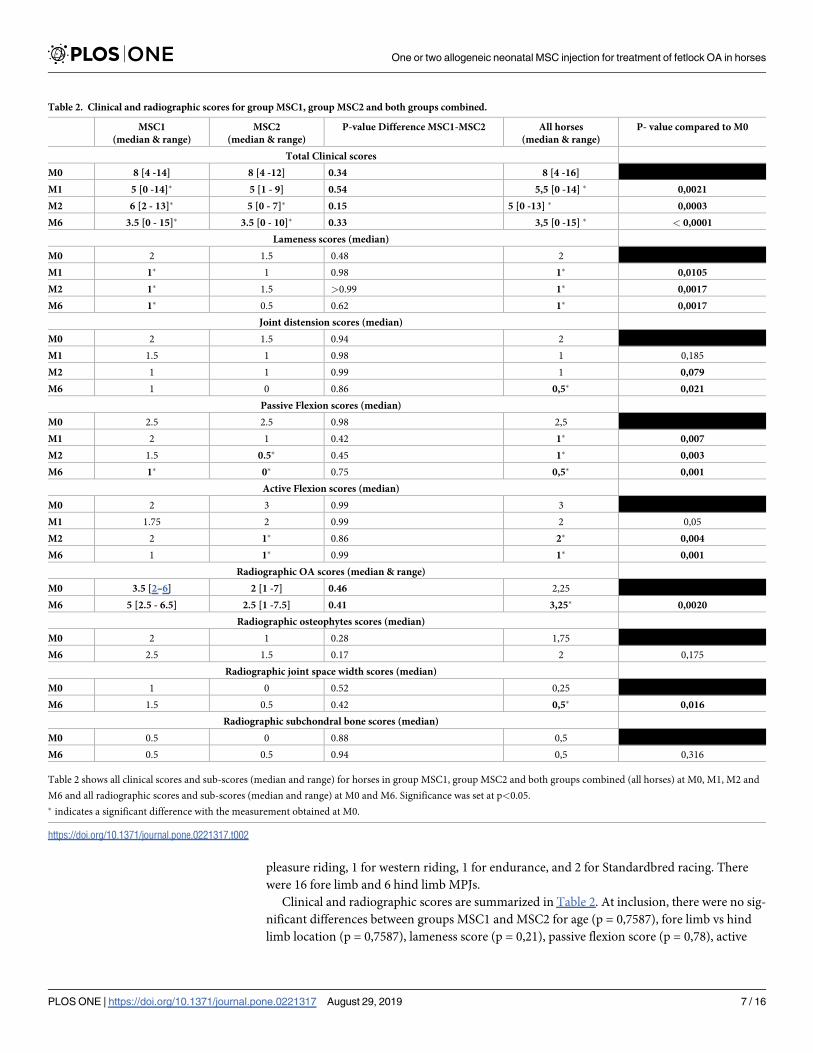
Clinical and radiographic scores are summarized in Table 2. At inclusion, there were no sig-
nificant differences between groups MSC1 and MSC2 for age (p = 0,7587), fore limb vs hind
limb location (p = 0,7587), lameness score (p = 0,21), passive flexion score (p = 0,78), active
Table 2. Clinical and radiographic scores for group MSC1, group MSC2 and both groups combined.
MSC1
(median & range)
MSC2
(median & range)
P-value Difference MSC1-MSC2 All horses
(median & range)
P- value compared to M0
Total Clinical scores
M0 8 [4 -14] 8 [4 -12] 0.34 8 [4 -16]
M1 5 [0 -14]� 5 [1 - 9] 0.54 5,5 [0 -14] � 0,0021
M2 6 [2 - 13]� 5 [0 - 7]� 0.15 5 [0 -13] � 0,0003
M6 3.5 [0 - 15]� 3.5 [0 - 10]� 0.33 3,5 [0 -15] � < 0,0001
Lameness scores (median)
M0 2 1.5 0.48 2
M1 1� 1 0.98 1� 0,0105
M2 1� 1.5 >0.99 1� 0,0017
M6 1� 0.5 0.62 1� 0,0017
Joint distension scores (median)
M0 2 1.5 0.94 2
M1 1.5 1 0.98 1 0,185
M2 1 1 0.99 1 0,079
M6 1 0 0.86 0,5� 0,021
Passive Flexion scores (median)
M0 2.5 2.5 0.98 2,5
M1 2 1 0.42 1� 0,007
M2 1.5 0.5� 0.45 1� 0,003
M6 1� 0� 0.75 0,5� 0,001
Active Flexion scores (median)
M0 2 3 0.99 3
M1 1.75 2 0.99 2 0,05
M2 2 1� 0.86 2� 0,004
M6 1 1� 0.99 1� 0,001
Radiographic OA scores (median & range)
M0 3.5 [2–6] 2 [1 -7] 0.46 2,25
M6 5 [2.5 - 6.5] 2.5 [1 -7.5] 0.41 3,25� 0,0020
Radiographic osteophytes scores (median)
M0 2 1 0.28 1,75
M6 2.5 1.5 0.17 2 0,175
Radiographic joint space width scores (median)
M0 1 0 0.52 0,25
M6 1.5 0.5 0.42 0,5� 0,016
Radiographic subchondral bone scores (median)
M0 0.5 0 0.88 0,5
M6 0.5 0.5 0.94 0,5 0,316
Table 2 shows all clinical scores and sub-scores (median and range) for horses in group MSC1, group MSC2 and both groups combined (all horses) at M0, M1, M2 and
M6 and all radiographic scores and sub-scores (median and range) at M0 and M6. Significance was set at p<0.05.
� indicates a significant difference with the measurement obtained at M0.
https://doi.org/10.1371/journal.pone.0221317.t002
One or two allogeneic neonatal MSC injection for treatment of fetlock OA in horses
PLOS ONE | https://doi.org/10.1371/journal.pone.0221317 August 29, 2019 7 / 16
flexion score (p = 0,95), joint distension (p = 0,59), total clinical score (p = 0,6360) and radio-
graphic score (p = 0,46). There were more females in group MSC1 than group MSC2
(p = 0,0062). Nineteen of the 22 horses had previously received other treatments which had
failed to improve lameness. These treatments had included stall rest (n = 10), intra-articular
injection of corticoids (n = 5) hyaluronic acid (n = 4), IRAP or PRP (n = 3), intravenous
administration of tiludronate (n = 3), oral NSAID (n = 1), chondroprotective nutritional com-
plements (n = 1) and arthroscopic debridement and joint lavage (n = 9). Of the 9 horses that
were included after a diagnostic arthroscopy, 4 ended up in the MSC1 group and 5 in the
MSC2 group.
The mean delay between diagnosis of OA and MSC treatment was 481 days (median 356
days; range 89–2573) in the 11/22 horses for which this information was available. Six of these
horses had not been used in equestrian sports or racing for at least 12 months preceding MSC
treatment.
At the time of first MSC injection, prophylactic intravenous anti-inflammatory medication
consisting of an NSAID alone (flunixin or phenylbutazone) was administered to 10/28 horses,
an NSAID (flunixin or phenylbutazone) with dexamethasone to another 10/28 horses and
dexamethasone alone to 1 horse. Five horses did not receive any anti-inflammatory medication
and the information was missing for 2 horses. At the time of the second injection at M1, 11/28
horses received an NSAID/dexamethasone combination, 7 horses only an NSAID, and 1 horse
only dexamethasone. Four horses did not receive any anti-inflammatory medication and the
information was missing for 4 horses. During the 6 months of the study, only 3 horses received
additional oral NSAIDs: 2 because of persistent joint effusion and pain (one at 2 days and one
at 15 days after injection) and another one for pain in the contralateral limb at M4.
A total of 4/34 (12%) MSC injections and 2/10 (20%) placebo injections resulted in owner-
reported adverse effects in 4 of 28 horses (H1, H2, H17, H23). H1 developed mild periarticular
heat for 48 hours after the first injection and heat and joint distension after the second injec-
tion (placebo) for 48 hours. H2 developed joint distension after the first injection but not after
the second injection (MSCs). H17 had no reported adverse effect after the first MSC injection
but developed mild periarticular heat for 48 hours after the second injection (placebo). Only
one horse (H23) experienced adverse effects at both injection times. This horse received two
MSC injections and developed marked articular and periarticular swelling with a grade 4/5
lameness after the second injection (M1). No prophylactic anti-inflammatory medication had
been administered to this horse. Cytology revealed a leukocyte concentration of 103,000 cells/
μL with 70% neutrophils but bacteriological culture of the synovial fluid was negative. This
prompted the attending practitioner to perform a through-and-through joint lavage under
standing sedation 24 hours after MSC injection. Strikingly all 4 horses with reported adverse
effects had a positive outcome. H23 was free of lameness, joint effusion or flexion responses at
M6 (total clinical score improved from 6 to 0) but was excluded from the study. No other
adverse effects were reported by the owners during a one-year follow up period. There was no
difference in the incidence of adverse effects between horses that received and horses that did
not receive prophylactic anti-inflammatory injections (p = 0.92). None of the 23 joint injec-
tions that were preceded by a prophylactic intravenous dexamethasone injection resulted in
any reported adverse effects. No correlation could be established between any characteristics
of each cell batch (cell viability, culture passage numbers and total viable cell number) and the
occurrence of reported adverse effects.
Clinical scores were available for 22 horses (Table 2). There was improvement of the clinical
score in 9 out of 10 horses from group MSC1 and in all 12 horses from group MSC2. There
was a significant improvement of the total clinical score for horses in group MSC1 between
M0 and M1 (p = 0,0361), M0 and M2 (p = 0,0128) and M0 and M6 (p = 0,0030). There was
One or two allogeneic neonatal MSC injection for treatment of fetlock OA in horses
PLOS ONE | https://doi.org/10.1371/journal.pone.0221317 August 29, 2019 8 / 16
also significant improvement of the total clinical score for horses in group MSC2 between M0
and M2 (p = 0,0342) and between M0 and M6 (p = 0,0008). However, there was no significant
difference in the total clinical score between groups MSC1 and MSC2 at any time point in the
study: M0 (p = 0,6792), M1(p = 0,6254), M2 (p = 0,3378) or M6 (p = 0,3087). When considered
individually for both groups and for all horses, lameness scores, flexion tests and joint effusion
were all significantly reduced at M2 and M6 (and lameness score and passive flexion also at
M1), and there were no differences between both groups (Table 2). Objective gait analysis was
available at M0 and M6 for 6 horses, 5 with fore limb lameness (H4, H7, H8, H20, H22) and
one with hind limb lameness (H21). There was improvement in the objective lameness param-
eters (vector sum, max diff head/pelvis and min diff head/pelvis) in 3 out of 6 horses, which
was in agreement with the subjective assessment for 5 of these horses. Radiographic scores
were available for 16 horses (Table 2), 5 horses from group MSC1 and 11 horses from group
MSC2. There was no significant difference in the total radiographic OA score, osteophyte
score, joint space width score and subchondral bone score between M0 and M6 (p = 0,5597) in
either group MSC1 or group MSC2, and there were no differences between both groups.
At the end of the study, 5 horses returned to their intended level of use, 8 to a lower level
and 9 remained lame. Of the 19 horses who were involved in competitive equestrian sports
before inclusion in the study, 13 (68%) resumed competition (12 showjumpers, 1 endurance)
while 6 (32%) did not (5 racehorses that failed to race; 1 horse developed contralateral limb
tendinitis). Three horses were included in the study at the insistence of their owners without
any work-related objectives but in order to improve the horses’ well-being by reducing
lameness.
Discussion
Several studies have investigated safety and efficacy of single or repeated intra-articular injec-
tions of allogeneic adult MSCs for the treatment of OA in horses ([8,11,13,17,32]). Whereas
studies have documented the safety of equine neonatal MSCs [26], thereby confirming results
obtained in other species [33,34], their clinical efficacy has not been investigated neither fol-
lowing a single nor repeated injections in horses. This study therefore, is the first to compare
clinical outcomes of two different treatment regimens using intra-articular injection of alloge-
neic neonatal MSCs in horses with MPJ OA.
The study was designed as a multicentered (n = 10) trial, involving both academic and pri-
vate practices, to minimize the risks of repeated bias related to inclusion, treatment or evalua-
tion that may occur in cell therapy studies limited to one or two participating clinical centers.
Biological products, including cell therapies, are inherently variable from one batch to another.
To reduce the impact of such variability, clinical studies with allogeneic cells are generally per-
formed with a single batch of cells to run the entire experiment [32,35] Such practice intro-
duces a batch-specific result which would translate poorly into clinical practice where cells
from different batches derived from different donors are necessarily used. Therefore we delib-
erately used several different batches of cells in our study, in order to reproduce better the real-
ity of clinical practice and increase the clinical relevance of the study. Similarly, anticipating
true clinical practice conditions where donor/patient MHC typing and matching would be dif-
ficult to implement, cells were not MHC-typed, and cells from a different donor than used for
the first injection were used for the second MSC injection. Few equine MSC studies have been
performed in a blinded fashion, which introduces a high risk of bias when subjective clinical
outcome parameters are used, like lameness grading and scoring of flexion test responses. Dif-
ferent methodologies to implement blindness can be used. In a study performed by Broeckx
et al., the practitioner evaluating clinical outcome was different to the one administering the
One or two allogeneic neonatal MSC injection for treatment of fetlock OA in horses
PLOS ONE | https://doi.org/10.1371/journal.pone.0221317 August 29, 2019 9 / 16

treatment [32]. In agreement with investigation sites in our study, this method was considered
difficult to implement and masking of the injectate was chosen instead.
The assessment of horses’ return to athletic performance was based on public performance
databases (letrot.com; francegalop.com; ffe.com) rather than relying on owners’ assessment.
Owner-detected adverse effects to MSC injection were recorded in 18% of the horses in our
study, three horses after the first injection and one horse after the second injection. Studies
performed with adult autologous MSCs have shown self-limiting local inflammation after a
single intra-articular injection of MSCs combined with fetal bovine serum (FBS) or hyaluronic
acid [8,36]. In one study, the concurrent use of hyaluronic acid was incriminated for a 9% inci-
dence of joint flares after injection of femorotibial joints with autologous bone marrow-
derived MSCs[8].
A recent study explored the safety of a single intra-articular injection of adult allogeneic
peripheral blood-derived MSCs in healthy horses [37]. There was transient joint heat and
lameness after injection but no differences were found between the treated group and a control
group of healthy horses undergoing the same injection protocol. In another study performed
by the same group, evaluating efficacy of the same allogeneic peripheral blood-derived MSCs
in horses with fetlock osteoarthritis, no adverse effects were observed after a single intra-artic-
ular injection. The authors included prophylactic administration of a systemic dose of an
NSAID, which may have led to an underestimation bias of adverse effects [32].
When using allogeneic MSCs in repeat injections, specific immune responses against MHC
antigens may occur. Induced cytotoxic antibodies to donor MHC antigens have been
described against adult allogeneic MSCs in horses [38,39]. Both inflammatory stimuli present
in the OA environment and cellular differentiation, have been shown in vitro to upregulate
MHC-II antigen expression by MSCs [40]. Therefore, the lack of detectable expression of
MHC-II antigens in cell batches, without priming, is not a definitive assurance against mount-
ing effective immune reactions. However neonatal MSCs have been shown to express less
MHC-II than their adult counterparts under inflammatory conditions [21]. Regardless, other
authors have not been able to show any difference in post-injection reaction between autolo-
gous and allogeneic MSCs, whether the cells where neonatal [26] or bone marrow-derived
adult cells [41]. Yet another study, evaluating neonatal MSCs in dogs with OA, failed to iden-
tify a consistent specific humoral response after single or repeated MSC injection [34]. The use
of cells from different donors and the removal of fetal calf serum residues by extensive washing
of cell cultures may further help reduce the risk of a local inflammatory reaction after a second
MSC administration. The adverse effects observed in 2 horses after placebo injection (20%)
suggests the possibility of non-specific inflammation occurring related to the arthrocentesis
procedure itself or to the potential hyperreactive state of OA joints.
Only one horse in our study experienced a severe inflammatory reaction following injection
of MSCs, resulting in severe swelling, heat and lameness (H23). Synovial fluid analysis
(103,000 white blood cells/μL) was indicative of joint sepsis, possibly related to a breach in
aseptic technique. Even so, this increased cell number might also be compatible with the pres-
ence of the injected MSCs in the joint fluid one day after injection or with an immune-medi-
ated non-septic inflammatory synovitis (‘joint flare’), especially as clinical signs of joint sepsis
usually take longer than 24 hours to appear. Unfortunately, no cytological evaluation of the
joint fluid was performed to determine whether the elevated cell count was caused by the pres-
ence of neutrophils, monocytes, lymphocytes or MSCs. Consequently, and even though a
bacteriological culture remained negative, a conclusive diagnosis as to the cause of the acute
inflammatory joint reaction was not possible.
It is important to note that the occurrence of adverse injection effects was not related to a
negative outcome in our study. All 4 horses had a favorable outcome and H23 had returned to
One or two allogeneic neonatal MSC injection for treatment of fetlock OA in horses
PLOS ONE | https://doi.org/10.1371/journal.pone.0221317 August 29, 2019 10 / 16

his previous performance level at 6 months. This observation, though anecdotal, is similar to a
previous report that suggested that horses with marked injection reactions had generally an
excellent outcome compared to others [42]. Interestingly this also agrees with findings from a
study revealing a link between a pro-inflammatory response early after MSC administration
and immunomodulatory functions of MSCs [43]. An inflammatory reaction observed after
intra-articular injection of MSCs, that is controllable medically with NSAIDs, may need to be
considered more as an expected side effect rather that an adverse reaction.
The results of our study failed to show an improved outcome after two MSC injections with
a 1-month injection interval, in comparison with a single MSC injection. The mechanism of
action of MSCs is currently thought to be immunomodulatory one rather than any regenera-
tive effects brought about by the MSCs’ differentiation into chondrocytes [21]. The immuno-
modulatory effects of MSCs are thought to alter the inflammatory environment in the joint
after joint injury through an anti-inflammatory activity of the MSCs as well of cells recruited
to the injury site by the MSCs through their paracrine effect [16]. It would therefore be reason-
able to expect a more profound or persistent beneficial anti-inflammatory effect from two
administrations of MSCs than from a single administration. However, we were unable to show
this. Several reasons may explain the apparent lack of benefit of a second MSC injection.
Group sizes may have been too small with regard to the large variation in lameness scores to
detect a significant difference between both groups. Also, the interval of 1 month between
both injections may not be adequate. It was empirically chosen to fit with the estimated resi-
dence time of MSCs in the joint [22,44]. The first injection could have resulted in a humoral
response with optimal antibody levels peaking at 1 month, leading to a rapid elimination of
the MSCs of the second injection. It is further possible that the effect of the second injection
was ‘diluted’ by the course of action of the first injection in other ways. As the mechanism of
action of MSCs is still incompletely understood, their effects may not merely be cumulative as
could be expected in a classical pharmacological dose-response effect of repeated intra-articu-
lar drug administration. For example, if inflammatory parameters of the joint were lowered by
the first injection, MSCs from the second injection may not have encountered the expected
threshold of inflammatory stimuli to be primed for optimal anti-inflammatory activity [14,32].
Therefore, it would be useful to test a longer inter-injection interval such as 6 months, as sug-
gested by recent studies [45]. Beneficial effect of a second intra-articular injection of equine
umbilical cord-derived MSCs with a 6-month inter-injection interval have been demonstrated
in a pilot study with canine OA patients[34].
It is striking to note that horses in both groups experienced a significant improvement in
lameness scores and total clinical scores, as early as M1, and over the entire duration of the
study (M6). This finding is in agreement with those from efficacy studies of intra-articular
administration of adult MSCs for the treatment of OA in horses [8,12,13,32,17]. A recent
study, Broeckx et al. managed to include a placebo group in a study evaluating clinical efficacy
of an intra-articular combination therapy with MSCs and equine allogeneic plasma. Unfortu-
nately the authors used a saline control injection for the placebo group, which prevents a defin-
itive distinction between the relative efficacy of the MSCs and the allogeneic plasma each in
their own right [32]. When comparing our outcome data with the saline placebo group from
this study, horses from both groups (MSC1 and MSC2) in our study had a significantly better
outcome (p = 0,019). Even though our results compare favorably with outcome data from
other MSC efficacy studies, a definitive conclusion about the purported beneficial effect of
equine neonatal allogeneic umbilical cord-derived MSCs would require a prospective study
including a group of horses with MPJ OA treated with an intra-articular placebo injection and
subjected to the same period of rest and rehabilitation. However, given the nature of clinical
practice, it is almost impossible to convince horse owners to collaborate with a 6-month study
One or two allogeneic neonatal MSC injection for treatment of fetlock OA in horses
PLOS ONE | https://doi.org/10.1371/journal.pone.0221317 August 29, 2019 11 / 16

in which there is a real chance that their horse will miss out on a potentially useful new thera-
peutic modality or drug.
The 8-week period of rest and rehabilitation may also have helped the horse’s lameness
grade improve through the study. It has been shown that it may be difficult to differentiate the
beneficial effects of a rest and rehab program from those caused by a therapeutic agent admin-
istered concurrently in the treatment of equine osteoarthritis [46]. However, the authors feel
this is unlikely, as at least 7 horses had already been rested for periods exceeding 8 weeks with-
out improvement before recruitment in the study, other horses had failed to respond to other
treatments combined with rest, and the median time between OA diagnosis and MSC treat-
ment was one year with a minimum of 3 months for horses in this study.
There were no significant changes in the radiographic scores over the duration of the study.
Other studies using models of equine OA also failed to show a significant change in radio-
graphic signs of OA between MSC- and placebo-treated joints [40,11]. Furthermore, it is
important to note there was a large variation of the total radiographic OA score of horses
included in the study, ranging from mild to severe OA. This could have limited the ability to
recognize a statistically significant improvement in OA scores at 6 months after MSC injection
(M6). Even so, radiographic changes of OA like osteophytes, joint space narrowing and sub-
chondral bone lysis tend to be irreversible and would not be expected to improve with any
treatment. Therefore, the absence of significant exacerbation of the OA grade in either group
over a period of 6 months could be considered as a positive finding.
Our study had some limitations inherent to equine multicenter clinical field trials. An
increasing number of investigational centers induces variability in clinical assessment and
uneven data collection. Objective lameness evaluation was only feasible in a limited number of
patients due to limited availability of the Lameness locator equipmentj. Not all horses were
evaluated after intra-articular analgesia of the MPJ to confirm the presence of intra-articular
pain associated with osteoarthritis. Even so, we feel that the combination of perineural analge-
sia, a positive flexion test and imaging findings can be conclusive for a diagnosis of osteoarthri-
tis, even without the benefit of a recorded response to intra-articular analgesia [1]. Drugs
selected for prophylactic treatment for prevention of adverse injection effects varied between
operators. Inconsistency in radiographic protocol meant that all views required for grading the
OA score were only present in 16 horses (MSC1 = 5 and MSC2 = 11). And finally, during the 6
months of the study, some horses were lost to follow up due to non-compliance of the owners.
The low number of 22 horses retained for the final analysis could consequently induce the risk
of a type II statistical error.
We conclude that lameness caused by MPJ OA improved significantly after treatment with
allogeneic neonatal umbilical cord-derived MSCs combined with 8 weeks’ rest and rehabilita-
tion over the 6-month duration of the study. Intra-articular administration of umbilical cord-
derived, allogeneic, neonatal MSCs carries a low risk of unexpected adverse injection effects.
There is no apparent clinical benefit of repeated intra-articular administration of MSCs at a
1-month interval in horses with MPJ OA when compared to the effect of a single injection.
Manufacturers’ addresses
a Pan Biotech Gmbh, Aidenbach, Germany
b Detogesic, Zoetis, Malakoff, France
c Rompun Bayer Healthcare, Loos, France
d Sedivet, Merial, Lyon, France
e Torbugesic, Zoetis, Malakoff, France
f Phenylarthrite, Vetoquinol, Lure, France
One or two allogeneic neonatal MSC injection for treatment of fetlock OA in horses
PLOS ONE | https://doi.org/10.1371/journal.pone.0221317 August 29, 2019 12 / 16

g Finadyne Intervet, Beaucouze, France
h Dexadreson Intervet, Beaucouze, France
i Graphpad software, San Diego, USA
j Lameness Locator, Equinosis, Columbia, USA
Supporting information
S1 Table. Description of the population.
(DOCX)
S2 Table. Clinical scores of the horses.
(XLSX)
S3 Table. Radiographic scores of the horses.
(XLSX)
Acknowledgments
We are grateful to C. Dubois and L. Wimel, the staff of the experimental farm of the French
Horse and Riding Institute (IFCE), 19370 Chamberet, for providing neonatal tissues from
selected mares and foals. We thank Dr. K. Pader, Dr JM. Betsch, Dr M. Hamon, Dr E. Oua-
chee, Dr A. Vitte, Dr N. Delalande, Dr F. Croisier, Dr JC. Meunier, Dr D. Lavorel, Dr T.
Bertholdy, Dr F. Martin and Dr H. Sorribas for providing patients and performing clinical
evaluations and data collections. We wish to thank Dr S. Sage for his contributions to the
study protocol.
Author Contributions
Conceptualization: Marine Febre, Fabrice Labadie, Nathalie Saulnier, Stephane Maddens.
Data curation: Carmelo Magri, Marine Febre.
Formal analysis: Marine Febre.
Funding acquisition: Stephane Maddens.
Investigation: Carmelo Magri, Eddy Cauvin, Ise Francois, Antoine Lechartier, David
Aebischer, Anne-Sophie Moncelet.
Methodology: Marine Febre, Fabrice Labadie, Nathalie Saulnier, Ise Francois, Stephane
Maddens.
Project administration: Stephane Maddens.
Resources: Carmelo Magri, Marine Febre, Ise Francois.
Supervision: Stephane Maddens.
Validation: Stephane Maddens.
Visualization: Marine Febre.
Writing – original draft: Carmelo Magri, Michael Schramme, Marine Febre.
Writing – review & editing: Carmelo Magri, Michael Schramme, Marine Febre, Stephane
Maddens.
One or two allogeneic neonatal MSC injection for treatment of fetlock OA in horses
PLOS ONE | https://doi.org/10.1371/journal.pone.0221317 August 29, 2019 13 / 16

References1. Richardson DW, Dyson SJ. Chapter 36—The Metacarpophalangeal Joint. In: Ross MW, Dyson SJ, edi-
tors. Diagnosis and Management of Lameness in the Horse ( Second Edition). Saint Louis: W.B. Saun-
ders; 2011. pp. 394–410. https://doi.org/10.1016/B978-1-4160-6069-7.00036–5
2. Fortier L. Chapter 73—Clinical Use of Stem Cells, Marrow Components, and Other Growth Factors.
In: Ross MW, Dyson SJ, editors. Diagnosis and Management of Lameness in the Horse ( Second Edi-
tion). Saint Louis: W.B. Saunders; 2011. pp. 761–764. https://doi.org/10.1016/B978-1-4160-6069-7.
00073–0
3. Godwin EE, Young NJ, Dudhia J, Beamish IC, Smith RKW. Implantation of bone marrow-derived mes-
enchymal stem cells demonstrates improved outcome in horses with overstrain injury of the superficial
digital flexor tendon. Equine Vet J. 2012; 44: 25–32. https://doi.org/10.1111/j.2042-3306.2011.00363.x
PMID: 21615465
4. Smith RKW, Korda M, Blunn GW, Goodship AE. Isolation and implantation of autologous equine mes-
enchymal stem cells from bone marrow into the superficial digital flexor tendon as a potential novel
treatment. Equine Vet J. 2003; 35: 99–102. PMID: 12553472
5. Watts AE, Yeager AE, Kopyov OV, Nixon AJ. Fetal derived embryonic-like stem cells improve healing
in a large animal flexor tendonitis model. Stem Cell Res Ther. 2011; 2: 4–4. https://doi.org/10.1186/
scrt45 PMID: 21272343
6. Colbath AC, Dow SW, Phillips JN, McIlwraith CW, Goodrich LR. Autologous and Allogeneic Equine
Mesenchymal Stem Cells Exhibit Equivalent Immunomodulatory Properties In Vitro. Stem Cells Dev.
2017; 26: 503–511. https://doi.org/10.1089/scd.2016.0266 PMID: 27958776
7. Murphy JM, Fink DJ, Hunziker EB, Barry FP. Stem cell therapy in a caprine model of osteoarthritis.
Arthritis Rheum. 2003; 48: 3464–3474. https://doi.org/10.1002/art.11365 PMID: 14673997
8. Ferris DJ, Frisbie DD, Kisiday JD, McIlwraith CW, Hague BA, Major MD, et al. Clinical Outcome After
Intra-Articular Administration of Bone Marrow Derived Mesenchymal Stem Cells in 33 Horses With Stifle
Injury. Vet Surg. 2014; 43: 255–265. https://doi.org/10.1111/j.1532-950X.2014.12100.x PMID:
24433318
9. Fortier LA, Potter HG, Rickey EJ, Schnabel LV, Foo LF, Chong LR, et al. Concentrated Bone Marrow
Aspirate Improves Full-thickness Cartilage Repair Compared with Microfracture in the Equine Model. J
Bone Jt Surg. 2010; 92: 1927–1937. https://doi.org/10.2106/JBJS.I.01284 PMID: 20720135
10. McIlwraith CW, Frisbie DD, Rodkey WG, Kisiday JD, Werpy NM, Kawcak CE, et al. Evaluation of Intra-
Articular Mesenchymal Stem Cells to Augment Healing of Microfractured Chondral Defects. Arthros-
copy. 2011; 27: 1552–1561. https://doi.org/10.1016/j.arthro.2011.06.002 PMID: 21862278
11. Frisbie DD, Kisiday JD, Kawcak CE, Werpy NM, McIlwraith CW. Evaluation of adipose-derived stromal
vascular fraction or bone marrow-derived mesenchymal stem cells for treatment of osteoarthritis. J
Orthop Res. 2009; 27: 1675–1680. https://doi.org/10.1002/jor.20933 PMID: 19544397
12. Broeckx S, Suls M, Beerts C, Vandenberghe A, Seys B, Wuertz-Kozak K, et al. Allogenic Mesenchymal
Stem Cells as a Treatment for Equine Degenerative Joint Disease: A Pilot Study. Curr Stem Cell Res
Ther. 2014; 9: 497–503. https://doi.org/10.2174/1574888X09666140826110601 PMID: 25175766
13. Broeckx S, Zimmerman M, Crocetti S, Suls M, Marien T, Ferguson SJ, et al. Regenerative Therapies
for Equine Degenerative Joint Disease: A Preliminary Study. PLoS ONE. 2014; 9: e85917. https://doi.
org/10.1371/journal.pone.0085917 PMID: 24465787
14. Saulnier N, Viguier E, Perrier-Groult E, Chenu C, Pillet E, Roger T, et al. Intra-articular administration of
xenogeneic neonatal Mesenchymal Stromal Cells early after meniscal injury down-regulates metallo-
proteinase gene expression in synovium and prevents cartilage degradation in a rabbit model of osteo-
arthritis. Osteoarthritis Cartilage. 2015; 23: 122–133. https://doi.org/10.1016/j.joca.2014.09.007 PMID:
25219668
15. Vidal MA, Kilroy GE, Lopez MJ, Johnson JR, Moore RM, Gimble JM. Characterization of Equine Adi-
pose Tissue-Derived Stromal Cells: Adipogenic and Osteogenic Capacity and Comparison with Bone
Marrow-Derived Mesenchymal Stromal Cells. Vet Surg. 2007; 36: 613–622. https://doi.org/10.1111/j.
1532-950X.2007.00313.x PMID: 17894587
16. Ursini TL, Amelse LL, Elkhenany HA, Odoi A, Carter-Arnold JL, Adair HS, et al. Retrospective analysis
of local injection site adverse reactions associated with 230 allogenic administrations of bone marrow-
derived mesenchymal stem cells in 164 horses. Equine Vet J. 2019; 51: 198–205. https://doi.org/10.
1111/evj.12992 PMID: 29992618
17. Mariñas-Pardo L, Garcıa-Castro J, Rodrıguez-Hurtado I, Rodrıguez-Garcıa MI, Nuñez-Naveira L, Her-
mida-Prieto M. Allogeneic Adipose-Derived Mesenchymal Stem Cells (Horse Allo 20) for the Treatment
of Osteoarthritis-Associated Lameness in Horses: Characterization, Safety, and Efficacy of Intra-Articu-
lar Treatment. Stem Cells Dev. 2018; 27: 1147–1160. https://doi.org/10.1089/scd.2018.0074 PMID:
29978736
One or two allogeneic neonatal MSC injection for treatment of fetlock OA in horses
PLOS ONE | https://doi.org/10.1371/journal.pone.0221317 August 29, 2019 14 / 16

18. Mensing N, Gasse H, Hambruch N, Haeger J-D, Pfarrer C, Staszyk C. Isolation and characterization of
multipotent mesenchymal stromal cells from the gingiva and the periodontal ligament of the horse. BMC
Vet Res. 2011; 7: 42. https://doi.org/10.1186/1746-6148-7-42 PMID: 21810270
19. Iacono E, Brunori L, Pirrone A, Pagliaro PP, Ricci F, Tazzari PL, et al. Isolation, characterization and dif-
ferentiation of mesenchymal stem cells from amniotic fluid, umbilical cord blood and Wharton’s jelly in
the horse. Reproduction. 2012; 143: 455–468. https://doi.org/10.1530/REP-10-0408 PMID: 22274885
20. Koch TG, Heerkens T, Thomsen PD, Betts DH. Isolation of mesenchymal stem cells from equine umbili-
cal cord blood. BMC Biotechnol. 2007; 7: 26. https://doi.org/10.1186/1472-6750-7-26 PMID: 17537254
21. Prasanna SJ, Gopalakrishnan D, Shankar SR, Vasandan AB. Pro-Inflammatory Cytokines, IFNγ and
TNFα, Influence Immune Properties of Human Bone Marrow and Wharton Jelly Mesenchymal Stem
Cells Differentially. PLOS ONE. 2010; 5: e9016. https://doi.org/10.1371/journal.pone.0009016 PMID:
20126406
22. Toupet K, Maumus M, Luz-Crawford P, Lombardo E, Lopez-Belmonte J, Lent P van, et al. Survival and
Biodistribution of Xenogenic Adipose Mesenchymal Stem Cells Is Not Affected by the Degree of Inflam-
mation in Arthritis. PLOS ONE. 2015; 10: e0114962. https://doi.org/10.1371/journal.pone.0114962
PMID: 25559623
23. Grady ST, Britton L, Hinrichs K, Nixon AJ, Watts AE. Persistence of fluorescent nanoparticle-labelled
bone marrow mesenchymal stem cells in vitro and after intra-articular injection. J Tissue Eng Regen
Med. 2019; 13: 191–202. https://doi.org/10.1002/term.2781 PMID: 30536848
24. Brooks A, Futrega K, Liang X, Hu X, Liu X, Crawford DHG, et al. Concise Review: Quantitative Detec-
tion and Modeling the In Vivo Kinetics of Therapeutic Mesenchymal Stem/Stromal Cells: Detection and
Modeling Kinetics of Stem Cells. STEM CELLS Transl Med. 2018; 7: 78–86. https://doi.org/10.1002/
sctm.17-0209 PMID: 29210198
25. Joswig A-J, Mitchell A, Cummings KJ, Levine GJ, Gregory CA, Smith R, et al. Repeated intra-articular
injection of allogeneic mesenchymal stem cells causes an adverse response compared to autologous
cells in the equine model. Stem Cell Res Ther. 2017; 8: 42. https://doi.org/10.1186/s13287-017-0503-8
PMID: 28241885
26. Carrade DD, Owens SD, Galuppo LD, Vidal MA, Ferraro GL, Librach F, et al. Clinicopathologic findings
following intra-articular injection of autologous and allogeneic placentally derived equine mesenchymal
stem cells in horses. Cytotherapy. 2011; 13: 419–430. https://doi.org/10.3109/14653249.2010.536213
PMID: 21105841
27. Schumacher J, Steiger R, Schumacher J, Graves F, Schramme M, Smith R, et al. Effects of Analgesia
of the Distal Interphalangeal Joint or Palmar Digital Nerves on Lameness Caused by Solar Pain in
Horses. Vet Surg. 2000; 29: 54–58. https://doi.org/10.1111/j.1532-950X.2000.00054.x PMID:
10653495
28. Keegan KG, Kramer J, Yonezawa Y, Maki H, Pai PF, Dent EV, et al. Assessment of repeatability of a
wireless, inertial sensor–based lameness evaluation system for horses. Am J Vet Res. 2011; 72: 1156–
1163. https://doi.org/10.2460/ajvr.72.9.1156 PMID: 21879972
29. Bartholomew S, Owens SD, Ferraro GL, Carrade DD, Lara DJ, Librach FA, et al. Collection of equine
cord blood and placental tissues in 40 Thoroughbred mares. Equine Vet J. 2009; 41: 724–728. https://
doi.org/10.2746/042516409X429446 PMID: 20095217
30. Rakic R, Bourdon B, Demoor M, Maddens S, Saulnier N, Galera P. Differences in the intrinsic chondro-
genic potential of equine umbilical cord matrix and cord blood mesenchymal stromal/stem cells for carti-
lage regeneration. Sci Rep. 2018; 8: 13799. https://doi.org/10.1038/s41598-018-28164-9 PMID:
30217993
31. Lang HM, Schnabel LV, Cassano JM, Fortier LA. Effect of needle diameter on the viability of equine
bone marrow derived mesenchymal stem cells. Vet Surg. 2017; 46: 731–737. https://doi.org/10.1111/
vsu.12639 PMID: 28328147
32. Broeckx SY, Seys B, Suls M, Vandenberghe A, Marien T, Adriaensen E, et al. Equine Allogeneic Chon-
drogenic Induced Mesenchymal Stem Cells Are an Effective Treatment for Degenerative Joint Disease
in Horses. Stem Cells Dev. 2019; https://doi.org/10.1089/scd.2018.0061 PMID: 30623737
33. Taroni M, Cabon Q, Fèbre M, Cachon T, Saulnier N, Carozzo C, et al. Evaluation of the Effect of a Sin-
gle Intra-articular Injection of Allogeneic Neonatal Mesenchymal Stromal Cells Compared to Oral Non-
Steroidal Anti-inflammatory Treatment on the Postoperative Musculoskeletal Status and Gait of Dogs
over a 6-Month Period after Tibial Plateau Leveling Osteotomy: A Pilot Study. Front Vet Sci. 2017; 4:
83. https://doi.org/10.3389/fvets.2017.00083 PMID: 28642867
34. Cabon Q, Febre M, Gomez N, Cachon T, Pillard P, Carozzo C, et al. Long-Term Safety and Efficacy of
Single or Repeated Intra-Articular Injection of Allogeneic Neonatal Mesenchymal Stromal Cells for Man-
aging Pain and Lameness in Moderate to Severe Canine Osteoarthritis Without Anti-inflammatory
One or two allogeneic neonatal MSC injection for treatment of fetlock OA in horses
PLOS ONE | https://doi.org/10.1371/journal.pone.0221317 August 29, 2019 15 / 16

Pharmacological Support: Pilot Clinical Study. Front Vet Sci. 2019; 6: 10. https://doi.org/10.3389/fvets.
2019.00010 PMID: 30805348
35. Hatsushika D, Muneta T, Nakamura T, Horie M, Koga H, Nakagawa Y, et al. Repetitive allogeneic intra-
articular injections of synovial mesenchymal stem cells promote meniscus regeneration in a porcine
massive meniscus defect model. Osteoarthritis Cartilage. 2014; 22: 941–950. https://doi.org/10.1016/j.
joca.2014.04.028 PMID: 24795274
36. Pigott JH, Ishihara A, Wellman ML, Russell DS, Bertone AL. Inflammatory effects of autologous, geneti-
cally modified autologous, allogeneic, and xenogeneic mesenchymal stem cells after intra-articular
injection in horses. Vet Comp Orthop Traumatol VCOT. 2013; 26: 453–460. https://doi.org/10.3415/
VCOT-13-01-0008 PMID: 24080668
37. Broeckx SY, Spaas JH, Chiers K, Duchateau L, Van Hecke L, Van Brantegem L, et al. Equine alloge-
neic chondrogenic induced mesenchymal stem cells: A GCP target animal safety and biodistribution
study. Res Vet Sci. 2018; 117: 246–254. https://doi.org/10.1016/j.rvsc.2017.12.018 PMID: 29329028
38. Pezzanite LM, Fortier LA, Antczak DF, Cassano JM, Brosnahan MM, Miller D, et al. Equine allogeneic
bone marrow-derived mesenchymal stromal cells elicit antibody responses in vivo. Stem Cell Res Ther.
2015; 6: 54. https://doi.org/10.1186/s13287-015-0053-x PMID: 25889095
39. Berglund AK, Schnabel LV. Allogeneic major histocompatibility complex-mismatched equine bone mar-
row-derived mesenchymal stem cells are targeted for death by cytotoxic anti-major histocompatibility
complex antibodies. Equine Vet J. 2017; 49: 539–544. https://doi.org/10.1111/evj.12647 PMID:
27862236
40. Barrachina L, Remacha AR, Romero A, Vitoria A, Albareda J, Prades M, et al. Assessment of effective-
ness and safety of repeat administration of proinflammatory primed allogeneic mesenchymal stem cells
in an equine model of chemically induced osteoarthritis. BMC Vet Res. 2018; 14: 241. https://doi.org/
10.1186/s12917-018-1556-3 PMID: 30119668
41. Ardanaz N, Vazquez FJ, Romero A, Remacha AR, Barrachina L, Sanz A, et al. Inflammatory response
to the administration of mesenchymal stem cells in an equine experimental model: effect of autologous,
and single and repeat doses of pooled allogeneic cells in healthy joints. BMC Vet Res. 2016; 12: 65.
https://doi.org/10.1186/s12917-016-0692-x PMID: 27029614
42. Peroni JF, Borjesson DL. Anti-Inflammatory and Immunomodulatory Activities of Stem Cells. Vet Clin
North Am Equine Pract. 2011; 27: 351–362. https://doi.org/10.1016/j.cveq.2011.06.003 PMID:
21872763
43. Hoogduijn MJ, Roemeling-van Rhijn M, Engela AU, Korevaar SS, Mensah FKF, Franquesa M, et al.
Mesenchymal Stem Cells Induce an Inflammatory Response After Intravenous Infusion. Stem Cells
Dev. 2013; 22: 2825–2835. https://doi.org/10.1089/scd.2013.0193 PMID: 23767885
44. Wood JA, Chung D-J, Park SA, Zwingenberger AL, Reilly CM, Ly I, et al. Periocular and Intra-Articular
Injection of Canine Adipose-Derived Mesenchymal Stem Cells: An In Vivo Imaging and Migration
Study. J Ocul Pharmacol Ther. 2012; 28: 307–317. https://doi.org/10.1089/jop.2011.0166 PMID:
22175793
45. Matas J, Orrego M, Amenabar D, Infante C, Tapia-Limonchi R, Cadiz MI, et al. Umbilical Cord-Derived
Mesenchymal Stromal Cells (MSCs) for Knee Osteoarthritis: Repeated MSC Dosing Is Superior to a
Single MSC Dose and to Hyaluronic Acid in a Controlled Randomized Phase I/II Trial. STEM CELLS
Transl Med. 2019; 8: 215–224. https://doi.org/10.1002/sctm.18-0053 PMID: 30592390
46. Gough MR, Thibaud D, Smith RKW. Tiludronate infusion in the treatment of bone spavin: A double blind
placebo-controlled trial. Equine Vet J. 2010; 42: 381–387. https://doi.org/10.1111/j.2042-3306.2010.
00120.x PMID: 20636772
One or two allogeneic neonatal MSC injection for treatment of fetlock OA in horses
PLOS ONE | https://doi.org/10.1371/journal.pone.0221317 August 29, 2019 16 / 16