
Comparison of Different Nanosuspensions as Potential ...
8
*Corresponding author: Mitra Jelvehgari, Tel: +98 41 33392585, Fax: +98 41 33344798, Email: [email protected] © 2016 The Authors. This is an Open Access article distributed under the terms of the Creative Commons Attribution (CC BY), which permits unrestricted use, distribution, and reproduction in any medium, as long as the original authors and source are cited. No permission is required from the authors or the publishers. Adv Pharm Bull, 2016, 6(3), 345-352 doi: 10.15171/apb.2016.046 http://apb.tbzmed.ac.ir Advanced Pharmaceutical Bulletin Comparison of Different Nanosuspensions as Potential Ophthalmic Delivery Systems for Ketotifen Fumarate Saieede Soltani 1,2 , Parvin Zakeri-Milani 3,4 , Mohammad Barzegar-Jalali 4 , Mitra Jelvehgari 1,4 * 1 Drug Applied Research Center, Tabriz University of Medical Sciences, Tabriz, Iran. 2 Student Research Committee, Tabriz University of Medical Sciences, Tabriz, Iran. 3 Liver and Gastrointestinal Diseases Research Center, Tabriz University of Medical Sciences, Tabriz, Iran. 4 Department of Pharmaceutics, Faculty of Pharmacy, Tabriz University of Medical Sciences, Tabriz, Iran. Introduction Eye is the most exclusive organ of the body and a wide range of drug delivery systems are employed to deliver the drug into the eye. Presently, conventional eye drops encompass more than 90% of the marketed ophthalmic formulations. However, after using an eye drop, normally up to 5% of the instilled drug passes the cornea and reaches the intraocular tissues. This happens because of quick and vast precorneal drop loss afforded by blinking and high tear fluid output. To this end, controlled drug delivery to the eye has been suggested as one of the remarkable fields of pharmaceutical research. The major problems associated with conventional systems consist of low drug contact time and poor ocular bioavailability as a result of drainage of drug solution, tear turnover and dilution or lacrimation. Moreover, the anatomical barriers and physiological conditions of the eye are also considerable criteria which dominate designing of drug delivery systems. Numerous novel ocular drug delivery systems such as nanoparticles (NPs), nanoemulsions (NEs), nanosuspensions (NSs) have been developed to achieve higher bioavailability, controlled ocular delivery, patient compliance, and less side effects. 1,2 NSs and polymeric NPs are more valuable approaches over the current methods in delivering the highly hydrophilic or highly lipophilic molecules across the ocular mucosa. For instance, nanocrystal drug suspensions (NS) entitle an increased dissolution velocity along with saturation solubility of poorly water soluble drugs which is accompanied by an increase in ocular bioavailability. 3 As a colloidal dispersion of nanosized particles, NSs are stabilized by other excipients like surfactants (as polyvinyl alcohol), viscosity enhancers, or charge modifiers. 4 NSs can also be described as a biphasic system consisting poorly water soluble drug particles dispersed in an aqueous media in which the diameter of the dispersed particles is below 1μm. Size reduction of drug particles conducts to increasing the dissolution rate (enhanced surface area and saturation solubility). The increment in the saturation solubility rate of nanoparticles is related to increase of vapour pressure of the particles. 5 A nanosuspension formulation like this can be prepared by pearl milling, high-pressure homogenization, and precipitation techniques. 6,7 The precipitation method is the most currently used technique in which the drug is solved in an organic solvent Article History: Received: 9 November 2015 Revised: 8 July 2016 Accepted: 11 July 2016 ePublished: 25 September 2016 Keywords: Ketotifen fumarate Nanosuspension Transcorneal Permeability Abstract Purpose: The objective of this study was to develop, characterize, and comparatively investigate the ketotifen fumarate (KF) nanosuspensions (NS S ) to enhance the permeability of KF. Methods: In the present work, the NS P and NS E were prepared by double-emulsion solvent evaporation/nanoprecipitation methods with poly (D,Llactide-co-glycolide) and Eudragit RL100 polymers, respectively. The loading efficiency, particle size, and polydispersity index of prepared different NSs were evaluated with scanning electron microscopy (SEM), X-ray diffraction, differential scanning calorimetry (DSC), Fourier transform infrared spectroscopy (FTIR), and in vitro release and transcorneal permeation . NSs were also compared on the basis of particle size and polydispersity index. Results: Particle size, polydispersity index, and loading efficiency of NS P1 and NS E3 showed the best value (158 nm, 117 nm, 0.21, 0.43 and 43%, 95.23%, respectively). SEM showed spherical globules and DSC results showed the reduction in crystallinity. The NS E3 formulations demonstrated significantly (p<0.05) higher drug release rates than the NS P1 due to increases in the surface area. Comparative studies showed that NS E release and permeability are higher than NS P . Conclusion: It is concluded that both NS P and NS E provide a useful dosage form for the ocular drug delivery which can enhance the permeability of KF. Research Article
Transcript of Comparison of Different Nanosuspensions as Potential ...

*Corresponding author: Mitra Jelvehgari, Tel: +98 41 33392585, Fax: +98 41 33344798, Email: [email protected] ©2016 The Authors. This is an Open Access article distributed under the terms of the Creative Commons Attribution (CC BY), which permits unrestricted use, distribution, and reproduction in any medium, as long as the original authors and source are cited. No permission is required from the authors or the publishers.
Adv Pharm Bull, 2016, 6(3), 345-352 doi: 10.15171/apb.2016.046
http://apb.tbzmed.ac.ir
Advanced
Pharmaceutical
Bulletin
Comparison of Different Nanosuspensions as Potential Ophthalmic
Delivery Systems for Ketotifen Fumarate
Saieede Soltani1,2
, Parvin Zakeri-Milani3,4
, Mohammad Barzegar-Jalali4, Mitra Jelvehgari
1,4*
1 Drug Applied Research Center, Tabriz University of Medical Sciences, Tabriz, Iran. 2 Student Research Committee, Tabriz University of Medical Sciences, Tabriz, Iran. 3 Liver and Gastrointestinal Diseases Research Center, Tabriz University of Medical Sciences, Tabriz, Iran. 4Department of Pharmaceutics, Faculty of Pharmacy, Tabriz University of Medical Sciences, Tabriz, Iran.
Introduction
Eye is the most exclusive organ of the body and a wide
range of drug delivery systems are employed to deliver the
drug into the eye. Presently, conventional eye drops
encompass more than 90% of the marketed ophthalmic
formulations. However, after using an eye drop, normally
up to 5% of the instilled drug passes the cornea and
reaches the intraocular tissues. This happens because of
quick and vast precorneal drop loss afforded by blinking
and high tear fluid output. To this end, controlled drug
delivery to the eye has been suggested as one of the
remarkable fields of pharmaceutical research. The major
problems associated with conventional systems consist of
low drug contact time and poor ocular bioavailability as a
result of drainage of drug solution, tear turnover and
dilution or lacrimation. Moreover, the anatomical barriers
and physiological conditions of the eye are also
considerable criteria which dominate designing of drug
delivery systems. Numerous novel ocular drug delivery
systems such as nanoparticles (NPs), nanoemulsions
(NEs), nanosuspensions (NSs) have been developed to
achieve higher bioavailability, controlled ocular delivery,
patient compliance, and less side effects.1,2
NSs and polymeric NPs are more valuable approaches
over the current methods in delivering the highly
hydrophilic or highly lipophilic molecules across the
ocular mucosa. For instance, nanocrystal drug suspensions
(NS) entitle an increased dissolution velocity along with
saturation solubility of poorly water soluble drugs which is
accompanied by an increase in ocular bioavailability.3
As a colloidal dispersion of nanosized particles, NSs are
stabilized by other excipients like surfactants (as polyvinyl
alcohol), viscosity enhancers, or charge modifiers.4 NSs
can also be described as a biphasic system consisting
poorly water soluble drug particles dispersed in an
aqueous media in which the diameter of the dispersed
particles is below 1µm. Size reduction of drug particles
conducts to increasing the dissolution rate (enhanced
surface area and saturation solubility). The increment in
the saturation solubility rate of nanoparticles is related to
increase of vapour pressure of the particles.5 A
nanosuspension formulation like this can be prepared by
pearl milling, high-pressure homogenization, and
precipitation techniques.6,7
The precipitation method is the most currently used
technique in which the drug is solved in an organic solvent
Article History:
Received: 9 November 2015
Revised: 8 July 2016 Accepted: 11 July 2016
ePublished: 25 September 2016
Keywords:
Ketotifen fumarate
Nanosuspension
Transcorneal
Permeability
Abstract Purpose: The objective of this study was to develop, characterize, and comparatively
investigate the ketotifen fumarate (KF) nanosuspensions (NSS) to enhance the permeability
of KF.
Methods: In the present work, the NSP and NSE were prepared by double-emulsion solvent
evaporation/nanoprecipitation methods with poly (D,Llactide-co-glycolide) and Eudragit
RL100 polymers, respectively. The loading efficiency, particle size, and polydispersity
index of prepared different NSs were evaluated with scanning electron microscopy (SEM),
X-ray diffraction, differential scanning calorimetry (DSC), Fourier transform infrared
spectroscopy (FTIR), and in vitro release and transcorneal permeation . NSs were also
compared on the basis of particle size and polydispersity index.
Results: Particle size, polydispersity index, and loading efficiency of NSP1 and NSE3
showed the best value (158 nm, 117 nm, 0.21, 0.43 and 43%, 95.23%, respectively). SEM
showed spherical globules and DSC results showed the reduction in crystallinity. The NSE3
formulations demonstrated significantly (p<0.05) higher drug release rates than the NSP1
due to increases in the surface area. Comparative studies showed that NSE release and
permeability are higher than NSP.
Conclusion: It is concluded that both NSP and NSE provide a useful dosage form for the
ocular drug delivery which can enhance the permeability of KF.
Research Article

346 | Advanced Pharmaceutical Bulletin, 2016, 6(3), 345-352
Soltani et al.
and this solution is admixed with a miscible anti-solvent.
In this method, mixing leads to precipitation of drug in the
solution, and producing a very fine amorphous or
crystalline drug. Precipitation has also been accompanied
with the high shear proceeding.5,8
Several NS formulations
have been developed and successfully used for topical
ocular drug delivery.9,10
Kassem et al. formulated NSs for prednisolone,
hydrocortisone, and dexamethasone for topical ocular
delivery and evaluated them.11
Studies on the in vivo tissue
distribution of the glucocorticoid NSs certified remarkably
higher levels in anterior chamber tissues in comparison
with solution and microcrystalline suspension of similar
compounds.12
Aksungur et al. demonstrated NPs of cyclosporine (CsA)
loaded PLGA and /or Eudragit RL-100 and PLGA coated
with Carbopol for intensive dry eye syndrome therapy.
The ultrafine NPs were supplied with Eudragit RL
polymer. It was obtained that the NPs size reduction with
Eudragit RL concentration increasing resulted from
physicochemical characteristics of the polymer.13
Mandal
et al. showed that cloricromene loaded Eudragit RL100
polymeric NPs enhance the ocular bioavailability. They
suggested cloricromene-loaded NPs system for clinical
trial.14
Gupta et al. supplied PLGA nanoparticles containing
sparfloxacin for ophthalmic delivery using
nanoprecipitation technique and showed modified
precorneal residence time and ocular penetration for NPs.
The improved lyophilized NPs were stable for longer
period of time than traditional commercial formulation.15
The use of ketotifen fumarate (KF) for the treatment of
allergic conjunctivitis behaves as a histamine H1-receptor
antagonist, mast cell stabilizer, and eosinophil inhibitor in
that it decreases the chemotaxis and activation of
eosinophils. Eudragit RL 100 polymers are referred to as
ammonium methacrylate copolymers, which are
synthesized from acrylic acid and methacrylic acid esters
with 10% of functional quaternary ammonium groups.16
Biodegradable poly (DL-lactic-co-glycolic acid) (PLGA)
copolymers have been broadly used as carriers of
bioactive molecules.17
The biocompatibility and
biodegradability of PLGA have been proved, and also
approved by the FDA for specific human clinical
applications.18
Polymeric carrier systems using Eudragit
and PLGA have been investigated for the ophthalmic
release of gentamicin,19
cloricromene,20
acetazolamide21
and non-steroidal anti-inflammatory drugs such as
ibuprofen.22
These carrier systems showed good
stabilizing properties and narrow size distribution.
NSs of KF may overcome the problems observed in
conventional drops. These nanocarriers may prolong the
corneal contact time (higher bioavailability), controlled
ocular delivery, rapid penetration of active ingredients,
patient compliance, and ocular effect of KF.
The preparation of KF-loaded NS systems and evaluating
the effect of polymer type and composition of
formulations on the nanocarriers formation have targeted
in this study. The feasibility of using the KF-loaded
nanoparticulate system as an ocular formulation was
demonstrated through extensive characterization of the
size, charge, loading efficiency, drug release, and
transcorneal permeability.
Materials and Methods
Materials
For this study, the KF was supplied by Behansar Co.
(Iran). Eudragit RL 100 was kindly a gift from Akbarie
Co. (from RÖhm Pharma GMBh, Weiterstadt, Germany).
PLGA polymer Resomer® 502 H (MW 7000-17000) was
purchased from Sigma-Aldrich (Sigma-Aldrich Co. US).
Polyvinyl alcohol (MW 72000), D-mannitol,
dichloromethane (DCM), ethanol, sodium chloride,
calcium chloride, and potassium chloride were obtained
from Merck (Germany). All solvents and reagents were of
analytical grade. Commercial eye drop (Zaditen®, 0.025%)
was purchased from Thea pharma (France).
Methods
Preparation of KF-NSP and KF-NSE
Two nanocarriers, NSP (Nanosuspension of PLGA
polymer) and NSE (Nanosuspension of Eudragit RL100
polymer) were produced using PLGA and Eudragit RL
100, described polymers.23
Briefly, NSE and NSP were
prepared by nanoprecipitation method and double
emulsion solvent evaporation technique (W1/O/W2) at
different drug to polymer ratios, respectively (Table 1).
Table 1. Selected preparation parameters and their investigated range
Preparation parameter Selected formulation Investigation range
NSP1 NSE3 NSP NSE
Polymer type PLGA Eudragit RL100 PLGA Eudragit RL100 Drug to polymer ratio 1:5 1:15 1:5 - 1:10 1:7.5 - 1:15 Amount of drug (mg) 10 10 10 10 Concentration of polymer (mg/ml) 5 12.5 5-10 6.25-12.5 PVA (1%w/v)/ NaCl (0.8 %w/v) (ml) 25 - 25 - PVA (1%w/v) (ml) - 25 - 25 Theoretical drug content (%) 16.67 6.25 9.10-16.67 6.25-11.76 Mean drug entrapped (%±SD) 10.58±0.85 9.52±6.35 4.07-10.58 9.39-9.52 Drug loading efficiency (%±SD) 43.00±8.00 95.23±8.45 43-55 93.95-95.23 Mean particle size (nm) 158±2.24 117±16.00 158-754.6 117-182 Zeta Potential (mV±SD) -3.30±3.21 +13.40±0.28 -3.30 - -2.99 +6.58-13.40 Polydispersity Index (±SD) 0.21±0.29 0.43±0.18 0.21-0.83 0.34-0.73

| 347
Nanosuspensions as potential ophthalmic delivery systems
Advanced Pharmaceutical Bulletin, 2016, 6(3), 345-352
Formulation of KF-NSP: An aqueous 0.5 % w/v KF
solution was added to 10 ml PLGA in organic solvent
(dichloromethane) by using an ultrasound probe
(Hielscher, UP200H, amplitude 80%) in an ice bath for 3
min. This solution was added drop by drop using syringe
needle to 25 ml aqueous phase of PVA (1%w/v), NaCl
(0.8% w/v) and sonicated for 3 min. Then this emulsion
was diluted in 50 ml distilled water. The organic solvent
was allowed to evaporate at room temperature under
magnetic stirring and NSP were collected by
centrifugation (Eppendorf, Centrifuge 5810 R, Germany)
at 12000 rpm, 4°C for 60 min and washed and freeze-
dried .
Formulation of KF-NSE: KF and Edragit RL 100 were
dissolved in 12 ml ethanol. The solution was mixed with
25 ml of 1% w/v PVA aqueous solution using ultrasound
probe (Hielscher, UP200H) for 3 min. Then the mixture
of drug and polymer was diluted in 50 ml distilled water.
Finally, the resulted nanosuspension was stirred at room
temperature to extract the organic solvent. NSE was
separated under the same conditions of NSP. Prepared
NPs were mixed with 10ml 5%w/v mannitol solution as
a cryoprotectant and then lyophilized.
Characterization of NSP and NSE
Particle size and zeta potential
Particle size and zeta potential of freshly prepared NSP
and NSE were determined by Dynamic Light Scattering
(Malvern, UK) using a Zetasizer. The zeta potential is
used to measure the electric charge at the surface of the
particles, showing the physical stability of colloidal
systems. For this study, the formulated NSP and NSE
were diluted with distilled water. Visual observations
were made immediately after dilution for evaluation of
NSP and NSE efficiency, appearance (transparency),
phase separation, and precipitation of drug. The
polydispersity index of the resulting NSP and NSE were
determined by dynamic light scattering with Zeta sizer.
Morphology
The outer macroscopic structure of NSP and NSE were
investigated by scanning electron microscopy (SEM).
SEM (MIRA3 TESCAN, Czech Republic) was used to
examine the surface morphology of Eudragit and PLGA
nanoparticles. The samples were stationed on a metal
stub with a double adhesive tape and coated with the
platinum/palladium alloy under the vacuum.
Drug loading and production yield of NSP and NSE
To determine the amount of drug loaded in prepared
nanocarriers, NSP and NSE, the supernatant was UV
analyzed for the unloaded drug at wavelength 298 nm.
Calibration curve was performed by means of KF in 1%
PVA aqueous solution. The drug loading efficiency was
determined using the following equations:24
Evaluation of physical state and polymer-drug
interaction of NSP and NSE
Physical state and polymer-drug interaction of NSP and
NSE were examined by XRD, DSC and FTIR analyses.
X-ray diffraction (XRD): XRD analysis was performed
using Bruker Axs, D8 Advance diffractometer with
nickel-filtered CuKα radiation (operating at 40KV,
20mA). The scanning rate was 4 °C/min over a 2θ range
of 10°-90°.
Differential scanning calorimetry: Differential scanning
calorimetry (DSC) (Shimadzu, Japan) measurements
were carried out on drug, polymers and different
formulations. The weighed samples were put in
aluminum pans and scanned for 30°C-300°C with
heating rate of 10°C/min.
Fourier transform infrared spectroscopy: The Fourier
transform infrared spectroscopy (FT-IR) spectra for KF
loaded nanocarriers, blank NPs, polymer and drug were
obtained by a computerized FT-IR (Bruker, Tensor 27,
and USA) operating in the scanning wavenumber range
of 400-4000 cm-1
at 1 cm-1
resolution.
In vitro release study
In vitro release
In vitro release experiments were performed on the NSP
and NSE using dialysis bag diffusion method.20
Fifty
milligrams of particles were suspended in 4 ml SLF
(simulated lacrimal fluid) buffer (pH 6.8) in the dialysis
bag (cutoff 12,000 Da), which was immersed in 300 ml
of the same buffer as dissolution medium. The medium
was preheated to 32±1°C and stirred at 100 rpm. At
preset intervals, 3.5 ml of medium were withdrawn and
replaced with 3.5 ml of fresh SLF to keep the sink
condition. The amount of KF in the samples was
determined by UV spectrophotometer (Shimadzu, Japan)
analysis at wavelength 298 nm. The experiments were
repeated for each formulation in triplicate.
Ex-vivo transcorneal permeation studies The in vitro permeation study of the KF-loaded NSP and
NSE through the bovine cornea was performed using
Franz diffusion cell at 32 °C. Freshly obtained scleral
layer was mounted between the donor and the recipient
compartments. The nanocarriers suspended in 5 ml
distilled water were placed on the epithelial faced surface
and the compartments were clamped together. The NSP
and NSE was stationed on the cornea, and the opening of
the donor compartment was sealed with a glass coverslip
and soaked with simulated lacrimal fluid (SLF,
composition: 8.3 g of NaCl, 0.084g of CaCl2⋅2H 2O,
1.4g of KCl, and distilled deionized water to 1000 mL).
The recipient compartment was filled with 22-25 ml SLF
at pH 6.8 and stirred with a magnetic bead at 200 rpm.25
Three milliliters of the sample were withdrawn at
predetermined time intervals and analyzed for drugs at
298 nm.
348 | Advanced Pharmaceutical Bulletin, 2016, 6(3), 345-352
Soltani et al.
Permeability coefficient was calculated using the
following equation:
0C
Where Jss is the steady state flux per unit area, Kp is
the permeability coefficient for a given solute in a
given vehicle (cm h-1
), and 0C is the concentration of
the solute in the donor compartment.
Statistical analysis Where appropriate, all results were evaluated using a
one-way ANOVA or t-test at the 0.05 level of error.
Results & Discussion
The composition of NSP and NSE formulations are
listed in Table1, where the amount of the different
compounds is expressed as % (w/w). As shown in the
table, NSP was prepared by using PLGA and DCM as
the organic phase, PVA as stabilizer and sodium
chloride as osmosis pressure agent.26
NSE was produced
by using Eudragit RL100 and ethanol as organic phase
and PVA as emulsifier. Both formulations, NSP and
NSE, were loaded with the same amount of KF (10 mg)
and stabilized with the same surfactant in the same
concentration (1% w/v).
In the NS, water was used as an anti-solvent whereas
polyvinyl alcohol was used as the surfactant.
Nanoprecipitation occurs at the interface of the organic
phase (ethanol) and anti-solvent phase (water) due to
diffusion of the solvent by forming local disturbances
followed by precipitation of nanoparticles which were
governed by surfactant system.
Under the sonication, the mixture of ethanolic was
injected into the aqueous phase so the nanosuspension
(polymeric solutes become aggregated to produce
nanosized particles) was formed by precipitation with
diluted organic solution in the aqueous phase leading to
the production of nanoparticles. The sonication was
operated for several minutes to let the system reach
equilibrium. The solvent displacement method for
fabrication of NS was adopted from the
nanoprecipitation method applied for polymeric
nanoparticles. The organic phase (O) was poured into
an aqueous phase (W) containing a surfactant to yield
nanosuspension.4
The highest loading efficiency for NSP and NSE
formulations were 43% and 95.23%, respectively. The
results showed that the drug was uniformly distributed
throughout the NSE formulations (Table 2). Table 2. Comparison of various release characteristics, flux and permeability coefficient of KF from different NPs formulations and commercial
® drop
Formulation code aRel0.25 (%)
bRel8 (%)
cDE
dT50% (min)
ef1
fFlux (mg/cm
2.min)*10
-3
gKp (cm/min) *10
-4
NSP1 15.81±3.04 82.57±4.87 74.49 248.84 44.68 0.2 5.82
NSP2 12.30±0.52 65.15±7.75 62.72 352.61 54.72 0.06 3.44
NSP3 10.67±0.26 58.77±8.53 54.04 383.74 63.38 0.06 4
NSE1 30.67±1.54 65.51±4.10 61.28 93 49.24 0.6 10.7
NSE2 55.74±5.28 80.31±3.48 77.77 45.64 28.19 0.6 16.78
NSE3 65.14±1.44 88.82±3.33 85.88 46.61 20.22 0.5 17.30
KF drop® 97.77±0.00 101.62±1.81 101.03 8.31 0 0.1 28.99
a Rel0.25 = amount of drug release after 0.25 h;
b Rel8 = amount of drug release after 8 h;
cDE = dissolution efficiency;
dt 50% = dissolution
time for 50% fractions; e f1 = Differential factor (0<f1<15),
fflux and
gpermeability coefficient.
Particle size diameter (Z-Ave), polydispersity index (PI),
and zeta potential of NSP and NSE were determined just
after preparation (reported in Table 1). Freshly prepared
NSP1 showed a Z-Ave of 158 nm (0.21 PI) and a zeta
potential of −3.30 mV while freshly prepared NSE3
presented a Z-Ave of 117 nm (0.43 PI) and a zeta
potential of +13.40 mV. As can be observed, NSP and
NSE, showed negative and positive zeta potential values,
respectively. The positive zeta potential of NSE may a
longer residence time of NPs on the corneal surface. As
shown in Table 2, freshly prepared NSP1 was well
homogeneously dispersed with reduced particle size and
PI, compared to the NPE3 that was briefly
homogeneously dispersed. However, Table 2 clearly
shows that NSE was less stable than the NSP. The low
stability of this formulation (NSE) was also confirmed by
the increasing of polydispersity index value from 0.34 to
0.73. On the basis of PDI, we found that KF-loaded NSP
was better than KF-loaded NSE. A higher value of
polydispersity index indicates a broad particle size
distribution.27
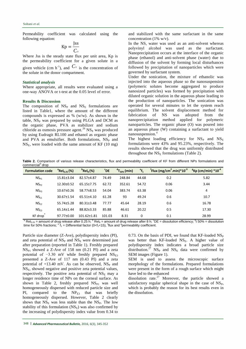
NSP and NSE data were confirmed by
SEM images (Figure 1).
SEM is used to assess the microscopic surface
morphology of the formulations. Prepared formulations
were present in the form of a rough surface which might
have led to the enhanced
dissolution rate.27
Moreover, the particle showed a
satisfactory regular spherical shape in the case of NSP,
which is probably the reason for its best results even in
the dissolution.
| 349
Nanosuspensions as potential ophthalmic delivery systems
Advanced Pharmaceutical Bulletin, 2016, 6(3), 345-352
Figure 1. SEM images of KF (A); NSE3 (KF:EU) 1:15 ratio (B); NSP1 (KF:PLGA) 1:5 ratio (C) at 1000× magnification.
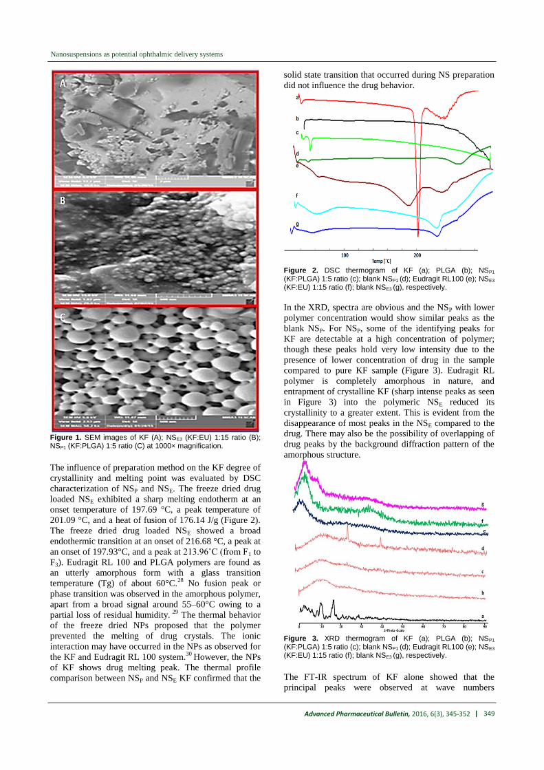
The influence of preparation method on the KF degree of
crystallinity and melting point was evaluated by DSC
characterization of NSP and NSE. The freeze dried drug
loaded NSE exhibited a sharp melting endotherm at an
onset temperature of 197.69 °C, a peak temperature of
201.09 °C, and a heat of fusion of 176.14 J/g (Figure 2).
The freeze dried drug loaded NSE showed a broad
endothermic transition at an onset of 216.68 °C, a peak at
an onset of 197.93°C, and a peak at 213.96˚C (from F1 to
F3). Eudragit RL 100 and PLGA polymers are found as
an utterly amorphous form with a glass transition
temperature (Tg) of about 60°C.28
No fusion peak or
phase transition was observed in the amorphous polymer,
apart from a broad signal around 55–60°C owing to a
partial loss of residual humidity. 29
The thermal behavior
of the freeze dried NPs proposed that the polymer
prevented the melting of drug crystals. The ionic
interaction may have occurred in the NPs as observed for
the KF and Eudragit RL 100 system.30
However, the NPs
of KF shows drug melting peak. The thermal profile
comparison between NSP and NSE KF confirmed that the
solid state transition that occurred during NS preparation
did not influence the drug behavior.
Figure 2. DSC thermogram of KF (a); PLGA (b); NSP1 (KF:PLGA) 1:5 ratio (c); blank NSP1 (d); Eudragit RL100 (e); NSE3 (KF:EU) 1:15 ratio (f); blank NSE3 (g), respectively.
In the XRD, spectra are obvious and the NSP with lower
polymer concentration would show similar peaks as the
blank NSP. For NSP, some of the identifying peaks for
KF are detectable at a high concentration of polymer;
though these peaks hold very low intensity due to the
presence of lower concentration of drug in the sample
compared to pure KF sample (Figure 3). Eudragit RL
polymer is completely amorphous in nature, and
entrapment of crystalline KF (sharp intense peaks as seen
in Figure 3) into the polymeric NSE reduced its
crystallinity to a greater extent. This is evident from the
disappearance of most peaks in the NSE compared to the
drug. There may also be the possibility of overlapping of
drug peaks by the background diffraction pattern of the
amorphous structure.
Figure 3. XRD thermogram of KF (a); PLGA (b); NSP1 (KF:PLGA) 1:5 ratio (c); blank NSP1 (d); Eudragit RL100 (e); NSE3 (KF:EU) 1:15 ratio (f); blank NSE3 (g), respectively.
The FT-IR spectrum of KF alone showed that the
principal peaks were observed at wave numbers

350 | Advanced Pharmaceutical Bulletin, 2016, 6(3), 345-352
Soltani et al.
stretching vibration N-H at 3424.64 cm−1, aromatic
stretching vibration C=C at 1649.70 cm−1, bending
vibration CH3 at 1476.99 cm-1, bending vibration
phenolic OH at 1397.14, and CH out of plane bending
vibrations in substituted ethylenic system (-C=CH- (cis)
at 754.15 cm-1
(Figure 4). The spectra obtained by FT-IR
for the PLGA are presented in Figure 4. The strong
bands in the region between 1760 and 1750 cm–1
could
be observed, in the spectra, due to the stretch of the
carbonyl groups within the PLGA. Moreover, stretching
bands are observed because of asymmetric and
symmetric C-C(=O)-O vibrations between 1300 and
1150 cm–1
. The presence of bands in these regions is of
benefit in the characterization of esters. The 3525 and
3459 cm–1
bands in the FT-IR spectra for lactide and
glycolide are ascribed to moisture in the sample (OH
group). The absorption bands between 3600 and 3400
cm–1
in the spectra presented in Figure 4, showing the
hydroxyl group, indicate that the PLGA copolymers are
hydrous. FT-IR studies showed characteristic peaks of
KF, confirming the purity of the drug. For NSP,
stretching vibration N-H is seen at 3400-3423, a stretch
of the carbonyl groups at 1760, asymmetric and
symmetric C-C(=O)-O vibrations at 1390 and bending
vibrations in substituted ethylenic system (-C=CH- (cis)
at 725-752 cm−1
.
For Eudragit RL 100, in the spectra, the strong bands are
observed in the region between 1150-1190 cm–1
and
1240-1270 cm–1
, due to the stretch of carbonyl (ester)
groups present in the Eudragit (Figure 4). There are also
stretching bands in view of the C (=O) ester vibration at
1734.01 cm–1
. The 1388.22, 1449.97, 2953, and 2992.11
cm–1
bands in the FT-IR spectra can be discerned to CHx
vibration. IR absorption frequency at 3437.91cm-1 (OH
stretch) presented in Figure 3 and showing the hydroxyl
group, indicates that the Eudragit RL100 is hydrous.29,31
FT-IR spectral studies showed that there was an
interaction between KF and polymers used. For NSE,
stretching strong band C-H (alkyne group) are seen at
3293.88-3298cm-1
, stretch band strong C-H (alkane
group) at 2936.61, 2937.50 and 2939.24 cm-1
, N-H
stretch band of the amine group at 3000 cm-1
, stretch
band of carbonyl group at 1728.54, 1728.79, and 1729.01
cm-1
, bending vibrations in -C-H at 1436.12, 1436.62,
and 1438.91 cm-1
, stretch band of ester group C-O at
1089.41, 1089.61, and 1090.74 cm-1
, and stretch band in
-C-Cl at 844.76, 845.12, and 845.57 cm−1
(Figure 4). The
available differences in the positions of the absorption
bands of KF were seen in spectra of the prepared
formulations, proving the presence of chemical
interactions in the solid state between the drug and the
polymers (PLGA and Eudragit RL100).
In vitro dissolution studies
In vitro dissolution data of all best formulations (NSP and
NSE) were compared together. The NSP formulation
showed that 82.57% of drug was released in 480 min
(NSP1). NaCl increased the solubility of drug by
entrapping the KF in the network interstitial spaces of
NaCl molecule and also reducing the particle size. In
NSs, increasing the osmotic pressure of W2 (external
phase of second emulsion) directs water migration from
W1 to W2 as well as a rapid shrinkage of the droplets.
This phenomenon results in smaller nanoparticles and
increases drug release.
In the NSE formulation 88.82% drug was released in 480
min (NSE3). Increasing the dissolution kinetics of KF
from NSE may be due to the conversion of the drug from
crystalline to amorphous state. Also presence of
surfactant (PVA) and co-surfactant (ethanol) in NSE
reduces the interfacial tension and helps to solubilize the
drug in the formulation of NSE. According to the
literature, the drug release amount and behavior as well
as the drug absorption are influenced by the particle size.
The particle size of NSE3 (117 nm) and NSP1 (158 nm)
are the smallest which may be the reason for their
highest releases (Figure 5).
Figure 4. FT-IR thermogram of KF (a); PLGA (b); NSP1 (KF:PLGA) 1:5 ratio (c); blank NSP1 (d); Eudragit RL100 (e); NSE3 (KF:EU) 1:15 ratio (f); blank NSE3 (g), respectively.
Figure 5. Cumulative percent release of KF from naoparticles with different polymer ratios and KF commercial drop.
KF delivery into and through the bovine cornea was
evaluated in ex vivo conditions, by the use of Franz
vertical diffusion cells. During this study, transcorneal

| 351
Nanosuspensions as potential ophthalmic delivery systems
Advanced Pharmaceutical Bulletin, 2016, 6(3), 345-352
delivery of prepared KF nanosuspensions was
compared with commercial (Zaditen®
) eye drop as
control. Comparison of data obtained from NSP and
NSE (Table 2) highlights a different KF delivery into
and through the bovine cornea. As expected, the NSE
showed a higher drug permeation and transcorneal
delivery than the NSP. However, differences of drug
permeability in two types of formulations (as NSP and
NSE) were statistically significant (p <0.05).
Comparison of data obtained from NSP and NSE
underlines the influence of different formulations on
the ex vivo drug availability; NSE is useful for
improving the transcorneal delivery. NSE is able to
favor KF permeation into the eye and at the same time
to prolong the contact time with the cornea and increase
the efficacy of drug delivery.
In NSs, solid drug is dissolved in the vehicle (lacrimal
fluid) and diffuses through the vehicle to the cornea. On
the other side, when a nanocarrier is applied onto the
eye, two consecutive physical events may limit corneal
absorption, namely, the drug release (from nanocarrier)
into lacrimal fluid and its penetration through the
corneal barrier. These two processes are intimately
intertwined, and both are due to the physicochemical
properties of drug (type of nanocarrier) and barrier.
The degree of partitioning of the drug into the cornea
relies on the relative affinity for the vehicle and for the
intercellular environment. In the present investigation,
the higher drug permeability may be due to the polymer
type (Table 2), surfactant, and the method of
preparation, which taken together act as penetration
enhancers.32,33
In addition, as shown for NSE, the small
particle size of the NSE (in comparison with NSp)
makes it an excellent carrier for promoting ex vivo
corneal KF permeation. Overall results show that NSE
is suitable nanoparticles for corneal delivery of KF.
NSs are almost exclusively formed from drug
nanoparticles with small amounts of biocompatible and
safe surfactants, such as PVA used in this work. This
leads to a highly fast dissolution process that favors
drug penetration into the cornea. Moreover, compared
to other colloidal carriers, NSs show extra advantages
such as simplicity, biodegradability of polymer
(PLGA), and scalable preparation methods.34,35
Conclusion On the whole, this work showed the high potential of
NSP and NSE in ocular drug delivery of KF. Indeed,
NSs has been established to be able to localize the drug
into the cornea ex vivo. Besides, the NSP was shown to
give comparable ocular KF delivery as the NSE, which
strongly enhanced in vitro ocular drug delivery.
Furthermore, the application of NSE in ocular KF
delivery showed the advantage of increasing
permeability and retention time of the drug in
comparison with the NSP. To conclude, results of this
work evinces that NSE formulation approach could be a
potentially valuable tool of use in the design of new KF
nanomedicines for the treatment of eye diseases.
Acknowledgments
The authors report that they have no conflicts of
interests. The financial support from the Drug Applied
Research Center of Tabriz University of Medical
Sciences under the grant No. 42 is greatly
acknowledged. Mitra Jelvehgari received the research
grant from Drug Applied Research Center, Tabriz
University of Medical Sciences, Tabriz, Iran.
Ethical Issues Not applicable.
Conflict of Interest
The authors declare that there is no conflict of interests
regarding the publication of this article.
References
1. Sahoo SK, Dilnawaz F, Krishnakumar S.
Nanotechnology in ocular drug delivery. Drug Discov
Today 2008;13(3-4):144-51. doi:
10.1016/j.drudis.2007.10.021
2. Patel A, Cholkar K, Agrahari V, Mitra AK. Ocular drug
delivery systems: An overview. World J Pharmacol
2013;2(2):47-64. doi: 10.5497/wjp.v2.i2.47
3. Ganta S, Paxton JW, Baguley BC, Garg S. Formulation
and pharmacokinetic evaluation of an asulacrine
nanocrystalline suspension for intravenous delivery.
Int J Pharm 2009;367(1-2):179-86. doi:
10.1016/j.ijpharm.2008.09.022
4. Adamczak M. Surfactants, polyelectrolytes and
nanoparticles as building blocks for nanocarriers.
Poland : AGH University of Science and Technology
in Krakow; 2013.
5. Jay KT. Nanosuspensions: Types of nanosuspension
methods and various applications. Available from:
http://www.biotecharticles.com/Nanotech/2011/May/N
anotechnology-Article/Nanosuspensions-Types-of-
Nanosuspension-Methods-and-Various-Applications-
893.htmlArticle. 2011.
6. Kumari A, Yadav SK, Yadav SC. Biodegradable
polymeric nanoparticles based drug delivery systems.
Colloids Surf B Biointerfaces 2010;75(1):1-18. doi:
10.1016/j.colsurfb.2009.09.001
7. Nagarwal RC, Kant S, Singh PN, Maiti P, Pandit JK.
Polymeric nanoparticulate system: A potential
approach for ocular drug delivery. J Control Release
2009;136(1):2-13. doi: 10.1016/j.jconrel.2008.12.018
8. Patel VR, Agrawal YK. Nanosuspension: An approach
to enhance solubility of drugs. J Adv Pharm Technol
Res 2011;2(2):81-7. doi: 10.4103/2231-4040.82950
9. Mudgil M, Gupta N, Nagpal M, Pawar P.
Nanotechnology: a new approach for ocular drug
delivery system. Int J Pharm Pharm Sci
2012;4(2):105-12.
10. Bangia JK, Om H. Nanoemulsions: a versatile drug
delivery tool. Int J Pharm Sci Res 2015;6(4):1363-72
doi: 10.13040/IJPSR.0975-8232.6(4).1363-72
11. Mygind T, Stiehler M, Baatrup A, Li H, Zou X,
Flyvbjerg A, et al. Mesenchymal stem cell ingrowth

352 | Advanced Pharmaceutical Bulletin, 2016, 6(3), 345-352
Soltani et al.
and differentiation on coralline hydroxyapatite
scaffolds. Biomaterials 2007;28(6):1036-47. doi:
10.1016/j.biomaterials.2006.10.003
12. James D. Pipkin, Rupert O. Zimmerer, John M.
Siebert, inventors; Cydex Pharmaceuticals, Inc.,
assignee. Nasal and ophthalmic delivery of aqueous
corticosteroid solutions. United States patent
US20090312724 A1.2009
13. Aksungur P, Demirbilek M, Denkbas EB, Vandervoort
J, Ludwig A, Unlu N. Development and
characterization of cyclosporine a loaded nanoparticles
for ocular drug delivery: Cellular toxicity, uptake, and
kinetic studies. J Control Release 2011;151(3):286-94.
doi: 10.1016/j.jconrel.2011.01.010
14. Mandal B, Alexander KS, Riga AT. Sulfacetamide
loaded eudragit(r) rl100 nanosuspension with potential
for ocular delivery. J Pharm Pharm Sci
2010;13(4):510-23.
15. Gupta H, Aqil M, Khar RK, Ali A, Bhatnagar A,
Mittal G. Sparfloxacin-loaded plga nanoparticles for
sustained ocular drug delivery. Nanomedicine
2010;6(2):324-33. doi: 10.1016/j.nano.2009.10.004
16.Oshlack B, Pedi Jr F, Chasin M., Inventors; Euro-
Celtique SA, assignee. Controlled release
formulations coated with aqueous dispersions of
acrylic polymers. United States patent US5580578 A.
1996
17. Mundargi RC, Babu VR, Rangaswamy V, Patel P,
Aminabhavi TM. Nano/micro technologies for
delivering macromolecular therapeutics using poly(d,l-
lactide-co-glycolide) and its derivatives. J Control
Release 2008;125(3):193-209. doi:
10.1016/j.jconrel.2007.09.013
18. Danhier F, Ansorena E, Silva JM, Coco R, Le Breton
A, Preat V. Plga-based nanoparticles: An overview of
biomedical applications. J Control Release
2012;161(2):505-22. doi:
10.1016/j.jconrel.2012.01.043
19. Al-Kassas R. Design and in vitro evaluation of
gentamicin-eudragit microspheres intended for intra-
ocular administration. J Microencapsul 2004;21(1):71-
81. doi: 10.1080/02652040310001619992
20. Pignatello JJ, Oliveros E, MacKay A. Advanced
oxidation processes for organic contaminant
destruction based on the Fenton reaction and related
chemistry. Crit Rev Environ Sci Tech 2006;36(1):1-84.
doi:10.1080/10643380500326564
21. Haznedar S, Dortunc B. Preparation and in vitro
evaluation of eudragit microspheres containing
acetazolamide. Int J Pharm 2004;269(1):131-40.
22. Pignatello R, Bucolo C, Ferrara P, et al. Eudragit
RS100® nanosuspensions for the ophthalmic
controlled delivery of ibuprofen. Eur J Pharm Sci
2002;16(1):53-61. doi:10.1016/S0928-0987(02)00057-X
23. Dillen K, Vandervoort J, Van den Mooter G, Ludwig
A. Evaluation of ciprofloxacin-loaded eudragit rs100
or rl100/plga nanoparticles. Int J Pharm
2006;314(1):72-82. doi:
10.1016/j.ijpharm.2006.01.041
24. Muthu MS, Rawat MK, Mishra A, Singh S. Plga
nanoparticle formulations of risperidone: Preparation
and neuropharmacological evaluation. Nanomedicine
2009;5(3):323-33. doi: 10.1016/j.nano.2008.12.003
25. Jantarat C. Application of Molecularly Imprinted
Polymer for Drug Delivery and Membrane
Separation of Chiral Drugs (PhD thesis). Southern
Thailand: Prince of Songkla University; 2009.
26.Meinel L, Illi OE, Zapf J, Malfanti M, Peter Merkle H,
Gander B. Stabilizing insulin-like growth factor-i in
poly(d,l-lactide-co-glycolide) microspheres. J Control
Release 2001;70(1-2):193-202.
27. Hu J, Johnston KP, Williams RO, 3rd. Spray freezing
into liquid (sfl) particle engineering technology to
enhance dissolution of poorly water soluble drugs:
Organic solvent versus organic/aqueous co-solvent
systems. Eur J Pharm Sci 2003;20(3):295-303.
28.Ignatious F, Sun L, inventors; Ignatious F, Sun L,
assignee. Electrospun amorphous pharmaceutical
compositions. United States patent US
20060013869A1. 2006.
29. Mandal B. Preparation and physicochemical
characterization of Eudragit® RL100
Nanosuspension with potential for Ocular Delivery of
Sulfacetamide (PhD thesis). Ohio: The University of
Toledo Digital Respository; 2010.
30. Swarnakar NK, Jain AK, Singh RP, Godugu C, Das
M, Jain S. Oral bioavailability, therapeutic efficacy
and reactive oxygen species scavenging properties of
coenzyme q10-loaded polymeric nanoparticles.
Biomaterials 2011;32(28):6860-74. doi:
10.1016/j.biomaterials.2011.05.079
31. Basu SK, Adhiyaman R. Preparation and
characterization of nitrendipine-loaded Eudragit RL
100 microspheres prepared by an emulsion-solvent
evaporation method. Trop J Pharm Res
2008;7(3):1033-41.
32. Kaur IP, Kanwar M. Ocular preparations: The
formulation approach. Drug Dev Ind Pharm
2002;28(5):473-93. doi: 10.1081/DDC-120003445
33. Ye T, Yuan K, Zhang W, Song S, Chen F, Yang X, et
al. Prodrugs incorporated into nanotechnology-based
drug delivery systems for possible improvement in
bioavailability of ocular drugs delivery. Asian J Pharm
Sci 2013;8(4):207-17. doi:10.1016/j.ajps.2013.09.002
34. Champion JA, Katare YK, Mitragotri S. Particle
shape: a new design parameter for micro-and
nanoscale drug delivery carriers. J Control Rel
2007;121(1):3-9. doi:10.1016/j.jconrel.2007.03.022
35. Reis CP, Neufeld RJ, Ribeiro AJ, et al.
Nanoencapsulation I. Methods for preparation of drug-
loaded polymeric nanoparticles. Nanomed: Nanotech,
Bio Med 2006;2(1):8-21.
doi:10.1016/j.nano.2005.12.003


