Comparison of self-report versus accelerometer – measured ...

www.wjpps.com Vol 7, Issue 10, 2018.
1
Hayder. World Journal of Pharmacy and Pharmaceutical Sciences
COMPARISON BETWEEN MESH VERSUS NON-MESH REPAIR OF
INGUINAL HERNIA
*Hayder Salim Shaker Al-Mindylawi
M.B.CH.B.M.S., AL-Kidney Collage of Medicine Baghdad University / Iraq.
ABSTRACT
Background: Hernias are abnormal protrusions of aviscus (part of it)
through a normal or abnormal opening in a cavity (usually the
abdomen). They are most commonly seen in the groin; a minority are
para-umbilical or incisional. In the groin, inguinal hernias are more
common than femoral hernias. Inguinal hernias occur in about 15% of
the adult population, and inguinal hernia repair is one of the most
commonly performed surgical procedures in the world. Approximately
800,000 mesh hernioplasties are performed each year in the United
States, 100,000 in France, and 80,000 in the United Kingdom. Aims of
Study: The present study was the first study that done at Baqubah
Teaching Hospital\Diyala\Iraq. The present study aims to comparing mesh with non-mesh
repair of inguinal hernias regarding:
1. Duration of surgery
2. Development of early post-operative complications such as wound infection, hematoma
and seroma formation.
3. Development of late post-operative complications such as persistent pain, testicular
atrophy and recurrence of inguinal hernia.
Materials and Methods: This is a prospective study for 200 patients underwent surgeries for
inguinal hernias at Baqubah Teaching Hospital. There were 192 (96%) male patients and 8
(4%) female patients. The surgeries were done over a period of 2 months (October–
November 2013) in the department of general surgery at Baqubah Teaching Hospital and
followed for 10 months. The patients are categorized into two groups: group 1 composed of
100 patients named the mesh group (the male patients were 96 and female patients were 4) as
they included (Lichtenstein mesh repair) for their hernias and group 2 which also composed
of 100 patients, named the non- mesh group (also the male patients were 96 and female
WORLD JOURNAL OF PHARMACY AND PHARMACEUTICAL SCIENCES
SJIF Impact Factor 7.421
Volume 7, Issue 10, 1-37 Research Article ISSN 2278 – 4357
Article Received on
23 July 2018,
Revised on 13 August 2018,
Accepted on 03 Sept. 2018
DOI: 10.20959/wjpps201810-12391
*Corresponding Author
Dr. Hayder Salim Shaker
Al-Mindylawi
M.B.CH.B.M.S., AL-Kidney
Collage of Medicine
Baghdad University / Iraq.

www.wjpps.com Vol 7, Issue 10, 2018.
2
Hayder. World Journal of Pharmacy and Pharmaceutical Sciences
patients were 4) in which their hernias are repaired using the (modified Bassini‘s repair) or
(Darn repair). The age range of the patients was (16-63 year) for group 1 and (16-70 years)
for group 2. The surgeries were randomly performed by senior surgeons. The two groups
were compared regarding development of early post-operative complications such as(wound
infection, hematoma and seroma formation) and development of late post-operative
complications such as (persistent pain, testicular atrophy and recurrence of inguinal hernia).
They are also compared regarding the duration of surgery. In the Lichtenstein mesh repair a
(7.5×15cm) piece of polypropylene mesh is commonly used for a Lichtenstein hernioplasty.
The non-mesh repair involve either (modified Bassini's repair) or (Darn repair). Results:
Regarding the development of early post-operative complications, the results were not
significantly different. Regarding the development of late post-operative complications
especially the persistent pain and recurrence of inguinal hernias, the results were significant.
There were more cases in the non-mesh group; the P-value was (0.030). Conclusion: The
mesh repair is superior to the non-mesh repair of inguinal hernias especially in terms of post-
operative persistent pain and recurrence of inguinal hernias; however, continuous clinical
trials have to be undertaken to find out the optimum surgical treatment of hernias.
KEYWORDS: Inguinal hernia, mesh hernioplasty, non-mesh repair of inguinal hernias.
INTRODUCTION
Hernias are abnormal protrusions of aviscus (part of it) through a normal or abnormal
opening in a cavity (usually the abdomen). They are most commonly seen in the groin; a
minority are Para umbilical or incisional (A.E.Kirk. et al, 1995).
In the groin, inguinal hernias are more common than femoral hernias. Inguinal hernias occur
in about 15% of the adult population, and inguinal hernia repair is one of the most commonly
performed surgical procedures in the world (Akyol C. et al, 2013).
Approximately 800,000 mesh hernioplasties are performed each year in the United States,
100,000 in France, and 80,000 in the United Kingdom (Albo D. et al, 2006).
The word "hernia" is derived from a Latin term meaning a "rupture". The earliest reports of
abdominal wall hernias date back to 1500 BC. During this early era, abdominal wall hernias
were treated with trusses or bandage dressings (Amid PK. et al, 1994).

www.wjpps.com Vol 7, Issue 10, 2018.
3
Hayder. World Journal of Pharmacy and Pharmaceutical Sciences
The first evidence of operative repair of a groin hernia dates to the first century AD. The
original hernia repairs involved wide operative exposures through scrotal incisions requiring
orchidectomy on the involved side. Centuries later, around 700AD, principles of operative
hernia repair evolved to emphasize mass ligation and en bloc excision of the hernia sac, cord,
and testis distal to the external ring (Awad SS. et al, 2002).
The modern surgical era began with Bassini, who in 1887 developed the first modern
anatomically based hernia treatment, this procedure spread worldwide, but was often
executed poorly, and hernia repair fell into a state of second hand surgery (Amid PK. et al,
2007).
The first true Cooper's ligament repair, which affixes the pectineal ligament to Poupart's
ligament and thereby repairs both inguinal and femoral hernia defects, was introduced by
Lotheissen in 1898(Butters M. et al, 2007) .
Darn repairs were first introduced in the early 20th century to reduce wound tension by using
either autologous tissue or synthetic suture to bridge the gap between fascial tissues. In 1918;
Handley introduced the first use of silk as a prosthetic darn and nylon followed several years
later. However, it was found that heavy prosthetic material increased the risk of wound
infection, and the silk suture ultimately lost its strength over time (Bisgaard T. et al, 2007).
The use of autologous or synthetic patches was also attempted in order to reduce wound
tension and improve rates of recurrence. The first patches, beginning in the early 20th
century, consisted of silver wire filigree sheets that were placed along the inguinal canal.
Over time, the sheets suffered from metal fatigue leading to hernia recurrence (Bay-Nielsen
M. et al, 2004).
In the late 1940, Canadian surgeon E.Shouldice developed a hernioplasty similar to the
Bassini's operation. This procedure became extremely popular as well as the standard of the
classic pure tissue hernioplasties; its only problem was that it was difficult to reproduce the
great results that Shouldice achieved with this repair (Butters M. et al, 2007).
In 1956 American surgeon Chester McVay and anatomist Barry Anson clarified the anatomy
of the groin and popularized the Cooper’s ligament Hernioplasty (Bringman S. et al, 2006).

www.wjpps.com Vol 7, Issue 10, 2018.
4
Hayder. World Journal of Pharmacy and Pharmaceutical Sciences
In 1984 Lichtenstein and colleagues published their results in repairing primary hernias using
a prosthetic mesh in a tension free manner, since then this repair has been accepted world
wide as the gold standard repair, this was based on the definite reduction in recurrence rates
as well as post-operative pain (Chastan P. et al, 2009).
Given the evidence that the use of mesh lowers the recurrence rate as well as the availability
of various prosthetic meshes for the reinforcement of the posterior wall of the inguinal canal,
most surgeons now prefer to perform tension-free mesh repair. Lichtenstein tension-free
hernioplasty is one of the most popular techniques used for inguinal hernia repair (Chung L.
et al, 2007).
AIMS OF STUDY
The hernia is a common problem and in Baqubah Teaching Hospital, the inguinal hernias
cases were common. So, there were a daily operative lists in the theatre including inguinal
hernias. The present study was the first study that done at Baqubah Teaching Hospital. I
would found that many surgeons in this hospital still use the non-mesh repair technique of
inguinal hernia repair.
The policy of many surgeons was the routine use of non- mesh repair of inguinal hernia
whereas the mesh repair technique done after taking the agreement of the patients before the
surgical operation. Therefore, the present study aims at comparing mesh with non-mesh
repair of inguinal hernias regarding:
1. Duration of surgery.
2. Development of early post-operative complications such as wound infection, hematoma
and seroma formation.
3. Development of late post-operative complications such as persistent pain, testicular
atrophy and recurrence of inguinal hernia.
Anatomic consideration
A useful learning tool for gaining a working knowledge of the inguinal region is to visualize
the region as it is surgically approached in the open technique of hernia repair (Chung RS. et
al, 1999).
The inguinal region is part of the anterolateral abdominal wall, which is made up of the
following nine layers, from superficial to deep: skin, Camper's fascia, Scarpa's fascia,

www.wjpps.com Vol 7, Issue 10, 2018.
5
Hayder. World Journal of Pharmacy and Pharmaceutical Sciences
external oblique aponeurosis, internal oblique muscle, transversus abdominis muscle,
transversalis fascia, preperitoneal fat and the peritoneum (DeBord JR. et al, -2007).
The first layers encountered in the inguinal region upon dissection through the subcutaneous
tissues are the Camper's and Scarpa's fasciae. Contained in this space are the superficial
branches of the femoral vessels, namely, the superficial circumflex and the epigastric and
external pudendal arteries, which can be safely ligated and divided when encountered
(Delikoukos S. et al, 2008).
The inguinal canal can be visualized as a tunnel traveling from lateral to medial in an oblique
fashion. It has a roof facing anteriorly, a floor facing posteriorly, a superior (cranial) wall,
and an inferior (caudal) wall. The canal contents (in men, cord structures; in women, the
round ligament) are the traffic that traverses the tunnel (Earle DB. et al, 2008).
Anatomy of the inguinal canal
The external oblique aponeurosis serves as the roof of the inguinal canal and opens just
lateral to and above the pubic tubercle. This is the external or superficial inguinal ring, which
allows the cord structures egress from the inguinal canal to the scrotum (Ferzli GS. et al,
2008).
The floor of the inguinal canal is composed of the transversus abdominis muscle and the
transversalis fascia. The entrance to the inguinal canal is through these layers, and this
entrance constitutes the internal or deep inguinal ring (Earle DB. et al, 2008).
The inferior wall of the inguinal canal is the inguinal (Poupart) ligament. This ligament is
formed by the lower edge of the external oblique aponeurosis and extends from the anterior
superior iliac spine to its attachments at the pubic tubercle and fans out to form the lacunar
(Gimbernat) ligament. The inguinal ligament folds over itself to form the shelving edge. This
folded-over sling of external oblique aponeurosis is the true lower wall of the inguinal canal
(Fingerhut A. et al, 2006).
The superior wall of the inguinal canal consists of a union of the internal oblique muscle and
transversus abdominis aponeurosis, which arches from its attachment at the lateral segment of
the inguinal ligament over the internal inguinal ring, ending medially at the rectus sheath and
coming together inferomedially to insert on the pubic tubercle, thus forming the conjoined
tendon (Fitzgibbons RJ Jr. et al, 2006).
www.wjpps.com Vol 7, Issue 10, 2018.
6
Hayder. World Journal of Pharmacy and Pharmaceutical Sciences
In males, the contents of the inguinal canal include the obliterated processus vaginalis
(which, when patent, forms the sac of the indirect inguinal hernia), the spermatic cord, and
the ilio-inguinal nerve (which comes out of the superficial inguinal ring along with the
spermatic cord).
In females, the inguinal canal contains the ilio-inguinal nerve and the round ligament of the
uterus (Earle DB. et al, 2008).
The coverings of the spermatic cord include the following: internal spermatic fascia, derived
from the transversalis fascia at the deep inguinal ring. Cremaster muscle, derived from the
internal oblique muscle at the deep inguinal ring. External spermatic fascia, derived from the
external oblique aponeurosis at the superficial inguinal ring (Fingerhut A. et al, 2006).
The contents of the spermatic cord include the following: vas deferens, testicular artery, and
artery of the ductus deferens, Cremasteric artery, pampiniform plexus, and genital branch of
the genito-femoral nerve, para-sympathetic and sympathetic nerves and lymphatic vessels
(Fitzgibbons RJ. et al, 2005).
The key nerves in the inguinal region are as follows: iliohypo- gastric nerve, ilio-inguinal
nerve and genital branch of the genito-femoral nerve (Franklin ME Jr. et al, 2004).
Anatomy of nerves of groin
The ilio-inguinal nerve runs medially through the inguinal canal along with the cord
structures traveling from the internal ring to the external ring. It innervates the upper and
medial parts of the thigh, the anterior scrotum, and the base of the penis (Franklin ME Jr. et
al, 2002).
The iliohypogastric nerve runs below the external oblique aponeurosis but cranial to the
spermatic cord, then perforates the external oblique aponeurosis cranial to the superficial
ring. It innervates the skin above the pubis (Franneby U. et al, 2006).
The genital branch of the genito-femoral nerve lies within the spermatic cord and travels with
the Cremasteric vessels through the inguinal canal. It innervates the cremaster muscle and
provides sensory innervation to the scrotum.Some variations in the anatomic distribution of
these nerves may be observed, for instance, the occasional absence of an ilioinguinal nerve
(Gianetta E. et al, 2000).
www.wjpps.com Vol 7, Issue 10, 2018.
7
Hayder. World Journal of Pharmacy and Pharmaceutical Sciences
The Hesselbach triangle is bounded by the inguinal ligament below, the lateral border of the
rectus abdominis medially, and the inferior epigastric vessels laterally. The sac of a direct
hernia lies in this triangle, whereas the neck of an indirect hernia sac lies outside the triangle
(lateral to the inferior epigastric vessels) (Grosz CR. et al, 2000).
Types of inguinal hernias
An indirect hernia is defined as a hernia protruding through the internal or deep inguinal ring,
whereas a direct hernia is a hernia protruding through the posterior wall of the inguinal canal.
To put it in a more anatomic way, an indirect hernia is lateral to the inferior epigastric artery
and vein, whereas a direct hernia is medial to these vessels (Franneby U. et al, 2006).
The Hesselbach triangle is the zone of the inguinal floor through which direct hernias
protrude, and its boundaries are the epigastric vessels laterally, the rectus sheath medially,
and the inguinal ligament inferiorly (Gianetta E. et al, 2000).
An incomplete hernia is confined to the inguinal canal, while a complete hernia comes out of
the inguinal canal through the external or superficial ring into the scrotum. Direct hernias are
always incomplete, while indirect hernias can be complete (Haapaniemi S. et al, 2004).
A sliding inguinal hernia is one in which a portion of the wall of the hernia sac is made up of
an intra-abdominal organ. As the peritoneum is stretched and pushed through the hernia
defect and becomes the hernia sac, retroperitoneal structures such as the colon or bladder are
dragged along with it and thus come to make up one of its walls (Hair A. et al, 2000).
Bilateral pediatric hernias are most commonly indirect hernias and arise because of the
patency of the processus vaginalis. Simple ligation of the hernia sac (herniotomy) alone is
enough (Haapaniemi S. et al, 2004).
Surgical treatment of indirect hernias in adults, unlike that in children, requires more than
simple ligation of the hernia sac. This is because the patent processus is only part of the story.
With time, the internal ring dilates, leaving an adult with what can be a sizable defect in the
floor of the inguinal canal; this must be closed in addition to division or reduction of the
indirect hernia sac (Hair A. et al, 2000).

www.wjpps.com Vol 7, Issue 10, 2018.
8
Hayder. World Journal of Pharmacy and Pharmaceutical Sciences
Types of inguinal hernia repair
Inguinal hernia repairs may be divided into the following 3 general types: The first type is
Herniotomy (removal of the hernial sac only): This is adequate for an indirect inguinal hernia
in children in whom the abdominal wall muscles are normal; formal repair of the posterior
wall of the inguinal canal is not required (Hakeem A. et al, 2011).
The second type is Herniorrhaphy (herniotomy plus repair of the posterior wall of the
inguinal canal): This may be suitable for a small hernia in a young adult with good abdominal
wall musculature; the Bassini's and Shouldice repairs are examples of herniorrhaphy (Hosgor
M. et al, 2004).
The third type is Hernioplasty (herniotomy plus reinforcement of the posterior wall of the
inguinal canal with a synthetic mesh): This is required for large hernias and hernias in middle
aged and elderly patients with poor abdominal wall musculature; the Lichtenstein tension-
free mesh repair is an example of hernioplasty (Hair A. et al, 2000).
Indications of inguinal hernia repair
Classically, the existence of an inguinal hernia, in itself, has been considered a reason enough
for operative intervention. However, some studies have shown that the presence of a
reducible hernia is not, in itself, an indication for surgery and that the risk of incarceration is
less than 1% (Junge K. et al, 2001).
Symptomatic patients (with pain or discomfort) should undergo repair; however, as many as
one third of patients with inguinal hernias are asymptomatic. The question of observation
versus surgical intervention in this asymptomatic or minimally symptomatic population was
addressed in two randomized clinical trials. The two trials yielded similar results: After long-
term follow-up, no significant difference in hernia-related symptoms was noted, and watchful
waiting did not increase the complication rate (Kark AE. et al, 1995).
In one study, the substantial patient crossover from the observation group to the surgery arm
led the authors to conclude that observation may delay but not prevent surgery. This
reasoning holds particularly true in the younger patient population. Thus, even an
asymptomatic patient, if medically fit, should be offered surgical repair (Kristin Masukawa.
et al, 2010).

www.wjpps.com Vol 7, Issue 10, 2018.
9
Hayder. World Journal of Pharmacy and Pharmaceutical Sciences
A long-term follow-up study determined that most patients with a painless inguinal hernia
will develop symptoms over time and concluded that surgery is recommended for medically
fit patients (Junge K. et al, 2001).
Koch et al found that recurrence rates were higher in women and that recurrence was 10
times more likely to be of the femoral variety in women than it was in men. Such findings
have led some to the conclusion that procedures providing coverage of the femoral space (e.g.
laparoscopic repair) at the time of initial operation are better suited for women as primary
repairs (Kark AE. et al, 1995).
Contraindications of inguinal hernia repair
Inguinal hernia repair has no absolute contraindications. Just as in any other elective surgical
procedure, the patient must be medically optimized (Koch A. et al, 2005).
Any medical issues (e.g. Upper respiratory tract or skin infection, poorly controlled diabetes
mellitus, chronic constipation, urinary obstruction, persisting cough, obstruction or
strangulation, or allergy to local anesthesia or prosthetic devices) should be fully addressed
and the operation delayed accordingly (Liem M.S. et al, 2003).
Patients with elevated American Society of Anesthesiologists (ASA) scores and high
operative risk should undergo a full preoperative workup and determination of the risk to
benefit ratio (Kark AE.et al, 1995).
Use of mesh for inguinal hernia repair
Emphasizing the Halstead principle of no tension, the Lichtenstein group advocated routine
use of mesh in 1984. The prosthesis used to reinforce the weakened posterior wall of the
inguinal canal is placed between the transversalis fascia and the external oblique aponeurosis
and extends well beyond the Hesselbach triangle (Lichtenstein IL. et al, 1988).
Mesh implants do not actively shrink, but they are passively compressed by the natural
process of wound healing. Mesh shrinkage occurs only to the extent to which the tissue
contracts. A mesh with a small pore size is likely to shrink more (McCormack K. et al, 2003).
Shrinkage of the different types of mesh in vivo is in the range of (20-40%), thus, it is
important for the surgeon to ensure that the mesh adequately overlaps the defect on all sides.
It is advisable to use a large (e.g. 7.5×15cm) sheet of mesh extending approximately (2cm)

www.wjpps.com Vol 7, Issue 10, 2018.
10
Hayder. World Journal of Pharmacy and Pharmaceutical Sciences
medial to the pubic tubercle,(3-4cm)above the Hesselbach triangle, and (5-6cm) lateral to the
internal ring so as to allow for mesh shrinkage (Lichtenstein IL. et al, 1988).
Although the use of traditional microporous or heavyweight polypropylene meshes over the
past 2 decades has reduced the recurrence rate after hernia surgery to less than 1%, a major
concern has been the formation of a rigid scar plate that causes patient discomfort and
chronic pain, impairing quality of life (Kark AE. et al, 1995).
More than 50% of patients with large mesh prosthesis in the abdominal wall complain of
paresthesia, palpable stiff edges of the mesh, or physical restriction of abdominal wall
mobility (Lichtenstein IL. et al, 1990).
It was assumed that the flexibility of the abdominal wall is restricted by implantation of
excessive foreign material and by excessive scar tissue formation (McCormack K. et al,
2003).
A better knowledge of the biomechanics of the abdominal wall and the influence of mesh on
those mechanics has led to the current understanding that “less is more". In other words, a
less dense, lighter-weight mesh with larger pores, though still stronger than the abdominal
wall and thus usable for the purposes of repair, will result in less inflammation, better
incorporation, better abdominal wall compliance, greater abdominal wall flexibility, less pain,
and possibly less scar contraction, therefore, its use will lead to a better clinical outcome
(Liem MS. et al, 1997).
Lightweight composite mesh was developed in the conviction that the ideal mesh should be
just strong enough to handle the pressure of the abdominal wall while remaining as low in
mass and as thin as possible.
The advantage of increasing the mesh pore size is that it makes it easier for tissue to grow
through the pores and thereby create a thinner, better-integrated scar (Malangoni MA. et al,
2007).
The newer lightweight composite meshes offer a combination of thinner filament size, larger
pore size, reduced mass, and increased percentage of absorbable material. Thus, less foreign
material is implanted, the scar tissue has greater flexibility (with almost physiologic

www.wjpps.com Vol 7, Issue 10, 2018.
11
Hayder. World Journal of Pharmacy and Pharmaceutical Sciences
abdominal wall mobility), there are fewer patient complaints, and the patient’s quality of life
is better (Liem MS. et al, 1997).
The use of lightweight mesh for Lichtenstein hernia repair has not been shown to affect
recurrence rates, but it has been found to improve some aspects of pain and discomfort 3
years after surgery. According to data from randomized, controlled trials and retrospective
studies, light meshes seem to have some advantages with respect to post-operative pain and
foreign body sensation (Milic DJ. et al, 2003).
Many manufacturers have now shifted toward lighter, more porous constructions that
maintain the strength of the repair but putatively reduce the inflammatory response. These
meshes may decrease long-term discomfort, but possibly at the cost of increased recurrence
rates (e.g. from inadequate fixation or overlap) (Nordin P. et al, 2002).
The question of absorbable versus permanent sutures to secure the mesh is based on surgeon
preference; to date, there has been no evidence conclusively favoring one type over the other.
Sutures made of polyglactin (Vicryl) or polypropylene are commonly used, with undyed
polyglactin often preferred for subcutaneous tissue. Theoretical advantage of absorbable
suture is that if nerve impingement is inadvertently caused, the suture material disappears
with time (Milic DJ. et al, 2003).
Intra-operative planning
For better hemostasis, sharp dissection is preferred to blunt dissection. This is one operation
in which every red blood cell must be caught. If a lipoma is present in the spermatic cord, it
should be excised to reduce the bulk of the cord (Nordin P. et al, 2002).
Some surgeons excise the cremaster muscle fibers in the cord; others prefer not to. With a
direct hernia, the sac is not dissected and opened, as is done with an indirect inguinal hernia.
Rather, it is inverted (pushed back) into the extraperitoneal space, sometimes with plication
of the transversalis fascia (Malangoni MA. et al, 2007).
Bilateral hernias can be repaired in a single procedure, especially with a Lichtenstein tension-
free mesh hernioplasty. Some surgeons, however, prefer to repair only one hernia at a time,
deferring repair of the other for about (4-6 weeks); this avoids the risk of bilateral infection
and the higher risk of penile and scrotal edema after bilateral inguinal hernia repair (Sanabria
A. et al, 2007).

www.wjpps.com Vol 7, Issue 10, 2018.
12
Hayder. World Journal of Pharmacy and Pharmaceutical Sciences
If the inguinal hernia is irreducible or obstructed, the sac should be opened first at its fundus,
before it is dissected up to its neck, to allow evacuation of toxic fluid and inspection of the
bowel for ischemia. If the conventional technique, in which the sac is first completely
dissected up to its neck, is followed, the ischemic bowel may slip back into the peritoneal
cavity before the sac is opened at its fundus and may then be difficult to retrieve for
inspection (Taylor EW. et al, 2004).
Ischemic bowel is blue-black and thick-walled, lacks luster, feels firm to the touch, and has
no peristalsis. The bowel must be wrapped in moist warm packs, and 100% oxygen should be
delivered for a few minutes. The bowel is then reassessed for viability. Any non-viable bowel
will have to be resected (Akyol C. et al, 2013).
Equipments
Standard operating room anesthesia equipment, outfitted for possible conversion to general
anesthesia and endotracheal intubation, is required. Inguinal hernia repair can be achieved
under local infiltration or field block and regional block spinal or epidural anesthesia (Terzi
C. et al, 2006).
Instruments and materials on hand may include the following: syringe, 25-Gauge needle,
surgical knife with blade, Mosquito forceps, dissecting scissors, polypropylene (Prolene) or
polyester mesh, Langenbeck retractors, Adson thumb forceps, needle holder, sutures
(absorbable or non- absorbable), Penrose drain or umbilical tape and non-crushing intestinal
clamps (in case bowel resection is required, in a strangulated hernia) (Bay-Nielsen M, et al,
2004).
The umbilical tape or Penrose drain may be used to retract the mobilized spermatic cord, but
a hernia ring forceps can also be used. If the neck of the hernia sac is particularly tight, the
use of a grooved probe or dissector may help minimize injury to the contents (Albo D. et al,
2006).
Monitoring and Follow-up
With the routine use of mesh for hernia surgery, the recurrence rate has fallen to less than
1%. Although some recurrences occur early, cases may be reported many years later.
Thorough clinical evaluation, a high degree of suspicion, and appropriate follow-up are
advised for keeping track of recurrences. A follow-up visit is routinely scheduled for 1 week

www.wjpps.com Vol 7, Issue 10, 2018.
13
Hayder. World Journal of Pharmacy and Pharmaceutical Sciences
after the procedure. Thereafter, follow-up is scheduled on an as-needed basis (Amid PK. et al,
2007).
Although the post-operative course is generally uncomplicated, patients must be routinely
instructed to recognize certain signs and symptoms that can alert them to potential
complications. Patients with chronic groin pain, post-operative neuralgia, paresthesia,
neurapraxia, or hypoesthesia for more than six months after surgery should be referred for
further evaluation, surgical exploration, and, if required, excision of the involved nerve (Albo
D. et al, 2006).
A multidisciplinary approach at a pain clinic is an option for the treatment of chronic post-
herniorrhaphy pain. Surgical means of treating specific causes of such pain include the
following: resection of entrapped nerves, mesh removal (in mesh-related pain), removal of
fixating sutures and burying the nerve endings in the internal oblique muscle (Franklin ME
Jr. et al, 2002).
Large-scale studies examining the convalescence period after elective inguinal hernia repair
convincingly demonstrated that the median length of absence from work was seven days
when patients were advised by their surgeons to limit the recuperation period and to resume
normal activities within one day after the procedure. Moreover, these studies confirmed that
the risk of recurrence was not increased by early resumption of activities. Thus, with
adequate analgesia, patients can safely return to their daily duties (Bringman S. et al, 2006).
Approach considerations
Open inguinal hernia repairs other than Lichtenstein hernioplasty are not merely of historical
interest. Surgeons must know and understand these repairs so that they can be carried out
when they are appropriate. Specifically, cases that involve a contaminated field (eg, necrotic
or perforated bowel secondary to hernia strangulation) are not amenable to prosthetic repair.
In such cases, either a primary tissue repair or a biologic implant repair is necessary (Albo D.
et al, 2006).
Lichtenstein tension-free mesh repair
Incision
The incision is placed about 1 cm above and parallel to the inguinal ligament, beginning from
the pubic tubercle and extending (5-6cm) laterally up to the mid-inguinal point. The

www.wjpps.com Vol 7, Issue 10, 2018.
14
Hayder. World Journal of Pharmacy and Pharmaceutical Sciences
subcutaneous fat is then opened along the length of the incision, and careful hemostasis is
achieved by ligating superficial pudendal and superficial epigastric vessels (Hakeem A. et al,
2011).
The Scarpa's fascia is similarly opened along the length of the incision, down to the external
oblique aponeurosis, and the external inguinal ring and the lower border of the inguinal
ligament are visualized. Although the risk is very low, routine exploration of the femoral
canal is advised in the absence of an inguinal hernia and in women. The external oblique
aponeurosis is then opened along the line of incision, starting from the external ring and
extending laterally for up to (5cm). The ilio-inguinal nerve, lying underneath the aponeurosis,
is safeguarded during this procedure (Chastan P. et al, 2009).
The superior and inferior flaps of the external oblique aponeurosis are gently freed from the
underlying contents of the inguinal canal and overturned and separated to expose the
cremaster with the cord structures, the ilioinguinal and iliohypogastric nerves, the uppermost
aponeurotic portion of the internal oblique muscle and conjoined tendon, and the free lower
border of the inguinal ligament. Wide separation of the two flaps provides ample space for
placement and fixation of mesh under vision while protecting the nerves (Hakeem A. et al,
2011).
Dissection of the spermatic cord
The spermatic cord, along with the cremaster, is then lifted up and separated from the pubic
bone for about (2cm) beyond the pubic tubercle to create space for extending the mesh well
beyond the pubic tubercle. When lifting the cord, the surgeon must be sure to include the ilio-
inguinal nerve, the genito-femoral nerve, and the spermatic vessels along with it. All of these
structures may then be encircled in a tape for ease of handling (Liem MS. et al, 1997).
The anatomic plane between the cremaster and the aponeurotic tissue attached to the pubic
bone is avascular, and cord structures encircled in the tape can be separated from the floor of
the inguinal canal up to the internal ring (Chastan P. et al, 2009).
A visible landmark for safeguarding the genito-femoral nerve is the external spermatic vein,
usually referred to as the “blue line". If the blue line is kept with the spermatic cord, the
surgeon can be sure that the genital branch of the genito-femoral nerve, which is always
adjacent to this vein, is well protected (Nathan JD. et al, 2003).

www.wjpps.com Vol 7, Issue 10, 2018.
15
Hayder. World Journal of Pharmacy and Pharmaceutical Sciences
Identification and management of the hernial sac
The cord structures and all of the nerves of the inguinal canal having been visualized, the
next step is to identify and isolate the hernia sac. The patient is asked to cough (if the
procedure undertaken under local or regional anesthesia), and the groin region is examined
for the presence of an indirect hernia, a direct hernia, a femoral hernia, a combined hernia, or
a spigelian hernia (McCormack K. et al, 2003).
A hernia sac can be managed by means of inversion, division, resection, or ligation.
Resection and ligation of a small hernia sac should not be performed unnecessarily, because
it causes post-operative pain. However, the hernia sac must be well separated from the
internal ring before it is invaginated (Chastan P. et al, 2009).
The risk of recurrence is not increased when a small or medium-sized indirect hernia sac is
not ligated. Excision of an indirect inguinal hernia sac is associated with a lower risk of
hernia recurrence than is division or invagination (Hair A. et al, 2000).
When the hernia sac is excised or divided, the proximal sac should never be left open; doing
so may lead to recurrence (McCormack K. et al, 2003).
The proximal sac is dissected free of cord structures well above the internal ring, and a high
ligation of the neck of the sac should be performed. The indirect hernia sac lies anterolateral
to the cord structures and is visualized by dividing the cremaster muscle longitudinally. The
cremaster muscle should not be divided transversely or excised, because doing so may result
in low-lying testes and dysejaculation (Hakeem A. et al, 2011).
The neck of a large hernia sac is transected at the midpoint of the inguinal canal, and the
proximal part is suture ligated. A high ligation of the proximal sac is recommended, and the
stump is reduced deep underneath the internal ring. The distal sac is left in place; however,
the anterior wall of the distal sac is incised to prevent post-operative hydrocele formation
(Nathan JD. et al, 2003).
A direct inguinal hernia lies posteromedial to the cord structures. The direct hernia sac is
isolated and dissected free. Its contents are reduced, and the peritoneal sac is inverted and
maintained in position with a purse-string suture. If a femoral hernia is suspected, the femoral
ring should be evaluated by incising the medial part of the iliopubic tract. If a sac is seen
entering the femoral ring, it is reduced and dealt with by inverting or ligating the neck of the

www.wjpps.com Vol 7, Issue 10, 2018.
16
Hayder. World Journal of Pharmacy and Pharmaceutical Sciences
sac. Sliding hernia is simply dissected free and inverted in the preperitoneal space (Vale L.et
al, 2004).
Placement and fixation of the mesh
A (7.5×15cm) piece of polypropylene mesh is commonly used for a Lichtenstein
hernioplasty. On the medial side, the sharp corners of the mesh are trimmed to conform to the
patient’s anatomy. For a femoral hernia, the mesh is tailored so that it has a triangular
extension from its lower edge on its medial side (Milic DJ. et al, 2003).
To compensate for future shrinkage, the mesh should be wide enough to extend (3-4cm)
beyond the boundary of the inguinal triangle. To compensate for increased intra-abdominal
pressure when the patient stands up, the mesh should be placed lax in the posterior wall of the
inguinal canal in such a way that it acquires a domelike wrinkle. The first medial most stitch
fixes the mesh (2cm) medial to the pubic tubercle, where the anterior rectus sheath inserts
into the pubic bone (Nathan JD. et al, 2003).
Care should be taken not to pass the needle through the periosteum of the bone or through the
pubic tubercle; this is one of the most common causes of chronic postoperative pain. The
same suture is then used as a continuous suture to fix the lower edge of the mesh to the free
lower border of inguinal ligament up to a point just lateral to the internal ring. Next, a slit is
made in the lateral end of the mesh to create a narrower lower tail (the lower one third) and a
wider upper tail (the upper two thirds). The slit extends up to a point just medial to the
internal inguinal ring. Lower edge of mesh sutured to inguinal ligament up to internal
inguinal ring (Milic DJ. et al, 2003).
To accommodate cord structures, lateral end of mesh is divided into wider upper (two thirds)
tail and narrower lower (one third) tail. The upper tail is then passed underneath the cord in
such a way as to position the mesh posterior to the cord in the inguinal canal, and the
spermatic cord is placed between the two tails of the mesh. The upper tail is then crossed over
the lower one, and the two tails are held in an artery forceps. With the mesh kept lax, its
upper edge is then fixed to the rectus sheath and the internal oblique aponeurosis with 2 or 3
interrupted non-absorbable sutures (Nilsson H. et al, 2007).
On occasion, the iliohypogastric nerve is found to be in the way of upper edge of the mesh. In
such cases, the mesh may be split to accommodate the nerve. The two tails are then tucked

www.wjpps.com Vol 7, Issue 10, 2018.
17
Hayder. World Journal of Pharmacy and Pharmaceutical Sciences
together and fixed to the inguinal ligament just lateral to the internal ring, thus creating a new
internal ring made of mesh. The tails are trimmed (5cm) beyond the internal ring and placed
underneath the external oblique aponeurosis (Nordin P. et al, 2003).
Suturing the mesh beyond the internal ring is unnecessary; doing so may cause injury to the
femoral nerve. Similarly, fixation of the tails of the mesh to the internal oblique muscle,
lateral to the internal ring, may cause entrapment of the ilio-inguinal nerve. Trying to suture
the two tails without crossing them or trimming the tails shorter than (5-6cm) beyond the
internal ring may result in recurrence at the deep inguinal ring (Nathan JD. et al, 2003).
If any of the inguinal nerves is injured or of doubtful integrity, it can be resected and its
proximal end ligated and buried within the fibers of the internal oblique muscle to keep the
stump of the nerve away from scarring. In male patients, the testes should always be gently
pulled back down to their normal scrotal position after fixation of the mesh (Nilsson H. et al,
2007).
Closure
Hemostasis is ensured in the inguinal canal, which is then closed by suturing the two flaps of
the external oblique aponeurosis, with care taken not to injure the underlying ilioinguinal
nerve (Nordin P. et al, 2003).
Suturing is started laterally and continued medially, where an adequate opening is left at the
newly created superficial inguinal ring so as not to occlude the emerging spermatic cord.
Subcutaneous tissue is approximated with interrupted sutures to obliterate any dead space,
and the skin is approximated with sutures, clips, or adhesive strips (Norrie J. et al, 2010).
A subcuticular continuous stitch with (3-0) absorbable sutures obviates any need for stitch or
clip removal and provides better cosmetic results. The operative site is cleaned and a sterile
dressing applied. Local infiltration of a long-acting anesthetic agent (e.g. bupivacaine or
ropivacaine) into the subcutaneous tissue around the incision provides good immediate
postoperative pain relief (O'Dwyer PJ. et al, 2005).
Plug-and-patch repair
The plug-and-patch repair adds a polypropylene plug shaped as a cone, which can be
deployed into the internal ring after reduction of an indirect sac. The plug then acts as a
toggle bolt to reinforce the defect (Milic DJ. et al, 2003).
www.wjpps.com Vol 7, Issue 10, 2018.
18
Hayder. World Journal of Pharmacy and Pharmaceutical Sciences
McVay repair
In the McVay repair the conjoined tendon is sutured to the inguinal ligament with interrupted
non-absorbable sutures (Nordin P. et al, 2002).
Bassini's repair
The Bassini's technique for inguinal hernia repair involves suturing the transversalis fascia
and the conjoined (transversus abdominis and internal oblique) tendon to the inguinal
ligament behind the spermatic cord with monofilament non-absorbable suture. It also
involves the so-called Tanner slide, which is a vertical relaxing incision in the anterior rectus
sheath intended to prevent tension (O'Dwyer PJ. et al, 2006).
Shouldice repair
The Shouldice technique is a 4-layer inguinal hernia repair performed with the patient under
local anesthesia. The transversalis fascia is incised from the internal ring laterally to the pubic
tubercle medially, and upper and lower flaps are created. These flaps are then overlapped
(double-breasted) with two layers of sutures. The conjoined tendon is then sutured to the
inguinal ligament, again in two overlapping layers. This reinforces the posterior wall and
narrows the deep inguinal ring. The Shouldice repair is classically done with a continuous
suture of (32-34 gauge) stainless steel wire, but synthetic monofilaments (e.g. poly-
propylene) can also be used. The external oblique aponeurosis is then closed in a double-
breasted fashion in front of the spermatic cord (Parviz K. et al, 2003).
Darn repair
A pure-tissue tensionless technique that is performed by placing a continuous suture in zigzag
way between the conjoined tendon and the inguinal ligament without approximating the two
structures (Paajanen H. et al, 2010).
Post-operative Care
Early mobilization is the key to rapid convalescence. Patients can safely ambulate on the
evening of the operation. If general or regional anesthesia is used, the patient may be
hospitalized for a few days. There is some pain in the post-operative period, and suitable
analgesics should be prescribed. The dressing is removed on post-operative day 5, and
stitches are removed on post-operative day 7 (Scott NW. et al, 2002).

www.wjpps.com Vol 7, Issue 10, 2018.
19
Hayder. World Journal of Pharmacy and Pharmaceutical Sciences
Patients should be advised to avoid strenuous activities for a few weeks. Typically, light work
can be resumed after one week, heavier jobs after six weeks. Male patients should be
monitored for testicular atrophy, which may occur as a result of venous or arterial injury or
obstruction in the spermatic cord (Parviz K. et al, 2003).
All patients should be monitored for the development of nerve pain from nerve entrapment in
suture material. Finally, patients should be monitored for recurrence, which may arise as a
consequence of inadequate repair, wound infection and chronic straining (e.g. From
coughing, constipation, or urination) (Sanders DL. et al, 2013).
Complications of inguinal hernia
Intra-operative complications
1. Vascular injuries
Superficial epigastric vessels in the incision may bleed. These vessels not only should be
identified when the incision is being made but also should be ligated and divided. Inferior
epigastric vessels may be injured during dissection of the spermatic cord in the inguinal
canal, dissection of an indirect inguinal hernia sac within the spermatic cord, plication of the
transversalis fascia, or transfixion of the hernial sac. These vessels should be identified at an
early stage and protected (Shamberger RC.et al, 1984).
External iliac or femoral vessels, especially veins, may be injured during fixation of the mesh
to the inguinal ligament in its lateral part. The tissue bites in the inguinal ligament should not
be very deep. Although less common than other intraoperative complications, vascular
injuries are potentially disastrous. They can be avoided by respecting the proximity of the
femoral vessels, particularly when suturing the mesh to the inguinal ligament (Shankar VG.
et al, 2010).
Hematoma formation can result from injury of the inferior epigastric vessels or pampiniform
plexus veins or from failure to ligate the superficial subcutaneous veins (Sanabria A. et al,
2007).
2. Injuries to abdomino-pelvic structures
Cord structures (e.g. testicular artery, pampiniform plexus of veins, and vas deferens) may be
injured during opening of the coverings of the spermatic cord or dissection of the indirect

www.wjpps.com Vol 7, Issue 10, 2018.
20
Hayder. World Journal of Pharmacy and Pharmaceutical Sciences
hernial sac within the spermatic cord. In particular, the surgeon should always be aware of
the vas deferens and should protect it from injury (Scott NW. et al, 2002).
Injury to the urinary bladder may occur during plication of the transversalis fascia. In
addition, injury to the urinary bladder, cecum, or sigmoid colon may occur during transfixion
of the hernial sac in a sliding indirect inguinal hernia (where these viscera are not contained
in the hernial sac but form a part of the wall of the sac) (Sanchez-Manuel FJ. et al, 2007).
A sliding hernia should be recognized early; if it is present, the entire hernial sac should not
be excised. Injury to the bowel may occur during transfixion of the neck of an indirect hernial
sac. The head end of the operating table can be lowered to ensure complete reduction of
contents of the sac, the sac can be twisted to push the contents into the peritoneal cavity, and
a tissue bite can be taken and the suture tied under vision (Simons MP. et al, 2009).
3. Nerve injuries
The ilio-hypogastric nerve, because it lies on the conjoined tendon outside the inguinal canal,
may be injured during dissection of the upper flap of the external oblique aponeurosis or
fixation of the mesh to the conjoined tendon. It may also become trapped in sutures during
closure of the external oblique aponeurosis (Starling JR.et al, 1989).
The ilioinguinal nerve, because it lies in the inguinal canal along with the spermatic cord,
may be injured during dissection of the cord. The genital branch of the genito-femoral nerve,
because it lies within the spermatic cord, may be injured during dissection of the hernia sac
(Milic DJ. et al, 2003).
Post-operative complications
1. Wound infection
Deep and persistent infection may necessitate removal of the mesh. Wound infection can also
weaken the repair and may be responsible for recurrence of the inguinal hernia (Stylianidis G.
et al, 2010).
2. Pain
Post-operative chronic pain is more frequent than was previously understood and has become
one of the most important primary endpoints in hernia surgery. In published reports, the
incidence of postherniorrhaphy pain has ranged from 0% to more than 30% (Taylor EW. et
al, 2004).
www.wjpps.com Vol 7, Issue 10, 2018.
21
Hayder. World Journal of Pharmacy and Pharmaceutical Sciences
Chronic inguinodynia is defined as pain persisting more than 3 months post herniorrhaphy,
after the process of wound healing is complete. On-fixation or inadequate mesh fixation
results in folding and rolling of the mesh, which can cause chronic pain and recurrence of the
inguinal hernia (Terzi C, et al, 2006).
Chronic pain after mesh hernioplasty also results from neuroma formation after accidental
division of the nerves. The ilio-inguinal, ilio-hypogastric, and genito-femoral nerves are
visualized and protected throughout the operation. They should not be dissected free from
their natural bed; doing so can lead to perineural fibrosis and chronic pain postoperatively
(Vale L. et al, 2004).
Deliberate sectioning of the nerves intra-operatively to prevent chronic groin pain has been
described but is still controversial. Current recommendations consist of nerve identification,
minimal handling, and preservation. Prevention of nerve injury is very important because
treatment of chronic neuralgias may not be successful. Entrapment of a nerve by suture or
mesh appears to be an important cause of postoperative pain. The groin nerves should be
identified and protected (Van Veen RN. et al, 2008).
Fibrin or biologic glues may be used instead of sutures to secure the mesh. It appears that
cyanoacrylate glue may be a viable alternative to sutures, and it is anticipated that the use of
fewer sutures may be associated with less inguinodynia. Another cause of significant post-
herniorrhaphy pain is the placement of a stitch into the periosteum at the pubic tubercle for
fixation of the mesh medially. This is often the point of maximal tenderness post-operatively.
Therefore, one should avoid taking a deep bite through the periosteum of the pubic tubercle;
tough, fibrous tissue in that region should instead be used for fixing the mesh (Stylianidis G.
et al, 2010).
The use of a low-density macroporous mesh with semi-resorbable, self-fixing properties
during tension-free repair may be a satisfactory solution to the clinical problems of pain and
recurrence after inguinal herniorrhaphy (Van Veen RN. et al, 2007).
3. Recurrence
The recurrence rate for Lichtenstein hernioplasty at specialist clinics in the United States is
consistently less than 1%. In an audit of Lichtenstein hernioplasty performed with local

www.wjpps.com Vol 7, Issue 10, 2018.
22
Hayder. World Journal of Pharmacy and Pharmaceutical Sciences
anesthesia by surgical residents, the recurrence rate was 2.1% over a ten years follow-up
period (Weyhe D. et al, 2007).
Recurrence in Lichtenstein hernioplasty may be due to inaccurate execution of the technique
(inadequate size or improper fixation of the mesh) or to an overlooked hernia at the primary
operation. To avoid the latter, the patient should be asked to cough, and the region should be
carefully examined for an indirect hernia, a direct hernia, a femoral hernia, or a combined
hernia (Wiese M. et al, 2010).
Recurrence may be more frequent in the presence of comorbid conditions such as chronic
obstructive pulmonary disease, obesity and the use of steroids. Other contributing factors may
be the use of too small pieces of mesh placed flat under tension, failure to achieve adequate
overlap [medially,(2cm) beyond the pubic tubercle; laterally,(5-6cm) beyond the internal
ring], or failure to cross the tails of the mesh (Wijsmuller AR. et al, 2007).
A thorough clinical evaluation, a high degree of suspicion, and diligent follow-up are advised
to keep track of recurrences. Women, because of the higher frequency of femoral hernias, are
at greater risk for recurrence (inguinal or femoral) after an open inguinal hernia operation
than men are. In female patients, the existence of a femoral hernia should always be excluded
by exposing the femoral canal (Liem MS. et al, 1997).
4. Seroma and Hematoma
Most seromas disappear spontaneously within (6-8weeks). If a seroma persists, it may be
aspirated. A small hematoma may be treated conservatively. For larger hematomas, which are
asymptomatic, evacuation under anesthesia should be considered. Meticulous dissection with
adequate hemostasis will reduce the incidence of seroma and hematoma formation (Woods B.
et al, 2008).
5. Ischemic orchitis and thrombosis
Ischemic orchitis leading to testicular atrophy is a rare but well-known complication of
inguinal hernia surgery. The patient may complain of pain and testicular swelling post-
operatively. Symptoms may last for 2-3 months, and testicular atrophy may occur. The rarity
of this complication notwithstanding, the surgeon should maintain a high index of suspicion.
Testicular ultrasonography and Doppler studies may facilitate early diagnosis and help avoid
orchiectomy (Zhao G. et al, 2009).

www.wjpps.com Vol 7, Issue 10, 2018.
23
Hayder. World Journal of Pharmacy and Pharmaceutical Sciences
Ischemic orchitis is thought to be secondary to venous thrombosis rather than arterial injury.
Thrombosis is caused by surgical trauma to the delicate veins of the pampiniform plexus and
disruption of the collateral blood supply to the testes during an attempt at complete removal
of a large hernia sac. It is also more likely in operations for recurrent hernia. It is thus
advisable not to attempt complete dissection and excision of a large hernia sac. The neck of
the hernia sac is transected at the midpoint of the inguinal canal, and the distal sac is left in
place; however, the anterior wall of the distal sac is incised to prevent post-operative
hydrocele (Scott NW. et al, 2002).
MATERIALS AND METHODS
This is a prospective study for 200 patients underwent surgeries for inguinal hernias at
Baqubah Teaching Hospital. There were 192 (96%) male patients and 8 (4%) female patients.
The surgeries were done over a period of 2 months (October–November 2013) in the
department of general surgery at Baqubah Teaching Hospital and followed for 10 months in
outpatient clinic and by personal communications (by phoning them) during period of follow
up.
The patients are categorized into two groups according to policy of the surgeons: group 1
composed of 100 patients named the mesh group (the male patients were 96 and female
patients were 4) as they included (Lichtenstein mesh repair) for their hernias and group 2
which also composed of 100 patients, named the non-mesh group (also the male patients
were 96 and female patients were 4) in which their hernias are repaired using the (modified
Bassini‘s repair) or (Darn repair).
The age range of the patients was (16-63 years) for group 1 and (16-70 years) for group 2.
The surgeries were randomly performed by senior surgeons. The policy of many surgeons
was routine use of non- mesh repair of inguinal hernia whereas the mesh repair technique
done after taking the agreement of the patients before the surgical operation.
The two groups were compared regarding development of many post-operative
complications: Early post-operative complications such as (wound infection, hematoma and
seroma formation) and late post-operative complications such as (persistent pain, testicular
atrophy and recurrence of inguinal hernia) as shown in (Figure-2) and (Table-2). They are
also compared regarding the duration of surgery (Figure-3) and (Table-3).

www.wjpps.com Vol 7, Issue 10, 2018.
24
Hayder. World Journal of Pharmacy and Pharmaceutical Sciences
The following patients are excluded from our study:
1. Patients under age of sixteen years old.
2. Patients who refuse the mesh repair.
3. Patients with contraindication to mesh repair such as strangulated inguinal hernia due to
higher risk of infection.
In the Lichtenstein mesh repair a (7.5×15cm) piece of poly- propylene mesh is commonly
used for a Lichtenstein hernioplasty. On the medial side, the sharp corners of the mesh are
trimmed to conform to the patient’s anatomy. To compensate for future shrinkage, the mesh
kept wide enough to extend (3-4cm) beyond the boundary of the inguinal triangle.
The first medial most stitch fixes the mesh (2cm) medial to the pubic tubercle, where the
anterior rectus sheath inserts into the pubic bone. The same suture is then used as a
continuous suture to fix the lower edge of the mesh to the free lower border of inguinal
ligament up to a point just lateral to the internal ring. Next, a slit is made in the lateral end of
the mesh to create a narrower lower tail (the lower one third) and a wider upper tail (the
upper two thirds). The slit extends up to a point just medial to the internal inguinal ring.
Lower edge of mesh sutured to inguinal ligament up to internal inguinal ring.
To accommodate cord structures, lateral end of mesh is divided into wider upper (two thirds)
tail and narrower lower (one third) tail. The upper tail is then passed underneath the cord in
such a way as to position the mesh posterior to the cord in the inguinal canal, and the
spermatic cord is placed between the two tails of the mesh.
The upper tail is then crossed over the lower one, and the two tails are held in an artery
forceps. With the mesh kept lax, its upper edge is then fixed to the rectus sheath and the
internal oblique aponeurosis (conjoint tendon) with 2 or 3 interrupted non-absorbable sutures.
On occasion, the iliohypogastric nerve is found to be in the way of upper edge of the mesh. In
such cases, the mesh may be split to accommodate the nerves. The two tails are then tucked
together and fixed to the inguinal ligament just lateral to the internal ring, thus creating a new
internal ring made of mesh. The tails are trimmed (5cm) beyond the internal ring and placed
underneath the external oblique aponeurosis.
The non-mesh repair involve either (modified Bassini‘s repair) or (Darn repair). The
Bassini's technique for inguinal hernia repair involves suturing the transversalis fascia and the

www.wjpps.com Vol 7, Issue 10, 2018.
25
Hayder. World Journal of Pharmacy and Pharmaceutical Sciences
conjoined (transversus abdominis and internal oblique) tendon to the inguinal ligament
behind the spermatic cord with monofilament nonabsorbable suture. It also involves the so-
called Tanner slide, which is a vertical relaxing incision in the anterior rectus sheath intended
to prevent tension. Darn technique for inguinal hernia repair performed by placing a
continuous suture in zigzag way between the conjoined tendon and the inguinal ligament
without approximating the two structures.
Statistical Methodology
Statistical data analysis was done using a computer, EP16 info, adopted by WHO and SPSS
7.5 (statistical analysis packages for social sciences version 7). The results were presented in
single measures for frequency, percentage, mean and standard deviation. Chi-square test is
used to test the significant difference between the proportions. P-value of equal or less than
(0.05) was considered as the level of significance.
RESULTS
In this study (200) patients were evaluated, the age range was (16-63 years) for group 1 and
(16-70 years) for group 2 as shown in (table-1). There were 192 (96%) male patients and 8
(4%) female patients and different types of inguinal hernias were operated as shown in
(figure-1). The P-value is considered significant if it is less than (0.05) and non- significant if
it is larger than (0.05). I would like to emphasis that the study needs more time of follow up
regarding the development of post-operative persistent pain and recurrence of inguinal
hernias.
Early post-operative complications
1. Wound infection
Three patients (3%) in group 1 developed postoperative superficial wound infection in form
of minor redness of skin edge and clear serous discharge that treated conservatively by
antibiotics and two patients (2%) in group 2 as shown in (table-2) and (Figure-2) developed
post-operative deep wound infection in form of pus discharge and partially opened wound
treated by daily dressing and antibiotics.
2. Wound hematoma
Two (2%) patients in group 1 and one (1%) patient in group 2 as shown in (table-2) and
(Figure-2) developed post-operative wound hematoma and treated conservatively by
analgesics and antibiotics.

www.wjpps.com Vol 7, Issue 10, 2018.
26
Hayder. World Journal of Pharmacy and Pharmaceutical Sciences
3. Wound seroma
One (1%) patient from each group developed post-operative wound seroma as shown in
(table-2) and (Figure-2) and also treated conservatively by follow up only.
Late post-operative complications
1. Persistent of pain
Two (2%) patients in group 1 developed post-operative persistent pain in comparison to five
(5%) patients in group 2 developed this complication as shown in (table-2) and (Figure-2).
2. Recurrence of inguinal hernia
Two (2%) patients in group 1 developed post-operative recurrence of inguinal hernia in
comparison to five (5%) patients in group 2 developed this complication as shown in (table-
2) and (Figure-2).
3. Testicular atrophy
There were no reported cases in both groups of patients regarding this post-operative
complication in this study as shown in (table-2) and (Figure-2).
Therefore, regarding the development of early post-operative complications, the results were
not significantly different-value (>0.05) which is statistically insignificant, whereas regarding
the development of late post-operative complications especially the persistent pain and
recurrence of inguinal hernias, the results were significant. P-value (<0.05) which is
statistically significant. There were more cases in the non-mesh group, the P-value was
(0.030) (table-2).
Operative time
They are also compared regarding the duration of surgery : as shown in (Table-3) and
(figure-3), the mean operative time was 30.4 minute in group 1 in comparison to 50.4 minute
in group 2. The operative time was calculated from the moment of the first incision to the end
of the last suture. So, the operative time in group 2 is longer than the operative time in group
1. P-value (>0.05) and so the difference is statistically insignificant.
www.wjpps.com Vol 7, Issue 10, 2018.
27
Hayder. World Journal of Pharmacy and Pharmaceutical Sciences
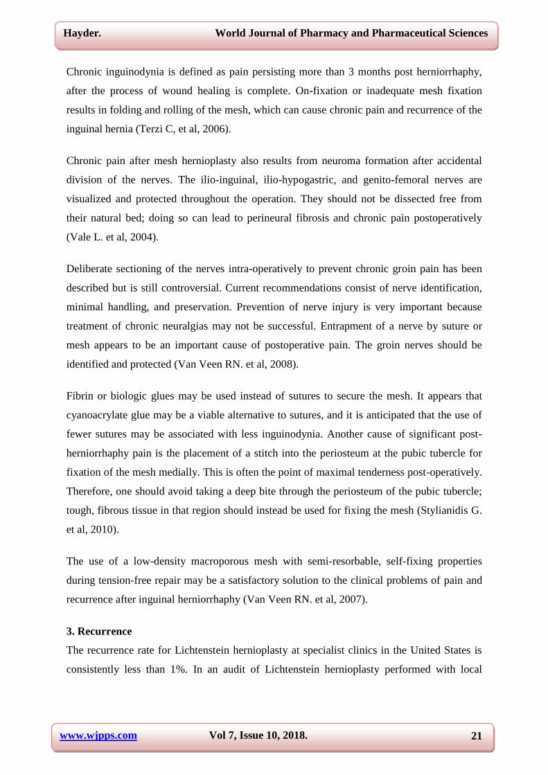
Figure 1: Shows the sex in both groups of the patients. The total number of male
patients was (192) and (8) were female patients (blue columns). In each group, the male
were 96 and the female were 4 patients (red and green columns).
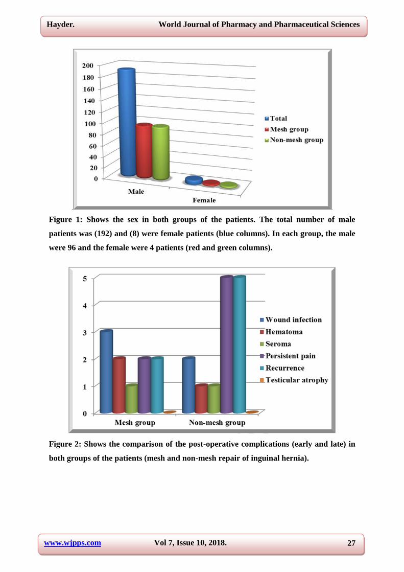
Figure 2: Shows the comparison of the post-operative complications (early and late) in
both groups of the patients (mesh and non-mesh repair of inguinal hernia).
www.wjpps.com Vol 7, Issue 10, 2018.
28
Hayder. World Journal of Pharmacy and Pharmaceutical Sciences
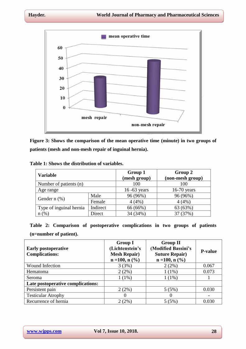
Figure 3: Shows the comparison of the mean operative time (minute) in two groups of
patients (mesh and non-mesh repair of inguinal hernia).
Table 1: Shows the distribution of variables.
Group 2
(non-mesh group)
Group 1
(mesh group) Variable
100 100 Number of patients (n)
16-70 years 16 -63 years Age range
96 (96%) 96 (96%) Male Gender n (%)
4 (4%) 4 (4%) Female
63 (63%) 66 (66%) Indirect Type of inguinal hernia
n (%) 37 (37%) 34 (34%) Direct
Table 2: Comparison of postoperative complications in two groups of patients
(n=number of patient).
Early postoperative Complications:
Group I (Lichtenstein’s Mesh Repair) n =100, n (%)
Group II (Modified Bassini’s
Suture Repair) n =100, n (%)
P-value
Wound Infection 3 (3%) 2 (2%) 0.067
Hematoma 2 (2%) 1 (1%) 0.073
Seroma 1 (1%) 1 (1%) 1
Late postoperative complications:
Persistent pain 2 (2%) 5 (5%) 0.030
Testicular Atrophy 0 0 -
Recurrence of hernia 2 (2%) 5 (5%) 0.030

www.wjpps.com Vol 7, Issue 10, 2018.
29
Hayder. World Journal of Pharmacy and Pharmaceutical Sciences
Table 3: The Mean Operative Time in both groups of patients (mesh and non-mesh
repair of inguinal hernia).
Variable Group 1 (Mesh
Repair) Group 2
(Non-mesh Repair) P-value
Mean operative
time (minute) 30.4 50.4 > 0.05
DISCUSSION
Tissue based suture repair by different techniques (Bassini’s, Shouldice etc.) has remained
conventional surgical treatment of inguinal hernia. These techniques shared many things in
common such as excessive tension on the suture line as well as the neighboring tissues, a lot
of dissection, trauma and undue operative time. These factors were found to be responsible
for a number of recurrences, persistent pain after surgery and morbidity leading to an undue
economical burden on the patient. This led to the introduction of mesh repair in the late’s
1980 with the concept of tension free repair of hernias. Since then a number of studies have
claimed improved results with mesh repair in terms of recurrence of hernia (Amid PK. et al,
2002).
This study compares and demonstrates the efficacy of non-mesh (Bassini’s) and mesh
(Lichtenstein) repair. The two groups were compared regarding the duration of surgery,
development of early post-operative complications such as (wound infection, hematoma and
seroma formation) and development of late post-operative complications such as (persistent
pain, testicular atrophy and recurrence of inguinal hernia).
Our study demonstrated that the tension-free mesh repair of inguinal hernia offer a significant
benefit over non mesh repair especially regarding post-operative persistent pain and
recurrence of inguinal hernia, P-value was (<0.05).
In our study, the gender distribution in the two groups of patients was 192(96%) male
patients and 8(4%) female patients. There were (96) male and (4) female in each group of
patients. These finding are agree with Arkan A. Al-Ogaili et al. (2009). In this study also the
male patients were found in majority which clarifies that the inguinal hernias are common in
males.
Another study done by Malik AM. Et al. (2007), also found an increase incidence of male
patients in the inguinal hernias.

www.wjpps.com Vol 7, Issue 10, 2018.
30
Hayder. World Journal of Pharmacy and Pharmaceutical Sciences
About characteristic of hernia, there were 66 patient indirect inguinal hernia in group 1 and
63 patient in group 2 whereas 34 patient direct inguinal hernia in group 1 and 37 patient in
group 2. These finding are agree with Parviz K. Amid (2003). In this study, also the indirect
inguinal hernias are more common than direct inguinal hernia in both groups of patients.
Regarding post-operative wound infection, our study resulted in three patients (3%) in group
I and two patients (2%) in group II. P-value (> 0.05) which is statistically insignificant. This
is agreeing with the results of the study done by Grant Am. (2005).
Also these results are agreed with study done by Scott et. al, (2002). This study was showed
that the rate of infection is more with mesh repair group of the patients which is similar to our
study.
Regarding post-operative wound seroma, there were one (1%) patient from each group
developed this complication. P-value (> 0.05) which is statistically insignificant. This result
is in agreement with Arkan A. Al-Ogaili et al. (2009).
Regarding post-operative wound hematoma, there were two (2%) patients in group I and one
(1%) patient in group II developed this complication. P-value (> 0.05) which is statistically
insignificant. Also this result is agreed with Arkan A. Al-Ogaili et al. (2009).
As shown from our result in (figure-2) and (table-2) that two (2%) patients in group I
developed post-operative persistent pain in comparison to five (5%) patients in group II
developed this complication-value was (<0.05) which is statistically significant. This result is
agreed with A.E. Kirk et al. (1995). A study done by Kristin and Masukawa et al. (2010) also
shows a similar result.
Recurrence is a major problem encountered by the surgeons. Tension is a cardinal factor in
the failure of a hernia repair. Tissue sutured under tension will tend to pull apart and the
suture creates an area of ischemic necrosis. (Liem M.S. et al. 2003).
In our study, two (2%) patients in group I developed post-operative recurrence in comparison
to five (5%) patients in group II developed this complication. P-value was (<0.05) which is
statistically significant. This result is agreed with study done by Malik AM. et al. (2007).
Another study done by W.W. Vrijland et al. (2002) also shows the same result.

www.wjpps.com Vol 7, Issue 10, 2018.
31
Hayder. World Journal of Pharmacy and Pharmaceutical Sciences
Bisgaar T et al and Butters (2007) mention a similar recurrence rate and found that the mesh
repair (Lichtenstein) was superior to suture repair of inguinal hernia.
Other complication which is not seen in our study is ischemic orchitis (testicular atrophy)
occur especially in recurrent inguinal hernia and this due to excessive dissection of the cord
lead to thrombosis of veins draining the testicle. This result is agreed with study done by
Wants GE. (1997).
Regarding the duration of surgical procedure, the overall operative time differs significantly
in two techniques and the mean operative time in mesh repair group (30.4 minute) is much
less than the mean operative time in non- mesh repair group (50.4 minute). P-value (>0.05)
which is statistically insignificant. This result is in agreement with Parviz K. Amid (2003).
Another study done by Malik AM. Et al. (2007), also found that the mean operative time in
the non-mesh group is longer than the mean operative time in the mesh group.
CONCLUSION
1. It was found that the use of traditional inguinal hernia repair (tissue repair or non-mesh
repair) is associated with considerable post-operative pain and more chance of recurrence
than mesh repair.
2. The mesh repair (tension-free repair) is superior to the non-mesh repair of inguinal hernias
especially in terms of post-operative recurrence and persistent pain.
3. Mesh repair technique of inguinal hernias takes shorter operative time than non-mesh
repair of inguinal hernias.
ACKNOWLEDGEMENTS
I would like to express my gratitude to everybody who has helped me during the long time I
have worked with this thesis. In particular I would like to thank Dr. Muqdad Fouad Abdul
Kareem (FICMS in General Surgery) for his support and help.
My deep gratitude to Assistant Professor Dr. Mustafa Khalil Hameed Khalaf ( FICMS in
General Surgery) and all teaching staff in College of Medicine \ Diyala University for
encouraging my scientific work.
My full respect and appreciation to Dr. Hussain Alwan Khalaf (FICMS in General Surgery)
for revising the scientific assessment of this thesis.

www.wjpps.com Vol 7, Issue 10, 2018.
32
Hayder. World Journal of Pharmacy and Pharmaceutical Sciences
Finally I would like to thank Diyala Health Directorate for their aids and the medical staff
members in surgical department in Baqubah Teaching Hospital and all the patients who
participated in my study and made it possible for the study to be completed.
REFERENCES
1. A.E.Kirk, M. Kurzer, K.J. Waiters. British hernia center tension free mesh hernia repair,
Journal Royal College of Surgery, 1995; 77(54): 299-304.
2. Akyol C, Kocaay F, Orozakunov E, Genc V, Kepenekci Bayram I, Cakmak A, et al.
Outcome of the patients with chronic mesh infection following open inguinal hernia
repair. J Korean Surg Soc., May 2013; 84(5): 287-91.
3. Albo D, Awad SS, Berger DH, Bellows CF. Decellularized human cadaveric dermis
provides a safe alternative for primary inguinal hernia repair in contaminated surgical
fields. Am J Surg., Nov. 2006; 192(5): e12-7.
4. Amid PK, Shulman AG, Lichtenstein IL. Local anesthesia for inguinal hernia repair step-
by-step procedure. Ann Surg., Dec. 1994; 220(6): 735-7.
5. Arkan A. Al-Ogaili et al. Open Mesh versus Non-Mesh Repair of Inguinal Hernia. A
prospective randomized trial at AI-Yarmouk Teaching Hospital and Al-Mussayb General
Hospital in a period between, April 2005–October 2009.
6. Awad SS, Fagan SP. Current approaches to inguinal hernia repair. Am J Surg., Dec. 2004;
188(6A Suppl): 9S-16S.
7. Amid PK. How to avoid recurrence in Lichtenstein tension-free hernioplasty. Am J Surg.,
Sep. 2002; 184(3): 259-60.
8. Amid PK. Lichtenstein tension-free hernioplasty. In: Fischer JE. Mastery of Surgery.
2.5th
. Lippincott Williams & Wilkins, 2007: 1933-9.
9. Butters M, Redecke J, Koninger J. Long term results of randomized clinical trial of
Shouldice, Lichtenstein and transabdominal preperitoneal hernia repairs. Br J Surg, 2007;
94(5): 562–569.
10. Bisgaard T, Bay-Nielson M, Christensen IJ, Kehlet H. Risk of recurrence 5 years or more
after primary Lichtenstein mesh and sutured inguinal hernia repair. Br J Surg, 2007; 94:
1038–40.
11. Bay-Nielsen M, Thomsen H, Andersen FH, et al. Convalescence after inguinal
herniorrhaphy. Br J Surg., Mar. 2004; 91(3): 362-7.

www.wjpps.com Vol 7, Issue 10, 2018.
33
Hayder. World Journal of Pharmacy and Pharmaceutical Sciences
12. Bringman S, Wollert S, Osterberg J, Smedberg S, Granlund H, Heikkinen TJ. Three-year
results of a randomized clinical trial of lightweight or standard polypropylene mesh in
Lichtenstein repair of primary inguinal hernia. Br J Surg., Sep. 2006; 93(9): 1056-9.
13. Chastan P. Tension-free open hernia repair using an innovative self-gripping semi-
resorbable mesh. Hernia., Apr. 2009; 13(2): 137-42.
14. Chung L, O'Dwyer PJ. Treatment of asymptomatic inguinal hernias. Surgeon., Apr. 2007;
5(2): 95-100.
15. Chung RS. Meta-analysis of randomized controlled trials of laparoscopic versus
conventional inguinal hernia repair. Surg Endosc., 1999; 7: 68-94.
16. DeBord JR, Whitty LA. Biomaterials in hernia repair. In: Fischer JE. Mastery of Surgery.
2.5th
. Lippincott Williams & Wilkins, 2007: 1965-8.
17. Delikoukos S, Fafoulakis F, Christodoulidis G, Theodoropoulos T, Hatzitheofilou C. Re-
operation due to severe late-onset persisting groin pain following anterior inguinal hernia
repair with mesh. Hernia., Dec. 2008; 12(6): 593-5.
18. Fingerhut A, Millet B, Veyrie N, et al. Inguinal hernia repair, update 2006. In: Edmund
AM, Neugebauer S, Fingerhut A, et al. EAES Guidelines for Endoscopic Surgery. 1.
Springer; 2006: 294-307.
19. Earle DB, Mark LA. Prosthetic material in inguinal hernia repair: how do I choose? Surg
Clin North Am., Feb. 2008; 88(1): 179-201.
20. Fitzgibbons RJ Jr,Giobbie-Hurder A, Gibbs JO, et al. Watchful waiting vs repair of
inguinal hernia in minimally symptomatic men: a randomized clinical trial. JAMA., Jan
18, 2006; 295(3): 285-92.
21. Fitzgibbons RJ, Filippi CJ, Quinn TH. Inguinal hernias. In: Brunicardi FC, Andersen DK,
Billiar TR, Dunn DL, Hunter JG, Pollock RE (Editors). Schwartz's Principles of Surgery.
8th
Edition. New York, NY: McGraw-Hill, 2005.
22. Franklin ME Jr, Gonzalez JJ Jr, Glass JL. Use of porcine small intestinal submucosa as a
prosthetic device for laparoscopic repair of hernias in contaminated fields: 2-year follow-
up. Hernia., Aug. 2004; 8(3): 186-9.
23. Franklin ME Jr, Gonzalez JJ Jr, Michaelson RP, Glass JL, Chock DA. Preliminary
experience with new bioactive prosthetic material for repair of hernias in infected
fields. Hernia., Dec. 2002; 6(4): 171-4.
24. Franneby U, Sandblom G, Nordin P, Nyren O, Gunnarsson U. Risk factors for long-term
pain after hernia surgery. Ann Surg., Aug. 2006; 244(2): 212-9.

www.wjpps.com Vol 7, Issue 10, 2018.
34
Hayder. World Journal of Pharmacy and Pharmaceutical Sciences
25. Gianetta E, Cuneo S, Vitale B, Camerini G, Marini P, Stella M. Anterior tension-free
repair of recurrent inguinal hernia under local anesthesia: a 7-year experience in a
teaching hospital. Ann Surg., Jan. 2000; 231(1): 132-6.
26. Grosz CR. Iliohypogastric nerve injury. Am J Surg., Nov. 1991; 142(5): 628.
27. Hair A, Duffy K, McLean J, Talor S, Smith H, Walker A et al. Groin hernia repair in
Scotland. BJS, 2000; 87: 1722–6.
28. Haapaniemi S, Nordin P, van der Linden W, Nilsson E. Choice of anesthesia and risk of
reoperation for recurrence in groin hernia repair. Ann Surg., Jul. 2004; 240(1): 187-92.
29. Hair A, Duffy K, McLean J, et al. Groin hernia repair in Scotland. Br J Surg., Dec. 2000;
87(12): 1722-6.
30. Hakeem A, Shanmugam V. Inguinodynia following Lichtenstein tension-free hernia
repair: a review. World J Gastroenterol, Apr. 14, 2011; 17(14): 1791-6.
31. Hosgor M, Karaca I, Ozer E, Suzek D, Ulukus C, Ozdamar A. Do alterations in collagen
synthesis play an etiologic role in childhood inguino-scrotal pathologies: an
immunohistochemical study. J Pediatr Surg., Jul. 2004; 39(7): 1024-9.
32. Junge K, Klinge U, Prescher A, Giboni P, Niewiera M, Schumpelick V. Elasticity of the
anterior abdominal wall and impact for reparation of incisional hernias using mesh
implants. Hernia., Sep. 2001; 5(3): 113-8.
33. Kark AE, Kurzer M, Waters KJ. Tension-free mesh hernia repair: review of 1098 cases
using local anesthesia in a day unit. Ann R Coll Surg Engl., Jul. 1995; 77(4): 299-304.
34. Kristin Masukawa, B. A, Samuel E. Wilson, Complication of mesh hernia repair USA,
2010.
35. Koch A, Edwards A, Haapaniemi S, Nordin P, Kald A. Prospective evaluation of 6895
groin hernia repairs in women. Br. J. Surg., Dec. 2005; 92(12): 1553-8.
36. Liem M.S. van Duyn E. B. van der Graaf Y. et al. A randomized comparison recurrence
after conventional laparoscopic inguinal hernia repair. American Journal of surgery,
2003; 237(1): 136-41.
37. Lichtenstein IL, Shulman AG, Amid PK, Montllor MM. Cause and prevention of
postherniorrhaphy neuralgia: a proposed protocol for treatment. Am J Surg., Jun. 1988;
155(6): 786-90.
38. Lichtenstein IL, Shulman AG, Amid PK, Montllor MM. The tension-free
hernioplasty. Am J Surg., Feb. 1990; 157(2): 188-93.
39. McCormack K, Scott NW, Go PM, Ross S, Grant AM, EU Hernia Trialist Collaboration.
Cochrane Database Syst Rev., 2003; (1): CD 001785.

www.wjpps.com Vol 7, Issue 10, 2018.
35
Hayder. World Journal of Pharmacy and Pharmaceutical Sciences
40. Liem MS, Halsema JA, van der Graaf Y, Schrijvers AJ, van Vroonhoven TJ. Cost-
effectiveness of extraperitoneal laparoscopic inguinal hernia repair: a randomized
comparison with conventional herniorrhaphy. Coala trial group. Ann Surg, 1997; 226:
668–76.
41. Malik AM. et al. Acomparative analysis between non-mesh (Bassini's) and mesh
(Lichtenstein) repair of primary inguinal hernia. A retrospective comparative analytical
study 2000 to December 2007 Liaquat University of Medical and Health Sciences,
Hyderabad/ Jamshoro, Pakistan.
42. Malangoni MA, Rosen RJ. Hernias. In: Townsend CM, Beauchamp RD, Evers BM, and
Mattox KL (Editors). Sabiston Textbook of Surgery. 18th
edition. Philadelphia, PA:
Elsevier, 2007.
43. Milic DJ, Pejic MA. Tension free procedures in the surgical treatment of groin hernias.
Spr Arh Cleok Lek, 2003; 131(1-2): 82–91.
44. Nordin P, Bartelmess P.Jansson C, Svensson C, EdlundG. Randomized trial of
Lichtenstein versus Shouldice hernia repair in general surgical practice. Br J Surg, 2002;
89: 45–9.
45. Nathan JD, Pappas TN. Inguinal hernias: an old condition with new solutions. Ann Surg.,
2003; 238(6 suppl): S148–57.
46. Nilsson H, Stylianidis G, Haapamaki M, Nilsson E, Nordin P. Mortality after groin hernia
surgery. Ann Surg., Apr. 2007; 245(4): 656-60.
47. O'Dwyer PJ, Alani A, McConnachie A. Groin hernia repair: postherniorrhaphy
pain. World J Surg., Aug. 2005; 29(8): 1062-5.
48. Nordin P, Zetterstrom H, Gunnarsson U, Nilsson E. Local, regional, or general anesthesia
in groin hernia repair: multicenter randomized trial. Lancet., Sep. 13, 2003; 362(9387):
853-8.
49. Norrie J, Chung L, O'Dwyer PJ. Long-term follow-up of patients with a painless inguinal
hernia from a randomized clinical trial. Br J Surg., Nov. 30, 2010; 5: 143-156.
50. O'Dwyer PJ, Norrie J, Alani A, Walker A, Duffy F, Horgan P. Observation or operation
for patients with an asymptomatic inguinal hernia: a randomized clinical trial. Ann Surg.,
Aug. 2006; 244(2): 167-73.
51. Parviz K.Amid: Lichtenstein tension–free hernioplasty for repair of primary and recurrent
inguinal hernias.Nyhus and Condon’s, PIONEERS IN HERNIA SURGERY. Lichtenstein
tension-free hernioplasty, March 2003.

www.wjpps.com Vol 7, Issue 10, 2018.
36
Hayder. World Journal of Pharmacy and Pharmaceutical Sciences
52. Paajanen H, Varjo R. Ten-year audit of Lichtenstein hernioplasty under local anesthesia
performed by surgical residents. BMC Surg., Aug. 2010; 10: 24.
53. Scott NW, McCormac K, Graham P, Go PM, Ross SJ, Grant AM. Open mesh versus non-
mesh for repair of femoral and inguinal hernia. Cochrane Database Syst Rev., 2002; (2):
CD002197.
54. Sanders DL, Waydia S. Asystematic review of randomized control trials assessing mesh
fixation in open inguinal hernia repair. Hernia., May 7, 2013; 98(5): 279-89.
55. Scott N, Go PM.N.Y.H, Graham P, McCormack K, Ross SJ, Grant AM, Open mesh
versus non-mesh repair of groin hernia: meta-analysis of randomized trials in 20
countries, 2002.
56. Shamberger RC, Ottinger LW, Malt RA. Arterial injuries during inguinal
herniorrhaphy. Ann Surg., Jul. 1984; 200(1): 83-5.
57. Shankar VG, Srinivasan K, Sistla SC, Jagdish S. Prophylactic antibiotics in open mesh
repair of inguinal hernia - a randomized controlled trial. Int J Surg., 2010; 8(6): 444-7.
58. Sanabria A, Dominguez LC, Valdivieso E, Gomez G. Prophylactic antibiotics for mesh
inguinal hernioplasty: a meta-analysis. Ann Surg., Mar. 2007; 245(3): 392-6.
59. Sanchez-Manuel FJ, Lozano-García J, Seco-Gil JL. Antibiotic prophylaxis for hernia
repair. Cochrane Database Syst Rev., Jul. 18, 2007; CD003769.
60. Simons MP, Aufenacker T, Bay-Nielsen M, Bouillot JL, Campanelli G, Conze J, et al.
European Hernia Society guidelines on the treatment of inguinal hernia in adult
patients. Hernia., Aug. 2009; 13(4): 343-403.
61. Starling JR, Harms BA. Diagnosis and treatment of genitofemoral and ilioinguinal
neuralgia. World J Surg., Sep-Oct. 1989; 13(5): 586-91.
62. Scott NW, McCormack K, Graham P, Go PM, Ross SJ, Grant AM. Open mesh versus
non-mesh for repair of femoral and inguinal hernia. Cochrane Database Syst Rev., 2002;
CD002197.
63. Stylianidis G, Haapamäki MM, Sund M, Nilsson E, Nordin P. Management of the hernial
sac in inguinal hernia repair. Br J Surg., Mar. 2010; 97(3): 415-9.
64. Taylor EW, Duffy K, Lee K, et al. Surgical site infection after groin hernia repairs. Br J
Surg., Jan. 2004; 91(1): 105-11.
65. Terzi C. Antimicrobial prophylaxis in clean surgery with special focus on inguinal hernia
repair with mesh. J Hosp Infect., Apr. 2006; 62(4): 427-36.

www.wjpps.com Vol 7, Issue 10, 2018.
37
Hayder. World Journal of Pharmacy and Pharmaceutical Sciences
66. Vale L, Grant A, Mc Cormac, Scott NW, EU Hernia Trialist collaboration. Cost
effectiveness of alternative methods of surgical repair of inguinal hernia. Int J Technol
Assess Health Care, 2004; 20(2): 192–200.
67. Van Veen RN, Mahabier C, Dawson I, et al. Spinal or local anesthesia in Lichtenstein
hernia repair: a randomized controlled trial. Ann Surg., Mar. 2008; 247(3): 428-33.
68. Van Veen RN, Wijsmuller AR, Vrijland WW, Hop WC, Lange JF, Jeekel J. Long term
follow up of a randomized clinical trial of non-mesh versus mesh repair of primary
inguinal hernia. Br J Surg, 2007; 94: 506–10.
69. W.W. Vrijland, M. P van den Tol, R. W Luijendijk, W.C. J Hop, J.J. V Busschbach,
D.C.D de Lange, D van Geldere, A.B. Randomized clinical trial of non-mesh versus mesh
repair of primary inguinal hernia, 293-297.
70. Wants GE. My experience without tension, primary inguinal hernia in man, Journal
international of surgery, 1997; 71(46): 111-6.
71. Wantz GE. Complications of inguinal hernial repair. Surg Clin North Am., Apr. 1984;
64(2): 287-98.
72. Weyhe D, Belyaev O, Müller C, Meurer K, Bauer KH, Papapostolou G, et al. Improving
outcomes in hernia repair by the use of light meshes--a comparison of different implant
constructions based on a critical appraisal of the literature. World J Surg., Jan. 2007;
31(1): 234-44.
73. Wiese M, Kaufmann T, Metzger J, et al. Learning curve for Lichtenstein
hernioplasty. Open Access Surgery., Jul. 2, 2010; 3: 43-6.
74. Wijsmuller AR, Lange JF, Kleinrensink GJ, et al. Nerve-identifying inguinal hernia
repair: a surgical anatomical study. World J Surg., Feb. 2007; 31(2): 414-20; discussion
421-2.
75. Woods B, Neumayer L. Open repair of inguinal hernia: an evidence-based review. Surg
Clin North Am., Feb. 2008; 88(1): 139-55, ix-x.
76. Zhao G, Gao P, Ma B, Tian J, Yang K. Open mesh techniques for inguinal hernia repair:
a meta-analysis of randomized controlled trials. Ann Surg., Jul. 2009; 250(1): 35-42.