Comparison and Characterization of Specific Fertilization ...
266
vk Comparison and characterisation of specific fertilisation proteins in human (Homo sapiens) and baboon (Papio anubis) spermatozoa / by RODI OMONDI OJOO DEPARTMENT OF VETERINARY ANATOMY AND PHYSIOLOGY UNIVERSITY OF NAIROBI University ot NAIROBI Library r03506854 N a ,*OBi *4*aTE UNl^ s ,r r A THESIS SUBMITTED IN FULFILMENT OF THE REQUIREMENTS FOR THE DEGREE OF DOCTOR OF PHILOSOPHY OF THE UNIVERSITY OF NAIROBI 2005
Transcript of Comparison and Characterization of Specific Fertilization ...

vkComparison and characterisation of specific fertilisation proteins
in human (Homo sapiens) and baboon (Papio anubis) spermatozoa /
by
RODI OMONDI OJOO
DEPARTMENT OF VETERINARY ANATOMY AND PHYSIOLOGY
UNIVERSITY OF NAIROBI
University ot NAIROBI Library
r03506854
Na ,*OBi*4*aTE UNl^ s , r r
A THESIS SUBMITTED IN FULFILMENT OF THE REQUIREMENTS FOR
THE DEGREE OF DOCTOR OF PHILOSOPHY OF THE UNIVERSITY OF
NAIROBI
2005

i i
DECLARATIONI
I, RODI OMONDI OJOO HEREBY DECLARE THAT THE WORK
CONTAINED IN THIS THESIS IS MY ORIGINAL WORK AND HAS NOT
BEEN PRESENTED FOR A DEGREE IN ANY OTHER UNIVERSITY
DATE:
THIS THESIS HAS BEEN SUBMITTED FOR EXAMINATION WITH OUR
APPROVAL AS UNIVERSITY SUPERVISORS
Prof. D.ODUOR-OKELO, B.V.Sc., D.V.M., M.Sc., Ph.D.
Signed:.. ...............
Date:. LJj..Q ./.' P.Q .'?/..........
Dr. P.A.ADOYO, B.Sc., Ph.D
Signed .
Date....... Q S U
Prof. H.D.M.MOORJE, B.Sc. Ph.D
Signed
Date...
UN"'f*srrrUfi«A«y

Ill
ACKNOWLEDGEMENTS
I am grateful to the World Health Organization (WHO) for financial
support through the Career Development Fellowship (Re: H9/181/R405) that
enabled me to carry out this work towards my PhD.
I am particularly grateful to Prof. H.D.M. Moore for his guidance and
support in the course of this work and especially for exposing me to current
techniques being used in research. I am grateful to Dr. PA. Adoyo for introducing
me to the molecular biology techniques, his supervision and kindly allowing me to
use his Laboratory. I thank my other supervisors Prof. Oduor-Okelo and
Prof.Wango for their dedicated supervision and encouragement as I did the work.
I am indebted to a number of people who assisted me in different ways in
the course of doing this work. I wish to mention Mr. Deya and Mr. Mwaura of
Institute of Primate Research and Mr. Mwasela of Department of Veterinary
Anatomy and Physiology who have assisted me greatly in my work while in
Kenya. I wish to thank Nick Jenkins, Mick Turton, Dr. Kath Carrol, Dr. Chi Wong
and Dr. Nadia Al-Eisa who were all then in the Department of Molecular Biology
and Biotechnology, University of Sheffield, for their assistance and patience as I
learnt some of the techniques..
I could not have done it without my dear wife Atieno, who endured my
long working hours and long periods of separation and my kids Aringo and
Anyango, who had to see little of ‘daddy’ as I was doing this work.

IV
Lastly I am thankful to my dad, mum and entire Ojoo family who
encouraged excellence and perseverance and for laying that foundation in my life
To God be the glory!
V
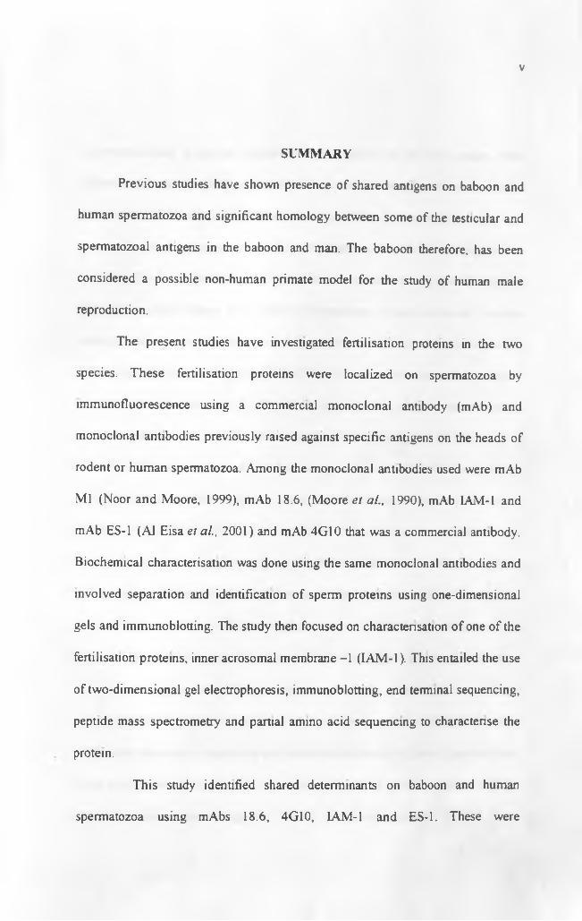
SUMMARY
Previous studies have shown presence of shared antigens on baboon and
human spermatozoa and significant homology between some of the testicular and
spermatozoa! antigens in the baboon and man. The baboon therefore, has been
considered a possible non-human primate model for the study of human male
reproduction.
The present studies have investigated fertilisation proteins in the two
species. These fertilisation proteins were localized on spermatozoa by
immunofluorescence using a commercial monoclonal antibody (mAb) and
monoclonal antibodies previously raised against specific antigens on the heads of
rodent or human spermatozoa. Among the monoclonal antibodies used were mAb
Ml (Noor and Moore, 1999), mAb 18.6, (Moore et al., 1990), mAb IAM-1 and
mAb ES-1 (Al Eisa et al., 2001) and mAb 4G10 that was a commercial antibody.
Biochemical characterisation was done using the same monoclonal antibodies and
involved separation and identification of sperm proteins using one-dimensional
gels and immunoblotting. The study then focused on characterisation of one of the
fertilisation proteins, inner acrosomal membrane -1 (IAM-1). This entailed the use
of two-dimensional gel electrophoresis, immunoblotting, end terminal sequencing,
peptide mass spectrometry and partial amino acid sequencing to characterise the
protein.
This study identified shared determinants on baboon and human
spermatozoa using mAbs 18.6, 4G10, LAM-1 and ES-1. These were

VI
immunolocalised to similar domains on spermatozoa in the two species. One
dimensional immunoblots corroborated the immunolocalisation studies
demonstrating similarity of determinants recognised by the same mAbs in the two
species. Some determinants like Ml were found in rodents but not in the two
primates. ES-1 was localized by immunofluorescence to the equatorial segment
and tail in both human and baboon spermatozoa. One-dimensional sodium
dodecyl sulphate polyacrylamide gel electrophoresis of human sperm protein
extracts and immunoblotting using mAb ES-1, revealed immunoreactive bands of
apparent molecular weights, 24, 28, 40, 47 and 50 kDa, while in baboon sperm
extracts immunoreactive bands were recognised at 34, 38, 47, and 50 kDa.
Characterisation of IAM-1 using one dimensional immunoblots
revealed it was a protein with four immunoreactive bands of apparent molecular
weights 28, 32, 39 and 45 kDa in man while in the baboon, 3 immunoreactive
bands of apparent molecular weights, 32, 38 and 44kDa were identified. Two-
dimensional gel electrophoresis and immunoblotting revealed IAM-1 has native
pi’s of between 3.86 - 4.0. Immunofluorescence localized this antigen to the
anterior acrosome and equatorial region of both human and baboon spermatozoa.
Treatment with different extraction solutions revealed that this protein
was an integral membrane protein or a lipid-anchored protein intimately attached
to the inner acrosomal membrane and equatorial segment of human spermatozoa.
Mass spectrometry and amino acid sequencing isolated several peptides and the
overall picture obtained suggested IAM-1 was end-terminally blocked, non-

VII
glycosylated and a trypsin-like protease. Taken together, immunological and
biochemical data obtained suggested that the protein IAM-1 could be a membrane
anchored trypsin-like protease most likely involved in sperm-zona penetration
The ultimate goal of this study was identification of suitable gamete-
specific proteins integrally involved in the fertilisation process, which can be used
for immunocontraception. Further work remains to be done to confirm the
suitability of the IAM-1 as a possible candidate protein for testing. This work
provides additional evidence for use of the baboon as a model for study of human
reproduction.

VII I
TABLE OF CONTENTS
Title.................................................................................................................i
Declaration..................................................................................................... ii
Acknowledgements...................................................................................... iii-iv
Summary.......................................................................................................v-vii
Chapter 1 General Introduction and Literature Review
1.1. Gametogenesis.................................................................................... 1
1.2. Gamete morphology..................................................................................2
1.2.1. Sperm morphology.................................................................................2
1.2.1.1 Plasma membrane structure................................................................. 3
1.2.1.2.Sperm nucleus.......................................................................................6
1.2.1.2.1. Nuclear envelope.............................................................................7
1.2.1.2.2. Sperm head cytoskeleton................................................................ 7
1.2.1.2.3. Acrosome........................................................................................ 8
12.1.3. Sperm tail........................................................................................... 9
1.2.1.3.1. Connecting piece.............................................................................. 10
1.2.1.3.2. Middle piece..................................................................................... 10
1.2.1.3.3. Principal piece.................................................................................. 11
1.2.1.3.4. End piece...........................................................................................12
1.2.2. Oocyte/Egg morphology...................................................................... 12
1.2.2.1. Cumulus layer......................................................................................12
1.2.2.2. Zona Pellucida......................................................................................13

1.3. Overview o f mammalian fertilisation.................................................... 14
1.3.1. Capacitational changes on sperm.........................................................14
1.3.1.1. Sperm plasma membrane changes.....................................................15
1.3.1.2. Intracellular changes of sperm........................................................... 18
1.3.1.3. Changes in sperm motility.................................................................19
1.3.2. Sperm-egg interaction.........................................................................20
1.3.2.1. Sperm-cumulus interaction................................................................20
1.3.2.2. Sperm-zona interaction...................................................................... 21
1.3.2.2.1. Zona proteins...................................................................................22
1.3.2.2.2. Primary Sperm-ZP interactions...................................................... 23
1.3.2.2.3. Secondary Sperm-ZP binding........................................................26
1.3.2.3. Sperm-oolemmal interaction............................................................28
1.3.2.3.1. Sperm fusion proteins..................................................................... 29
1.3.2.3.2. Egg fusion proteins......................................................................... 33
1.3.2.3.3. Fusion mechanisms......................................................................... 37
1.3.2.3.4. Egg activation..................................................................................37
1.4. Contraception possibilities and use o f Non-human primates.........38
1.4.1. Qualities of an ideal immunocontraceptive..........................................39
1.4.2 Immunocontraception antigens............................................................40
1.4.2.1. Zona pellucida antigens..................................................................... 40
1.4.2.2. Sperm antigens................................................................................... 42
1.4.3. Non-human primate............................................................................48

1.5. Aims ofStudy............................................................................................. 51
Chapter 2 Inimunolocalisation, 1-D PAGE and Immunoblotting
of fertilisation proteins
2.0. Introduction...............................................................................................52
2.1. Sperm-egg interactions..............................................................................52
2.1.1. Sperm-oolemmal interactions................................................................ 53
2.2. Monoclonal antibodies in fertilisation studies..........................................56
2.3. Specific aims o f this chapter......................................................................59
2.4. Materials and Methods............................................................................. 60
2.4.1. Sperm preparations.................................................................................60
2.4.1.1. Human sperm preparation...................................................................60
2.4.1.2. Sperm count determinations............................................................... 60
2.4.1.3. Baboon sperm preparations.................................................................61
2.4.1.4. Sperm Swim-up technique.................................................................. 62
2.4.2. Baboon testis preparations......................................................................62
2.4.3. One dimensional SDS-PAGE................................................................ 64
2.4.3.1. Sample loading.................................................................................... 65
2.4.4. Western blotting......................................................................................66
2.4.4.1. Semi-dry protein transfer.....................................................................66
2.4.4.2. Tank transfer (Wet protein transfer)....................................................67
2.4.5. Immunoblotting.......................................................................................68
2.4.5.1. Protocol 1............................................................................................ 68
X

2.4.5.2. Protocol II.......................................................................................... 70
2.4.5 3. Immunodetection.................................................................................71
2.4.5.3.1. Alkaline phosphatase detection....................................................... 71
2.4.5.3.2. Enhanced chemiluminescence detection (ECL)............................. 71
2.4.5.3.3. Enhanced chemiluminescence glycoprotein detection.................. 72
2.4.6. Total protein staining for gels............................................................... 74
2.4 6.1. Silver staining protocol..................................................................... 74
2.4.6.2. Coomassie blue staining..................................................................75
2.4.7. Total protein staining for western blots................................................75
2.4.7.1. Colloidal gold total protein stain......................................................... 75
2.4.8. Differential extraction of human sperm proteins..................................76
2.4.9. Indirect Immunofluorescence technique (IIF).......................................77
2.4.9.1. Human sperm slide preparations....................................................... 77
2.4.9.2. Antibody labeling of sperm slides.....................................................78
2.4.9 3. Baboon testicular slide preparations..................................................78
2.4 9 3.1. Labeling of baboon testicular slides............................................... 79
2.4.9.5. Microscopy......................................................................................... 79
2.5. Results................................................................................................. 81
2.5.1. Indirect Immunofluorescence (IIF).....................................................81
2.5.1.1. Phosphotyrosine proteins.................................................................. 81
2.5.1.1.1. Human spermatozoa....................................................................... 81
2.5.1.1.2. Baboon sperm................................................................................. 82

XII
2.5.1.2. Monoclonal antibody 18.6.................................................................82
2.5.1.2.1 Human and Baboon sperm............................................................... 82
2.5.1.2.2. Baboon testis....................................................................................82
2.5.1.3. Monoclonal antibody IAM-1 ........................................................... .82
2.5.1.3.1. Human sperm....................................................................................82
2.5.1.3 2. Baboon sperm...................................................................................83
2.5.1.3.3. Baboon testis....................................................................................83
2.5.1.4. Monoclonal antibody ES-1 ................................................................ 83
2.5.1.5. Monoclonal antibody Ml .................................................................. 83
2.5.2. Immunoblots........................................................................................84
2.5.21. IAM-1.................................................................................................. 84
2.5.2 2. ES-1................................................................................................... 84
2.5.2.3. Phosphotyrosine proteins................................................................... 84
2.5.2.4. Differential extraction of human sperm proteins................................85
2.5.2.5. Glycoprotein detection.........................................................................87
2.6. Discussion.................................................................................................. 88
2.6.1. IAM-1................................................................................................. 88
2.6.1.1. Immunoblotting and immunolocalisation........................................... 88
2.6.1.2. Biochemical characterisation.............................................................. 90
2.6.2. ES-1....................................................................................................... 92
2.6.3. Phosphotyrosine proteins........................................................................94
2.6.4. M l............................................................................................................ 98
NAIROBI UNlVTffsrTY ^ • C T E UttRARY

X1U
2.6.5. 18.6..........................................................................................................99
2.6.6. Conclusions........................................................................................... 100
Chapter 3 2-D PAGE and Biochemical Analysis of IAM-1 by
Mass Spectrometry.
3.0. Introduction............................................................................................... 101
3.1. One vs two-dimensional gel electrophoresis............................................ 101
3.2. 2-D gel electrophoresis............................................................................. 102
3.2.1. Principles of 2-D gel electrophoresis................................................... 102
3.2.2. 2-D gel electrophoresis of sperm proteins............................................ 104
3.2.3. Membrane protein isolation...................................................................106
3.3. Mass spectrometnc identification of proteins.........................................107
3 .4. Specific aims o f this chapter....................................................................109
3.5. Materials and methods............................................................................110
3.5.1. Sample preparation................................................................................. 110
3.5.1.1. Alkaline extraction of sperm proteins..................................................111
3.5.1.2. Non-alkaline extraction........................................................................ I l l
3.5.2. 2-D electrophoresis...............................................................................112
3.5.2.1. Rehydration of immobilized pH gradient strips................................ 112
3.5.2.2. Isoelectric focusing.............................................................................. 112
3.5.2 3. 2nd dimension SDS-PAGE.................................................................109
3.5.3. Visualization of results......................................................................... 114
3.5.3.1. Total protein detection on gels.................................................. . 115

xiv
3.5.3.2. Colloidal Coomassie gel staining......................................................115
3.5.3.3. 2-D Semi-dry protein transfer........................................................... 117
3.5 3.4. Total protein detection on blots..........................................................117
3.5.3 5. Excision of protein bands or spots.................................................. 118
3.5.3.51. Gel proteins...................................................................................... 118
3.5.3.5.2. Blot proteins................................................................................... 119
3.6. Results........................................................................................................ 120
3.6.1. IAM-1 extraction.................................................................................... 120
3.6.2. 2-D electrophoresis................................................................................. 121
3.6.2.1. 2-D gels................................................................................................ 121
3.6.2.2. 2-D immunoblots................................................................................. 121
3 6.2.2.1. Non-alkaline protein immunoblots...................................................121
3.6.2.2.2. Alkaline protein immunoblots..........................................................121
3.6.3. N-termmal sequencing............................................................................ 122
3.6.4. Peptide mass fingerprinting....................................................................122
3.6.5. Internal peptide sequencing....................................................................123
3.7. Discussion...................................................................................................124
3.7.1. IAM-1 extraction..................................................................................... 124
3.7.2. 2-D gels and immunoblots....................................................................... 129
3.7.3. N-terminal sequencing............................................................................. 131
3.7.4. Peptide mass fingerprinting..................................................................... 133
3.7.5. Internal peptide sequencing.................................................................... 133
XV
3.7.5.1. Keratin..................................................................................................
3.7.5.2. Trypsin-like proteins........................................................................... 135
3.7.5.3. Glutathione S-transferases..................................................................138
3.7.5.4. Tubulin alpha 3/7................................................................................ 139
3.7.5.5. Heat shock 27kDa protein...................................................................140
3.7.5.6. Proteasomes..........................................................................................141
3.7.6. Conclusions...........................................................................................142
Chapter 4 General Discussion and Conclusion
4.1. Fertilisation antigens............................................................................... 143
4.1.1. ES-1......................................................................................................145
4.1.2. IAM-1......................................................................................................147
4.2. Conclusions..............................................................................................157
4.2.1. Future directions.................................................................................. 158
Appendix
References

1
Chapter 1
GENERAL INTRODUCTION AND LITERATURE REV IEW
1.1. Gametogenesis
Gametogenesis or the formation of gametes occurs in the male and
female gonads resulting in haploid gametes in the two sexes. In the male the
process is referred to as spermatogenesis. This is a well orchestrated process of
cellular proliferation, meiotic division and differentiation that occurs when
sexual maturity is attained and results in the formation of spermatozoa. It
involves a series of mitotic divisions by spermatogonial cells that result in the
formation o f other spermatogonia, a subset of which enter meiosis to become
primary spermatocytes. The resultant primary spermatocytes undergo first
meiotic division giving rise to secondary spermatocytes which then progress
through second meiotic division to give rise to haploid round spermatids. The
last mitotic and subsequent meiotic divisions during spermatogenesis are
characterised by incomplete cytokinesis, a feature that ensures that the resultant
daughter cells remain connected via cytoplasmic bridges and develop
synchronously until spermiation. Each round spermatid undergoes a
differentiation process termed spermiogenesis during which it is transformed
into a polarised spermatozoon with a distinct head and tail. The spermatozoon
is structurally and functionally adapted to deliver the genetic material in its
nucleus to the female egg (Eddy and O'Brien, 1994; de Krester and Kerr, 1994;
Johnson and Everitt, 1995).
2
In the female gonad, the oogonia undergo oogenesis. In most mammals
during foetal life there is an initial phase of oogonial mitosis followed by a
period when all the oogonia enter first meiotic division to become primary
oocytes. These oocytes then develop through the stages of meiotic prophase I
to diplotene (or dictyate) stage before birth where they remain arrested until the
period preceding ovulation at sexual maturity. At this time, the oocyte resumes
first meiotic division and undergoes an asymmetrical cell division. In this
process most cytoplasm, half of the chromosomes and practically all the
organelles are retained in the oocyte while the rest is extruded in the smaller
polar body. In most mammals meiotic division in the ovulated oocyte is then
arrested again in metaphase II. Therefore at ovulation there is a metaphase II
oocyte delimited by the oolemma, the first polar body in the perivitelline space,
the zona pellucida which is an acellular matrix enclosing both the oocyte and
first polar body, all invested by the outermost cumulus layer. The oocyte has
the substrates and mRNAs needed for synthesis of bio-molecules required for
initial embryonic development. Completion of meiosis marked by the extrusion
of a second polar body occurs after the sperm fuses with the egg (Johnson and
Everitt, 1995).
1.2. Gamete morphology
1.2.1. Sperm morphology
The sperm has a head and tail or flagellum, enveloped by the plasma
membrane. The head is attached to the tail at the neck. The head comprises an

3
acrosome, the nucleus, cytoskeletal components and the cytoplasm. Most
mammalian sperm have spatulate shaped heads that are flattened in the
anterior-posterior axis. However, in most rodents sperm have a falciform
shaped head with the acrosome overhanging the convex margin of the nucleus.
The tail is composed of a connecting piece, middle piece, principal
piece and the end or terminal piece. There are species differences in the shapes
and sizes o f the heads of mammalian spermatozoa and the lengths and relative
sizes of the flagella components. (Fouquet and Kann, 1994; Eddy and O’Brien,
1994; Breed, 1997)
1.2.1.1. Plasma membrane structure
This is the external envelope of both the head and tail components of
sperm. The sperm plasma membrane has been subdivided into several domains
(Fig. 1) based on the evidence that different regions of the sperm surface have
different organization and structure of membranes (Primakoff and Myles,
1983; Holt, 1984). The sperm head plasma membrane is subdivided into,
acrosomal (anterior head) and post-acrosomal (posterior head) regions (Fig. 1).
The acrosomal region is further subdivided from the anterior aspect caudally
into, marginal segment (or peripheral rim, anterior band, apical segment)
domain found on the anterior acrosomal margin, the principal segment
(acrosomal segment) domain that covers the major part of the acrosome and the
equatorial segment (posterior acrosome) domain that forms the posterior part of
the acrosome (Fig. 1). The marginal together with the principal segment
domains are often also termed the anterior acrosome or acrosomal cap (Eddy
and O’Brien, 1994). The post-acrosomal region (posterior head, post-

PLOAM
IAM
Fig.l. Structure o f the human spermatozoon showing the different domains on the head and tail. The dotted-bold lines with arrows indicate transverse sections made at that level.A.Ac: anterior acrosome, Eq.S: equatorial segment, P.Ac: post acrosomal region, M.Pi:middle piece, P.Pi: principal piece, E.Pi: end piece.PL: plasma membrane, Ax: microtubules of axoneme, FS: Fibrous sheath, ODF: Outer dense fiber, Mit: mitochondria, OAM: Outer acrosomal membrane. Ac.M:acrosomal matrix, IAM: inner acrosomal membrane, N:nucleus, Nc:Neck (After Fawcett, 1975; Eddy and O'Brien, 1994)

4
acrosomal segment) domain spans the region between the posterior acrosomal
margin and the sperm neck. The acrosomal and post-acrosomal regions are
separated by the serrated band or sub-acrosomal ring along the posterior
margin of the equatorial segment. The plasma membrane of the head region is
de-limited from that of the tail by the posterior ring (nuclear or striated ring).
The tail plasma membrane is subdivided into a middle piece domain overlying
the middle piece and a posterior tail domain overlying the principal and end
pieces of the tail. The two domains are separated by the annulus. The plasma
membrane adheres tightly to the underlying structures at the posterior ring and
annulus. The process of establishing plasma membrane domains commences
during spermiogenesis and is completed during maturation in the epididymis. It
is thought that structures involved in spermatid re-modelling during
spermiogenesis like the manchette, tubulo-bulbar complexes and Sertoli cell
ectoplasmic specializations may be involved in formation of plasma membrane
domains on sperm. (Russell, 1980; 1984; Eddy and O ’Brien, 1994, Toshimori,
1998).
Evidence of the plasma membrane being partitioned into domains was
first gathered from electrophoretic studies on whole sperm from the rabbit, bull
and ram. In these experiments sperm migrated towards the anode tail-first
(Bangham, 1961; Nevo et al., 1961). These results suggested presence of a
negative charge on the sperm surface that was more concentrated on the sperm
tails. This has been confirmed using positively charged colloidal iron
hydroxide that bound more to the flagellum of ram and stallion sperm
(Yanagimachi et al., 1972; Lopez et al., 1987).

5
Studies using different lectins as probes for polysaccharide chains had
variable results with regard to their distribution on the sperm surface. Although
there were more binding sites on the heads, there was more tail to tail
aggregation observed as a result of the overall greater surface area of the tails
(Koehler, 1981; Lee and Ahuja, 1987). Freeze-fracture, freeze-etch and surface
replica studies have shown differences in the plasma membrane of different
regions corresponding to separate domains. The intra-membranous particles
visualized using these techniques vary in number, size and distribution in
different plasma membrane domains (Friend and Fawcett, 1974).
Within species there are altered patterns, sizes and distribution of intra-
membranous particles during maturation, capacitation and acrosome reaction
(Eddy and O’Brien, 1994). Regional specializations in the plasma membrane of
the sperm head are seen at the equatorial and marginal segments of the
acrosome, post-acrosomal region and posterior ring. The plasma membrane
specializations at the posterior ring, especially the tight apposition with the
nuclear envelope, may effectively separate the sperm into two compartments
with differences in ionic balance and metabolism (Eddy and O’Brien, 1994).
Along the tail there are modifications seen in the plasma membrane of
the middle piece in association with the mitochondria. The plasma membrane
adheres tightly to the annulus, possibly separating the middle piece as a distinct
domain from the caudal parts of the tail (Friend and Fawcett, 1974). Other
studies using filipin to demonstrate sterol presence and distribution, and
polymixin to demonstrate presence of anionic lipids, confirm regional

6
differences of surface components (Bearer and Friend, 1980; Lopez and de
Souza, 1991).
Antibodies have been used to localise the surface antigens on sperm
and to define their roles using specific bioassays. Antisera against germ cells or
spermatozoa components have been used but tend to lack specificity. To
overcome this, monoclonal antibodies against specific epitopes of surface
antigens have been raised to further enhance the study of specific sperm
surface antigens. These have been useful in localisation and characterisation of
several sperm surface antigens in different mammals (see Eddy and O'Brien,
1994 for review). However, they also have limitations as they may recognise
similar epitopes shared by different proteins (Villaroya and Scholler, 1986).
The sperm plasma membrane proteins are modified along the
epididymis by: addition of new components, unmasking or modification of pre
existing sperm moieties or loss of sperm-surface components. Most
modifications could involve alterations in the glycoproteins and the glycolipids
and this takes place in specific regions of the epididymis (Eddy el al., 1985).
1.2.1.2. Sperm nucleus
This contains the haploid paternal set of chromosomes in the form of
highly condensed chromatin associated with protamines. The latter are low
molecular weight proteins that are very basic and rich in arginine and cysteine.
They are synthesized in the spermatids and due to abundant cysteine content,
form covalent disulphide bonds stabilizing the DNA by cross-linkages. Other
nucleoproteins could be involved in the stabilization o f chromatin too (Warrent
and Kim, 1978).

7
1.2.1.2.1. Nuclear envelope
This encloses the sperm nucleus. Unlike most somatic cell nuclear
membranes that have 40-60 nm separation of the membrane bilayer, the sperm
nuclear envelope has a 7-10 nm separation only. Most of the envelope lacks
nuclear pores except for the area caudal to the posterior ring or redundant
nuclear envelope where there are abundant hexagonally arranged pores
(Fawcett, 1975). The inner surface of the nuclear membrane has a protein
meshwork lining called the nuclear lamina that forms the nuclear skeletal
framework anchoring the chromatin. It is generally made up of four related
proteins namely, lamin A, Bi, B2 and C in mammals (Mckeon et al., 1986;
Eddy and O'Brien, 1994).
1.2.1.2.2. Sperm head cytoskeleton
This comprises sub-acrosomal, post-acrosomal and para-acrosomal
components. The sub-acrosomal component lies between the inner acrosomal
membrane and the nuclear envelope and is equivalent to the perforatorium of
rodent sperm, while the post-acrosomal cytoskeleton is delimited by the
nuclear envelope, the post-acrosomal plasma membrane and the posterior ring.
The two parts form the perinuclear theca (Bellve et al., 1992). The third
component, the para-acrosomal cytoskeleton lies between the acrosome and the
plasma membrane. It has been described in the hamster but could be present in
other species too (Olson and Winfrey, 1985; Eddy and O'Brien, 1994). The
sperm cytoskeleton has proteins ranging from 8 - 8 0 kDa. Though some of
these proteins could be structural in function others are now known to be
cytosolic proteins, basic proteins and nuclear-like histones (Oko et al., 2001).

8
Some of these proteins could be involved in spermatid differentiation events
like, nuclear and acrosomal shaping, acrosomal-nuclear attachment and later
on, sperm- egg interaction (Eddy and O’ Brien 1994; Oko et al, 2001).
1.2.1.2.3. Acrosome
This overlies the anterior aspect of the sperm nucleus and is therefore
indented on the caudal aspect where it is attached to the nucleus. The acrosome
comprises an inner acrosomal membrane that immediately invests the anterior
portion o f the outer layer of the nuclear envelope, an outer acrosomal
membrane which is continuous with the inner acrosomal membrane at the
equatorial region and the acrosomal contents enclosed by the two membranes
(Fig. 1). The outer acrosomal membrane is in turn invested by the plasma
membrane. The acrosome is subdivided into an acrosomal cap or anterior
acrosome and the posterior acrosome or equatorial segment. These regions are
further subdivided into segments corresponding to those found on the plasma
membrane namely the apical, principal and equatorial segments of the
acrosome. Generally, the marginal segment is the acrosomal portion extending
beyond the nucleus, the principal segment overlies the nucleus and the
equatorial segment overlies the equator of spatulate sperm or lateral surfaces of
falciform-shaped sperm. There are a few exceptions to these general divisions
as seen in some species where the sperm head is neither spatulate nor falciform
shaped, like the woolly opossum. In the latter species, there is no identifiable
equatorial segment (Phillips, 1970). There is species variation in size, shape
and relative proportions of the acrosomal cap to the equatorial segment.
Acrosomal contents have ordered arrangements seen in different

9
species varying from lamellar to crystalline to cobblestone-like (Flechon, 1974;
Friend and Fawcett, 1974). The acrosomal membrane has an ordered
arrangement of intra-membranous particles peculiar to different segments. The
outer acrosomal membrane is composed of 12 - 290 kDa proteins including
glycoproteins, calmodulin binding proteins and phosphorylated proteins (Olson
et al., 1985). The inner acrosomal membrane is connected to the outer
membrane by bridges in boar spermatozoa (Russell et al., 1979). Acrosomal
contents include proteases such as proacrosin, acrosinin, acrosin inhibitors, P-
galactosidase and hyaluronidase among other proteins. It is notable that
acrosomal hyaluronidase and proacrosin are spermatogenic cell-specific
isozymes. Using monoclonal and polyclonal antibodies, many antigens of
unknown function have been localised in the acrosome (Goldberg, 1977; Eddy
and O'Brien, 1994).
1.2.1.3. Sperm tail
The components of the tail or flagellum include the neck (connecting
piece), middle piece, principal and end piece (Fig. 1). The base of the tail is
attached to the nucleus by the connecting piece. The tail contains a centrally
located axoneme (Fig. 1). The axoneme extends from the connecting piece to
the end piece of the tail. It is composed of a central pair of microtubules
surrounded by nine peripheral doublets (Fawcett, 1981). Each doublet has a
complete microtubule A, and a C-shaped microtubule B. Each A microtubule
has two dynein arms extending to the next B microtubule of the adjacent
doublet in a clockwise manner, viewed from the tail base to the end piece.
There are spokes that radiate from the central microtubules to the outer

10
peripheral pairs. The peripheral doublets are named 1 to 9 from the
microtubule pair on the plane bisecting the central pair. The axoneme is
encircled by nine outer dense fibres which have a wrapping of helically
arranged mitochondria in the middle piece while in the principal piece the
axoneme is surrounded by the outer dense fibres that are then covered by the
fibrous sheath (Fig. 1). The end piece lacks any peri-axonemal components.
The outer dense fibres and the fibrous sheath form the cytoskeleton of the
sperm tail (Fawcett, 1975; Eddy and O’Brien, 1994; Fouquet and Kann, 1994).
1.2.1.3.1. Connecting piece
This is formed by the capitulum or the neck portion that fits into the
implantation fossa on the sperm head and the segmental columns. The outer
part of the nuclear envelope is adherent to the basal plate at the implantation
fossa. The capitulum fits caudal to the basal plate. From the capitulum, two
major and five minor segmental columns arise. The major columns split into
two each and together with the five minor columns are continuous with the
nine outer dense fibres. The distal centriole gives rise to the axoneme of the
sperm tail. The two centrioles may regress during spermiogenesis, or the
proximal one may be retained in some species. The connecting piece
terminates at the beginning of the middle piece (Fawcett and Phillips, 1969;
Fawcett, 1975).
1.2.1.3.2. Middle piece
This is the part of the tail between the connecting piece and the
beginning of the principal piece at the annulus. It has the axoneme centrally
ringed by nine outer dense fibres giving the 9+9+2 arrangement found in

11
mammalian sperm (Fig. 1). Outer dense fibres differ between species in size
and shape but also among themselves. They are named according to the
corresponding microtubule doublet. Generally numbers 1, 5, 6 and 9 are bigger
than the rest. They taper from the middle piece to the distal principal piece. The
smaller fibres terminate before the larger ones in the principal piece (Telkka et
al., 1961; Eddy and O ’Brien, 1994). Outer dense fibers are thought to be
involved in providing elastic recoil during flagellar motion, due to abundant
disulfide linkages in their protein composition. The mitochondrial sheath
surrounds the outer dense fibres of the middle piece. These are helically
wrapped end to end along it and invested externally by plasma membrane.
There are species differences in numbers of mitochondria and lengths of the
middle piece. The caudal limit of the mitochondria along the middle piece is
marked by the annulus (Fawcett, 1975).
1.2.1.3.3. Principal piece
It is the longest segment of the flagellum. It is characterised by the
fibrous sheath, formed by two longitudinal columns attached together by semi
circular ribs, in mammalian and some avian spermatozoa. The fibrous sheath
surrounds the axoneme and the outer dense fibers except numbers 3 and 8. It is
invested by the plasma membrane externally. The longitudinal columns run
peripheral to microtubules 3 and 8 and are attached to the outer dense fibers 3
and 8 in the proximal part of the principal piece and microtubule doublets 3
and 8 distally. The longitudinal columns are in the dorso-ventral axis of the
sperm tail defined by the plane perpendicular to the central microtubule
doublets o f the axoneme. The ribs attach to the longitudinal columns and

12
branch to attach to each other. The fibrous sheath is thought to modulate
flagellar movement (Fawcett, 1975; Eddy and O’Brien, 1994).
1.2.1.3.4. End piece
This forms the terminal part of the flagellum. The beginning of the end
piece is marked by the termination of the fibrous sheath of the principal piece.
It is formed by the axoneme and the overlying plasma membrane. It lacks peri-
axonemal structures. The microtubules of the axoneme terminate at different
levels caudally within the end piece (Fawcett, 1975; Fouquet and Kann, 1994).
1.2.2. Oocyte/Egg morphology
At ovulation in most mammals, the oocyte/egg comprises ooplasm
containing the maternal chromosomes arrested in metaphase II enveloped in an
oolemma. Surrounding the oolemma is the perivitelline space where the 1st
polar body is found. Enclosing the perivitelline space is the zona pellucida
which is invested by the cumulus layer. The egg plasmalemma (oolemma) has
microvilli along its surface except for the area overlying the mitotic spindle
(Phillips and Shalgi, 1980). Underlying the area with microvilli, are the cortical
secretory granules (Wassarman and Albertini, 1994).
I.2.2.I. Cumulus Layer
This comprises cells that are remnants of the follicular granulosa cells
in an extracellular matrix secreted by these cells, rich in hyaluronic acid
(Camaioni et al., 1996). This layer is shed off in some species like the bovine
and ovine, prior to mammalian gamete interaction. It may also be modified
during oviductal transport by interaction with the oviductal epithelium.

13
Cumulus cells may have paracrine influence on the fallopian tubes by secreting
steroids, peptides and cytokines (Yanagimachi, 1994; Hunter, 2002; Talbot et
al., 2003).
1.2.2.2. The Zona Pellucida (ZP)
This is the egg extracellular matrix that envelopes the ovum together
with the perivitelline space around it, in all mammalian oocytes. It is thought to
be secreted solely by the developing oocyte in the course of oogenesis
(Yanagimachi, 1994; Eberspaecher et al, 2001) or both the oocyte and the
surrounding granulosa cells of the corona radiata (Lee and Dunbar, 1993). It
varies in thickness from, less than 2pm in some marsupials to 27pms in the
bovine (Dunbar and Wolgemuth, 1984). It is composed of 3 sulphated
glycoproteins ZP1, ZP2 and ZP3 in most species, (Wassarman, 1988; Harris et
al., 1994). This nomenclature was based on the migration of these
glycoproteins on SDS gels. There is another nomenclature based on length of
the coding region of genes for zona glycoproteins, ZPA, ZPB and ZPC with
ZP1 corresponding to ZPB, ZP2 to ZPA and ZP3 to ZPC of mice and humans
(Harris et al., 1994). Homologues of ZP proteins have been isolated in lower
vertebrates like fish (Lyons et al., 1993; Chang et al., 1996). The zona
pellucida functions in mediation of species specificity in gamete interaction
(O'Rand, 1988), blocking polyspermy (Barros and Yanagimachi, 1972) and
embryo protection prior to implantation (Mcleskey et al., 1998). Studies using
knock-out mice without ZP have indicated it is essential for fertilisation
(Rankin et al., 1996).
14
1.3. Overview of mammalian fertilisation
Mammalian fertilisation involves several sequential steps including,
deposition o f sperm in the female tract, transport or motility during which there
is capacitation, cumulus penetration, sperm-egg recognition and primary ZP
binding, acrosome reaction, secondary binding and sperm penetration of the
ZP, sperm-oolemmal binding then fusion and egg activation (Yanagimachi,
1994).
1.3.1. Capacitational Changes on Sperm
Following spermiation in the testis, there is progressive alteration in
the biochemical composition o f the sperm plasma membrane along the
epididymis. This involves changes of the lipid composition and membrane
protein components of the sperm plasma membrane. The membrane proteins
may be integral or surface adsorbed proteins and may be replaced or modified
as the sperm traverse the epididymis (Yanagimachi, 1994). In the course of
sperm maturation in the epididymis, there is overall increase in the net surface
negative charge and glycosylation of sperm membrane proteins, changes
thought to be associated with stabilization of the plasma membrane preventing
premature acrosomal reaction and hypermotility.
Ejaculated mammalian spermatozoa are initially unable to fertilize
and require a period of conditioning in the female tract during which they
attain functional competence for fertilisation as a result of physiological
changes that occur on and within them. This process is known as capacitation

15
(Yanagimachi, 1994; Topfer-Petersen et al., 2000) and was first described
independently by Austin (1951) and Chang (1951). Capacitation is required for
acrosome reaction that is induced by physiological stimuli like zona
glycoproteins (Florman and First, 1988). Furthermore, capacitation primes
sperm receptors to respond to the oocyte vestments and initiates signal
transduction pathways leading to acrosome reaction (Topfer-Petersen, 2000).
Capacitation is accompanied by membrane changes like, increased membrane
fluidity, sperm surface antigen expression and modification, protein
phosphorylation, membrane hyperpolarization and intracellular changes like
Ca” concentration and pH (Storey, 1995; Brewis and Moore, 1997). The exact
relationship between these modifications is not clear and neither is there
synchronous capacitation of all spermatozoa in a given semen sample in vitro
(Baldi et al., 2000). It is also known that capacitation is modulated by
molecules of seminal plasma origin such that spermatozoa undergo
capacitational changes while at the same time there is inhibition of spontaneous
acrosome reaction (Fraser et al., 2003)
1.3.1.1. Sperm plasma membrane changes
The sperm plasma membrane is directly exposed to the capacitating
environment within the female reproductive tract and undergoes changes that
alter the stability of the membrane, making it more sensitive to the egg and its
vestments (Yanagimachi, 1994). Two groups of decapacitation proteins have
been identified:
a) those of seminal plasma origin with low molecular weights, 5-23 kDa
16
b) and those of epididymal origin with high molecular weights, 129-259
kDa.
The decapacitation proteins are either adsorbed on the sperm surface during
epididymal transit or from seminal plasma during ejaculation. They are either
structurally modified or removed during capacitation of sperm within the
female reproductive tract (Yanagimachi, 1994). Addition of decapacitation
proteins found in seminal fluid is associated with the inhibition of capacitation
in human sperm (Luconi et al., 2000). Some of these proteins could act by
activating a Ca2 Atpase that maintains low intracellular Ca: concentration in
epididymal sperm (Adeoya-Osiguwa and Fraser, 1996). Other antigens are
masked or unmasked on the sperm plasma membrane and yet others
redistributed on the plasma membrane, during capacitation (Yanagimachi,
1994).
Other changes during capacitation are in plasma membrane
glycoproteins and peripheral membrane proteins. These may include removal
of the terminal sialic acid in glycoproteins. The removal of
sialoglycoconjugates from spermatozoa occurs during capacitation in vivo
under the influence of sialic acid binding protein which is secreted by the
uterus (Yanagimachi, 1994; De Jonge, 2005). Involvement of glycolipids like
seminolipid of seminal vesicle origin has also been documented. Seminolipid is
initially sulphated and localised over the anterior acrosomal surface, soon after
ejaculation where it is believed to be involved in stabilizing the plasma
membrane. With capacitation, it migrates to equatorial segment where it is in

17
de-sulphated form and could take part in the sperm-egg fusion later in
fertilisation (Flesch and Gadella, 2000).
Freeze fracture studies of acrosomal membranes in the hamster,
human and guinea pig sperm demonstrated capacitation associated alteration in
distribution of integral membrane proteins, seen as intra-membranous particles
(IMP). The IMP-free areas were found to have few or no sterols and anionic
lipids. The guinea pig middle piece plasma membrane also showed alteration in
IMP distribution following capacitation (Bearer and Friend, 1990).
Fluorescence photobleaching technique has been used on mouse
sperm to demonstrate fluidity alterations of the plasma membrane. While the
overall content of phospholipids in the plasma membrane may not change
during capacitation, their distribution within the lipid bilayer is altered and
there is also increased phospholipid methylation and synthesis of
phosphatidylcholine from phosphatidylethanolamine (Yanagimachi, 1994;
Baldi et al. , 2000).
Altered fluidity of the plasma membrane improves aggregation of
sperm surface proteins, increasing avidity for zona ligands (O'Rand et al
1988). Cholesterol content of mammalian sperm in most species studied affects
capacitation in vitro (Cross, 1998). It has been suggested that loss of
cholesterol from sperm membranes during capacitation increases fusogenicity
of the acrosomal and plasma membranes. However, experiments testing this
hypothesis indicate that fusogenicity changes may not be directly attributable
to efflux of cholesterol from the plasma membrane.

18
Loss of cholesterol from the sperm membranes is however,
associated with overall increase in the intracellular pH and concomitant
increased acrosomal responsiveness in vitro (Cross, 1998). Cholesterol efflux
from the plasma membrane in vitro has also been found to be correlated with
transmembrane signalling events involving cAMP and protein tyrosine kinases
resulting in protein tyrosine phosphorylation during capacitation (Visconti et
al.t 1999). It is noteworthy that species with low cholesterol content of the
mature sperm plasma membrane, like boar and ram have shorter capacitation
times (1-2 hours) while those with higher content like human and horse sperm
have relatively longer times (6-8 hours) (Flesch and Gadella, 2000). Efflux of
cholesterol in vitro has been mediated by albumin or other compounds like
cyclodextrin with high affinity for cholesterol; however in vivo correlates of
these compounds have not been clarified (Flesch and Gadella, 2000). These
authors have also proposed involvement of intracellular lipid transport proteins
in movement of sterols or lipids from plasma membrane to the acrosomal
membranes, as an alternative hypothesis to that of cholesterol efflux.
1.3.1.2. Intracellular Changes o f Sperm
Among the changes that occur within sperm during capacitation is
the increase in intracellular concentrations of Ca"\ This has been observed in
sperm of several mammals including human (Baldi et al., 2000). Concentration
of Ca2' in sperm is regulated by ionic pumps in the plasma membrane, a Ca“ -
ATPase, a Ca: 7H~ exchanger system and a NaVCa2* antiporter. Although
these ionic pumps are found in sperm, how they regulate intracellular Ca“ has
not been elucidated. Ca‘ are believed to be sequestered intracellularly, though

19
their precise location is unclear as mature sperm lack an endoplasmic reticulum
while the acrosome that was thought to be a possible store does not retain
significant amounts of Ca: (Kirkman-Brown et al., 2000; Baldi et al., 2000).
Recently, it has been suggested that prostasomes of seminal fluid origin could
have a role in Ca: ~ availability to spermatozoa during capacitation in vivo
(Arienti et al., 2004; De Jonge, 2005). The other internal changes during
capacitation include increase in intracellular Na* and decrease in zinc
concentrations. Bicarbonate ions have been shown to regulate adenyl cyclase
activity and cAMP metabolism in hamster sperm and are also required for
protein tyrosine phosphorylation during capacitation. Furthermore, the
involvement of bicarbonate ions in capacitation is protein kinase A dependent
and alters fluidity of the sperm plasma membrane (Visconti et al., 1990; Baldi
et al., 2000; De Vries et al., 2003).
1.3.1.3. Changes in Sperm Motility
Hyperactivated motility has been defined as the kind of swimming
pattern exhibited by most sperm in the ampulla of the oviduct at fertilisation,
while activated motility is that shown by cauda epididymal sperm when
released into physiological medium, where the progress is in a straight
trajectory (Yanagimachi, 1994). Hyperactivated motility on the other hand, is
characterised by pronounced flagellar movements, marked lateral excursion of
the sperm head and a non linear trajectory.
Hyperactivated movement of sperm was first reported by Yanagimachi,
(1969) who observed that it was related to acquisition of fertilizing ability by
spermatozoa, and also proposed its role in zona penetration. Other possible
20
benefits o f this motility include, release of fertilizing sperm from the oviductal
isthmus epithelial cells (Demott and Suarez, 1992), penetration of mucous
found in the oviductal isthmus of various species (Jansen, 1980; Suarez, et al,
1992; 1997) and traversing the cumulus mass (Ho and Suarez, 2001).
Various factors including progesterone, follicular fluid and hormonal
changes in blood supply have been proposed as triggers to hypermotility in
vivo (Ho and Suarez, 2001). Calcium ions are required for axonemal function
and together with other factors like bicarbonate ions and cAMP may be crucial
for initiation and maintenance of hyperactivated motility (Lindemann et al,
1991; Suarez and Ho, 2003; Ho and Suarez, 2003). However, the precise
details of their involvement are not yet elucidated.
1.3.2. Sperm - egg interaction
Sperm - egg interactions occur at various levels on the egg surface.
Each level involves different receptors on the sperm and egg surface. The
sperm has to pass consecutively through the egg vestments to penetrate into the
egg. These are successively, the cumulus oophorus, the zona pellucida and the
oolemma (Fig. 2).
1.3.2.1. Sperm-Cumulus interaction
Sperm-cumulus interaction is poorly understood despite this being the
first egg vestment to be penetrated by the fertilizing spermatozoon in most
mammals (Kirkman-Brown et al., 2002). The cumulus layer comprises outer
cumulus cells and inner corona radiata cells in a hyaluronic acid matrix. The
corona cells send processes to each other and through the zona to the oocyte.
These processes form gap junctions that are thought to be involved in cell-cell

21
communication particularly during oocyte growth. Passage through the
cumulus possibly primes sperm for acrosome reaction (Tesarik et al., 1988,
Kirkman-Brown et al., 2002). Sperm passage through this vestment is physical
involving hyperactivated motility and possibly enzymatic activity due to sperm
surface hyaluronidases like PH-20 (Hunnicutt et al., 1996; Primakoff and
Myles, 2002). The cumulus may also act as a selective barrier excluding
uncapacitated and acrosome-reacted sperm (Saling, 1991). Another function
attributed to this layer is selection of abnormal and some normal sperm that are
then phagocytised by leukocytes found among the cumulus cells (Nottola et al.,
1998).
1.3.2.2. Sperm - zona interaction
Mammalian sperm-zona interaction is thought to be species specific
mediated by the glycan-recognising receptors on the sperm surface and their
complementary ligands on the zona pellucida. This interaction occurs in two
steps namely, primary sperm-zona binding and secondary sperm-zona binding.
Briefly, it involves primary binding between acrosome-intact sperm and the
ZP, an interaction that is mediated by ZP3 which is one of the zona
glycoproteins of the unfertilised egg and specific ZP3 receptors on the sperm
plasma membrane. The binding of sperm surface receptors to ZP3 triggers
acrosomal exocytosis, an event that is followed by secondary sperm-ZP
interaction between molecules on the acrosome-reacted sperm and another
zona glycoprotein, ZP2 (Mcleskey et al., 1998; Tulsiani and Abou-Haila, 2001;
Wassarman, 2005).

22
1.3.2.2.1. Zona Proteins
ZP3
ZP3 is responsible for species-specific binding of spermatozoa to the
zona pellucida i.e. sperm-egg recognition and primary binding resulting in
adhesion of the sperm to the zona pellucida and induction of acrosome
reaction, a pre-requisite for fertilisation (Bleil and Wassarman, 1980;
Wassarman, 1990; Saling, 1991). Sperm binding to mouse ZP3 has been
investigated and found to be determined by O-linked oligosaccharides
specifically linked to serine residues 332 and 334 on the C-terminal half of the
ZP3 molecule (Florman and Wassarman, 1985; Chen et al., 1998; Rosiere and
Wassarman, 1992). The ZP3 polypeptide backbone may either have a role in
supporting and orienting the oligosaccharide chains for interaction with the
sperm receptors or have a direct role in the actual sperm binding (Bagavant et
al, 1993 a, b). Following primary binding, sperm undergo acrosomal reaction
characterised by multiple point fusions between the outer acrosomal
membranes and overlying plasma membrane, formation of hybrid vesicles and
exposure o f the acrosomal contents and inner acrosomal membrane
(Yanagimachi, 1994; Brewis and Moore, 1997). Following fertilization,
glycosidases of oocyte cortical granule origin cleave the oligosaccharides on
ZP3 responsible for sperm-zona recognition (Dean, 2004).
ZP2
Following acrosome reaction, the inner acrosomal membrane is
exposed to the ZP matrix. At this stage sperm binding to the egg is mediated by
ZP2 which binds sperm molecules in the acrosomal matrix or inner acrosomal
23
membrane: it is responsible for maintenance of sperm binding to the eau,
functioning thus as a secondary receptor (Bleil et al., 1988). Recent work
however, suggests ZP2 may be an important component of a three dimensional
lattice formed by ZP glycoproteins, required for sperm binding to the ZP
Following fertilization, ZP2 is cleaved by enzymes of cortical granule
origin altering the structure of this lattice. This modification alters the binding
affinity of the zona matrix to other spermatozoa. (Moller and Wassarman,
1989; Dean, 2004).
ZPI
In murine eggs it is thought to have a structural role, cross-linking ZP2-
ZP3 heterodimers to form a three dimensional ZP matrix (Greve and
Wassarman, 1985). Porcine and rabbit ZPI however, show sperm binding
properties similar to murine ZP3 (Yonezawa, et al., 1995; Prasad et al., 1996)
In porcine, ZPI binds to sperm membrane receptors via N-linked
oligosaccharides (Yonezawa et al., 1995). A combination of ZPI and ZP3 is
still required for optimal sperm binding (Yurewicz et al., 1993).
I.3.2.2.2. Primary Sperm-ZP interactions
Among the important sperm proteins implicated in primary zona
binding are J3 1-4 galactosyltransferase, sp56, a receptor tyrosine kinase known
as Zona Receptor Kinase (ZRK) and spermadhesins. While most of the other
putative ZP3 receptors have been isolated in non-primate mammals, it is only
ZRK that has been isolated in human sperm too.

24
3 1-4 Galactosvltransferase (Galtase)
3 1-4 galactosyltransferase was first reported as a primary zona binding
candidate by Shur and Hall, (1982). It is found intracellularly as a
glycosyltransferase in a number of cell types though it is a cell surface protein
in others including mouse sperm (Evans et al., 1995; Shaper et al., 1990). The
surface antigen acts as a lectin, binding N-acetylglucosamine sugar residues.
Use of galactosylated ZP proteins indicated interaction of Galtase with ZP3
(Miller et al., 1992a) and transgenic mice over-expressing surface Galtase on
sperm bound more ZP3 compared to controls (Youakim et al., 1994). Sperm
from Galtase null mice are unable to undergo acrosome reaction in presence of
mouse ZP3 or anti-Galtase antibodies, unlike normal mouse sperm, indicating
involvement of this protein in acrosome reaction induction in this species (Lu
and Shur, 1997; Talbot et al., 2003).
Sp56
This is a 56 kDa peripheral membrane protein found on the dorsal
surface of the mouse sperm head. It is also found on hamster sperm. It
covalently associates with purified mouse ZP3 (Bleil and Wassarman, 1990)
and |;T labelled ZP3 glycopeptides (Cheng et al., 1994). Purified Sp56 binds
ZP of unfertilized mouse eggs but not fertilized ones and inhibits sperm-egg
binding in vitro (Bookbinder et al., 1995). Sp56 has been cloned and
sequenced; the sequence obtained confirming it could be a peripheral
membrane protein. It is however not found in guinea pig or human sperm
(Bookbinder et al, 1995).

25
Receptor Tvrosine Kinases
Several sperm proteins that bind to zona glycoproteins and undergo
autophosphorylation on exposure to zona pellucida glycoproteins have been
identified (Naz and Ahmad, 1994). These proteins have molecular weights of
95 kDa, 63 kDa, 51 kDa and 14 - 18 kDa (Saling, 1991; Naz and Ahmad,
1994). The importance of tyrosine phosphorylation in sperm-zona interactions
was demonstrated by experiments in which inhibition of protein tyrosine kinase
prevented acrosomal exocytosis, effectively blocking fertilisation (Leyton et
al., 1992).
A 95kDa mouse sperm surface protein was identified as the major
phosphotyrosine protein. It had tyrosine kinase activity, was implicated in ZP3
binding, and was involved in events leading to acrosomal exocytosis in sperm
(Saling, 1991; Leyton et al., 1992). This mouse 95 kDa protein designated
Zona Receptor Kinase (ZRK) was also recognised on human sperm by a
monoclonal antibody (mAb) 97.25 and shown to be involved in human sperm-
zona interaction (Moore et al., 1987). The gene coding for the human
homologue of this protein was cloned and its sequence determined (Burks et
al., 1995). Recombinant human ZP3 induced phosphorylation of ZRK on
human spermatozoa, a process that is linked to a signal transduction pathway
which culminates in acrosomal exocytosis (Brewis et al., 1995; Mcleskey et al,
1998). Furthermore, acrosomal exocytosis was inhibited by specific antibodies
targeting ZRK (Moore et al., 1995). The precise details of the signal
transduction pathway are however not yet fully elucidated.

26
Spermadhesins
These are 12-16 kDa proteins some of which bind ZP glycoproteins.
They include AWN-1, AQN-l and AQN-3. The ones that bind to the ZP
glycoproteins are similar in not being N-glycosylated at the 50th Asparagine
amino acid, a feature that has been related to primary ZP binding (Calvete et
al., 1993 a, b; 1994). These antigens are localised on the acrosomal cap and
have been identified on pig, equine and dog sperm (Topfer-Petersen et al
1995).
Work done in different species indicates involvement of more than one
specific sperm receptor in primary sperm-zona binding. It is possible that
alternate pathways or receptors have evolved to ensure fertilisation in the
absence of a particular one (Barber and Fayrer-Hosken, 2000) or the receptors
act together or independently in the process to enhance chances of successful
sperm-zona binding (Chapman and Barratt, 1996). Furthermore, it has been
suggested that ZP3 has multivalent low and high affinity interactions with
several sperm based receptors during primary sperm-zona interaction forming a
fertilisation complex required for this phase of gamete interaction (Thaler and
Cardullo, 1996).
I.3.2.2.3. Secondary Sperm-ZP binding
Secondary binding occurs following acrosome reaction and the
resultant exposure of the acrosomal contents and inner acrosomal membrane. It
has been suggested that acrosome reaction may facilitate secondary binding in
three ways; by extemalization of ligand proteins, by promoting protein
migration across the fluid membrane to access the binding sites such as for PH-

27
20, or by alterations on pre-existing membrane proteins possibly by the
acrosomal contents (Fenichel and Durand-Clement, 1998). Among the sperm
molecules identified as possible receptors for ZP2 binding are PH-20,
Proacrosin, Rabbit sperm autoantigens (RSAs) and Sp 17.
PH-20
This was initially identified as a membrane protein, anchored by
glycosyl phosphatidylinositol to the sperm plasma membrane of the post-
acrosomal region in guinea pig sperm (Phelps et al., 1988). It has since been
localised within the acrosome in the same species. This protein has both ZP
binding and hyaluronidase domains, with the latter possibly involved in
cumulus matrix dispersal at fertilisation (Primakoff et al., 1988; Lin et al.,
1994). A monoclonal antibody against the ZP binding domain inhibited
adhesion of acrosome reacted sperm to the ZP (Primakoff et al., 1985). PH-20
has been cloned in other species like mouse (Lathrop et al., 1990), humans and
cynomolgous monkeys (Lin et al., 1993).
Proacrosin
This is a zymogen found intra-acrosomally in all mammalian sperm.
During acrosome reaction it is cleaved to yield the active form, acrosin.
Localization within the acrosome, ZP binding properties and proteolytic
activities are suggestive of a role in ZP secondary binding and penetration
(Topfer-Petersen, 1996). Molecular analysis has identified residues on
proacrosin that could be involved in ZP binding (Mcleskey et al., 1998) which
was supported by findings of Tsubamoto et al., (1996) showing binding of
porcine ZP2 to proacrosin. However proacrosin-null mice are fertile indicating

28
that the role played by acrosin during fertilisation may not be essential (Baba et
al., 1994).
Rabbit Sperm Autoantizens (RSAs)
These are low molecular weight proteins found on sperm and
spermatogenic cells identified by ZP binding properties. Antibodies against
these proteins inhibit sperm-egg interaction in vitro and in vivo (O’Rand,
1981; O ’Rand et al., 1984). Two clones that were isolated from a rabbit testis
cDNA library and sequenced, yielded a predicted 17kDa protein named Sp 17
The latter is specific to the testis and has also been isolated in human and
mouse testis (Kong et al, 1995; Lea et al, 1996). It was also isolated and
sequenced in the baboon testis (Adoyo et al., 1997). Sequencing of the cDNA
clones obtained for the three species revealed a high degree of conservation of
Sp 17 between the species. Antisera against Sp 17 localised the protein on the
acrosome and it has further been suggested that the protein remains on the
remnants of acrosomal and plasma membranes at the equatorial region
following acrosomal exocytosis to be involved in secondary binding to the ZP
(Richardson et al., 1994).
1.3.2.3. Sperm-Oolemmal Interactions
The later stages of mammalian gamete interaction prior to fertilisation,
involve adhesion and eventual fusion of membranes of the gametes. The
precise details of mechanism of membrane fusion in eukaryotes remain
undetermined. There is however growing evidence that specific sperm ligands
and corresponding oolemmal receptors could be involved in sperm-oolemmal
adhesion and fusion. It has been reported that only acrosome-reacted sperm

29
naturally reach the perivitelline space to interact with the egg oolemma (Moore
and Bedford, 1983) and that the sperm plasma membrane which persists at the
equatorial region following acrosome reaction, is the initial point of sperm-egg
fusion (Moore and Bedford, 1978; Bedford et al, 1979). The membranous
portion of sperm acrosome persisting after acrosome reaction, the equatorial
segment, due to its role in initial fusion, has been the focus of investigations
regarding sperm-oolemmal interaction (see illustration in Fig.2).
1.3.2.3.1. Sperm fusion proteins
A number of putative spermatozoal fusion proteins of testicular and
epididymal origins, have been reported by various investigators. Some of these
proteins are localised at the equatorial segment following acrosome reaction
like equatorin/MN9, GII/M13, DE, while others like fertilin are located on the
post-acrosomal plasma membrane of the sperm head (Primakoff et al, 1987,
Allen and Green, 1995; Toshimori et al., 1998; Noor and Moore, 1999).
Equatonn (MN9)
This is a 38-48 kDa protein complex in mice and in rat sperm. It is also
found in human sperm where it is predominantly localised at the equatorial
segment, between the inner and outer acrosomal membranes. It persists at the
equatorial segment even following acrosome reaction (Toshimori et al, 1992;
Toshimori, 2001). Antibodies against equatonn do not affect sperm-egg
binding but significantly inhibit the fusion step in vitro (Toshimori et al, 1998;
Toshimori, 2001). Intra-oviductal administration of mouse mAb MN9 also
significantly reduced fertilisation in vivo (Yoshinaga et al., 2001).

A pv
Fig. 2 Schematic diagrams of successive stages in mammalian sperm-egg interactions. (A) The fertilizing sperm penetrates the cumulus layer to reach the surface of the zona pellucida (ZP). At the zona surface, the acrosome intact sperm (AI) undergoes primary binding that induces acrosome reaction involving multiple fusion sites along the plasma and outer acrosomal membranes leading to vesiculation and release of acrosomal matrix contents (All). The matrix contents and inner acrosomal membrane proteins could participate in secondary sperm-zona binding and alignment of the head leading to zona penetration. The actual penetration of the zona pellucida (AIII) could be mechanical (arrows) and may also involve hydrolytic enzymes in the matrix and membrane proteases. Once in the perivitelline space (B) the sperm adheres to the oolemma, motility ceases and fusion commences at the plasma membrane overlying the equatorial segment (BI) and spreads to involve the rest of the inner acrosomal membrane and sperm head (BII). ZP: zona pellucida, pv: perivitelline space. (After Brewis & Moore, 1997).

30
G U M 13
This is a guinea pig sperm 34kDa protein that is translocated to the
equatorial region plasma membrane following acrosome reaction. It is thought
to acquire fusogenicity in the process. Antibodies to this protein inhibited
guinea pig sperm-egg fusion (Allen and Green, 1995).
M l
This is a 34 or 37.5 kJDa protein localised at the equatorial segment
between the outer acrosomal membrane and the plasma membrane of hamster
sperm but following acrosome reaction is localised to plasma membrane
overlying the equatorial segment. The monoclonal antibody Ml also inhibited
sperm-egg fusion but not binding or motility (Noor and Moore, 1999).
DE
This is a 37 kDa glycoprotein of epididymal origin, acquired by sperm
in the course of epididymal transit. It is a member of the family of Cysteine-
Pich Secretory Proteins (CRISPs) and acidic epididymal glycoprotein (AEG).
This is an evolutionarily conserved group of proteins found in the male
reproductive tract among other tissues (Xu and Hamilton, 1996). DE has been
identified in mouse, rat and human spermatozoa using molecular cloning
techniques. The human cDNA form of this protein is also referred to as the
acidic epididymal glycoprotein related protein (ARP) (Hayashi et al., 1996).
Monoclonal antibody against DE inhibits fertilisation in vivo, and
sperm-oolemma fusion but not binding. (Cohen et al, 2000). Though initially
located on the dorsal sperm head, it is later translocated to the equatorial
segment during capacitation (Toshimori, 2001). Additionally, specific DE

31
binding sites were localised on the mouse oolemmal surface implying possible
involvement of DE in sperm-oolemmal fusion despite having no disintegrin
domain or fusion motif within its structure (Cohen et al, 2000). More recent
findings have indicated that DE-mediated gamete interactions are dependent on
the native conformation of the molecule which is in turn influenced by
formation o f disulfide bonds (Ellerman et al., 2002).
The ADAM family
The ADAM (a disintegrin and metalloprotease) family comprises cell
membrane-associated multidomain zinc dependent proteases that also have
adhesive properties. At least 29 members of this family of proteins have been
identified and have high sequence homology and domain organization. Apart
from the key structural domains namely the disintegrin and metalloproteinase
domains, ADAMs also have the following domains within their structure, a
pro-domain, a cysteine-rich domain, epidermal growth factor-like domain,
transmembrane domain and cytoplasmic tail domains (Cho et al, 1998;
Kaushal and Shah, 2000). Five of the ADAMs, ADAM2 (fertilin (3), 3
(Cyritestin), 5, 16, 18 are expressed as proteins in male germ cells and
spermatozoa.
Fertilin
Fertilin (formerly PH-30) is among the putative fusion proteins that
have been investigated intensively. It is a heterodimeric protein that belongs to
the ADAM family made of two subunits a (ADAM 1) and (3 (ADAM 2).
Fertilin a and (3 which are initially in precursor form (with all domains found
in ADAMs) during spermatogenesis. They undergo proteolytic processing in

32
the course of testicular and epididymal transit, removing the pro and
metalloprotease domains, leaving the disintegrin domain as the N-terminal
domain in both subunits (Cho et al, 1998).
The Fertilin (3 disintegrin domain has an ECD (glu-cys-asp) tri-peptide
motif instead of the RGD (arg-gly-asp) motif found at the same position, in
closely related Snake Venom Metalloproteinases (SVMPs). It is this tri-peptide
motif that is associated with integrin binding, especially the presence of the
terminal aspartic acid (Zhu et al., 2000; Mclaughlin et al., 2001).
Fertilin was first identified on guinea pig sperm where it is located on
the post-acrosomal plasma membrane (Primakoff et al., 1987). It has
subsequently been cloned and sequenced in the mouse, rat, rabbit, bovine,
macaque and human (Frayne and Hall, 1999). The monoclonal antibody
against fertilin (3 inhibited sperm-egg fusion (Primakoff et al., 1987). The
disintegrin domain of fertilin (3 is thought to bind to a complementary integrin
on the egg oolemma that are associated with specific tetraspanin proteins, to
effect sperm-egg adhesion (Chen et al., 1999). However, there are indications
that fertilin (3 may be involved in sperm-zona interaction too (Cho et al, 1998).
CD9 the tetraspanin associated with the integrin a6(31, in murine eggs,
mediates binding of a 57kDa protein (possibly fertilin (3) or recombinant
peptides o f fertilin p to the egg (Chen et al, 1999). Recombinant peptides to the
binding region of the fertilin (3 disintegrin domain, inhibit sperm-egg adhesion
and fusion (Primakoff and Myles, 2000).

33
Fertilin a , by virtue of having both a disintegrin and fusion peptide is
thought to have a role in sperm-oolemma adhesion and fusion respectively. The
fusion component is in the form of a hydrophobic cysteine rich sequence
within the structure of fertilin a that has similarities with a virus fusion peptide
and probably behaves like one during sperm oolemma fusion (Martin et al,
1998; Wolfe et al, 1999). Functional fertilin a is however not expressed on
human sperm implying that it may not be essential for human sperm-oolemmal
interactions (Jury et al, 1998).
Cvritestin (ADAM 3)
This is a 55kDa protein with a disintegrin domain. The protein is
located at the equatorial segment and inner acrosomal membrane of sperm
following acrosome reaction. Anti-cyritestin antibodies strongly inhibit sperm-
oolemmal binding in the mouse (Yuan et al, 1997). Peptide analogues of
disintegrin domain of mouse ADAM3 also inhibit gamete binding and fusion
(Linder and Heinlein, 1997; Yuan et al., 1997). It has been proposed that
Fertilin (3 and ADAM3 may act sequentially or concurrently in sperm-egg
adhesion (Cho et al., 1998). The antibody inhibition assays and location o f this
protein at the equatorial segment of sperm, suggest possible involvement in the
binding and fusion process of egg and sperm.
I.3.2.3.2. Egg fusion proteins
Sperm bind to the egg oolemma at the region with microvilli (Shalgi
and Phillips, 1980). However, microvilli are not necessarily associated with
sperm receptors on the egg oolemma as growing hamster oocytes have fusion
ability before microvilli development and changes in pH affecting microvilli do

34
not alter sperm-oolemma binding (Yanagimachi et al., 1980; Zuccotti et al. ,
1991).
Oolemmal fusibility is transient and is lost after fertilisation occurs
This is almost immediate in the rabbits, mice and humans and gradual in others
like the hamster (Austin, 1961; Wolf, 1978; Zuccotti et a l 1991, Sengoku et
al., 1995). Among the egg receptors that are being investigated in relation to
sperm-oolemmal fusion are the integrins. These are associated with tetraspanin
proteins.
Intezrins
These are proteins found on the surface of different cell types that
function as receptors for cell-cell and cell-matrix interactions. Over 24
heterodimeric integrins have been identified so far. An integrin comprises a
and (3 subunits. The a subunit is composed of 3-4 divalent cation binding
domains, a single transmembrane portion, and a short cytoplasmic region
(Ruoslahti, 1996 a, b; Evans, 2001). The (3 subunit comprises relatively large
extracellular domains o f 48-56 cysteine residues that are often clustered in four
repeated motifs (Hogervorst et al., 1990). Integrins bind ligands like collagen,
laminin, vitronectin, fibronectin and RGD (Arg-Gly-Asp)-containing peptides.
It has been noted that for binding most integrins require an acidic residue like
the aspartate in the RGD peptides of cognate ligands (Evans, 2001). However
some integrins bind more than one ligand whilst others are associated with dual
functions like secretion and other cellular functions (Ruoslahti, 1996 a,b).
Their activation triggers phosphorylation of intracellular proteins, increased

35
gene transcription, changes in cytoskeletal arrangement among other changes
in cells (Mcleskey et al., 1998).
Integrins are involved in the sperm-oolemma interaction. Evidence for
this was obtained from observations that RGD peptides, which are known
integrin ligands, inhibit fusion o f zona free hamster eggs to human and hamster
sperm (Bronson and Fusi, 1990; Fusi et al., 1993). Secondly, integrins have
also been identified distributed on microvilli at the fusogenic area of the egg
oolemma and lastly though indirectly, is the fact that there has been
identification of an integrin binding disintegrin domain in fertilin, one of the
putative sperm fusion proteins (Blobel et a/, 1992).
One of the integrins, a6|3l had been implicated in the murine sperm-
oolemmal fusion process by experiments that showed that:
a) antibodies against the integrin inhibit sperm-egg binding; b) peptides that
bind other integrins do not inhibit sperm-egg binding or fusion and; c) sperm
binds transfected cells expressing a6pl and such binding is inhibited by
peptides containing the disintegrin domain of fertilin (Almeida et al., 1995).
Recent experimental evidence however contradict these earlier findings
suggesting that the integrins ot6pi may not be crucial in the sperm-oolemmal
interaction (Miller et al., 2000).
Tetraspanins " a»»obi UNiwnsrrrUttftAfiy
These comprise a family of integral plasma membrane proteins with
four transmembrane domains, two extracellular loops and three short
cytoplasmic segments. This group of proteins are associated with integrins,
MHC class II glycoproteins and with each other (Chen et al, 1999) They have

36
been implicated in cellular processes like, growth control, intracellular
signalling, migration and adhesion though their precise mode of action is
unclear (Hemler, 1998; Chen et al., 1999).
CD9
CD9 is a member of the transmembrane tetraspan superfamily that
interacts with the integrins, especially (31 integrins and other membrane
proteins (Miller et al., 2000; Kaji et al, 2000). It is expressed on egg microvilli
at the fusogenic area of the egg oolemma (Kaji et al., 2000). It is also
expressed in other somatic cells such as the muscle cells (Tachibana and
Hemler, 1999). The role of this tetraspan has been investigated using knock-out
mice. CD9 null female mice were infertile when mated with normal males.
This was attributed to the marked reduction in sperm-egg fusion ability as a
result of the absence of this protein. The sperm-egg binding was however,
unaffected (Kaji et al, 2000). Antibodies against CD9 inhibited sperm-egg
binding and fusion and additionally, the antibodies also inhibited the binding of
the disintegrin domain of fertilin (3 to murine eggs (Miller et al., 2000; Chen et
al, 1999).
Other Oolemmal Tetraspanins
Other tetraspanins that are also associated with pi integrins and CD9
have also been identified on the mouse egg surface. These are CD81 and CD98
(Takahashi et al., 2001). Antibodies to CD98 significantly inhibited sperm-egg
binding and binding of ADAM3 disintegrin domain. It is thought that
tetraspanins interact with each other, with integrins and with other integrin-
associated proteins forming a tetraspan web that defines and maintains the site

37
of sperm fusion in murine fertilisation (Takahashi et al., 2001). The tetraspan
web has been hypothesized to be involved in cell signalling at raft-like micro
domains o f the plasma membrane (Rubinstein et al, 1996).
1.3.2.3.3. Fusion mechanisms
Fusion results in the plasma membranes of the sperm and egg becoming
continuous. It has been hypothesized from viral fusion experiments that fusion
proteins share common fusion peptides that are short sequences of hydrophobic
amino acid residues of a helical structure. Peptides of this structure, due to
their hydrophobic nature, are able to enter the lipid bilayer of cell membranes
causing destabilization and eventual fusion (Snell and White, 1996; Pecheur et
a l 1999). Among the putative sperm fusion proteins is fertilin a that has a
hydrophobic motif with a possible fusion role. However, sperm-egg fusion still
occurs in knockout mice lacking detectable levels of fertilin a (Talbot et al.,
2003).
1.3.2.3.4. Egg activation
Egg activation is triggered off by spcrm-oolemmal fusion. The actual
fusion occurs in the region of the egg oolemma with microvilli and sub-
plasmalemmal cortical secretory granules (Mcleskey et al., 1998).
Consequently there is cytosolic mobilisation of Ca‘ from the point of fusion in
oscillating waves to the rest of the egg, exocytosis of Ca: dependent cortical
granules into the perivitelline space to prevent polyspermy, resumption of
meiosis, pronuclei formation, syngamy, initiation of DNA replication and
cleavage (Yanagimachi, 1994; Topfer-Petersen, 1999).

38
It has been proposed that the trigger for the events that take place in the
course of egg activation could be due to a soluble sperm factor introduced in
the egg following fusion of the gametes (Parrington, 1996; Swann and
Parrington, 1999). Nitric oxide synthase has been identified as a possible sperm
factor introduced into the egg at fertilization. Moreover, nitric oxide produced
by activity o f this enzyme was shown to trigger egg activation events (Kuo et
al., 2000). However more recently, another possible factor, a gamete specific
form of phospholipase C called PLC£ (zeta) has been implicated in egg
activation. Its introduction into the egg by sperm is associated with calcium
oscillations which are in turn associated with egg activation (Swann et al,
2006). It remains to be determined conclusively, which of the factors is
responsible for egg activation.
1.4. Contraception possibilities and use of non-human primates
At the present growth rates, the world population is estimated to reach
28 billion people by the year 2050 (Aitken, 2001). This presents a problem of
overpopulation with grave implications for the future. Population growth rates
are at present highest in the developing countries where there is no
corresponding increase in resources like health facilities or food production to
cater for the numbers. These great numbers could also lead to environmental
degradation and a myriad of social and economic problems as many compete
for limited resources available (Aitken, 2001). Effective, affordable and
acceptable contraception may be one of the solutions to the global problem of
overpopulation.

39
A number of methods of contraception are currently available but they
have varying acceptability due to, having undesirable side effects, offering
inadequate protection against pregnancy, unsuitability or unavailability for a
proportion of the population due to cultural, economic or personal reasons
(Frayne and Hall, 1999). One o f the possible methods of contraception that is
receiving increasing attention is immunocontraception. This method makes use
of the body’s immune system to block an indispensable step in the process of
gamete interaction during fertilisation. Events in the fertilisation sequence
including, sperm capacitation, sperm-cumulus interaction, sperm-zona binding,
the acrosome reaction, zona penetration and gamete fusion present potential
targets for immunological interception (Bedford, 1994). The feasibility of
immunocontraception as a potentially viable alternative method of fertility
regulation is emphasized by the presence of large numbers of overtly healthy
men and women that are infertile due to presence of anti-sperm antibodies
(Diekman et al., 2000).
1.4.1. Qualities of an ideal immunocontraceptive
The suitability of an antigen for use in human immunocontraception is
determined by its tissue specificity, involvement in human fertility and
immunogenicity. A good contraceptive vaccine would therefore have to be
safe, reliable, affordable, easy to administer, capable of evoking a homologous
response in most or all individuals and acceptable to a wide range of the
population. Further, it should have a long lasting but reversible contraceptive
effect that can be assessed by simple assays (Frayne and Hall, 1999).

40
1.4.2. Immunocontraception antigens
Immunisation o f women (Baskin, 1932), female rabbits (Menge, 1971)
and mice (Tung et al., 1979) with isologous sperm or testicular extracts
resulted in reduced fertility levels. Similarly, immunisation of females using
porcine ZP extracts reduced fertility in a number of animal species including
rodents (Hasegawa et al., 1992), equidae (Liu et al, 1989), non-human primates
(Sacco et a l 1983; Dunbar et al., 1989; Bagavant et al., 1994) and elephants
(Fayrer-Hosken et al., 1999). It is also known that antisperm antibodies (ASAs)
may be responsible for up to 30% of male or female cases of infertility (Naz
and Menge, 1994). Additionally, 70% of vasectomies are associated with
formation o f AS As (Liskin et al., 1983) and vasovasostomy done to restore
fertility is often associated with persisting infertility largely implicating ASAs
(Goldberg et al., 2001). These findings together, indicate the potential for using
isologous or heterologous sperm or zona antigens for immunocontraception. A
number o f gamete antigens have been tested or are being tested as
immunocontraceptives. They include zona-derived and sperm antigens.
1.4.2.1. Zona pellucida antigens
Autoantibodies against zona antigens were proposed to be a cause of
infertility in women (Shivers and Dunbar, 1977). Antisera against ZP inhibited
sperm binding to homologous eggs in hamsters, mice and rats (Gwatkin et al.,
1977; Tsunoda and Chang, 1976). These observations triggered interest in use
of ZP antigens for immunocontraception. Subsequently, immunisation of
female squirrel monkeys (Satmirt sciureus) and cynomolgous monkeys
(Macaca fasctculans) with solubilized porcine ZP glycoproteins, resulted in

41
reduced fertility of the animals and depletion of ovarian follicles by the
immune response that was generated (Sacco et al., 1983; Gulyas et al., 1983)
Similar observations were made on baboons immunised with porcine ZP
(Dunbar et a l , 1989).
The induction of ovarian pathology could not be attributed to the sugars
on the ZP glycoproteins, as deglycosylated ZP antigens had similar effects on
the ovary. It was then realised that the pathology arose due to presence of T-
cell epitopes in the ZP antigens and these were responsible for T-cell mediated
immunity, resulting in oocyte destruction in the ovaries (Mahi-Brown, 1996).
Distinction of B-cell epitopes responsible for humoral immune response from
T-cell epitopes on the ZP antigens was achieved, offering the possibility of
using specific peptides for immunocontraception (Millar et a i , 1989).
Immunisation of female cynomolgous monkeys (Macaca fascicularis) with
synthetic peptides of a human ZP3 epitope and a macaque homologue of the
same ZP3 epitope, resulted in induction of a humoral response to the epitope
and infertility. There was no ovarian pathology seen in the study (Mahi-Brown,
1996). The duration of infertility and reversibility o f it were not determined
conclusively in the latter experiment. However, the findings supported possible
use of zona glycoprotein peptides in female immunocontraception.
It has been suggested that immunisation of female animals with ZP
antigens may alter surface carbohydrate moieties of the zona proteins, affecting
sperm-zona recognition and binding (Barber and Fayrer-Hosken, 2000).
Alternatively, IgG the major class of immunoglobulin involved in the humoral
immune response generated, may sterically hinder sperm receptorfs) from

42
binding to the zona glycans. IgG may also bind to the zona glycoproteins
achieving the same effect, especially when serum and oviductal levels of the
antibodies are high (Paterson and Aitken, 1990). However, in vitro penetration
of canine zonae by homologous sperm is inhibited by anti-porcine ZP
polyclonal and monoclonal antibodies whereas the sperm-binding is not (Mahi-
Brown et al., 1985; Bamezai et a l 1988). Monoclonal antibodies against
mouse ZP2 and ZP3 also inhibit penetration but not binding of mouse sperm to
the zonae (East et al., 1985). These findings suggest that there are additional
mechanisms responsible for immunocontraception using ZP antigens other than
those proposed above including possible premature conformational changes of
ZP ligands akin to the zona block that occurs after egg activation (Barber and
Fayrer-Hosken, 2000).
1.4.2.2. Sperm Antigens
Various sperm specific antigens with immunocontraceptive potential
have been identified using antibodies generated following immunisation of
rodents with extracts of whole sperm or purified sperm components. A number
of the antigens of immunocontraceptive interest have been shown to be
involved in sperm-zona recognition like, SP10, SP17, FA-1 and PH-20 and
sperm-oolemmal interactions like, fertilin a, fertilin p and cyritestin.
SP10
This protein was first identified in the human sperm localised within the
acrosome (Herr et al., 1990a). Later it was identified in other species including
mouse, fox, bovine, porcine and non -human primates like the baboon and
43
rhesus monkey using immunological and molecular biology techniques (Herr
et al., 1990b, Freemerman et al., 1993; Beaton et al., 1995; Reddi et al, 1995,
Coonrod et al., 1996). Antibodies against SP-10 inhibit, in vitro mouse
fertilisation, bovine sperm-zona binding and human sperm penetration of zona-
ffee hamster eggs (Coonrod et al., 1996; Liu et al., 1989; Anderson et al.,
1987). More recently, a monoclonal antibody pep-SPIO against a peptide of
human SP-10 localised SP-10 to the equatorial region of human sperm after
acrosome reaction and inhibited human sperm to zona free hamster egg binding
but not human sperm-zona binding (Hamatani et al., 2000). The findings that
SP-10 is specifically localised in the maturing germ cells of the testis and on
sperm coupled with the observation that it remains associated with the
equatorial segment of the sperm head following acrosomal reaction, make SP-
10 a prime vaccine candidate. Furthermore, immunisation of mice and
macaques with human SP-10 causes antibody secretion in the female
reproductive tract (Srinivasan et al., 1995; Kurth et al., 1997a). Similarly,
immunisation of macaques with recombinant macaque SP-10 generated
oviductal antibodies that cross-reacted with human sperm SP-10 (Kurth et al.,
1997b).
Sp l7
This is a member of the rabbit sperm autoantigens (RSAs) (see RSAs in
section 1.3.2.2.3). Immunisation of female cynomolgous monkeys with human
Spl7, that shares >96% homology with the macaques, induced an immune
response with IgG class antibodies expressed in the oviductal fluid that
recognised macaque Sp 17 on sperm ( Lea et al., 1998a). However, in mice,

44
immunisation with a chimeric synthetic Sp 17 peptide reduced fertility in one
strain that was not correlated to serum or vaginal antibody levels. In another
strain of mice, it induced no immune response (Lea et al, 1998b). Recent work
has localised Sp 17 in the fibrous sheath of sperm tail and in other body tissues
in mice and humans (Lea et al., 2004). Presence of Sp 17 in other body tissues
renders it unsuitable as an immunocontraceptive agent.
FA-1
Fertilisation antigen 1 (FA-1), a sperm specific glycoprotein, has been
isolated in murine, rabbit, bull, macaque and human sperm. The murine FA-1
has been cloned and sequenced (Naz, 1996a). In all species mentioned, the
antibodies to FA-1 inhibit fertilisation in vitro at the level of sperm-zona
interaction (Naz et al., 1992). Furthermore, recombinant FA-1 caused reduced
fertility of mice immunised with the protein. This contraceptive effect was
reversible (Naz and Zhu, 1998). These findings are indications of the potential
of this protein for use as a possible immunocontraceptive.
PH-20
This sperm antigen is involved in secondary sperm - ZP binding. PH-
20 has been discussed briefly earlier, in section 1.3.2.2.3. Antibodies against
PH-20 inhibit sperm - zona binding in vitro (Primakoff et al., 1985) and there
is significant infertility induced following immunisation with PH-20, in both
male and female guinea pigs (Primakoff et al., 1988). In males, the infertility is
accompanied by autoimmune orchitis (Tung et al., 1997). There are no further
investigations of female immunocontraception using the molecule though it
was promising.

45
SP56
This has so far been identified in the mouse spermatids and sperm (see
section 1.3.2.2.2). In the latter it has high affinity for mouse ZP3 glycoprotein
and is thought to be involved in sperm -zona binding (Bookbinder et a i ,
1995). It was earlier on thought to be a peripheral membrane protein but recent
work has demonstrated that it is an intracrosomal protein (Kim et a i , 2001).
The limitation of Sp56 is that it has been isolated only in the mouse sperm
(Bookbinder et a i, 1995).
P 1-4 galactosyl transferase
Like SP56 this sperm protein is found in mouse sperm and is involved
in sperm -zona binding. It has been discussed in section 1.3 2.2.2 in more
detail. It is however an unlikely immunocontraceptive agent as there is a
related somatic cell isoenzyme that has a high degree of homology with the
germ cell protein (Frayne and Hall, 1999).
Lactate Dehydrogenase (LDH- Cf)
Sperm specific LDH-C4 is an isozyme that shares antigenic epitopes
with a somatic cell form lactate dehydrogenase (Gupta and Chaturvedi, 2000).
Synthetic B-cell epitopes of the enzyme have been used in immunisation of
female baboons with significant reduction in fertility (O’Hem et a i , 1995).
However, the mechanism of immunocontraception is unclear. In male baboons,
immunisation with a B-cell epitope of the synthetic peptide successfully
reduced fertility without autoimmune orchitis. The infertility involved reduced
sperm - zona binding though it is not clear whether this protein is normally
involved in sperm -zona interactions (Goldberg et a i , 2001). This antigen has

46
been tested in animal models and the results are promising. The only
constraints may be that, the precise function of LDH-C4 in the fertilisation
process is unclear and the presence of a somatic cell isoenzyme with close
sequence homology
The ADAM Family
Among the members o f the ADAM family that have been considered
for immunocontraceptive use due to their possible involvement in sperm -
oolemmal interaction are fertilin a, fertilin (3 and cyritestin (see also section
1.3.2.3.1).
In man, macaque and rodents fertilin a and (3 have been cloned and
sequenced. The tissue distribution of fertilin and cyritestin differs between the
rodents and the macaque, with both fertilin {3 and cyritestin being
spermatogenic cell specific in the rodents while in the macaque fertilin [3 is
sperm specific but cyritestin is found in other somatic cells. Fertilin a was
however found distributed widely in other somatic tissues in both primates and
the rodents. Furthermore, though both fertilin a and cyritestin genes exist in
man, the proteins are not expressed on germ cells (Jury et al, 1997; Frayne and
Hall, 1998). Taken together, these findings imply that only fertilin 3 may be
considered as a candidate for primate immunocontraception. Although in vitro
trials have indicated its involvement in sperm - oolemmal binding, in vivo
immunisation trials of female guinea pigs with fertilin 3 were not promising
(Ramarao et al., 1996)

47
Other proteins have been considered like those o f epididymal origin that
possibly function in sperm-egg interaction such as protein DE (see section
1.3.2.3.1). It is known that antibodies against DE inhibit sperm penetration of
zona-free eggs in vitro (Cuasnicu et al., 1990) and in vivo insemination with
sperm exposed to DE reduces fertilisation rates in female rats (Hall and Tubbs,
1997) . Male rats immunised with purified DE have reduced fertility and no
apparent autoimmune reaction along the reproductive tract (Ellerman et al.,
1998) . These findings suggest that proteins acquired in the epididymis
participate in fertilisation and could be important for immunocontraception.
As evidenced in the number of proteins considered in relation to
immunocontraception using sperm antigens, there is no definite single protein
that has been identified to be essential in the fertilisation process. Furthermore,
there are species variations in expression or non-expression of some of the
proteins considered. It is also not clear to date, whether sperm proteins function
in concert or sequentially or whether some are redundant in different species. It
is likely that they have concerted action to maximise fertilising ability and
therefore loss of individual proteins may not significantly affect sperm
fertilising ability (Frayne and Hall, 1999). This could explain the limited effect
some knock-out experiments may have on fertilisation. On the other hand, it
could also make interpretation of some of the results from these experiments
difficult.
48
1.4.3. Non- human primates
Laboratory rodents have been used extensively to study various aspects
of reproduction. A lot of the foregoing literature review on important and basic
aspects of fertilisation is based on results of studies done on mice, rats, guinea
pigs and hamsters. However, the findings from these experimental systems
may not necessarily be representative of that in humans. Since the eventual
target of immunocontraception benefits is humans, it therefore would be ideal
if information regarding safety, efficacy, and nature of immune response
evoked by a particular candidate protein could be as representative as possible
of the situation in humans. Use of human subjects for experimentation is
however restricted due to ethical and legal implications and as pointed out by
Wango (1990), experimental results obtained from laboratory and other non
primate animals need to be validated in non-human primates before extension
of the same studies to human subjects.
Non-human primates are closer to man with regard to structure and
functions o f several biological systems. Evolutionarily, the apes like gorillas,
chimpanzees and orang-utans are closer to man than the Simian primates like
baboons and macaques. However, the apes are endangered species therefore,
generally unavailable for research endeavours (Graham, 1981; Mahi-Brown,
1996). The focus of non-human primate studies in reproduction has been on the
simian primates particularly the baboon and macaques.
Various studies in reproduction have shown the suitability of the
baboon as a model for studying human reproduction. Morphological findings
show that spermatogenesis in the olive baboons (Papio cynocephalus) is

49
intermediate between the pattern in the rodents and man (Chowdhury and
Steinberger, 1976). Several sperm antigens have now been identified in the
baboon using monoclonal antibodies (Isahakia, 1988; 1989; Isahakia and
Bambra, 1990). A number of these monoclonal antibodies against baboon
sperm antigens were shown to cross react with antigens on human spermatozoa
(Isahakia and Bambra, 1990). Molecular biological techniques used in
characterization of sperm or testicular antigens for use as immunogens, have
demonstrated significant homology between some of these in the baboon and
human, like LDH-(C4) with 99.3% homology (O’Hern et al., 1995). Baboon
SP17 predicted protein, was found to have 97% similarity to the human, 74%
homology to mouse and 77% similarity to that of the rabbit (Adoyo et al.,
1997). Sequencing of SP-10 in different species revealed 85% homology
between baboon and human SP-10 and 60% homology between the mouse and
the human form (Freemerman et al., 1993). These data are strong indicators of
the suitability of the baboon as a model for studies in human male
reproduction. The studies on the baboon highlighted above indicated
i) significant homology of isolated sperm proteins in this species with
those of humans
ii) existence of cross-reactivity between human antisera and baboon
sperm antigens
and
iii) presence of significant morphological correlates in human and
baboon spermatogenesis
50
Based on this background this particular study was carried out. It is
hoped that results obtained from this study would strengthen the case for use of
the baboon as suitable non-human primate model for study of various
immunocontraceptive agents (particularly sperm antigen based agents), prior to
use in humans.

SI
1.5. Aims of Study
1. To identify fertilisation antigens common to the baboon and human sperm.
2. To characterise some of the common sperm antigens.
3. To specifically characterise Inner Acrosomal Membrane -1 (IAM-1), one of
the putative fertilisation antigens, on human spermatozoa.

52
CHAPTER 2
IMMUNOLOCALISATION, 1-D PAGE AND
IMMUNOBLOTTING OF FERTILISATION ANTIGENS
2.0. Introduction
2.1. Sperm-egg interactions
Mammalian fertilisation is a complicated process involving a number of cell-
matrix and cell-cell interactions. The fertilising spermatozoon has to penetrate the
egg vestments successively from the cumulus mass externally, to the zona pellucida
and finally the egg oolemma (Yanagimachi, 1994; Brewis and Moore, 1997). The
sperm has proteins complementary to egg receptors that are of importance in
gamete interaction during fertilisation. These are found on its plasma membrane,
outer acrosomal membrane, acrosomal matrix, inner acrosomal membrane and
equatorial segment.
For sperm to penetrate the egg in vivo, they undergo a series of poorly
defined events within the female reproductive tract collectively called
capacitation.In most species, capacitation is associated with hyperactivated motility
of sperm, protein tyrosine phosphorylation, plasma membrane destabilization,
modification and expression of various sperm surface antigens (Brewis and Moore,

53
1997; de Lamirande et al., 1997). Only capacitated spermatozoa can undergo zona
pellucida induced acrosome reaction and fertilise zona-intact eggs (Yanagimachi,
1994; De Jonge, 2005).
Sperm penetration of the cumulus is thought to be mechanical due to the
hyperactivated motility and possibly enzymatic too due to at least one sperm
surface hyaluronidase, PH-20 (Bedford, 1998; Primakoff and Myles, 2002). The
zona pellucida presents the next barrier to sperm. Acrosome-intact sperm-zona
interaction is species specific due to presence of specific ligands on sperm and
complementary receptors on the zona (Wassarman, 1999). It involves initial loose
binding, specific tight binding, induction of a signal transduction cascade in sperm
that results in acrosomal exocytosis, then secondary binding leading to zona
penetration (Brewis and Wong, 1999). The oolemma of the egg presents the final
barrier to sperm. Its penetration involves sperm antigens at the equatorial segment
and those on the inner acrosomal membrane interacting with the oolemmal
receptors leading to sperm-egg fusion.
2.1.1. Sperm - Oolemmal Interactions
Sperm-oolemmal fusion is a key event during fertilisation that involves close
approximation of the gamete membranes and their subsequent fusion. It is now
known from mammalian gametes studied, that fusion at the surface of the oolemma
is initiated at the region of the sperm head called the equatorial segment (Moore
54
and Bedford, 1978; Santhananthan et al., 1986; Taggart et al., 1993). This region
acquires fusion competence following acrosome reaction (Yanagimachi, 1988; Arts
et al., 1993) hence is of importance in the study of sperm-egg fusion.
Recently, attempts have been made to identify specific molecular
components involved in the fusion process. So far among the possible sperm
molecules identified include fertilin a and (3, Cyritestin and a protein of epididymal
origin, DE (see reviews by Primakoff and Myles, 2002 and Cuasmcu et al., 2001).
Other putative fusogenic proteins localised at the equatorial segment of mammalian
sperm have been identified and include M29 (Saling et al., 1985), equatorin (MN9)
(Toshimori et al., 1992), G11 and G13 (Allen and Green, 1995) and Ml (Noor and
Moore, 1999). Fertilin a, P and cyritestin have all been well characterised and
belong to the ADAM (A Disintegnn And Metalloproteinase) protein family. Initial
findings that fertilin a had a fusion peptide in its structure were suggestive of a role
in sperm-oolemmal fusion (Blobel et al, 1992). However, doubts were cast on its
significance in the fusion process by the recent findings indicating that fertilin a may
not be expressed on human and gorilla sperm (Jury et al., 1998). Furthermore, even
though this family of proteins may be involved in sperm-egg interaction, recent
evidence from knock-out experiments in mice indicate that they may not be
involved at the oolemmal binding and fusion step which in turn suggests the
likelihood of other more significant molecules determining this step (Yamagata et
al., 2002; Primakoff and Myles, 2002).

55
DE is a 32 kDa rat epididymal protein associates with the sperm surface in
the epididymis and is detectable to the point of fertilisation. It becomes localised to
the equatorial segment during capacitation of sperm hence could play a part in
sperm-egg fusion. It has also been found to have 40% homology to human ARP an
epididymal protein with similar localization on human sperm. Both of these proteins
are classified under the CRISP (Cysteine Rich Secretory Proteins) family.
Antibodies against the human ARP protein significantly inhibit sperm to zona free
hamster egg penetration. Furthermore, complementary binding sites for DE and
ARP have been localised on the egg plasmalemma of mice and humans respectively.
Work done on these two proteins as reviewed recently by Cuasmcu et al.t (2001),
indicates they could be promising leads. However, further experiments like the use
of knock-out mice for these proteins could contribute to better understanding of
their specific roles in the gamete fusion process.
Another molecule that is being considered is equatorin, an antigen of 38-48
kDa recognised by a mAb MN9 that is localised on inner matrix of the equatorial
segment in rodent and human sperm (Toshimori et al., 1998). In mice, mAb MN9
blocks in vitro fertilisation at the level of sperm-egg fusion and not binding.
Following gamete fusion in the same species, equatorin was still detectable in the
egg up to the commencement of cleavage of the embryo (Manandhar and
Toshimori, 2001). Ml and M29 resemble equatorin in terms of the molecular
weights obtained in the different studies and the ability of antibodies to block
56
sperm-egg fusion without affecting the binding process (Saling et al., 1985, Noor
and Moore, 1999). While it appears that there may be differences in ultrastructural
localization of equatonn compared to Ml the methods used for ultrastructural
studies in each case were different and there is still the possibility that Ml and
equatorin could still be the same molecule. Further characterisation including
isolation and peptide sequencing could resolve these doubts. From the foregoing it
is evident that the equatorial segment may have a number of putative fusogenic
proteins and is still open to investigations in the different species.
2.2. Monoclonal Antibodies in Fertilisation Studies
Ever since this technology was first developed by Kohler and Milstein
(1975), it has found wide applicability in various fields of scientific research
including reproduction. The mammalian spermatozoon has functionally important
sperm specific proteins that are also potent autoantigens (Caron and Saling, 1991).
Antisperm antibodies are known to impair sperm function at various levels
including, maturation, transport, acrosome reaction and fertilisation (Diekman and
Goldberg, 1995). It is with the aim of developing methods of disrupting normal
gamete function that a lot of effort has been directed towards identifying various
sperm specific antigens. Monoclonal antibodies have been used extensively in the
selective process of identifying and characterising various sperm antigens
(Anderson et al., 1987).

57
Our laboratory has over the recent past, raised various monoclonal
antibodies against determinants found on sperm in different mammals. Among these
were mAb 18.6 that recognises a determinant that so far appears conserved in the
anterior acrosome of most mammals (Moore et al, 1990), mAb Ml whose cognate
antigen is possibly involved is sperm-egg fusion (Noor and Moore, 1999). More
recently, Al Eisa et al, (2001) raised monoclonal antibodies against purified human
sperm heads. Some of these mAbs recognised antigens associated with the
equatorial segment of acrosome reacted human sperm indicating, that they could
possibly have a role in sperm-egg interactions in humans. Among mAbs raised were
/nner Acrosomal Membrane-1 (IAM-1), that recognises an antigen located on the
inner acrosomal membrane and equatorial region of human sperm. It is testis and
sperm specific and has so far been identified in human but not rodent sperm. It
inhibited in vitro human sperm - zona free hamster egg binding but not fusion (Al-
Eisa et al., 2001). The other mAb was equatorial segment-1 (ES-1). The cognate
ES-1 antigen was also detected at the equatorial segment of human sperm. ES-1
mAb inhibited both human sperm-egg binding and human sperm- zona free hamster
egg fusion (Al Eisa et al., 2001).
In the present study, there was further biochemical characterisation of the
cognate antigens to the two mAbs IAM-1 and ES-1 on human and baboon sperm
and baboon testis. Both of these antigens are thought to be involved in sperm-egg
interaction particularly the adhesion and fusion process. The expression patterns of
58
antigens recognised by some of the monoclonal antibodies previously raised against
determinants in other species, was also examined in the baboon testis and sperm.

59
2.3. Specific aims of this chapter
1. Immunolocalisation of the cognate antigens to anti-phosphotyrosine
antibodies, mAbs IAM-1, ES-1, Ml and 18.6 on human and baboon
spermatozoa and baboon testis, using indirect immunofluorescence
technique (IIF).
2. Biochemical characterisation of the fertilisation antigens mentioned in 1
above using one dimensional polyacrylamide gel electrophoresis.

60
2.4. Materials and Methods
2.4.1. Sperm Preparations
2.4.1.1. Human sperm preparation
Fresh semen samples were obtained from proven healthy donors. Following
liquefaction for a minimum of 30 minutes at 37°C, some of the semen was used for
“swim-up” (see 2.4.1.4 below) while the rest was processed further. Of the latter,
1.5 ml samples were aliquoted into micro-centrifuge tubes and centrifuged at 960 g
for 8 minutes. The supernatants were discarded and the pellets resuspended in 1.5
mis of phosphate buffered saline (PBS) pH = 7.4 (prepared as detailed in
Appendix). These were centrifuged at 960g for 8 minutes, the supernatants
discarded and pellets resuspended in PBS. The washing with PBS was then
repeated twice. The resultant pellet was well resuspended in 200pls of PBS. The
sperm concentration was determined with a Neubauer Haemocytometer (see
2.4.1.2) and the sample either used immediately or stored at -20°C until required.
2.4.1.2. Sperm count determination
The concentration of spermatozoa was determined using a Neubauer
haemocytometer according to the protocol provided in the WHO Laboratory
Manual for Examination of Human Semen and Sperm-Cervical Mucus Interaction
(1999). Briefly this was determined as follows:
61
Sperm counts were made in 5 smaller squares in the central part of the Neubauer
chamber giving a total count N. To obtain the sperm concentration the following
formula was applied:
N x 5 x 104 = Number of sperms /lml.
Where:
N is the sum of the sperm counts determined in the 5 central squares in the
chamber.
The factor 5 is used as the central chamber of the haemocytometer has 25 small
squares.
104 is the volume of the central chamber
2.4.1.3. Baboon sperm preparation
Male Olive baboons (Papio anubis) were anaesthetised with a mixture of
Ketamine hydrochloride (10% Ketaset®, Bristol laboratories) and Xylazme
hycrochloride (2% Rompun®, Bayer, Leverkeusen) mixed in a ratio of 100:50mg
respectively (the actual dosage rate used was 8mg/kg body weight of Ketamine
hydrochloride and 0.4mg/kg of Xylazine hydrochloride in the mixture). Semen was
collected from the anaesthetised baboons by rectal electro-ejaculation using a
standard rectal probe (Standard Precision Electronics Inc., Littleton, Co., USA).
The semen was allowed to liquefy at 37°C for at least 30 minutes. Some of the
semen was used for “swim-up" that was performed as detailed in section 2.4.1.4.

62
The rest was washed, concentration determined and stored as detailed for human
spermatozoa in section 2.4.1.1 above.
2.4.1.4. Sperm s>vim-up technique
1 ml Biggers, Whitten and Whittingham (BWW) medium (see Appendix)
with added 0.3% BSA was pre-warmed to 37°C in a 15 mis falcon tube 1ml of
semen was carefully pipetted under the BWW The tube was incubated at 37°C, 5%
CO2 tilted at an angle of 45°. After 1V2 hours swim-up, the top 0.75 mis of medium
was collected and washed with PBS three times, each time pelleting the
spermatozoa at 960g. The sperm pellet was then well resuspended in 200pls PBS
and the concentration determined. The spermatozoa were stored frozen at -20°C
until required for further experiments. For some of the washed spermatozoa,
concentration was adjusted with PBS to 5 million per ml. lOpls of the resuspended
sperm was placed on a multispot slide and air dried in preparation for subsequent
Indirect Immunofluorescence staining.
2.4.2. Baboon Testis Preparation
Baboon (Papio anubis) testes were collected following orchiectomy on
anaesthetised (as detailed in 2.4.1.3 above) healthy adult male animals that were
being culled at the Institute of Primate Research, Karen, Kenya. The testes were
immediately stored in liquid nitrogen and later at -70 °C until required.

63
Table 1. Reagents used in preparation of stacking and different percentages of
resolving gels for SDS polyacrylamide gels.
Stacking Gel Resolving Gels
4% 7.5% 10%
40% Bis Acrylamide 1.0ml 1.88mls 2.5mls
0.5M Tris-HCl pH=6.8 2.52mls - -
1.5M Tris-HCl pH=8.8 - 2.5 mis 2.5mls
10% SDS lOOpls lOOpls lOOpls
Distilled Water 6.36mls 5.47mls 4.8mls
10% APS 50pls 50pls 50pls
TEMED lOpls 5 pis 5 pis
APS and TEMED were the last two reagents added during gel preparation. APS -
Ammonium persulphate (BDH), TEMED - N N N' N' - Tetramethylethylene
diamine (Sigma), SDS - Sodium dodecyl sulphate (BDH), Bis Acrylamide - N N'-
methylene-bisacrylamide (Promega).
♦

64
2.4.3. One dimensional SDS Polyacrylamide Gel Electrophoresis (ID SDS-
PAGE)
One dimensional SDS - PAGE was done according to the method of
Laemmli (1970). Briefly, electrophoresis reagents were prepared according to the
protocol given in Table 1. The gels were either 7.5% resolving gels for the
separation of high molecular weight proteins or 10% gels for separation of the
medium to low molecular weight proteins. The reagents were mixed carefully,
taking care not to form air bubbles and poured to set in a prepared Mini Protean™
II electrophoresis system (BIO-RAD). Immediately, the top of the gel was layered
with 70% industrial methylated spirit (IMS) and the gel was allowed to set. After
the resolving gel had set, the 4% stacking gel ingredients (Table 1) were mixed
together, the EMS poured off, and the stacking gel layered on top of the resolving
gel. A comb was immediately inserted in the stacking gel to create loading wells.
As soon as the stacking gel had set, the comb was removed, the mini protean
apparatus assembled for electrophoresis, and electrode running buffer (25mM Tris,
192mM Glycine , 0.5% SDS) added to fill the inner and outer chambers of the
apparatus. Molecular weight standards and samples (see 2.4.3.1. below) were
loaded in the wells and electrophoresis carried out (at 150V and 40mA) for 60
minutes or till dye front migrated to the bottom of the resolving gel.

65
Samples loaded onto the wells were prepared as follows. Each well was
loaded with sperm proteins extracted from at least 250,000 sperm. This required
amount of spermatozoa in PBS was mixed with reducing or non-reducing loading
buffer (Table 2) at a ratio of 1: 4, sample: loading buffer. The sample-buffer mixture
was boiled for 5 minutes. The mixture was centrifuged at 10,600g for 10 minutes.
Table 2. Reagents used in preparation, of non-reducing and reducing sample
loading buffers for One Dimensional Sodium Dodecyl Sulphate Polyacrylamide Gel
Electrophoresis (SDS-PAGE)
2.4.3.1. Sample loading
Loading buffers
Reagents Reducing Non-reducing
Distilled Water 4.0mls 4.4mls
0.5mis TrisHCl pH=6.8 1. Omls l.Omls
Glycerol 800(ils 800pls
10% w/vSDS 1.6mls 1.6mls
2-P mercaptoethanol 400pls -
0.05% w/v Bromophenol blue 200pls 200pls
About 25pis of the supernatant was loaded into each of the wells of the
prepared gel. Molecular weight markers were diluted 1:3 with reducing sample

66
loading buffer, boiled for 1 minute then loaded alongside the samples onto the
prepared wells on the gel. For ES-1 and anti-phosphotyrosine cognate antigen
determinations, reducing loading buffer (Table 2) was used to treat the sperm
sample while for IAM-1 non-reducing buffer (Table 2) was used for protein
extractions.
2.4.4. Western Blotting
This was carried out according to a modified version of the method by
Towbin et a l.,( 1979).
2.4.4.1. Semi-Dry Protein Transfer
This mode of protein transfer involved the use of the Trans-blot19 semi-dry
transfer cell (BIO-RAD). Protein transfer was done onto Immobilon-P (Millipore)
polyvinylidene fluoride (PVDF) or Trans Blot (BIO-RAD), nitrocellulose
membrane.
The PVDF membrane was prepared as follows:
A piece of the membrane cut to the size of the 1 -D gel that was run was immersed
in absolute methanol for 15 seconds, then 2 minutes in distilled water and then
equilibrated in Towbins transfer buffer (25 mM Tris, 19 mM glycine, 20%
methanol) for at least 5 minutes before use.
Alternatively, nitrocellulose membrane cut to the size of the gel was prepared by
equilibration in Towbins transfer buffer for 5 minutes.

67
Following electrophoresis, the gel was briefly equilibrated in Towbtns transfer
buffer for 5 minutes. Five Whatman' filter papers cut to the size of the gel were
soaked in transfer buffer and placed on the anodic plate of the semi-dry transfer
unit. The piece of nitrocellulose or PVDF membrane, previously cut to the size of
the gel and prepared for protein transfer as detailed above, was then placed on the
filter papers. Immediately, the gel was placed carefully on the membrane and its
orientation marked on the same. Another set of five Whatman’ filter papers,
previously cut to gel size and soaked in transfer buffer were placed on the gel while
taking care to expel any air bubbles trapped between them. The cathodic plate of
the Trans-blot transfer unit was placed on top of the filter papers completing the
assembly for protein transfer. Protein transfer was done at a constant voltage of
10V for 60 minutes.
2.4.4.2 Tank Transfer (Wet Protein Transfer)
In this mode of protein transfer following electrophoresis, the gel was
equilibrated in Towbins transfer buffer for 5 minutes. A protein transfer cassette
had been prepared as follows:
A clean scotch brite pad larger in size than the gel was placed on the anodic plate
(light coloured). Then five Whatman' filter papers cut to the size of the gel (that
had been soaked in transfer buffer) were placed on it, taking care to expel any air
bubbles trapped between them with a clean glass rod. A piece of nitrocellulose or
PVDF membrane cut to the gel size prepared and pre-equilibrated in Towbins

68
transfer buffer (as in 2.4.41. above) was then placed on the filter papers.
Immediately, the gel was placed carefully on the membrane and its orientation
marked on the same. The sandwich was completed by placing five other filter
papers, pre-wetted in the transfer buffer, on the gel, and finally covering these with
the scotch bnte pad and then the cathodic plate (black colour). The transfer
sandwich unit was then immersed in the Towbins transfer buffer within the Trans-
Blot Electrophoretic Transfer Cell (BIO-RAD). Cooling of the apparatus was achieved
by running circulating water and use of a stirrer in the transfer cell. The transfer
was done at 200V, 400mA and 200W for 3 hours.
2.4.5. Iminuno-blotting
2.4.5.1. Protocol I
Following protein transfer to PVDF or nitrocellulose membranes, the
membranes were carefully removed and the lanes with the samples cut out and
placed in blocking solution (blocking done to prevent non-specific antibody binding
to the membrane) of 3% w/v Bovine Serum Albumin (BSA) (Sigma) in PBST
(0.05% v/v Tween-20* (Sigma) in Phosphate buffered saline), on a rocking
platform (Biometra) at room temperature for 2 hours. Alternatively, the membrane
was placed in blocking solution made up of 5% w/v non-fat dried milk in PBST, at
4°C overnight (12 hours). All subsequent steps were done at room temperature

69
with the membranes agitated on the rocking platform. After blocking, the
membranes were washed three times in PBST. Each wash was performed for 5
minutes. This was followed by incubation of the membranes in primary antibody for
1 Vi hours. Primary antibodies were monoclonal antibodies prepared using
hybndoma technology following immunisation of mice against specific sperm
components. Negative controls not treated with primary antibody but treated with
secondary antibody, were incorporated in the experiments. After incubation in
primary antibody, the membranes were washed three times (10 minutes for each
wash) in PBST. They were then incubated for 2 hours in secondary antibody
(diluted 1:1500 with 3% BSA in PBST). The secondary antibodies used were either
Anti-Mouse IgG Alkaline Phosphatase Conjugate (Sigma) or Anti-Mouse IgG
Horseradish peroxidase conjugate (Amersham Lifescience). For the primary
antibody ES-1, the secondary antibody used was Goat Anti-mouse IgM (Mu chain)
(ICN Biomedicals Inc.) diluted 1:1500 (with 3% BSA in PBST). Secondary
antibody incubation was followed by five consecutive washes of 10 minutes each in
PBST. Lanes with the molecular weight markers were stained separately for total
protein as detailed in section 2.4.7.

70
Total protein staining and immunodetection was combined according to the
protocol of Chevallet et al., (1997). All the subsequent steps were performed on a
rocking table (Biometra). This method involved the removal of the PVDF
membrane following protein transfer; washing 3 times for 5 minutes (3 x 5) each in
30% ethanol, followed by washing 3 x 5 in 0.05% v/v Tween20 in distilled water
Staining for total protein was carried out overnight in colloidal gold total protein
stain (Bio-rad) diluted 1:3 with 0.05% Tween20 in distilled water. The membrane
was then rinsed 3 x 5 minutes in PBS containing 0.05% Tween20 and 1% w/v,
polyvinylpyrrolidone (Sigma). Blocking was then done in the same solution for 2
hours. Then primary antibody incubation was performed with IAM-1 supernatant
for 2 hours. The membrane was then rinsed 6 x 5 minutes in PBS with 0.05%
Tween20. This was followed by 1 hour incubation with secondary antibody that
was Anti-mouse IgG (raised in Sheep) conjugated to horse radish peroxidase
(Amersham Pharmacia Biotech) and diluted 1:1,500 with PBS containing 0.05%
Tween20. Finally there were 6 x 5 minutes washes in PBS containing 0.05%
Tween20. Visualization of Immuno-reactions was done using Enhanced
Chemiluminescence (ECL+Plus) Western blotting detection kit (Amersham
Pharmacia Biotech) according to the manufacturers protocol described below.
HA1ROBI UN IVFRSrrr KAftETE LIBRARY
2.4.S.2. Protocol II

71
2.4.5.3.1. Alkaline-phosphatase detection
When the secondary antibody used was Anti-Mouse IgG Alkaline
Phosphatase Conjugate, the substrate buffer (0.1M Tris-HCl pH = 9.5, 0.2M NaCl
and 0.001M MgCh) was used to wash the membranes once after the last wash with
PBS, following incubation with secondary antibody. The membranes were then
incubated in a substrate solution made of 5 mis of substrate buffer, 33 pi of NBT
(50 mg/ml Nitro blue tetrazolium) and 16.5 pi of BCIP (50mg/ml of 5-bromo-4-
chloro-3-indolyl-phosphate) for 15 minutes to allow for colour development. The
immunoblots were scanned into a Computer that had Image master 1-D Software
(Pharmacia Biotech) that enabled detection and calculation of apparent molecular
weights of the immunoreactive bands.
2.4.5.3.2. Enhanced Chemiluminescence detection
Where the secondary antibody was Anti-Mouse IgG Horseradish peroxidase
conjugate (Amersham Lifescience), detection involved use of the Enhanced
Chemiluminescence+Plus (ECL+Plus) Western blotting detection kit (Amersham
Pharmacia Biotech) according to the manufacturers protocol. This involved mixing
the two solutions provided in the kit: ECL+Plus substrate solution in Tris buffer
(Solution A) and Acridan solution in dioxane and ethanol (Solution B) in the ratio
of 40:1 respectively in a container covered with aluminium foil. In the dark room,
about 4 mis of the solution was spread on the membrane that was placed on clean
2.4.5.J. Immuno-detection

72
cling film and covered by the same to prevent drying. The reaction was given 5
minutes to develop and excess mixture blotted off; the membrane was immediately
wrapped in clean cling film and exposed to Kodak XAR -5 film (Anachem Ltd) that
was carefully positioned on the blot and marked in pencil (for orientation
purposes), at 20, 40, 60 seconds then at 3, 10 or 15 minutes to determine the
optimal exposure. The films were developed for 2 - 5 minutes in Kodak Industrex
manual developer (Kodak Industrie, France), diluted 1: 2.33 in distilled water The
films were then fixed in Kodak industrex manual fixer (Kodak Industrie, France)
diluted 1:2.33 (in distilled water), for 2 - 5 minutes. The films were washed in
running water for 5 minutes and air -dried. Images were scanned into a computer
and analysed with the help of the Image master ID software (Pharmacia Biotech).
2.4.5.3.3. Enhanced Chemiluminescence Glycoprotein Detection
This was done using the ECL™ glycoprotein detection system (RPN 2190,
Amersham Pharmacia Biotech), according to the manufacturers instructions.
Briefly, it involved the preparation of human sperm sample for SDS-PAGE gel
electrophoresis as outlined in section 2.4.1.1 above, using SDS non-reducing
loading buffer for protein extraction and solubilisation. For sample preparation, at
least 250,000 human spermatozoa were used for protein extraction per well on a 1-
° gel prepared “ detailed m section 2.4.3.1 above. A positive protein control,
transferrin (at least 500 ng/lane) was run in one of the lanes on the gel. A negative
control was included which had sperm protein that did not undergo the periodate

73
oxidation step but was treated with non-reducing loading buffer. The gel was run as
detailed above and proteins transferred to Immobilon - P (Millipore) PVDF
membrane using the Tank transfer umt (BIO-RAD). The membrane was removed
following successful protein transfer and dried as follows: it was placed in 100%
methanol for 15 seconds then placed to air dry on a clean filter paper at room
temperature for 15 minutes. It was then stored in a fridge at 4°C till the
glycoprotein detection was done. Solutions required were provided in the
glycoprotein detection kit. Dilutions of these solutions were made according to the
manufacturer’s protocol. All subsequent incubations were performed on a rocking
table (Biometra). The detection for glycoprotein was preceded by rehydration of
the PVDF membranes by placing in 100% methanol for 15 seconds then double
distilled water for 2 minutes and then PBS for 10 minutes. Thereafter, they were
incubated for 20 minutes (in the dark) in a solution of lOmM sodium metaperiodate
(this step oxidized the carbohydrate moieties) dissolved in lOOmM acetate buffer,
pH = 5.5. This was followed by two rinses in PBS then 3 washes of 10 minutes
each in PBS. The membranes were then incubated for 60 minutes in a solution
made of 4 pi of 0.125mM biotin hydrazide (this reagent biotinylated any
carbohydrates present) dissolved in 20mls of lOOmM acetate buffer, pH =5.5. This
was followed by two brief rinses and 3 washes of 10 minutes each in PBS.
Subsequently there was blocking by incubation for 60 minutes in 20 mis of 5%
blocking agent in PBS. Washing was done as had been outlined in the previous step
74
with PBS. Thereafter the membranes were incubated for 30 minutes in streptavidin
horseradish peroxidase conjugate (diluted 1:6000 in PBS) and then a washing step
carried out as outlined above in PBS. Detection was done with the ECL+Plus kit
according to the protocol in section 2.4.5.3.2 above.
2.4.6. Total Protein Staining for Gels
2.4.6.1. Silver Staining Protocol
In this protocol the Silver Staining Kit (Amersham Pharmacia Biotech) was
used according to the manufacturer’s instructions. All the incubations and washings
were done on a rocking table (Biometra) at room temperature. Briefly, following
ID SDS-PAGE, the gel was placed in fixmg solution (40% v/v ethanol, 10% glacial
acetic acid and 50% distilled water) for 30 minutes. This was followed by a five
minutes wash in distilled water, repeated twice. The gel was then incubated in
sensitizing solution (30% v/v ethanol, 0.125% w/v glutaraldehyde, 0.83M sodium
acetate, 0.2% w/v sodium thiosulphate) for 30 minutes. It was then washed with
distilled water for five minutes, this repeated twice. The gel was then placed in
silver solution (in 0.25% w/v silver nitrate, 0.014% formaldehyde and 90% v/v
distilled water) for 20 minutes. It was washed twice in distilled water, each time for
a minute, then placed in developing solution (0.24M sodium carbonate and 0.0074
w/v formaldehyde) for 2-15 minutes. When there was visually sufficient
development of the protein spots, the gel was transferred to stop solution (40mM

75
EDTA in distilled water) for 10 minutes then washed three times in distilled water
for 5 minutes each. The images of the gels were scanned into a computer.
2.4.6.2. Coomassie Blue staining
Following SDS-PAGE, gels were covered well with excess Coomassie blue
stain (0.1% w/v Coomassie Blue R-250 (Sigma) in 40% methanol, 10% acetic acid
and 50% distilled water) and placed on a rocking platform for 1 hour. The
Coomassie stain was decanted off and the gel destamed in several changes of
destaining solution (40% methanol, 10% acetic acid in distilled water) until the
protein bands were well defined against minimal background. A digital image of the
gel was acquired using a scanner connected to a computer.
2.4.7. Total protein staining for Western blots
2.4.7.1. Colloidal Gold total protein stain
Colloidal Gold Total Protein Stain (BIO-RAD) was used according to
manufacturer’s instructions for the purpose of detection of protein bands following
western blotting. Briefly, the blot was treated as follows: three washes of 10
minutes each in PBST then three rinses of 2 minutes each, in distilled water. The
stain was diluted 1:4 in distilled water and staining done until the protein bands
were clearly visible. The blot was washed 3 times for 10 minutes each in distilled
water. Images were scanned into a computer.

76
2.4.8. Differential extraction of human sperm proteins
This was done to determine the optimal extraction solution(s) for IAM-1.
Human sperm was prepared as detailed in section 2.4.1.1. Approximately 85 million
sperm were diluted in 3.25mls of PBS in a sterile bijou bottle that was placed on ice
and sonicated at an amplitude of 15p for 2 minutes. The suspension was vortex
mixed and equal amounts aliquoted into four microfuge tubes. These were each
centrifuged at 17,000g for 30 minutes. The supernatant was aspirated and
discarded. 200pl of different extraction solutions (Table 3 under results) were
added to each microfuge tube and the sperm suspension mixed well. Extraction was
done for 12 hours at room temperature whilst shaking the tubes. Sperm proteins
were extracted from approximately 20 million spermatozoa per microfuge tube.
After 12 hours of extraction, the tubes were centrifuged at 13,000g for 10
minutes, the supernatants aspirated and aliquoted into clean sterile labeled
microfuge tubes. 25 ils of the urea containing protein extracts were aliquoted into
two separate tubes then 10% glycerol and a grain of bromophenol blue added. The
different solutions were mixed well and each loaded on a separate well on a 4%
stacking, 10% resolving gel (prepared as outlined previously in section 2.4.3).
The 1M NaCl sperm supernatant (after centrifugation) was desalted using a
Micron* YM-3 Centrifugal filter device (Millipore) according to the manufacturer's
instructions. The resultant protein solution was diluted with non-reducing loading
buffer and treated as outlined in section 2.4.3.1 above before loading onto the

77
prepared resolving gel. Low molecular weight markers were loaded then ID SDS-
PAGE run and then semi-dry protein transfer onto PVDF membranes carried out as
outlined previously (see 2.4 4.1). The subsequent combination of colloidal gold
staining and immunoblotting using mAb IAM-1 as the primary antibody and a
secondary anti-mouse IgG antibody was done as detailed previously in section
2.4.5.2.
2.4.9. Indirect Immunofluorescence Technique (IIF)
Comparative IIF staining was done on human and baboon spermatozoa and
baboon testis using a set of monoclonal antibodies (mAbs) as primary antibodies.
These were either commercially obtained or had been raised in mice against specific
epitopes on mammalian sperm. The secondary antibodies used were anti-mouse
IgG or anti-mouse IgM (for mAb ES-1 only) conjugated to fluorescein
isothiocyanate (FITC).
2.4.9.1. Human sperm slide preparations
Swim-up sperm washed as detailed in section 2.4.1 were used for IIF The
sperm concentration was determined with a Neubauer haemocytometer as detailed
in 2.4.1.2 above then adjusted to 5-10 million cells/ml using PBS.
A lOOpl of adjusted sperm suspension was then used to make a thin smear that was
spread evenly on a slide and air dried. For multispot slides, 10 pi per spot was

78
spread evenly on each spot and air dried. The slides were fixed in methanol for 15
seconds at room temperature, air dried then stored at 4 °C until used.
2.4.9.1 Antibody labeling of sperm slides
The slides with sperm smears were covered with supernatant of mAbs
(primary antibody). A cover slip was placed carefully on top of the supernatant and
the slides were incubated for 1 Vi hours at 37°C in a humidified chamber. Negative
controls (incubated with PBS instead of supernatant) were included.
Following incubation all the slides were gently washed 3 times with PBS to remove
unbound primary antibody. They were then incubated for 1 hour in a humidified
chamber at 37°C, with FITC conjugated Anti-mouse IgG (whole molecule) (Sigma)
diluted 1:200 with 3% BSA in PBST. At the end of the incubation all slides were
rinsed gently 5 times with PBS and prepared for microscopy as detailed in 2.4.9.4.
below.
2.4.9.3. Baboon testicular slide preparations
Baboon testicular pieces were removed from storage at -70°C and
immersed in liquid nitrogen. The tissues were mounted in OCT compound (Tissue-
Tek, Miles, IN) and 6 im sections were cut in a cryomicrotome. The cut sections
were mounted on poly-L-lysine coated slides and were placed briefly at room
temperature for the sections to adhere to the slides. They were then fixed in 100%
methanol for 1 minute followed by 100% Acetone for 15 seconds, air-dried and
stored at -20 °C till processed.

79
2.4.9.3.I. Labeling of baboon testicular slides
Testicular slides prepared as detailed above, were removed from -20 °C and
allowed to equilibrate at room temperature for 5 minutes. The slides were then
flooded with supernatants of mAbs 18.6 and IAM-1. Negative control slides
(without primary antibody) were flooded with PBS. The slides were incubated for 1
hour in a humidified chamber kept at 37 °C. After 1 hour the slides were gently
rinsed 3 times with PBS, then flooded with secondary antibody that was FITC
conjugated Anti-mouse IgG (whole molecule, Sigma) diluted 1:1500 in 3% BSA in
PBST. These were then incubated at 37 °C in a humidified chamber for 1 hour and
then rinsed with PBS and prepared for microsocopy as detailed in 2.4.9.4 below.
2.4.9.4. Microscopy
After 3 washes in PBS (following incubation with secondary antibody) the
slides were mounted in Mowiol-Dabco anti-fade preparation, and sealed with a
coverslip. The pattern and degree of immunofluorescence was examined with an
Olympus BH2-RFCA phase-contrast microscope fitted with a UV lamp and filter.
The Images were captured with the aid of a CCD camera linked to a computer with
Scion software (Scion Corp.,USA).
For Confocal microscopy, following secondary antibody incubation the
slides were gently washed with PBS and briefly air-dried. They were then mounted
in Vectashield* Mounting Medium with Propidium Iodide (Vector Laboratories
Incorporation) and sealed with a coverslip. The slides were viewed in a Leica TCS

80
4D Confocal microscope using a 400 mW laser, with a range from 457- 675nm
(visible light ), Krypton - Argon and Images captured with Scanware software
v5.1a (Leica).

81
2.5. Results
2.5.1. Indirect Immunofluorescence (IIF)
2.5.1.1. Phosphotyrosine Proteins
2.5.1.1.1. Human Spermatozoa
Similar patterns of fluorescent labeling were observed using the two
antiphosphotyrosine monoclonal antibodies (mAbs), PY20 and 4G10. On both
human and baboon sperm there appeared to be significantly more intense
fluorescence with mAb 4G10 than with mAb PY20, hence the former was preferred
for further experiments. Depending on the time-lapse during capacitation, human
sperm probed with mAb 4G10 as the primary antibody showed regional differences
in patterns of immunofluorescence.
Freshly ejaculated human sperm (Fig. 3a) had fluorescence of the annulus,
and the caudal parts of the principal piece. Following swim-up, most sperm had
flourescence of the annulus and entire principal piece (Fig.3b). In some
spermatozoa, there was additional fluorescence of the equatorial band and anterior
acrosome. Following about 3 hours of capacitation in vitro, there was significant
increase in the intensity of fluorescence of the principal piece and a portion at the
neck - sperm head junction (Fig. 3c), most likely, the centrioles or segmental
columns of the neck. At this stage the annulus could not be readily distinguished
from the rest of the principal piece.

Fig. 3a, 3b Confocal pictures of human sperm showing initial anti-phosphotyrosine labelling of the annulus region and caudal principal piece of the tail in freshly ejaculated sperm that progresses to involve the entire principal piece in swim-up sperm . In some sperm there was also labeling of the anterior acrosome and equatorial segment. Each scale bar = 5pm
Fig. 3c Confocal image of human sperm capacitated for 3 hours showing intense epifluorescence of the principal piece of sperm tails. Scale bar = 5pm

82
2.5.1.1.2. Baboon Sperm
Baboon sperm used for UF were swim-up sperm. Under these conditions,
baboon sperm probed with mAb 4G10 had fluorescence of the equatorial band,
anterior acrosome and post acrosomal regions of the head and parts of the middle
piece (Fig. 4a). Other sperm had fluorescence of the entire principal and end pieces
of the tail (Fig. 4b).
2.5.1.2. Monoclonal Antibody IS. 6
2.5.1.2.1. Human and Baboon Sperm
Human sperm probed with mAb 18.6 had intense fluorescence of the entire
anterior acrosome (Figs. 5a, 5b). Baboon sperm probed with the same mAb had
similar results, with epifluorescence of the anterior acrosome (Figs. 6a, 6b).
2.5.1.2.2. Baboon Testis
Indirect immunofluorescence of baboon testis was carried out using mAb
18.6. The cognate antigen was localised only on the acrosomes of luminal
spermatozoa (Figs. 7a, 7b) within the seminiferous tubules.
2.5.1.3. Monoclonal Antibody I AM-1
2.5.1.3.1. Human Sperm
Human sperm probed with IAM-1 as the primary antibody had
epifluorescence of the anterior acrosome and the equatorial band (Figs. 8a, 8b).
These findings were similar in both capacitated and non-capacitated human sperm.

<
Fig. 4a Baboon sperm probed with anti-phosphotyrosine 4G10 showing epifluorescence of the equatorial band , anterior acrosomal and post acrosomal region of the head and the middle piece. Scale bar = 2.8 pm
Fig. 4b Confocal picture of swim-up Baboon sperm probed with anti-phosphotyrosine 4G10, showing epifluorescence of the principal and end pieces of the tail. Scale bar = 2.8pm

Fig. 5a Phase contrast micrograph of human sperm. Scale bar = 1.9nm
Fig. 5b Epifluorescence of the anterior acrosome of human sperm probed with mAb 18.6. Scale bar = 1.9gm

Fig. 6a. Phase contrast micrograph of baboon sperm. Scale bar = 2.2^m
Fig. 6b Baboon sperm probed with mAb 18.6 showing epif- luorescence of the anterior acrosome. Scale bar = 2.2nm

Fig.7a Phase contrast micrograph o f luminal spermatozoa in baboon seminiferous tubules. Scale bar = 1.6gm
Fig.7b Luminal spermatozoa of the baboon seminiferous tubules probed with mAb 18.6. There is fluorescence of the anterior acrosomes of the spermatozoa. Scale bar = 1.6nm

83
2.5.1.3.2. Baboon Sperm
There were two patterns of staining observed, one where there was
epifluorescence of the equatorial band together with patchy staining of the anterior
acrosome (Fig. 9a) and a second pattern where there was intense fluorescence
along the equatorial band only (Fig. 9b). Predominantly more of the swim-up
baboon sperm had second pattern of epifluorescence with only the equatorial band
stained.
2.5.1.3.3. Baboon Testis
Baboon testis probed with IAM-1 had fluorescence concentrated toward
the luminal aspect of the seminiferous epithelium around the maturing spermatids
and on luminal spermatozoa (Figs. 10a, 10b).
2.5.1.4, Monoclonal antibody ES-1
Baboon sperm probed with mAb ES-1 had fluorescence of the equatorial
band, part of the post-acrosomal region of the head and the tail especially the
middle piece (Fig. 1 la). Human sperm on the other hand probed with the same
antibody had almost similar pattern of staining with fluorescence of the equatorial
band, post-acrosomal region and most of the tail (Fig. 11 b).
2.5.1.5. Monoclonal antibody M l
Baboon and human sperm probed with mAb Ml had no fluorescence (Figs.
12a, 12b) implying lack of this particular antigen on sperm in both species.

Figs. 8a, 8b Confocal images of human sperm at different planes, demonstrating equatorial segment and anterior acrosome localisation of the cognate antigen of mAb IAM-1. Each scale bar = 1.5nm
Figs. 9a, 9bConfocal Images o f swim-up baboon sperm probed with mAb IAM-1. The cognate antigen was localized predominantly at the equatorial segment of the spermatozoa but some (arrows) had both anterior acrosome and equatorial segment localisation. Each scale bar = 2^m

1
Fig. 10a Phase contrast photomicrograph of baboon seminiferous epithelium. BM: Basement membrane, L:Lumen. Scale bar= 5^m
Fig. 10b Baboon seminiferous epithelium showing immunolocali- sation of the cognate antigen to IAM-1 in the part of the epithelium adjacent to the lumen (L) of the tubule. BM:Basement membrane, L:Lumen. Scale bar = 5^m

Fig. 11a Confocal image of baboon sperm probed with mAb ES-1. The cognate antigen is mainly localised at the equatorial segment (arrow). Scale bar = 1.9^m
Fig. 11 b Confocal Image of human sperm that has been probed with mAb ES-1. The antigen is localised mainly at the equatorial segment (arrow) and along the sperm tail. Scale bar = 2.5\im

Fig. 12a Phase contrast micrograph of baboon sperm. Scale bar
Fig. 12b Micrograph of the baboon sperm in the figure above pi with monoclonal antibody M 1. There is no epiflouresence of the \ or the tail of the sperm. Scale bar = 3.6pm

84
2.5.2. Immunoblots
2.5.2.1. I AM-1
Monoclonal antibody IAM-1 (mAb IAM-1) recognised four bands of
apparent molecular weights 28, 32, 39 and 45 kDa on immunoblots of human
sperm proteins (Figs. 13 and 15), under non-reducing conditions. On baboon sperm
immunoblots, mAb IAM-1 recognised 3 protein bands at apparent molecular
weights, 32, 38 and 44 kDa under non-reducing conditions (Fig. 15). The three
immunoreactive bands recognised by mAb IAM-1 on baboon sperm immunoblots
appeared similar to three bands of approximately the same apparent molecular
weights found on the human sperm immunoblots (Fig. 15).
2.5.2.1 ES-1
Monoclonal antibody ES-1 (mAb ES-1) was immuno reactive with several
bands of protein on both human and baboon sperm immunoblots (Fig. 15). In
baboon sperm, mAb ES-1 recognised bands of apparent molecular weights, 24, 28,
40, 47, 50 and 71 kDa. In human sperm, immunoreactive bands were at 34, 38, 47
and 50 kDa. The mAb ES-1 recognised similar protein bands of apparent molecular
weights 47 and 50 kDa m both human and baboon sperm (Fig. 15).
2.5.2.3. Phosphotyrosine Proteins
Immunoblots of baboon testis and sperm probed with anti-phosphotyrosine
monoclonal antibodies PY20 and 4G10 revealed presence of several
phosphotyrosine containing proteins of low, medium and high apparent molecular

Fig. 13 Immunoblot o f human sperm probed with mAb IAM-1 and detected with alkaline phosphatase. Four immunoreactive bands were evident between ~ 26 and 44 kDa under non-reducing conditions.Lane 1- molecular weight marker, lane 2 - human sperm extract and lane 3 - control.
85
weights. In baboon testis immunoblots, bands of apparent molecular weights, 32,
41, 45, 55, 62, 90, 107 and 129 kDa were observed (Fig. 14a). Baboon sperm
immunoblots had phosphotyrosine containing proteins of apparent molecular
weights 29, 37, 44, 47, 57, 66, 82, 105, 118 and 145 kDa (Fig. 14b).
2.5.2.4. Differential extraction of human sperm proteins
Different protein bands were resolved on Western blots of extracts of
human sperm proteins obtained using various solutions (Table 3). A strongly ionic
solution of 1M NaCl extracted several proteins ranging from 1 6 - 7 0 kDa
However, the cognate protein bands recognised by mAb IAM-1 that would have
fallen within this range were not represented in this group (Table 3, Figs 16a, 16b)
Solutions with high concentration of Chaotropes, 9M Urea and 2M Thiourea and
detergents Triton X-100 and ASB-14 extracted the IAM-1 protein that was
resolved on immunoblots as 2 bands of apparent molecular weights 24 and 28 kDa
Using the 7M Urea lysis solution, it appeared as if too little of the IAM-1 protein
was extracted to be detectable by ECL on the blot though other protein bands were
detectable at 28 and 29 kDa in the same sample (Table 3, Figs. 16a, 16b). Indirect
Immunofluorescence performed on sperm following extraction of sperm proteins in
the various solutions, indicated presence of the IAM-1 protein at the inner
acrosomal membrane and equatorial band of sperm heads that had been sonicated
then treated with IM NaCl solution and no epifluorescence on sperm treated with
the other extraction solutions (Table 3).

Fig. 14a Baboon testis immunoblot probed with the antiphosphotyro- sine mAb PY-20 and detection done using ECL. Several bands ranging from ~ 32kDa to 128kDa were rec- ognized.Lane 1 - molecular weight markers ,Lane 2 - ECL overlay of baboon testis extract, Lane 3 - ECL overlay of control lane.

Fig. 14b Im-munoblot of Baboon sperm probed with antiphospho- tyrosine mAb 4G10 and detection done using alkaline phosphatase.Several im- munoreactive bands were evident between ~ 28 and 145 kDa. Lane 1- molecular weight markers and lane 2 - the baboon sperm lane.

Fig. 15 Immunoblots of human and baboon sperm showing immunoreactivity to two monoclonal antibodies, IAM-1 and ES-1. Lanes 1 and 7 are low molecular weight markers with the corresponding weights in kilodaltons adjacent to Lane 1. Lane 2-1 AM -1 on human sperm using alkaline phosphatase detection, Lane 3-IAM-1 on human sperm using ECL detection, Lane 4-baboon sperm IAM-1 using ECL detection, Lane 5-ES-l on baboon sperm using ECL detection and Lane6- ES-1 on human sperm using ECL detection. Arrows on lanes 5 and 6 show common bands.
6
Fig. 16a Colloidal gold total protein staining of a western blot following extraction of whole human sperm proteins using different solubilizing agents. Lanes 1 & 6 are low molecular weight markers, Lanes 2-5 are sperm extracts using different trer-ments, 2-1M NaCl, 3 - 7M Urea lysis solution, 4 - 9M Urea lysis solution, 5 - 9M Jrea and Thiourea lysis solution.
Fig. 16b ECL Kodak film overlay of the aboveproteins showing detection of bands inmninoreacttverowheads) in only lanes 4 and 5 at ~ 23kDa an

86
Table 3 . Summary of total protein staining, immunoblot and QF results on human
sperm fo llo w in g protein extraction using different solutions.
Extraction Solutions Bands of Proteki
extracted (kflodaftons)
Bands
Immunoreactive
to IAM-1
Sperm Indirect
Immunofluorescence
(UF) after protein
extraction
lMNaCl 16, 23, 29, 32, 42, 45,
49,70
7M Urea, 2 % ASBI4
, 8mM PM SF, 0.5%
CA, 1% Triton X-
100,2M Thiourea
28,49
9 M Urea, 2%
ASB14, 8mM
PMSF, 0 .5% C A 1%
Triton X-lOO
24,26,28,48 24,28
•
9 M Urea, 2%
ASB14, 8mM
PMSF, 0 .5% CA
1% Triton X-100,
2 M Thiourea
24,28,38,43 24,28
1-D S D S -P A G E was performed and resultant Western Blots stained with Colloidal
G old to ta l p ro tein stain then subsequent immunoblotting done on the same blots
using m A b IAM-1 with ECL detection. HF was done on the sperm following
protein extraction. ASB14 - Amidosulfobetaine detergent , PMSF

Fig. 17 Total carbohydrate detection on human sperm protein extract using the ECL glycoprotein detection system (Amersham ). Lane 1- low molecular weight markers, lane 2 - positive control ECL overlay for transferrin, lane 3 - ECL overlay o f lysis buffer extract of human sperm. The white arrowheads show bands positive for glycoproteins

87
Phenylmethylsulfonyl fluoride, CA- Carrier Ampholyte pH 4 - 7., + indicates
positive IEF. - negative results.
2.5.2.5. Glycoprotein Detection
Immunoblots of human sperm proteins extracted under non reducing
conditions revealed immunoreactivity of proteins of molecular weights greater than
66 kDa. There were no glycoproteins detected at the level of the bands representing
IAM-1 (Fig. 17).

88
2.6. Discussion
There is a lot of similarity in results between human and baboon sperm
immunoblots and Indirect Immunofluorescence (IIF) labeling, using different
primary monoclonal antibodies.
2.6.1. LAM-1
2.6.1.1. Immunoblotting and Immunolocalisation
mAb IAM-1 recogmses epitopes of 4 bands of proteins of apparent
molecular weights, 28, 32, 39, and 45 kDa on human sperm immunoblots. Similar
results were observed on human sperm by Al-Eisa et al., (2001) with a ladder of 3
to 4, 32-42 kDa proteins on immunoblots. The bands immunoreactive to this
antibody in baboon sperm immunoblots, were of similar molecular weights to those
of human sperm. However, the band with the lowest molecular weight (28kDa)
was not evident in baboon sperm immunoblots. Therefore, based on similarity in
apparent molecular weight of immunoreactive proteins, there was similarity in the
proteins recognised by mAb IAM-1 in baboon and human spermatozoa.
Presence of heterogenous peptides immunoreactive to the same antibody
had been observed in SP-10, an mtra-acrosomal protein in human sperm and sperm
agglutination antigen-1 (SAGA-1) or CD52 that is found on the sperm plasma
membrane (Herr et al., 1992; Diekman et al., 1997; Diekman. 2003). These
researchers attributed this phenomenon to either the hydrolysis of the protein by

89
acrosomal proteases or to alternative splicing mechanisms occurring during
synthesis of the protein. However, despite the inclusion of protease inhibitors in the
extraction solutions, there was the presence of multiple mAb IAM-1
immunoreactive bands as a consistent feature in different human sperm samples
tested, suggestive of the existence of different isoforms of the cognate protein,
most likely due to alternative splicing.
Indirect Immunofluorescence of baboon and human spermatozoa using the
same mAb demonstrated similarity m the pattern of labeling in sperm from the two
species, with epifluorescence of the anterior acrosome and equatorial band
However in the baboon spermatozoa used in this study, there was predominantly
epifluorescence of the equatorial band unlike in human spermatozoa where there
was intense labeling of both the anterior acrosome and equatorial segment.
Confocal microscopy revealed greater intensity of FITC labeling at the equatorial
segment in human sperm compared to the anterior acrosome. It is not clear whether
the pattern of staining in baboon sperm is as a result of changes resulting from
commencement of the capacitation process during the swim-up process or whether
in this species this antigen is actually predominantly localised in the equatorial
segment of sperm. IEF of baboon testis also revealed presence of IAM-1 in the
germinal epithelium especially within the seminiferous epithelium around maturing
spermatids and on the luminal spermatozoa. This is consistent with findings in
human testicular sections by AI-Eisa et al., (2001) who observed that this antigen
91
confirmed presence of I AM-1 labeling on sperm heads that had been subjected to
NaCl treatment and no labeling on sperm that had been subjected to the detergent -
chaotrope combination. These results suggest that IAM-1 is a protein with
relatively hydrophobic amino acid residues, possibly intimately related to the lipid
portion of the sperm acrosomal membranes as an integral membrane protein or
well-anchored to the membrane, hence the inability of strongly ionic solutions to
extract it (Santoni et al., 2000). Detergent-chaotrope lysis solution however,
overcame this due to the amphipathic nature of detergent molecules that disrupt the
arrangement of the bilipid layer of the sperm membranes and concomitantly the
bonding of this protein to the lipid layer (Rabilloud, 1996).
Glycoprotein detection done on human sperm protein extract under non
reducing conditions revealed presence of glycoproteins of molecular weights
greater than 66 kDa. IAM-1 under these conditions, had polypeptides of molecular
weights ranging from 26 -4 4 kDa. This was observed on parallel immunoblots of
the same samples probed with the mAb IAM-1. These results suggest that IAM-1 is
not a glycoprotein. Work done by Al-Eisa et al., (2001) on IAM-1 demonstrated its
location on the inner acrosomal membrane of acrosome reacted human sperm.
Sperm glycoproteins are particularly associated with its plasma membrane as
peripheral and integral membrane proteins although some are found in the
acrosomal matrix. They are important in the interactions of sperm and their
immediate environment during; the maturation process in the epididymis.

92
capacitation within the female tract and finally sperm-zona interactions
(Yanagimachi, 1994; Diekman, 2003). The finding that IAM-1 is non-glycosylated
would be consistent with its location on the inner acrosomal membrane and inner
side of the equatorial segment, parts of the sperm that are not in direct contact with
its immediate environment until after acrosome reaction. It therefore appears from
our data, that IAM-1 is a polymorphic, non-glycosylated protein that is an integral
membrane protein or a protein well-anchored to the lipid component of the inner
acrosomal membrane and equatorial segment.
2.6.2. ES-1
mAb ES-1 recognised several bands on immunoblots of both baboon and
human spermatozoa under reducing conditions. Among the bands that appeared
common to both species were those at molecular weights 38 - 40, 47 and 50 kDa.
Earlier findings (Al-Eisa et ai, 2001) indicated this antibody recognised a single
band of an apparent molecular weight of 40kDa in human sperm. In our
experiments there appear to be at least four bands on human sperm and about six in
baboon. EIF of baboon spermatozoa revealed labeling predominantly at the
equatorial segment. This pattern was also observed in human spermatozoa.
However, the middle piece was also labeled in baboon spermatozoa while in human
sperm the entire tail was also labeled. The proteins responsible for the tail labeling
could possibly account for the other bands observed in immunoblots of sperm in the

93
two species. It should however be noted that in both baboon and human
spermatozoa the predominant labeling was that of the equatorial band. The tail
epifluorescence was not as intense as that of the equatorial band. Though Al-Eisa et
al., (2001) had observed a single band of - 40kDa our data indicated more than
one band on immunoblots m both species. This could have arisen due to use of
different batches of supernatant of the mAb ES-1 for our experiments. It is
therefore possible that our supernatant was not entirely monospecific to the
particular epitope localised at the equatorial segment but additionally, recognised
other epitopes of tail proteins in both human and baboon spermatozoa. It is
however noteworthy that there was close similarity in the molecular weights of
immunoreactive bands recognised by the ES-1 supernatant in man and baboon
sperm immunoblots and there was definite labeling for this antigen at the equatorial
segment of spermatozoa in both species. ES-1 has been implicated in sperm-oocyte
interaction, specifically sperm-oolemmal interaction with mAb ES-1 significantly
inhibiting human sperm-zona free hamster oocyte interaction (AI-Eisa et al, 2001).
The antigen ES-1 is localised on the surface of the outer acrosomal membrane in
human spermatozoa although it migrates to the plasma membrane surface following
acrosome reaction (Al-Eisa et al., 2001). This antigen could therefore be an
important antigen in human and possibly baboon sperm-oolemmal binding and
fusion process.

94
2.6.3. Phosphotyrosine Proteins
Protein tyrosine phosphorylation is one of the key intracellular events that
take place during capacitation and acrosome reaction of mammalian spermatozoa
affecting subsequent sperm-egg interactions (Naz, 1996b). The present study
demonstrated presence of phosphotyrosine containing proteins in baboon
spermatozoa and testis through immunoblots and also through indirect
immunofluorescence of baboon sperm. Indirect immunofluoresence localised
tyrosine phosphorylated proteins on human sperm too. Anti-phosphotyrosine
monoclonal antibodies 4G10 and PY20 used were specific for phosphotyrosine
residues in protem.
There was similarity in patterns of I1F labeling of human and baboon
spermatozoa using anti-phosphotyrosine monoclonal antibodies implying similarity
of proteins involved in the two species. In human spermatozoa there were
progressive changes in the anti-phosphotyrosine labeling of sperm right from freshly
ejaculated state through swim-up to capacitated state after 3 hours incubation. In
non-capacitated spermatozoa of both species, the annulus and caudal parts of the
principal pieces were labeled, with some additionally having labeling of the anterior
acrosome and equatorial segment.
In a number of swim-up baboon spermatozoa, the principal and end pieces
of the flagellum were labeled. It was also noted that capacitation in human
spermatozoa is associated with certain changes including, increased intensity of

95
labeling of the principal piece and labeling in the region of the centrioles. Carrera et
al., (1996) also observed labeling in the neck region of capacitated human sperm
though they did not expound on its significance. The centnolar apparatus of
spermatozoa is known to be structurally related to the axoneme and possibly the
latter is also involved in hypermotility observed in capacitated sperm. It has been
reported that some phosphotyrosine contaming proteins could be related to the
axoneme in the boar (Berruti and Martegam, 1989). It is therefore possible that
some phosphotyrosine proteins could be related to the centrioles too and are
involved in capacitational changes in the sperm flagellum. The other possible
alternative is labeling of the segmental columns in the neck region or connecting
piece. These columns are structurally continuous caudally as the outer dense fibres
that are also related to the fibrous sheath of the principal piece of the tail. It would
be expected therefore that the same proteins contribute to the formation of these
structures and hence the presence of phosphotyrosine containing proteins in the
neck segment. However, during capacitation, the middle piece did not show
labeling by OF. The reason for this is unclear if the foregoing is true. It is possible
that presence of a helical mitochondrial sheath around the outer dense fibres in the
middle piece reduces accessibility of the antibodies to the phosphorylated proteins
of the outer dense fibres. This has been corroborated by findings ot other
experiments in the past which indicated that the mitochondria of the middle piece
could form a barrier reducing access of chemicals to the outer dense fibers here
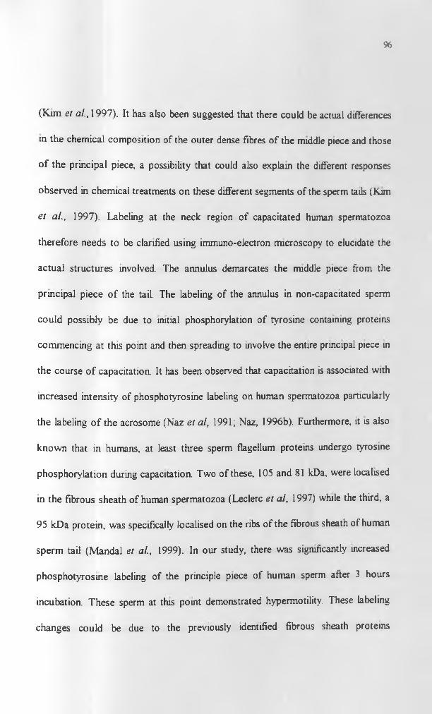
96
(Kim et al., 1997). It has also been suggested that there could be actual differences
in the chemical composition of the outer dense fibres of the middle piece and those
of the principal piece, a possibility that could also explain the different responses
observed in chemical treatments on these different segments of the sperm tails (Kim
et al., 1997). Labeling at the neck region of capacitated human spermatozoa
therefore needs to be clarified using immuno-electron microscopy to elucidate the
actual structures involved. The annulus demarcates the middle piece from the
principal piece of the tail. The labeling of the annulus in non-capacitated sperm
could possibly be due to initial phosphorylation of tyrosine containing proteins
commencing at this point and then spreading to involve the entire principal piece in
the course of capacitation. It has been observed that capacitation is associated with
increased intensity of phosphotyrosine labeling on human spermatozoa particularly
the labeling o f the acrosome (Naz et al, 1991; Naz, 1996b). Furthermore, it is also
known that in humans, at least three sperm flagellum proteins undergo tyrosine
phosphorylation during capacitation. Two of these, 105 and 81 kDa, were localised
in the fibrous sheath of human spermatozoa (Leclerc et al, 1997) while the third, a
95 kDa protein, was specifically localised on the ribs of the fibrous sheath of human
sperm tail (Mandal et al., 1999). In our study, there was significantly increased
phosphotyrosine labeling of the principle piece of human sperm after 3 hours
incubation. These sperm at this point demonstrated hypermotility. These labeling
changes could be due to the previously identified fibrous sheath proteins
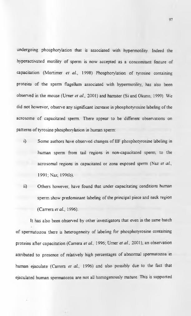
97
undergoing phosphorylation that is associated with hypermotility. Indeed the
hyperactivated motility of sperm is now accepted as a concomitant feature of
capacitation (Mortimer et al., 1998) Phosphorylation of tyrosine containing
proteins of the sperm flagellum associated with hypermotility, has also been
observed in the mouse (Urner et al., 2001) and hamster (Si and Okuno, 1999). We
did not however, observe any significant increase in phosphotyrosine labeling of the
acrosome of capacitated sperm. There appear to be different observations on
patterns of tyrosine phosphorylation in human sperm:
i) Some authors have observed changes of IIF phosphotyrosine labeling in
human sperm from tail regions in non-capacitated sperm, to the
acrosomal regions in capacitated or zona exposed sperm (Naz et al.,
1991; Naz, 1996b).
ii) Others however, have found that under capacitating conditions human
sperm show predominant labeling of the principal piece and neck region
(Carrera et al., 1996).
It has also been observed by other investigators that even in the same batch
of spermatozoa there is heterogeneity of labeling for phosphotyrosine containing
proteins after capacitation (Carrera et al., 1996; Urner et al., 2001), an observation
attributed to presence of relatively high percentages of abnormal spermatozoa in
human ejaculate (Carrera et al., 1996) and also possibly due to the fact that
ejaculated human spermatozoa are not all homogenously mature. This is supported

98
by recent data that has shown existence of substantially different human sperm
subpopulations in each normal ejaculate, each with varied rates of tyrosine
phosphorylation in response to in vitro capacitating conditions (Buffone et al.,
2004). However, under the capacitation conditions used in our study there were
predominant changes in labeling of the sperm flagellum in human spermatozoa.
Immunoblots of baboon testis and spermatozoa identified low, medium and
high molecular weight phosphotyrosine containing proteins. Some of the proteins
had almost similar apparent molecular weights in baboon testis and spermatozoa
and possibly were the same proteins. Naz and Ahmad, (1994) reported presence of
14-18 kDa, 51kJDa, 63kDa and 95kDa phosphotyrosine containing proteins. Our
studies did not however include demonstration of changes in degree of tyrosine
phosphorylation of these protein bands following capacitation of baboon sperm.
This, coupled with possible effects of zona proteins on the baboon sperm protein
tyrosine phosphorylation, would be a possible area for further study in an
endeavour to understand capacitational changes in spermatozoa of this species.
2.6.4. M l
mAb Ml was tested by indirect immunofluorescence on human and baboon
sperm where it did not recognise any determinant in both species. This particular
antibody had been raised against purified hamster sperm heads on which it
recognised an epitope at the equatorial segment of hamster sperm (Noor and

99
Moore, 1999). This antibody cross-reacted with spermatozoa from a number of
mammals (Noor and Moore, 1999). However, in our experiments there was no
evidence of cross-reactivity with human and baboon spermatozoa implying that the
particular determinant was not conserved in these primates.
2.6.5. 18.6
Monoclonal antibody 18.6 labeled the anterior acrosome in both baboon
and human spermatozoa. Baboon testis probed with the same antibody had FITC
labeling on luminal spermatozoa and none on the germinal epithelium of the
seminiferous tubules. The mAb 18.6 has been used as a specific marker of the
acrosome (Moore et al., 1990). The cognate antigen is localised to the acrosomal
membranes and contents of human spermatozoa. This antigen appears to be one
that is conserved in spermatozoa of eutherian mammals as it has been observed in a
number of species including rodents. However, a precise role of this antigen in the
fertilisation process is still unclear as the antibody does not affect sperm-zona free
hamster and sperm-zona (intact) binding interactions in hamster and humans
(Moore et al, 1990). Nevertheless its conservation in mammalian sperm and
localization on acrosomal membranes and acrosomal matrix is suggestive of a role
in the fertilisation process. Our results confirm earlier observations on localization
of the antigen at the anterior acrosome of human spermatozoa and additionally
demonstrate presence of the same antigen at a similar locus in baboon spermatozoa.

100
Presence of the antigen on luminal spermatozoa and not the seminiferous
epithelium could imply that the epitope for mAb 18.6 becomes “unmasked” just at
the time of spermiation or the final modifications to the antigen resulting in the
immunoreactive epitope occurs almost at spermiation.
2.6.6. Conclusions
The studies demonstrate presence of shared antigens like IAM-1, ES-1,
Phosphotyrosine proteins and 18.6 in man and the baboon spermatozoa using both
indirect immunofluorescence (ILF) and immunoblots of 1-D gels. Some antigens
found in rodents like Ml appear not to be conserved in primates. IAM-1 is a non-
glycosylated integral membrane protem or a protein well anchored to the inner
acrosomal and equatorial segment membranes.

101
CHAPTER 3
2-D PAGE AND BIOCHEMICAL ANALYSIS OF IAM-1 BY
MASS SPECTROMETRY
3.0. Introduction
3.1. One vs. Two-dimensional gel electrophoresis
One dimensional sodium dodecyl sulphate polyacrylamide gel
electrophoresis (SDS-PAGE) offers a means of separating proteins on the basis
of their molecular weight. The drawback with this mode of separation is the
limited resolution it offers making it impossible to distinguish between proteins
of similar molecular weight and post-translationally modified forms of the same
protein (Graves and Haystead, 2002). Two-dimensional (2-D) SDS-PAGE
offers a reproducible method of resolving individual proteins from a complex
mixture on the basis of both their charges and molecular weights (Dove, 1999)
thus providing a solution to some of the problems inherent in the one
dimensional separation. Furthermore, 2-D gels enable separation of isoforms
and post-translationally modified proteins on the basis of their charge
differences (Brewis, 1999).

102
3.2. 2-D gel electrophoresis
3.2.1. Principles of 2-D gel electrophoresis
Two-dimensional (2-D) gel electrophoresis is a technique for separation
o f proteins involving a combination of a first dimension where proteins are
separated along a pH gradient on the basis of differences in their charges
(isoelectric focusing) and a second dimension where focused proteins are further
separated on an SDS polyacrylamide gel on the basis of their molecular weights
(O ’Farrel, 1975; Fichmann and Westermeier, 1999).
Isoelectric focusing (DEF) is possible as amino acids are amphoteric
molecules that either possess a net negative, positive or zero charge depending
on the pH of the solution they are contained in (Berkelman et al., 1998). The
net charge a protein possesses is contributed by its amino acid side chains, its
carboxyl and amino termini and groups contributed as a result of post-
translational modifications. At pH values below their isoelectric points (pi)
amino acids have a net positive charge and at pH values above their pi they
have a net negative charge. At their pi, they have no net charge. In IEF,
proteins migrate along a pH gradient under the influence of an electric field.
Therefore, a protein with a net positive charge migrates towards the cathode
and as the pH increases along the gradient it becomes progressively less positive
until it reaches its pi where it stops moving. Likewise, a protein with net
negative charge migrates towards the anode until it reaches its pi, where it
becomes stationery in the electric field as it has no net charge (Berkelman et al.,
1998). Thus proteins can be separated on the basis of very small charge

103
differences using EEF. The individual proteins are concentrated at their different
isoelectric points under the influence of the electric field, hence the term
“isoelectric focusing”.
The concept of 2-D gel electrophoresis was first documented by Macko
and Stegemann (1969). However, the actual 2-D methods were run successfully
only after Klose (1975) and O’Farell (1975) separately developed effective
sample preparation protocols. The basic method initially involved use of a tube
gel rod that had mixtures of carrier ampholytes (small soluble amphoteric
molecules) that had high buffering capacity close to their pi. When an electric
field was applied across the rod the ampholytes established the pH gradient
when they migrated to their pis. The sample was focused in this gradient and
then transferred to a vertical slab gel for the second dimension. This method
became the ‘IsoDalt’ system when it was improved by Anderson and .Anderson
(1978), who optimized running conditions for replicate samples.
The technique of Immobilized pH gradients (IPG's) was introduced by
Bjellquist et al. (1982). The IPG’s differed from the earlier pH gradients in that
the acrylamido buffers (these comprise a single basic or acidic group linked to
an acrylamide monomer) that form the pH gradient are covalently incorporated
into a polyacrylamide gel when it is cast (Berkelman et a l 1998). The
proportion of acidic or basic acrylamido buffer content defines the resultant pH
gradient. The acrylamide monomers of the buffers link to the bisacrylamide and
acrylamide monomers of the gel and undergo simultaneous polymerization and
incorporation into the gel matrix as it sets. The gel is supported by casting on a

104
plastic backing. Gorg et al., (1988) pioneered application of IPG technology to
2-D electrophoresis. Use of IPG’s in the first dimension of 2-D electrophoresis
(also called IPG-Dalt) resulted in enhanced reproducibility and resolution,
improved ease o f handling of the gel and stability of the pH gradient (Berkelman
et al., 1998; Fichmann and Westermeier, 1999). These features coupled with the
high loading capacity of IPGs have led to rapid spot identification enabling
microsequencing, amino acid analysis and mass spectrometry (Gorg and Weiss,
1999).
3.2.2. 2-D gel electrophoresis of sperm proteins
Sperm proteins from various species have been separated using 2-D gel
electrophoresis with different objectives in mind:
2-D gel electrophoresis has been applied to general studies of sperm proteins
such as in the determination of electrophoretic maps of plasma membrane
proteins in boar sperm (Russell et al., 1983) and human sperm (Naaby-Hansen,
1990). It has also been applied to more specific studies like those investigating
phosphorylated sperm plasma membrane proteins in bovine epididymal sperm
(Noland et al., 1984), identifying specific proteins such as actin in rabbit sperm
(Welch and O ’Rand, 1985) or in the isolation of cognate proteins for antisperm,
auto and iso-antibodies (Shetty et al., 1999). Recently, post-vasectomy
autoantibodies that were predominantly against outer dense fibre proteins ot rat
sperm were characterised by 2-D electrophoresis (Flickinger et al., 2001).

105
When plasma membranes from human sperm were analysed, up to 260
proteins between pis 4.5 - 7.8 were identified (Naaby-Hansen, 1990). The
limitations of 2-D techniques using human sperm include the small protein
recovery per ejaculate, staining variability of extracted proteins and the limited
samples available per individual. Kritsas et al, (1992), described an improved
technique using mini-gels, for limited sperm sample protein analysis. The
preponderance of acidic proteins in boar and bovine sperm plasma membrane
extracts was observed (Peterson et al., 1983; Fenner et al., 1992).
Several specific sperm proteins have been characterised by 2-D SDS-
PAGE. Among the proteins characterised are:
(i) P26h, which is a sperm protein of epididymal origin involved in gamete
interaction in the hamster. This protein was shown to be relatively hydrophilic
with a basic pi o f about 8.3 (Coutu et al., 1996).
(ii) SP-10, a polymorphic intra-acrosomal protein of molecular weights 18-
45kDa has been analyzed using 2-D gel electrophoresis (Freemerman et al.,
1994). The protein has been recognised in several species including, humans,
baboons, macaques, pigs, foxes, mice and rats. The SP-10 peptides in human
sperm were found to have pis clustered around 4.9 (Freemerman et al., 1994).
(iii) MSA-63, an antigen of molecular weight 24-84 kDa and pi range of 4-6 has
been characterised in the mouse using 2-D (Liu et al., 1992).

106
3.2.3 Membrane protein isolation
Molloy, (2000) reviews various techniques used by different
investigators for 2-D membrane protein analysis. It has been noted that 2-D
electrophoretic separations of proteins in both eukaryotes and prokaryotes is
characterised by poor representation of the membrane and other hydrophobic
proteins (Santoni et al., 2000; Molloy, 2000). In prokaryote cell preparations
using carrier ampholyte tube gels and IPG's for 2-D runs, there was poor
representation of the hydrophobic membrane proteins compared to those
predicted in genomes (Wilkins et al, 1998; Molloy, 2000). This was attributed
to poor initial solubilisation of the proteins using conventional solubilising
solutions, reduced solubility, and loss of protein at pi due to precipitation and
adsorption (Rabilloud, 1998; Molloy, 2000). In spite of using the current range
of detergents, incorporation of thiourea and reducing agents in the solubilisation
buffer, resolution o f certain more hydrophobic membrane proteins is still not
possible (Molloy et al, 2000). There is so far no universal protocol that works
effectively in extraction of both eukaryotic and prokaryotic membrane proteins.
Moreover, almost all the protocols are best suited to isolating denatured or
reduced proteins. There is therefore, need to test and adapt specific protocols
depending on the nature of the particular membrane protein of interest.

107
3.3. Mass spectrometric identification of proteins
Mass spectrometry is becoming increasingly applied in identification of
purified proteins due to its sensitivity of analysis and high sample throughput
(Griffin et al„ 1992; Aebersold and Mann, 2003). It can be used in concert with
other techniques of protein purification like 2-D gel electrophoresis that can
separate specific proteins in a complex mixture of proteins (Aebersold and
Mann, 2003). Separated or purified proteins are digested and then subjected to
mass spectrometry. Mass spectrometers can analyse peptide masses providing a
peptide mass fingerprint or peptides to give an amino acid sequence by tandem
mass spectrometry (MS/MS) (Graves and Haystead, 2002). There are different
types of mass spectrometers (MS) but they all essentially have three
components:
(i) an ion source that can be Electrospray ionization (ESI) or Matrix assisted
/aser t/esorption/zonization (MALDI),
(ii) a mass analyser which could be an ion trap, time of flight (TOF), quadrupole
and Fourier transform ion cyclotron analysers,
(iii) a detector that records the number of ions at each mass/charge (m/z) value.
Therefore based on different combinations of these basic components, there
different mass spectrometers like the Triple quadrupole MS, Quadrupole time of
flight MS, Fourier transform MS and MALDI-TOF MS (Aebersold and Mann,
2003). Peptides are analyzed in gas phase where they are ionized and charged.
Using MALDI-TOF (time of flight) mass spectrometer the result is a mass
spectrum depicting mass/charge (m/z) ratios of the peptides in the digest. The

108
m/z data obtained are used to search the protein databases using peptide-
database searching algorithms (peptide matching/peptide mass fingerprinting) to
identify the specific protein or family of related proteins (Figeys et al., 2001;
Aebersold and Mann, 2003; Steen and Mann, 2004). It is also possible to obtain
the internal peptide sequence of the digested protein using tandem mass
spectrometry in Triple Quadrupole MS and Quadrupole TOF MS and other
MS. In these MS a specific peptide can be selected and its peptide bonds
sequentially broken giving the partial sequence of amino acids making up the
protein (Graves and Haystead, 2002). There are different algorithms available
for searching sequence databases such as Peptide search, Sequest, Protein
prospector and Mascot. These make use of the partial sequence data to identify
matching proteins from predicted and known sequences within databases (Steen
and Mann, 2004).

109
3.4. Specific aims of this chapter
1. To identify the specific protein recognised by mAb IAM-1 on human
sperm using 2-D gel electrophoresis techniques.
2. To obtain sufficient quantities of the protein for characterisation, using
peptide mass fingerprinting and/or N-terminal sequencing and/or internal
peptide sequencing.
110
3.5. Materials and Methods
2-D electrophoresis was performed according to a modified version of
the protocol by Berkelman et al., (1998) to optimize conditions for isolation of
non-reducing proteins. Briefly, it involved the following steps depicted in the
flow chart (Table 4):
- Sample preparation
- Rehydration o f IPG strips
- Isoelectric Focusing
- 2nd Dimension SDS PAGE
- Visualization and analysis of results
3.5.1. Sample preparation
Human sperm was prepared according to the protocol given earlier in
Chapter 2 (2.4.1.1). Following this initial treatment, 50 - 150 million sperm
were used for protein extraction from sperm for 2-D gels. The sperm were
resuspended in 3.25 mis of Phosphate buffered saline (PBS) pH=7.4, placed on
ice and sonicated in a Soniprep 150 Ultrasonic disintegrator (Sanyo Gallenkamp
PLC) at an amplitude of 10.5 p for 2 minutes. The resultant solution was
centrifuged at 10,600g for 10 minutes and the pellet resuspended in 200pls of
PBS.

Table 4. Flow chart of protocol used in isolation and characterization of IAM-1 from
human sperm.

I l l
3.5.1.1. Alkaline extraction of sperm proteins
200pls of sonicated washed sperm (prepared as in 3.5 .1) was mixed with
2.5mls of pre-chilled 0.1M sodium bicarbonate solution and placed on a rotary
mixer at 4 °C. for 1 hour. The suspension was centrifuged at 115,000 g for 20
minutes. The supernatant was removed and the pellet re-suspended in 2.5mls of
PBS then centrifuged at 115,000g for a further 20 minutes. The pellets were
collected and re-suspended in 350pls of lysis solution plus 1 pi of reconstituted
creatinine phosphokinase (CPK) pi range 4.9 - 7.1 (from the Carbamylate pi
calibration kit for 2-D electrophoresis Amersham Pharmacia Biotech) and left to
stand at room temperature for 1 hour. The solution was centrifuged at 17,000g
for 10 minutes. 350pls of the supernatant was used for rehydration of IPG
strips. Table 5 highlights the various alternative solutions used for protein
extraction from sonicated sperm to determine the optimal extraction solution for
the IAM-1 protein. It was found that the optimal extraction solution for IAM-1
was that in treatment vii (Table 5) and therefore solutions constituted in a
similar manner were used for further investigations.
3.5.1.2. Non- alkaline extraction
The washed sperm suspension in PBS (see end of 3.5.1) was centrifuged
at 960 g for 8 minutes and the pellet resuspended in 0.5 mis of lysis buffer and
placed on rotary mixer for a minimum of 4 hours at room temperature. After
this period of time, 250 pis of rehydration solution (8M Lrea, 2% CHAPS,
0.5% v/v IPG buffer and few grains of bromophenol blue in distilled water) was
added to the 100 pis of lysis solution (treatment (iv) in Table 5) -sperm

Table 5. Various solutions used for extraction of sperm proteins for 1-D gel
electrophoresis and observed efficacy in IAM-1 extraction of each as assessed
using intensity of ECL on immunoblots.
Treatment solution used Immunodetection using ECL
i) SDS reducing loading buffer —
ii) SDS non-reducing loading buffer ++++
iii) 1M Sodium Chloride extraction
then SDS (non-reducing)
loading buffer to supernatant
—
iv) Lysis solution with 9.5M Urea, 4% Chaps,
0.5%IPG pH4 - 7, PMSF, Distilled Water. +++
v) Lysis solution with 9.5M Urea. 4% Chaps,
0.5%IPG pH4-7, PMSF, DTT, Distilled Water, —
vi) Lysis solution with 9.5M Urea,
0.5-2% Triton X-100, 0.5% IPG,
PMSF, Distilled water
+
vii) Lysis solution with 9M Urea,
2M Thiourea, 2% ASB-14,
l%Triton X-100, 0.5% IPG
pH4-7, PMSF, Distilled water
- Indicates nothing detected using ECL
+ Indicates weakly positive response
+++ Strong positive response
++++ Very strong positive responseCHAPS (3-(3-cholamidopropyl)-dimethylammonio)-l-propanesulIonate)
PMSF- Phenylmethane sulfonyl fluoride(Sigma)
DTT - Dithiothreitol

112
suspension plus 1 pi of reconstituted creatinine phosphokinase and kept
standing for a further 1 hour. The sample was then centrifuged at 10,600 g and
the supernatant removed and loaded on to a reswelling tray to rehydrate an
immobilised pH gradient (IPG) strip. For the treatment vii in Table 5, having
ASB-I4 as the detergent, the same lysis extract was used for rehydration of the
IPG strip with no additional rehydration solution.
3.5.2. 2-D electrophoresis
3.5.2.1. Rehydration of Immobilized pH Gradient Strips (IPGs)
Rehydration and sample loading of EPG strips was done at the same
time. 350pls or 300pls of rehydration solution (containing the sample) was
aliquoted onto the slots on the Immobiline DryStrip Reswelling Tray
(Amersham Pharmacia Biotech UK Limited). The 350pls was for 18 cm IPG
strips (Amersham Pharmacia Biotech.) while 300pls was for 17cm IPG strips
(Bio-rad). The IPG strips were carefully placed on the rehydration solution gel
face downwards and overlain with 2 mis of IPG cover fluid ( Amersham
Pharmacia Biotech). The strip was allowed to rehydrate for a minimum of 12
hours at room temperature.
3.5.2.2. Isoelectric Focusing (IEF)
This step required preparation of the Immobiline Dry Strip Kit
(Amersham Pharmacia Biotech) comprising an Immobiline DryStrip Tray,
DryStrip aligner, electrodes, electrode strips, sample cup bar and sample cups
The kit was set up on a Multiphor II electrophoresis unit (Amersham Pharmacia

113
Biotech.) cooling plate. The MultiTemp III thermostatic circulator (Amersham
Pharmacia Biotech.) connected to the electrophoresis unit, was set to 20 °C.
Rehydrated IPG strips were removed from the reswelling tray, rinsed with
distilled water briefly and excess moisture blotted off. They were placed on the
Immobiline drystrip aligner, gel face upwards within the tray, with the anodic
side facing the positive side of the cooling plate. Two IEF electrode strips, each
110 mm long, were cut and soaked in 500 pis of distilled water. The IEF strips
were aligned in contact with the ends of the gels on either ends of the IPG
strips. The appropriate electrodes were placed on top of the electrode strips at
the ends of the IPG strips.
Where additional sample solution was loaded to the IPG strips, sample
cups were placed on the sample bar, positioned on the Immobiline drystrip tray
in contact with the cathodic electrode. The sample cups were carefully adjusted
in contact with the LPG strips. IPG cover fluid was poured to cover the IPG
strips and sample cups. lOOpls of sample was loaded onto the sample cups and
it gravitated into the sample cups.
IEF was done for a total of 75,000 - 80,000 Volt Hours in gradient mode as in
Table 6.

114
Table 6. Programme used for Isoelectric focusing for 18 cm IPG strips
Phase Voltage(V) Current(mA) Power(W) VH1 500 1 5 12 500 1 5 35003 3,500 1 5 10,0004 3,500 1 5 72,500
The electrode strips were changed twice during LEF. At the end of IEF, the IPG
strip was either immediately stored at -20 °C or equilibrated (see Appendix for
equilibration solution) for 30 minutes before the second dimension SDS PAGE.
3.5.2.3. 2nd dimension SDS PAGE
The thermostatic circulator was set to 15 °C. A precast ExcelGeF SDS,
gradient 8-18 polyacrylamide gel (Amersham Pharmacia Biotech), 110 x 245 x
0.5 mm, was opened and air-dried for 8 minutes then carefully placed on about
4 mis of cover fluid on the cooling plate. A Multiphor buffer strip positioner
(Amersham Pharmacia Biotech.) was set in place with the cathodic and anodic
buffer strips positioned on either side, in contact with the gel. The IPG strip was
removed from the equilibration solution and excess solution blotted off the ends.
It was then positioned, gel face downwards, on the SDS gel parallel to the
cathodic buffer strip. Sample application pieces were placed at either end of the
IPG strip. 3pis of low molecular weight markers (Dalton Mark VII-L TM,
SIGM A), 14,200 - 66,000 kDa, was diluted with 7pls of equilibration solution
and 5 pis of the resulting solution loaded on two sample application pieces. The
two pieces were placed one on either side of the IPG strip, at the same level, on
the SDS gel.
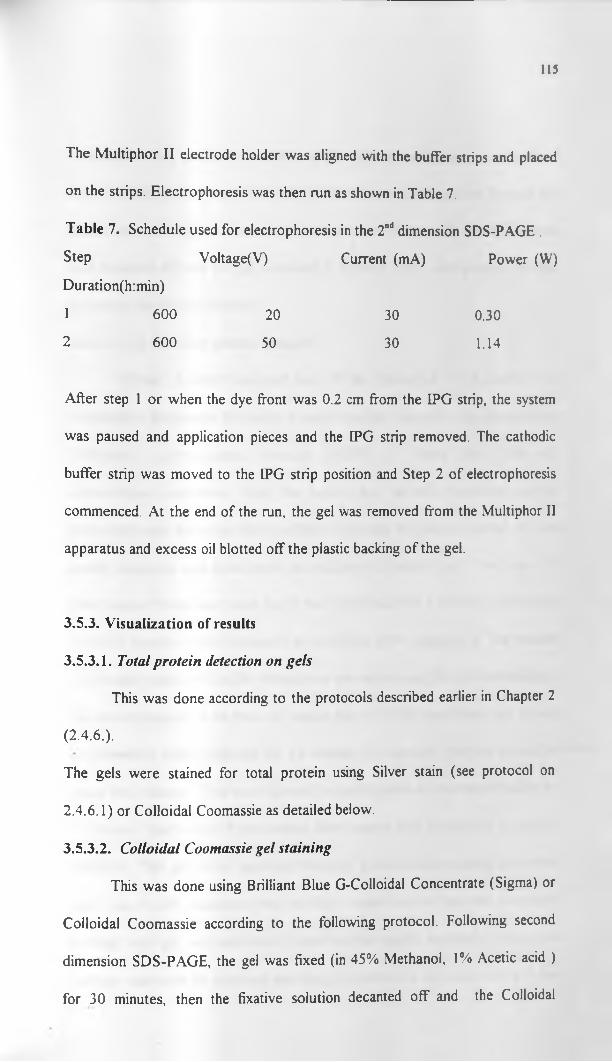
115
The Multiphor II electrode holder was aligned with the buffer strips and placed
on the strips. Electrophoresis was then run as shown in Table 7.
Table 7. Schedule used for electrophoresis in the 2nd dimension SDS-PAGE .
Step
Duration(h:min)
Voltage(V) Current (mA) Power (W)
1 600 20 30 0.30
2 600 50 30 1.14
After step 1 or when the dye front was 0.2 cm from the IPG strip, the system
was paused and application pieces and the IPG strip removed. The cathodic
buffer strip was moved to the IPG strip position and Step 2 of electrophoresis
commenced. At the end of the run, the gel was removed from the Multiphor II
apparatus and excess oil blotted off the plastic backing of the gel.
3.5.3. Visualization of results
3.5.3.1. Total protein detection on gels
This was done according to the protocols described earlier in Chapter 2
(2.4.6.).
The gels were stained for total protein using Silver stain (see protocol on
2.4.6.1) or Colloidal Coomassie as detailed below.
3.5.3.2. Colloidal Coomassie gel staining
This was done using Brilliant Blue G-Colloidal Concentrate (Sigma) or
Colloidal Coomassie according to the following protocol. Following second
dimension SDS-PAGE, the gel was fixed (in 45% Methanol, 1% Acetic acid )
for 30 minutes, then the fixative solution decanted off and the Colloidal
116
Coomassie stain, prepared according to the manufacturers instructions (diluted
first 1:4 with distilled water then 4:1 in absolute methanol), added The gel was
placed on a rocking platform at room temperature, for 18 - 24 hours. The stain
was decanted off and the gel destained in distilled water until suitable contrast
of protein spots was obtained.
3.5.3.3. 2-D Semi-dry protein transfer
Protein Transfer involved use of the Multiphor II Novablot Unit
(Amersham Pharmacia Biotech). Protein transfer was done to Immobilon-P
(Millipore), polyvinylidene fluoride (PVDF) or Trans Blot (Bio-rad),
nitrocellulose membrane. Both the anodic and cathodic novablot graphite
electrodes were saturated with distilled water then the excess blotted off. The
anodic electrode was fitted onto the multiphor II buffer tank. Two sets of 9
filter papers (Whatmans) were cut to the dimensions 110 x 240 mm, and wetted
in CAPS transfer buffer (prepared as described in the Appendix). The transfer
buffer used was CAPS buffer where there was subsequent protein sequencing of
the spots obtained on the blots. A similar size of PVDF membrane was cut and
pre-wetted in 100% methanol for 15 seconds. It was then hydrated in distilled
water for 2 minutes. This was followed by equilibration in the transfer butfer for
5 minutes. One set of 9 pre-wetted filter papers was placed on the anodic
electrode. The gels were separated from the plastic backing using a novablot
unit. The PVDF membrane was carefully placed on the gel and the plastic
backing with gel and membrane placed on the anodic electrode. The plastic
backing was carefully removed and the gel overlain by the other set ot 9 pre
117
wetted filter papers. The cathode electrode was then fitted over the sandwich,
the safety lid placed and the unit connected. The transfer was done at 150mA
(about 0.8mA /cm2 per hour) for 2 hours.
3.5.3.4. Total protein detection on blots
Total protein detection and immunodetection was combined according
to the protocol of Chevallet et al., (1997) to facilitate accurate localisation of
immunoreactive spots. Briefly this involved: removal of the PVDF membrane
from the Novablot unit following protein transfer. All the subsequent steps were
performed on a rocking table; washing, 3 times for 5 minutes ( 3 x 5 ) each in
30% ethanol, followed by washing 3 x 5 in 0.05% v/v, Tween20 in distilled
water; staining overnight in colloidal gold total protein stain (Bio-rad) diluted
1:3 with 0.05% Tween20 in distilled water for total protein; The membrane was
then rinsed 3 x 5 minutes in PBS containing 0.05% Tween20 and 1% w/v,
polyvinylpyrrolidone (Sigma); blocking was then done in the same solution for
2 hours followed by primary antibody incubation with I AM -1 supernatant tor 2
hours. The membrane was then rinsed 6 x 5 minutes in PBS with 0.05%
Tween20. This was followed by a 1 hour incubation with Anti-mouse IgG
(raised in Sheep) conjugated to horse radish peroxidase (Amersham Pharmacia
Biotech), diluted 1:1,500 with PBS containing 0.05% Tween20. Finally there
was a 6 x 5 minutes wash in PBS containing 0.05% Tween20.
Visualization of Immunoreactions was done using Enhanced
Chemiluminescence (ECL+Plus) Western blotting detection kit (Amersham
118
Pharmacia Biotech) according to the manufacturers protocol described earlier in
Chapter 2 (see 2.4.5.3.2).
3.5.3.5. Excision o f protein bands or spots
3.5.3.5.I. Gel proteins
This was done following Colloidal Coomassie staining of the gel, for
MALDI-TOF (A/atrix Assisted laser Desorption /onisation Time Of Flight
mass spectrometry) and for Q-TOF (Quadrupole/orthogonal time of flight)
tandem mass spectrometry. The bands of interest were identified, carefully
excised with sterile scapel blades and placed in separate sterile microfuge tubes.
A part of the gel without protein (a control spot) was cut and placed in a
microfuge. The samples were then kept at 4 °C until analyzed. Peptide mass
fingerprinting was performed using a Voyager-DE PRO MALDI-TOF mass
spectrometer at the Microchemical Facility of Babraham Institute in Cambridge
UK.
For tandem mass spectrometry, peptides resulting following trypsin
digestion were separated by reverse phase HPLC in a 15 cm PepMap C l8
column then internal peptide sequencing was performed using a
Quadrupole/orthogonal acceleration time of flight (Q-TOF) mass spectrometer
(Micromass).

119
3.5.3.5.2. Blot proteins
Western blots were performed on human sperm 2-D gels and these were
stained for total protein using Coomassie blue stain (protocol similar to 2.4.6.2).
Spots corresponding to IAM-1 were carefully excised and placed in sterile
microfuge tubes. They were stored at 4 °C until analyzed. N-terminal
sequencing was performed by Edman degradation at the Babraham Institute in
Cambridge, UK.

120
3.6. Results
3.6.1 IAM-1 extraction
Extraction of human sperm proteins using various lysis solutions shown
in Table 5 (treatments i - vii) provided further information regarding the
physico-chemical properties of IAM-1 and its interactions with mAb IAM-1.
Inclusion of reducing agents whether (3-mercaptoethanol as in treatment (i) or
dithiothreitol as in (v) resulted in non-recognition of the antigen by the mAb
IAM-1. Extraction solutions containing the anionic detergent SDS and the
zwitterionic amido sulfobetaine detergent ASB-14 realized the most efficient
extraction of IAM-1 from the sperm membranes. Furthermore, when the
detergent ASB-14 was used, efficiency of extraction was enhanced at high urea
concentrations (9M) and by inclusion of the additional chaotrope thiourea as in
treatment (vii). Extraction solutions with the non-ionic detergent Tritron X-100
as the only detergent resulted in poor recovery of IAM-1 (treatment vi). Use of
strongly ionic solutions of sodium chloride alone did not extract IAM-1 from
the sperm membranes (treatment iii).
It was also noted that despite strong immunoreactivity using ECL
detection on 2-D blots, the protein IAM-1 was barely discernible on 2-D blots
stained with colloidal gold total protein stain (Figs. 19, 20, 21). In some of these
blots other sperm proteins were intensely stained indicating that a good amount
of other sperm proteins were actually transferred onto the membranes (Fig. 19).
NAIROBI UNIVERSITY KABETE LIBRARY

121
3.6.2. 2-D electrophoresis
3.6.2.1. 2-D gels
Several human sperm proteins were resolved following 2-D
electrophoresis. These proteins were mainly clustered between pi’s 4.2 and 8.7.
They were relatively low molecular weight proteins of between 1 0 - 8 0 kDa
(Fig. 18).
3.6.2.2. 2-D immunoblots
3.6.2.2.1. Non-alkalineprotein immunoblots
Immunoreactive spots were accurately assigned on Western blots
according to the method of Chevallet et al., (1997). Immunoprobing with mAb
IAM-1 on 2-D blots of the human sperm proteins (extracted without prior
alkaline incubation with lOOmM sodium carbonate) revealed immunoreactive
spots of apparent p i’s 3.86 - 4.0. These spots had apparent molecular weights
of 23, 27 and 32 kDa (Fig 19).
3.6.2.2.2. Alkaline protein immunoblots
When human sperm proteins were extracted following incubation under
alkaline conditions (with lOOmM sodium carbonate) and 2-D separation
performed, immunoblots of the resolved proteins probed with mAb IAM-1 had
immunoreactive spots with pi's of 5.8 and 6.2 and apparent molecular weights
o f about 25 kDa (Fig. 20).
When narrower range Immobilised pH gradient strips ot pH 4-7 were
used for the 2-D separation of the same proteins, the resolved I AM-1
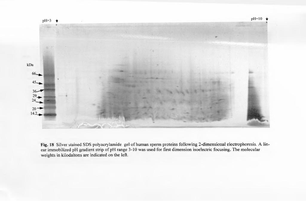
Fig. 18 Silver stained SDS polyacrylamide gel of human sperm proteins following 2-dimensional electrophoresis. A linear immobilized pH gradient strip of pH range 3-10 was used for first dimension isoelectric focusing. The molecular weights in kilodaltons are indicated on the left.

Fig. 19 Immunoblot of whole human sperm, lysis buffer protein extract (without alkaline treatment ), stained with colloidal gold total protein stain. The blot was overlain with ECL Kodak film (see overlay) and the arrow indicates the position of protein spots immunoreactive to mAb 1AM-I of apparent pi ~3.86- 4.0 and apparent molecular weights of ~ 23, 27 and 32 kDa.
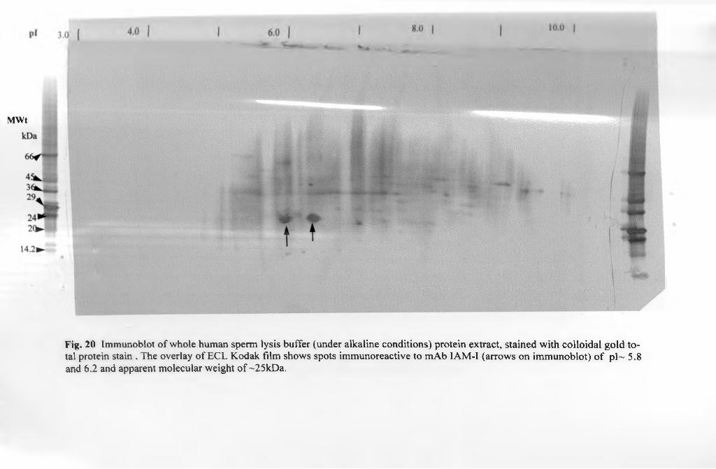
MWt
Fig. 20 Immunoblot of whole human sperm lysis buffer (under alkaline conditions) protein extract, stained with colloidal gold total protein stain . The overlay of ECL Kodak film shows spots immunoreactive to mAb IAM-I (arrows on immunoblot) of pl~ 5.8 and 6.2 and apparent molecular weight of ~25kDa.

p i 4 .0 j 5.0 6.0 7.0I
I
MWt
Fig. 21 Immunoblot of whole human sperm lysis buffer protein extract, following alkaline treatment and 2-dimensional gel electrophoresis, stained with colloidal gold total protein stain. Arrows show position of the proteins immunoreactive to mAb IAM-1, of mean apparent pis -5.32, 5.61 and 6.07 and mean apparent molecular weights of - 24kDa.
4 »

122
immunoreactive spots had pi's of 5.27 - 5.37, 5.5 - 5.72 and 5.98 - 6.16 and
corresponding apparent molecular weights of 23.5, 24.5 and 23.3 kDa
respectively (Fig. 21). Therefore it was apparent that alkaline incubation prior to
extraction shifted the pi of IAM-1 to make it less acidic.
3.6.3. N-terminal sequencing
An N-terminal amino acid sequence was obtained from protein spots on
human sperm western blots (Table 8). The selected spots corresponded to
immunoreactive IAM-1 protein spots. The sequence of 10 amino acids obtained
was not complete as there were no amino acids detected at positions 4, 6 and 8
Furthermore, in some cases there were changes in the amino acid residues with
each cycle performed as in 2, 3 and 9 (Table 8). A search of the protein
databases using the possible combinations of the amino acids did not yield any
significant match.
Table 8. N-terminal sequence of IAM-1
1 2 3 4 5 6 7 8 9 10
s L G X A X I X G R
V L L
G
3.6.4. Peptide mass fingerprinting
Following trypsin digestion and MALDI-TOF mass spectrometry, mass
spectra were obtained for two spots corresponding to IAM-1 immunoreactive

123
proteins excised from the western blots (Figs 22a, 22b). The resultant masses
representative o f peptides from trypsin digestion of the two spots are listed in
Table 9. Various protein databases were interrogated using these masses, in an
attempt to identify the proteins in the two spots. The search engine used was the
MS-Fit (http.y/www-tbsc.ncifcrf.gov/ucsfhtml3 2/mstit.htni) while the protein
databases interrogated included SWISS-PROT (Tables 10 and 11) and NCBInr
databases. However for both of the spots there was low percentage of peptide
masses matched to actual proteins in the databases with the highest for Spot 1
being 23% (Table 10) and for Spot 2 being 33% (Table 11) in the SWISS-
PROT database.
3.6.5. Internal peptide sequencing
Internal peptide sequencing of two immunoreactive spots yielded a
number of peptides. These were matched in the protein databases to Trypsin
like proteins, Keratin, Glutathione transferase mu 3, alpha 3/7 tubulin, Ras
related protein and others (Table 12). It was noteworthy that keratin, trypsin
like proteins and alpha tubulin were detected in both of the spots. Keratin was
matched to that of human and sheep. The trypsin however was matched to
trypsin found in three different species namely, human, porcine and canine.

PE B iosystem s Voyager System 6022ap*c 7.,mctbp . air*.
Fig.22a. MALDI mass spectrum of one of the spots immunoreactive to IAM-1 of apparent molecular weight 23,419 Da and pl~ 6.0
Fig.22b. MALDI mass spectrum of one of the protein spots immunoreactive to IAM-1 of apparent molecular weight 25,941 Da and pl~ 5.6

T a b le 9 . M asses o f P ep tid es obtained from M A L D I-T O F mass spectrom eter
following trypsin digestion of two spots of IAM-1 immunoreactive proteins.
Masses of Peptides from Spot 1 Masses of Peptides from Spot 2
846.3955 856.6282
855.0765 862.5609
871.1054 878.4478
872.3176 882.7488
877.0589 884.7136
883.0702 894.4957
886.5982 918.6918
900.1572 1166.8822
917.3644 1973.3489
923.5960 1994.2490
1066.1256 2283.1312
1121.5903 2399.9055
1207.5269 2565.71 18
1209.6414 2705.4805
1253.6869 3122.4889
1307.7012
1708.8802
2283.1831
2338.1567
2367.2086
2566.2191

T a b le 10. D ata ob tained fo r Spot 1 masses using M S-Fit search engine on the SwissProt
database. T he top 5 m atches are shown in the low er part o f table.
Sample ID (comment): Magic Bullet digestDatabase searched: SwissProt. 11.02.2001Molecular weight search (1000 - 50000 Da) selects 73280 entries.Full pi range: 100225 entries.Species search ( MAMMALS ) selects 18164 entries.Combined molecular weight, pi and species searches select 11425 entries. MS-Fit search selects 74 entries (results displayed for top 5 matches).Considered modifications: | Peptide N-terminal Gin to pyroGlu Oxidation of M Acetylated |
Protein N- terminus
Min. # 3eptides ® Match
4
Peptide Mass Tolerance
(+/-)500.000 ppm
PeptideMasses
aremonoisoto
pic
DigestUsed
Trypsin
Max. # ^ , • Peptide .. . , Cysteines v. Missed . N ModifiedCleavag , terminuses , y .. Hydroge , acrvlamide 1 n (H)
Peptide C terminus Free Acid
(OH)
Input # Peptide Masses
21
Result Summary
. MOWSE * ( % ) oak Masses ScoreMatched
Protein MW (Da)/pl
SwissProt.
Species 1 1 0 2 *2001 Protein Name v Accession
#
ADIPOPHILIN (ADIPOSEI 1.12e+03 5/21 (23%) 46664.3/6.42 MOUSE P43883 DIFFERENTIATION-RELATED
PROTEIN) (ADRP)ACETYLCHOLINESTERASE COLLAGENIC TAIL PEPTIDE PRECURSOR (ACHE Q
: 479 4/21 (19 % ) 47911.9/8.92 RAT 03516, SUBUNIT)(ACETYLCHOLINESTERASE- ASSOCIATED COLLAGEN) DELTA3,5-DELTA2,4-
0354^ DIENOYL-COA ISOMERASE,414 4 /2 1(19 %) 36118.7/7.60 MOUSE - 5— * MITOCHONDRIAL
PRECURSORSODIUM/POTASSIUM- TRANSPORTING ATPASE BETA-1 CHAIN
4 261 4/21 (19 % ) 35071.5/8.72 SHEEP P05028 (s q d IUM/POTASSIUM-DEPENDENT ATPASE BETA-1 SUBUNIT)
43294.3 / 5.69 PIG04640 APOLIPOPROTEIN A-IV
5 244 4/21 (19 % ) 9 PRECURSOR (APO-AIV)

T a b l e 11. Data ob tained from masses derived from Spot 2 using M S-Fit search engine on
the Sw issProt. database. T h e top 5 matches are listed in the lower part o f the table.
Sample ID (comment): Magic Bullet digestDatabase searched: SwissProt. 1 1.02.2001Molecular weight search (1000 - 50000 Da) selects 73280 entries.Full pi range: 100225 entries.Species search ( MAMMALS ) selects 18164 entries.Combined molecular weight, pi and species searches select 11425 entries.MS-Fit search selects 20 entries (results displayed for top 5 matches).Considered modifications: | Peptide N-terminal Gin to pyroGlu Oxidation of M Protein N-terminus Acetylated |
Min. # Peptides
toMatch
4
Peptide Mass
Toleran ce (+/-) 500.000
ppm
Peptide « . Max. # CysteinesMasses Missed Modified
are SC . Cleavages bymonoisotopic OP510 j acrylamide
PeptideN
terminus Hydroge
n (H)
PeptideC
terminus Free Acid
(OH)
Rank MOWSEScore
#(%)Masses
Matched
Result SummarySwissProt.
Protein Species 11 02-2001 MW (Da)/pl v Accession
#Protein Name
Input # Peptide Masses
15
I
3
4
5
480
411
353
182
177
4/15(26 %) 36720.4/7.13 BOVIN
4/15(26 %) 26908.9/6.75 BOVIN
5 /15 (3 3 % ) 47578.9/9.17 HUMAN
4 /15(26%) 41834.7/8.91 HUMAN
4/15(26%) 43135.8/6.28 HUMAN
P05980
P09611
P09543
000532
P2~361
PROSTAG LAND IN-F SYNTHASE 1 (PGF SYNTHASE 1) (PGF 1) (PROSTAGLANDIN-D2 11 REDUCTASE 1) (PGFSI)PLACENTAL LACTOGEN I PRECURSOR (BPLP-I)2\3'-CYCLIC NUCLEOTIDE 3'- PHOSPHODIESTERASE (CNP) (CNPASE)SERIN E/THREONINE- PROTEIN KINASE KKIALRE (CYCLIN- DEPENDENT KINASELIKE 1)MITOGEN-ACTIVATED PROTEIN KINASE 3 (EXTRACELLULAR SIGNAL-REGULATED KINASE 1) (ERK-1) (INSULIN- STIMULATED MAP2 KINASE) (MAP KINASE 1) (MAPK 1) (P44-ERK1) (ERT2) (P44-MAPK) (MICROTUBULE- ASSOCIATED PROTEIN-2 KINASE)

T a b l e 12. Internal peptide sequence o f various pro teins obtained from tandem m ass spectrom etry o f 2 presum ptive IAM-1 proteinspots and corresponding matches derived from the protein database searches
Spot 1Peptide Sequences Database Match Score Peptides
M atchedM olecular weight (Da)
P*A c c e s s io n
LGEHNIDVLEGNEQFINAAKLSSPATLNSRVATVSLPR
Trypsin precursor (Porcine) (EC 3.4.21.4) 428.6 3 24,391 7.4 P00761
Ql.VESDINGLR Keratin, type 1 cuticular HA5 (Hair keratin) 135.4 1 47,578 4.8 Q92764VDIIENQVMDFR LKPQY LEELPGQLK
Glutathione S-transferase mu 3 119.2 2 26,670 5.4 P21266
E1VDLVLDR Tubulin alpha-3/alpha7 chain 72.6 1 49,941 5.0 P05214l.FDQAFGLPR Heat shock 27 kDa protein (HSP27) 71.3 1 22,920 6.6 P42929VGEYSLYIGR Serum amyloid p-component precursor 53.3 1 25,369 6.5 P3743VVL1GDSGVGK Ras related protein Rab-1 1 a (RAB-11) 42.9 1 24,375 6.5 P24410FQELESETl.K Prostatic acid phosphatase precursor 39.4 1 44,548 6.2 PI 5309NKATSFHLGQLK Hypothetical 40.7 kDa protein in Pykl-S 33.4 1 40,683 8.7 P39729
Spot 2
LGEHNIDVLEGNEQFINAAKLSSPATLNSRIITHPNFNGNTLDNDIML1KVATVSLPR
Trypsin precursor (Porcine) (EC3.4.21.4) 438.9 4 24,391 7.4 P00761
QLVESDINGLRDSLENTLTETEAR
Keratin, type I microfibrillar 48 kDa 234.6 2 46,656 4.8 P02534
TQNPMVTGTSVLGVK Proteasome beta chain precursor 83.4 1 29,174 5.9 P28373VLEGNEQFINAAK Trypsin III precursor (Human) (EC 3.4.21.4) 61.1 1 26,758 6.3 PI 5951Tl.LDIDNTR Keratin, type I cytoskeletal 9 53.0 1 61,969 5.2 P35527It DELTLCR Keratin, type I cuticular HA3-I 31.6 1 45,917 4.9 076009VATISLPR Trypsin, anionic precursor (Canine) (EC3.4.21.4) 31.2 1 26,404 4.7 P06872IIATASVLGVK 6-phosphofructokinase beta subunit 26.3 1 10,4970 6.9 Q03216EIVDLVLDR Tubulin alpha chain, testis-specific 24.0 1 49,975 5.0 PI 8288

124
3.7. Discussion
Two-dimensional gel electrophoresis (2-D) is a powerful tool that
enables separation of complex mixtures of proteins into purified constituent
polypeptides. 2-D reference maps of human spermatozoal proteins have recently
been established (Naaby-Hansen, 1990; Xu et al., 1994; Naaby-Hansen et al.,
1997). However, despite availability of this data it is just a few of the proteins
that have actually been identified on the maps and of these, fewer still have
undergone molecular characterisation. Among the subset of sperm proteins so
far identified using 2-D are a number of sperm surface proteins including those
of seminal plasma origin (Naaby-Hansen et al., 1997) and those found on the
sperm membranes (Xu et al., 1994). Sperm surface proteins overlying the inner
acrosomal membrane and equatorial segment such as IAM-1 would be of
particular interest as they could be important in gamete interaction at
fertilisation
3.7.1. LAM-1 extraction
Three-dimensional conformation of proteins is important functionally
and is dependent on non-covalent interactions between regions along the
polypeptide chain. It is also known that some antibodies are directed against the
resultant conformational epitopes on the protein molecules (Rabilloud, 1996).
Our experimental results using reducing agents in extraction solutions indicate
that there is the presence of at least one conformational epitope on the I AM-1
125
protein as treatment of sperm proteins with these agents resulted in non-
recognition of the cognate protein by mAb LAM-1. For solubilisation of proteins
to take place, there has to be disruption of its non-covalent interactions
(Rabilloud, 1996). The combination of chaotropes like urea and thiourea with
reducing agents completely unfold proteins, with chaotropes breaking
intermolecular hydrogen and hydrophobic bonds while reducing agents break
any disulfide bonds.
In our study, inclusion of reducing agents like the alkyl thiol,
mercaptoethanol or cyclizable thiols like dithiothreital (DTT) in the extraction
solution irreversibly affected the subsequent binding of the mAb IAM-1 to the
cognate antigen. This was most likely due to presence of a conformational
epitope whose integrity was maintained by the presence of one or more disulfide
bonds. It is therefore hypothesized here that inclusion of the reducing agents
reduced the disulfide bond(s) leading to the loss of native conformation of the
protein and with it, the epitope for mAb IAM-1, resulting in non-recognition by
the antibody.
Use o f a combination of colloidal gold total protein stain which is a
relatively sensitive protein stain (detects as low as lng of protein), coupled with
ECL detection methods demonstrated that the mAb IAM-1 had a very strong
affinity for the cognate protein as it immunoreacted with quantities of the
antigen that were barely discemable on western blots of human sperm. The
intense staining of other sperm proteins on the same 2-D blots where IAM-1

126
was barely visible indicated that insufficient amounts of IAM-1, unlike many
other sperm proteins, were transferred and bound to the blots.
This could have arisen due to:
i) low amounts of the protein being initially available on sperm
membranes or
ii) poor extraction of IAM-1 from the sperm membranes or
iii) loss of the protein during the 2-D electrophoresis
iv) loss o f the protein during the processing of the blots.
Loss o f membrane and other poorly soluble proteins during isoelectric
focusing (IEF) occurs during sample entry into the IEF gel due to stacking of
proteins and aggregation resulting in precipitation and more so during IEF when
proteins near their isoelectric point. At this crucial stage loss of proteins can
either arise from adsorption of the proteins to the relatively hydrophobic matrix
of the Immobilized pH gradient strip or aggregation due to reduced repulsive
charges on the individual proteins close to isoelectric point leading to
precipitation (Rabilloud, 1996; Rabilloud, 1998). Attempts were made to
optimize extraction of this membrane protein and reduce loss of the same during
IEF as follows:
i) Addition of thiourea to solubilisation buffer as was done enhances
protein solubility even close to isoelectric point and improves the
overall protein loading capacity of the gel (Rabilloud, 1998).
ii) Urea denatures proteins including relatively hydrophobic ones, in
aqueous solutions exposing hydrophobic residues to the solvent.

127
ASB14
IIS — 0 “ Na
llo
SDS
0 ----- (CH2CH20)9----- H
Triton X-100
Fig. 23. Structural formulae of the detergents used in extractton of IAM-1 from
human sperm. ASB14 and CHAPS are zwitteriomc detergents while SDS is an
anionic detergent and Triton X-100 is a non-ionic detergent

128
iii) Detergents are used to prevent hydrophobic interactions of the exposed
residues that could lead to protein aggregation and precipitation.
Both ASB-14 and CHAPS are sulfobetaine zwitterionic detergents, with
ASB-14 having an amido group added between the sulfobetaine head and a
linear alkyl hydrophobic tail while CHAPS has the amido group between the
polycyclic hydrophobic tail and the sulfobetaine head (Fig. 23).
Our studies demonstrated improved recoveries of IAM-1 when
extraction solutions contained the detergent ASB-14 or SDS compared to non
ionic (Triton X-100) and another zwitterionic detergent (CHAPS). It is known
that SDS is a particularly effective anionic detergent useful in solubilising a wide
range of proteins. In the solubilisation process at least two SDS molecules bind
to each amino acid imparting an overall negative charge to the denatured
protein molecules (Molloy, 2000). This curtails the use of this detergent in
Isoelectric focusing as this separation technique exploits differences in the
intrinsic surface charge of protein molecules (Santoni et al., 2000).
ASB-14 provided equally efficient extraction of IAM-1 in solubilising
solutions. This particular detergent has also been used successfully in extraction
of relatively hydrophobic bacterial membrane proteins (Molloy et al., 2000). Its
greater efficacy as a sulfobetaine detergent has been attributed to the presence
of its long alkyl hydrophobic tail unlike the polycyclic hydrophobic tail in
CHAPS (see Fig.23). Indeed the findings that ASB-14 and SDS provide
relatively more efficient extraction of IAM-1 compared to Tritron X-100 and
CHAPS would be consistent with the hypothesis that IAM-1 is a relatively

129
hydrophobic integral membrane protein or a membrane protein well anchored to
the inner acrosomal membrane.
Strong solutions of NaCl disrupt bonding due to ionic or electrostatic
interactions between peripheral membrane proteins and other membrane
proteins or with the phospholipids of the membranes (Rabilloud, 1996; Santoni
et al., 2000). The same occurs when solutions with high pH are used as was the
case during sodium carbonate treatment of sperm in our extractions. In fact
Fujiki et al., (1982) state that carbonate treatment discriminates between
peripheral and integral proteins with the latter remaining attached to cell
membranes after carbonate treatment at pH=l 1. Therefore failure of carbonate
treatment coupled with concentrated NaCl solutions to extract IAM-1
confirmed that the protein was not a peripheral membrane protein but one
intimately associated with the acrosomal membranes possibly as an integral
membrane protein or protein well anchored to the lipid component of the
membranes.
3.7.2. 2-D gels and immunoblots
Earlier 2-D work on sperm proteins using carrier ampholyte gel rods for
the first dimension had preponderance of neutral and acidic proteins between
pi’s 5.6-6.9. These were relatively low molecular weight proteins of less than
lOOkDa (Naaby-Hansen, 1990; Naaby-Hansen et al., 1997). Similar findings
were obtained using immobilised pH gradient gels tor the first dimension in 2-D
separation o f human sperm proteins (Xu et al., 1994). It was also noted in the

130
latter study that membrane proteins constituted less than 15% of proteins
isolated by 2-D electrophoresis of human sperm. However, so far there are no
studies reporting separation of human sperm proteins under non-reducing
conditions as were used in our experiments. Our findings on 2-D gels of human
sperm proteins were in agreement with the earlier findings where there was
more representation of the lower molecular weight proteins (<80kDa) and
proteins with p i’s of less than 9 (Naaby-Hansen, 1990).
Other techniques such as use of Non Equilibrium pH gradient
electrophoresis (NEPHGE) have improved the resolution of more basic sperm
proteins (Naaby-Hansen et ai, 1997). However, for the purposes of our study,
use of conventional immobilized pH gradients were adequate to localize the
protein of interest, IAM-1, on 2-D gels.
Other earlier 2-D SDS PAGE studies have also recorded the presence of
proteins that formed “charge trains” or different isoforms (Naaby-Hansen, 1990;
Xu et a i , 1994; Diekman et a i , 1997; Naaby-Hansen et al., 1997). These were a
series of spots that were isoforms of the same protein on the gels with almost
similar apparent molecular weights but differing pis. They are thought to arise
as a result o f variations in the extent of post-translational modifications on the
particular proteins (Naaby-Hansen et a l 1997). From our study, IAM-1 is an
acidic protein that in native form has a pi of about 4.0. Treatment of this protein
with an alkaline solution of sodium carbonate (pH=l 1) altered its pi to between
5.3 and 6.0. It is likely that the acidic nature of the native protein could be partly
contributed to by post-translational groups linked to the protein.
131
Post-translational modifications of proteins are important in determining:
their eventual location within the cell, lifespan within the cell like acetylation and
regulating activity of cellular proteins as in phosphorylation. Post-translational
modification o f proteins like phosphorylation, are also known to alter migration
patterns of proteins on gels (Hao et a l., 2002). It is therefore conceivable that
addition of alkali to IAM-1 hydrolyzes the post-translationally added groups
responsible for acidity of the protein, with the resultant isoforms of the protein
having higher or more basic pi values. Predominance of negatively charged
amino acid residues like aspartic acid and glutamic acid within the structure of
IAM-1 could also contribute to its acidic pi.
It is unlikely that alteration of the pi of IAM-1 was due to dissociation
of subunits of the protein as there were no significant changes in the apparent
molecular weights of the isoforms before and after alkaline treatment.
Therefore, the proposed mechanism involving hydrolysis of post-translational
groups by the alkaline treatment is the more likely mechanism by which there
was alteration in the pi value of IAM-1.
3.7.3. N-terminal sequencing
N-terminal sequencing was performed on protein spots excised from
human sperm western blots. The sequence obtained for 10 cycles was
incomplete as no amino acids were detected in some cycles. In other cycles the
precise identification of the amino acid obtained was uncertain as more than one
type of amino acids was obtained at the same position. Inconsistency in the
132
amino acids obtained at certain positions during sequencing was attributed to
low quantity o f sample protein that necessitated use of higher sensitivity settings
for detection. The higher settings could have contributed to variations due to
background interference.
Apparent lack of amino acids at certain positions on the other hand
could have arisen as a result of blocked N-terminal amino acids. It is known that
many eukaryotic proteins have blocked N-terminal amino acids due to post-
translational modification and as a result are refractory to Edman degradation
particularly the initial coupling step of the process (Brown and Roberts, 1976;
Brown, 1979). Therefore blockage of N-termini by glycosylation, acetylation,
formylation, fatty acid acylation and cyclization or other modifications could
have contributed to the missing amino acids at some of the positions in the
protein. Under such circumstances internal peptide sequencing can be carried
out to get a partial sequence for identification or the protein can be processed
further to remove the groups blocking the N-termini (LeGendre et al., 1993). A
search of the protein data bases using the various permutations of the sequence
data we obtained was inconclusive as there were no significant matches.
Therefore, peptide mass fingerprinting and internal peptide sequencing of the
protein was carried out to characterise I AM-1.

133
3.7.4. Peptide mass fingerprinting
Database searches using mass spectra obtained from two spots
corresponding to IAM-1 were inconclusive. The percentage of peptides
matched to particular proteins was generally less than 45% for each of the
spots. Identification of a protein in peptide mass fingerprinting is dependent on
availability of matches within the protein or translated genomic databases
(Aebersold and Mann, 2003). Furthermore, the more the peptides that match
(higher % matching) the more likely a protein can be related to a particular
family of proteins or to a specific protein from the database (Figeys et a/.,
2001). Despite obtaining a large number of peptides in digests for both spots,
the percentage of peptides matched per protein spot was relatively low possibly
indicating that IAM-1 could be a novel sperm membrane protein hitherto
undefined in the protein databases.
3.7.5. Internal peptide sequencing
Data obtained from internal peptide sequencing of 2 spots
immunoreactive to mAb IAM-1 revealed a number of peptide sequences with
homology to trypsin-like proteins in both spots. The other types of protein
common to both spots were the keratin related proteins and alpha tubulin
proteins. Other proteins were identified in only one of the two spots that were
sequenced.
The sequencing of multiple peptides of different proteins in each of the
spots could also be attributed to the non-reducing conditions used in our

134
experiments that resulted in horizontal smears rather than distinct spots in the 2-
D gels. It is known that the presence of reducing agents during isoelectric
focusing improves resolution of protein spots in the second dimension of
separation (Gorg et al., 2000). Due to the constraints of the non-reducing
conditions required for IAM-1 isolation it was possible that other adjacent
proteins with close pis’, were also excised for analysis from the resolving gel,
contributing to the peptides of several proteins obtained in each spot sequenced
Some of the proteins that had high scores, appeared in both protein spots and
had a few similar biochemical characteristics to IAM-1 are discussed below.
3.7.5.1. Keratin
There were a number of peptides isolated that were matched to various
keratin types in humans and sheep. Keratins are proteins expressed in body cells
particularly epidermal cells as cytokeratins or soft alpha keratins while in hair
and other keratinized structures they are expressed as hair or hard alpha keratin.
Both cytokeratins and hair keratin exist as heterodimers of type I (acidic) and
type II (basic to neutral) proteins forming intermediate filaments (Rogers et al.,
1998). Cytokeratins have also been identified in germ cells, particularly the
sperm flagellum (Hinsch et al., 2003).
In our experiment four keratin types were isolated with three being of
human origin and one of sheep wool origin. It is likely that the two human
cuticular keratins (Q92764 and 076009) and one sheep wool keratin (P025j 4)
were contaminants introduced during the processing ot the samples. The other

135
keratin (Pj 5527) isolated could have also been a contaminant from skin cells or
could have been a sperm protein.
Recent work involving antibodies raised against purified recombinant
peptides of epidermal K9 recognised testicular K9 found associated with
manchette microtubules. Furthermore, significant homology in cDNA and amino
acid sequences o f testicular and epidermal K9 was observed when the gene was
cloned in the two tissues (Mochida et al., 2000). However, following spermatid
remodelling and disappearance of the manchette the fate of K9 is unclear.
Although keratins have been isolated as components of the mature sperm
cytoskeleton, they are found in the outer dense fibres and the fibrous sheath of
the flagellum (Kierszenbaum, 2001; Hinsch et al., 2003). Furthermore the
spermatid K9 has an apparent molecular weight of 62-64 kDa. (Mochida et al.,
2000). Therefore while sperm cytokeratins are found in the flagellum of
spermatozoa, IAM-1 is found associated with the acrosomal membranes and
equatorial region of the spermatozoon. The biochemical characteristics further
suggest that I A M -1 is not K9 and therefore the latter could have been either a
contaminant or a sperm protein.
3.7.5.2. Trypsin-like Proteins
It is noteworthy that there were different peptides obtained during the
sequencing of both spots, with a number corresponding to trypsin-like proteins
in database searches. The entries with the best matches for both spots were
however, homologous to a trypsin-like serine protease ot porcine origin
(accession number: P00761). This could imply that the porcine trypsin used for

136
digestion prior to Q-TOF MS/MS, underwent autolysis and the resultant
peptides were sequenced.
On the other hand, the matching of certain peptides obtained to human
and canine trypsin-like proteins complicates the picture further. While the
peptide that was matched to the human trypsin III precursor (P35030) is similar
to a segment o f one of the peptides that was matched to porcine trypsin
(P00761), the short peptide that was matched to canine trypsin anionic
precursor differed from either human or porcine trypsin by one amino acid
isoleucine substituting for valine in the canine trypsin. However, it is known that
partial sequence data cannot guarantee a 100% identity as some proteins differ
by a small number of amino acids that may not be in the segment sequenced
(Charbonneau, 1993). The import of these findings would be that there could be
a trypsin-like protein in human sperm that has certain structural characteristics
similar to canine and porcine trypsins.
Expression of trypsin-like proteins in the testis especially on haploid
germ cells and epididymal spermatozoa has been observed in the mouse and pig
(Akama et a l, 1994; Ohmura et al., 1999). Trypsin-like proteases are serine
proteases with Arg/Lys-Xaa cleavage specificity. Among the serine proteases
with such catalytic function found in the mammalian sperm acrosome are
proacrosin/acrosin (Crosby et al, 1998), trypsin-like proteases (Akama et al.,
1994) and some of the testicular .serine proteases 1-5 (TESP 1-5) identified in
mouse sperm (Honda et al., 2002b) among others. In both the mouse and
human sperm, serine protease activity during sperm-zona interaction appears to

137
have a crucial function as inhibitors of these proteases actually prevent sperm-
zona penetration in vitro (Fraser, 1982; Liu and Baker, 1993).
It is thought that trypsin-like proteases in mammalian sperm may be
involved in enzymatic dispersal of acrosomal contents during acrosome
reaction, limited hydrolytic activity on the zona pellucida during sperm
penetration, cleavage and subsequent activation of certain acrosomal matrix
constituents and lectin-like carbohydrate binding properties and hence
involvement in sperm-zona pellucida secondary binding (Ohmura et al., 1999;
Honda et al., 2002a). It has indeed been suggested that among the proteases
activated following acrosome reaction are a group of serine proteases that are
associated with the acrosomal membranes and this latter group may
subsequently be involved in sperm-zona pellucida interactions (Honda et al.,
2002b).
An inner acrosomal membrane-associated trypsin-like protein in human
sperm would therefore be an attractive prospect as IAM-1 is also associated
with the inner acrosomal membrane. Furthermore, following preparation ot
human sperm under alkaline conditions in our experiments, we resolved proteins
of pi’s between 5.3 - 6.2 and apparent molecular weights between 23-25kDa.
The theoretical calculated molecular weights for trypsins range between 24 -
27kDa in the different species with pi s of 4.7 - 7.4. It would therefore be in
keeping with these expected results and with the known functions of trypsin-like
proteases in acrosomes of spermatozoa to have such a protein in the human
sperm acrosome. Therefore, on the basis ot the biochemical parameters ot

138
trypsin-like proteins, the functional characteristics of serine proteases that form
acrosomal contents in other species, and the relatively high scores obtained in
matches for trypsin-like proteins in our experiments and the proposed functions
of trypsin-like serine proteases in the acrosome, it appears that IAM-1 is a
membrane-anchored trypsin-like protein.
3.7.5.3. Glutathione S-transferase
Two of the peptides sequenced following the trypsin digestion were
matched to human glutathione S-transferase mu3. Glutathione S-transferases
exist in several forms and are expressed in many body tissues. They are
primarily cytosolic detoxification enzymes that offer protection against reactive
oxygen species of exogenous or endogenous origin (Hayes and Pulford, 1995;
Kidd et al., 2003). Reactive oxygen species include the superoxide anion (O2),
hydrogen peroxide (H2O2), hydroxyl radical ( OH) and hydroxide anion (’OH)
which are produced during metabolic redox reactions in body cells (Loscalzo,
2004). Though there are three classes of this enzyme in the body, it is only the p
class that is expressed in brain and testicular tissue (Campbell et al., 1990). The
testicular Glutathione S-transferase p class isoenzyme has a pi of about 5.2,
similar to the brain one and both are thought to be associated with maintaining
the integrity o f the blood-testicular and blood-brain barriers in the respective
organs (Campbell et al., 1990).
Catalytically active p class Glutathione S-transferases have also been
identified on goat sperm plasma membrane where they also appear to be
involved in sperm-egg interaction particularly sperm-zona binding (Hemachand
139
et al., 2002). Furthermore, introduction of solubilised zona pellucida to
spermatozoa causes aggregation of Glutathione S-transferases and induces
acrosome reaction. The sperm membrane associated Glutathione S-transferase
had molecular weights of 23 -26 kDa but were removed by treatment with
strongly ionic solutions and immunolocalised to the sperm surface indicating
they were peripheral membrane proteins (Hemachand et al., 2002). Our findings
of peptides matching to p class Glutathione S-transferase in our 2-D gels is in
agreement with the earlier findings regarding presence of this enzyme on the
membranes associated with sperm. While earlier investigations pointed to its
presence in the human testis-blood barrier (Campbell et al., 1990), this study
demonstrates that it is one of the protein constituents of the ejaculated human
sperm. Further immunolocalisation work needs to be done to determine its
specific location on human sperm. It is however unlikely that Glutathione S-
transferase mu3 is IAM-1, judging from its biochemical characteristics
(Hemachand et al., 2002) that identify it as a peripheral protein on the plasma
membrane.
3.7.5.4. Tubulin alpha-3/7
This was first identified in mice where it is exclusively expressed in the
testis (Villasante et al., 1986). Genes encoding for the same protein were found
in humans. The alpha-3 and alpha-7 tubulins are identical though coded for by
different genes (Villasante et al., 1986). It is also known that alpha and beta
tubulins form dimers (Fanarraga et al., 1999). Subsequently other studies have

140
shown that alpha-3/7 tubulins are testis and sperm specific in humans. In the
sperm they are components of the axoneme while in the testis they have been
localised to the manchette, a structure associated with morphogenesis of sperm
during spermiogenesis (Mochida et al., 1998; 1999). Therefore, Tubulin alpha-
3/7 are cytoskeletal proteins found in germ cells.
The expected molecular weight of these proteins is about 49kDa with a
pi of about 5.0. From our experiment it had an apparent molecular weight of
25kDa. It could be that the hydrolysis of the tubulin dimers by the alkaline
conditions used in the experiment resulted in much smaller molecules of close to
25kDa. IAM-1 on the other hand is not a cytoskeletal protein and is localised to
the acrosome and equatorial segment and not the tail of sperm. Inclusion of
tubulin alpha3/7 among the digested peptides in both protein spots may be due
to the constraints in our study where reducing agents were not used in the 2-D
separation of proteins.
3.7.5.5. Heat shock 27kDa protein
Heat shock proteins are produced in various mammalian cells in
response to elevated temperature or exposure to stressful environments (Sarge,
1995). In these cells they act as molecular chaperones involved in protein
folding and transport and mediate development of tolerance to the physico
chemical stress (Bohring et al., 2001). Among the heat shock proteins that have
been localised on the surface of spermatozoa are 95kDa and 70 kDa proteins
(Miller et al., 1992b). More recently analysis of the human genome has revealed

141
that one of the outer dense fiber proteins HspBlO, has a lot of similarity to the
27kDa human heat shock protein (Fontaine et al., 2003). Our finding of a 27
kDa heat shock protein is therefore in agreement with earlier work that heat
shock proteins are found on sperm despite the inactive state of the sperm
genome. The precise function of these proteins however is still undetermined as
it is unlikely that they have similar functions in germ cells to that in somatic
cells.
3.7.5.6. Proteasomes
One of the peptides sequenced was matched to proteasome beta chain
precursor. Proteasomes are multicatalytic complexes of cellular proteases
involved in ATP dependent degradation of ubiquinated proteins (Tipler et al.,
1997). Proteasomes have been found in all eukaryotic cells so far studied
(Wojcik et al., 2000). Work done on human and mouse spermatozoa
demonstrated presence of 26S proteasomes in the gametes (Tipler et al., 1997).
Furthermore, proteasomes have been localised on the acrosome, post-acrosomal
region, nuclear vacuoles of neck region, mid-piece and cytoplasmic droplet of
normal human spermatozoa (Ziemba et al., 2002). Other work has localised
proteasomes particularly at the centriolar region in the neck and acrosome of
human sperm (Wojcik et al., 2000). It has been suggested that these enzymes
could be involved in degradation of histones leading to nuclear condensation
during spermiogenesis (Wojcik et al., 2000).

142
With regard to mature spermatozoa, proteasomes are thought to be
involved in degradation of paternal mitochondria during mammalian fertilisation
and in sperm-zona interaction (Sutovsky et al., 2003; 2004). While IAM-1 is
testis and sperm specific, proteasomes are generally found in many somatic cells
other than sperm. It is therefore unlikely that IAM-1 could be a proteasome
However, the finding of proteasomes in our 2-D studies on human sperm is
consistent with other studies that have localised these enzymes on mammalian
sperm (Tipler et al., 1997; Ziemba et al., 2002; Sutovsky et al., 2004)
The Ras related protein R ab-lla (RAB-11) like proteasomes is also
found in a number of somatic cells in human and also mouse tissues (Gromov et
al., 1998). On the same basis of tissue and species specificity it is unlikely that
IAM-1 is Rab-1 la.
3.7.6. Conclusion
Investigations to characterise IAM-1 were inconclusive with regard to
the precise identity of the protein. Examination of data obtained from the
internal peptide sequencing however, pointed to IAM-1 being a trypsin-like
protein more than any of the other proteins that were isolated. Biochemical data
supported its association with the acrosomal membranes as a membrane
anchored protein or integral membrane protein which in its native state has a pi
of 3.86-4.0 and apparent molecular weight of between 23-25 kDa.

143
Chapter 4
GENERAL DISCUSSION AND CONCLUSION
4.1. Fertilisation Antigens
A number of sperm antigens conserved among several mammalian species
have been studied (Feuchter et al.. 1981; Isahakia and Alexander, 1984; Eddy and
O'Brien, 1994; Naz, 1999). The initial observations were made using antisera
against whole sperm preparations (Hansen. 1972; D’Almeida and Voisin, 1977).
Later on, with the development of the hybridoma technique by Kohler and
Milstein (1975), monoclonal antibodies raised against specific epitopes of sperm
antigens confirmed the presence of common antigens on mammalian spermatozoa
(Feuchter et al., 1981; Isahakia and Alexander, 1984; Eddy and O'Brien, 1994;
Naz, 1999).
The present study on various fertilisation antigens in human and baboon
spermatozoa provided insight into the nature of some of the determinants found in
the two species. The primary antibodies used were monoclonal antibodies (mAbs)
previously raised against specific components of rodent or human spermatozoa.
Some of the mAbs like Ml that was raised against a determinant on hamster
spermatozoa (Noor and Moore. 1999), failed to recognise any determinant on both
human and baboon sperm. Others like mAbs ES-1 and IAM-1 raised against
determinants on purified human sperm heads (Al-Eisa et al., 2001), recognised
determinants at identical domains on baboon and human spermatozoa.

144
Furthermore, there was homology in the corresponding proteins on immunoblots
of sperm in the two species. Phosphotyrosine proteins were not restricted to
particular domains on spermatozoa in both species but were found in both the head
and tail of sperm in the two species. Furthermore, the patterns of phosphotyrosine
staining of baboon and human spermatozoa were similar. mAb 18.6 that
recognises an antigen found on the anterior acrosome of spermatozoa in a number
of mammals (Moore et al., 1990) localised similar determinants on the anterior
acrosome on both human and baboon sperm.
The import of all these findings taken together is that there is similarity in a
number of fertilisation antigens localised on baboon and human sperm. Earlier
work using rabbit antisera against testicular tissue demonstrated the presence of
shared testicular antigens among human, chimpanzee, rhesus monkey and the
baboon (Menge and Fuller, 1975). Moreover, further work using monoclonal
antibodies confirmed presence of similar epitopes on sperm from human, monkey,
dogs, rabbits, rats and mice as shown by cross-reacting antibodies in these species
(Isahakia and Alexander, 1984). Other studies have also confirmed presence of
shared antigens of testicular origin on baboon and human sperm (Isahakia, 1988).
Therefore, on one hand, our results particularly with mAb 18.6 and anti-
phosphotyrosine mAbs demonstrate presence of antigens, which conserved in a
number of mammalian species, in human and baboon sperm. On the other hand,
failure of mAb Ml to recognise any antigen on baboon and human sperm is
indicative that this particular determinant is not conserved in these species and

145
therefore may not be of importance with regard to fertilisation in these and
possibly other primates.
4.1.1. ES-1
ES-1 was one of the antigens that was localised on both baboon and human
sperm. Our studies demonstrated identical localisation of the ES-1 antigen on the
equatorial segment of both baboon and human sperm. Furthermore, immunoblots
of baboon and human sperm revealed cross-reacting antigens of similar molecular
weights in the two species. Electron microscopic immunolocalisation had shown
ES-1 on the plasma membrane overlying the equatorial segment of acrosome-
reacted human sperm (Al-Eisa et al., 2001). Moreover, in vitro fertilisation
inhibition studies using human sperm and zona-free hamster oocytes indicated that
the mAb ES-1 significantly inhibited sperm-oolemmal fusion process (Al-Eisa et
al., 2001). Taken together, our present findings and those of earlier studies (Al
Eisa et al., 2001) indicate that the determinant ES-1 may be involved in sperm-
oolemmal fusion in the two pnmates and suggests homology of the protein in the
two species. The extent of homology would need to be determined by sequencing
of the isolated purified proteins as the monoclonal antibody recognises a specific
epitope found on the protein in the two species.
In mammals, the plasma membrane overlying the apical equatorial
segment is the part of the acrosome-reacted sperm that initially fuses with the
oolemma (Moore and Bedford, 1978; Bedford et al., 1979; Sathananthan et al.,

146
1986). As in sperm-zona pellucida interactions it is thought that there maybe initial
recognition followed by adhesion events likely to involve molecules of the plasma
membrane at the equatorial segment and corresponding ligands on the oolemma
(Brewis and Moore, 1997; Evans et al., 2001). Furthermore, fusogemcity of the
equatorial segment unlike other domains of acrosome-reacted sperm has been
shown using liposomes (Arts et al., 1993). Though poorly understood, the
acquisition of fusion competence by the plasma membrane overlying the
equatorial segment is thought to be the result of action by some of the acrosomal
contents during acrosome reaction (Takano etal., 1993; Yanagimachi, 1994). The
presence of ES-1 on the plasma membrane of the equatorial segment of acrosome-
reacted sperm makes it a suitable candidate for further investigation regarding
sperm-oolemmal interactions.
Among the other sperm equatorial segment associated proteins that have
been isolated in mammals, are the 40kDa mouse protein recognised by M29
(Saling et al., 1986), the 30-5lkDa proteins for G11 and M l3 (Allen and Green,
1995), the 37.8 and 34 kDa hamster protein, Ml (Noor and Moore, 1999), a 36
kDa protein of pi—5.5 that was identified on human sperm (Auer et al., 2000), a 38
- 48 kDa protein called equatorm recognised by mAb MN9 that has been isolated
from sperm in rodents and man (Manandhar and Toshimori, 2001) and equatorial
segment protein of 34-38kDa and pi of 5.1-5.4 (Wolkowicz et al., 2003). One
common finding for these protein constituents of the equatorial segment is that

147
specific antibodies against certain epitopes on these proteins inhibit sperm-
oolemmal fusion (Wolkowicz etal., 2003).
Another interesting observation is that the molecular weights of the
proteins associated with the equatorial segment as reported by the different
investigators, tend to be clustered around 38-40kDa. However, most of them have
not been fully characterised hence it is difficult to know whether some could just
be the same phylogenetically conserved proteins isolated in different species. ES-1
has five bands including a 38kDa band and 40 kDa band in baboon and humans
respectively. Isolation and sequencing of the protein may be necessary to
characterise it further and determine whether the immunoreactive bands seen on 1-
D SDS gels are isoforms of the same protein and whether they could be related to
any of the other equatorial segment proteins that have been isolated in other
species and in humans.
4.1.2. IAM-1
This protein was localised on both human and baboon sperm. mAb IAM-1
localised the determinants to the equatorial segment and the anterior acrosome in
both species. Specific isolation and characterisation of this particular protein
through the protocols used in our experiments were not entirely unequivocal. This
was attributed to a number of inherent constraints due to the physical
characteristics of the cognate protein that have been discussed in the previous

148
chapter. Nevertheless, the following properties of the protein became evident from
our experimental data:
i) The protein was either an integral membrane protein or a protein that is well
anchored to the acrosomal membranes by a lipid anchor (Fig.24)
ii) The protein first appears during spermiogenesis in the seminiferous epithelium
associated with developing round spermatids
iii) The IAM-1 protein had a number of immunoreactive bands of molecular
weights of 28, 32, 39 and 45 kDa in human while it was 32, 38 and 44 kDa in
baboon sperm 1 -D immunoblots.
iv) The IAM-1 protein from human sperm separated by 2-D electrophoresis had a
pi of 3.86 - 4.0 and molecular weight 23-32 kDa while under alkaline treatment it
had a pi of 5.3-6.2 and molecular weight of 23-25 kDa. This data suggests that
LAM-l could be post-translationally modified making it more acidic in its native
state.
v) IAM-1 had at least one conformational epitope that was (or were) irreversibly
altered on introduction of reducing agents to the extraction solutions.
vi) IAM-1 is a non-glycosylated membrane protein.
vii) IAM-1 could be a trypsin-like protein.
Other than transmembrane proteins, eukaryotic proteins are attached to the
membranes by lipid anchors (Fig. 24), the most abundant being those anchored by
glycosylphosphatidylinositol (GPI) (Udenfriend and Kodukula, 1995). There are
reports in literature of lipid anchored sperm membrane proteins in the acrosomal
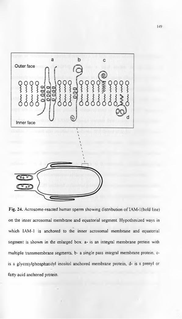
149
\
\
\\\\
\
Fig. 24. Acrosome-reacted human sperm showing distnbution of IAM-l(bold line)
on the inner acrosomal membrane and equatorial segment. Hypothesized ways in
which LAM-1 is anchored to the inner acrosomal membrane and equatorial
segment is shown in the enlarged box. a- is an integral membrane protein with
multiple transmembrane segments, b- a single pass integral membrane protein, c-
is a glycosylphosphatidyl inositol anchored membrane protein, d- is a prenyl or
fatty acid anchored protein.

150
region of mammalian sperm Recently, a 14 kDa sperm acrosomal /wembrane-
associated protein (SAMP 14) that has a lipid anchor in the form of
glycosylphosphatidylinositol was isolated in human sperm (Shetty et al., 2003).
While SAMP 14 is a 14 kDa protein with a pi of about 4.8 and is dissimilar to
IAM-1 in these respects, it also has characteristics similar to LAM-1 and in certain
ways could shed light on possible characteristics of IAM-1. PIPLC treatment
resulted in liberation of SAMP14 charge variants from sperm while for IAM-1
alkaline treatment also resulted in isoforms of different charges. SAMP 14 has a
domain with 10 cysteine residues on the amino terminus. Although it is not
presently clear how many cysteine residues are present on IAM-1 our data
indicated that it also has disulfide bonds in its structure contnbuting to the
formation of the conformational epitope recognised by the cognate antibody.
Another GPI anchored acrosomal protein that has been characterised is PH-
20 (Pnmakoff et al., 1985) which is localised to the inner acrosomal membrane of
human sperm following acrosome reaction (Sabeur et al., 1997). However, its
localization on the entire human sperm head plasma membrane before acrosome
reaction and molecular weight differences are basic differences PH-20 has when
compared to IAM-1 (Sabeur etal., 1997).
IAM-1 bears considerable similarity to SP-10 another acrosomal protein
that has been identified in human sperm. SP-10 is reported as being first seen in
the human testis where it is associated with the round and maturing spermatids of
the adluminal compartment (Herr et al., 1990a). In the present study, IAM-1 was

151
observed on the adluminal spermatids undergoing spermiogenesis within the
seminiferous tubules of the baboon. A similar pattern of IAM-1 distribution had
been observed in the human testis. On human spermatozoa, 1AM-1 was localised
on the inner acrosomal membrane and equatorial segment following lonophore
induced acrosomal reaction (Al-Eisa et a l, 2001). This distribution is identical to
that of SP-10 after ionophore induced acrosome reaction (Herr et al., 1990a). It has
been reported that there is a pool of SP-10 that is anchored to the acrosomal
membranes by TX-114 resistant anchors and that this pool is extracted only by
extreme pH conditions and strong detergents (Foster and Herr, 1992). This
compares favourably with the present data on IAM-1 which is not extracted by
strong ionic solutions, very poorly extracted in TX-100 another non-ionic
detergent and requires extreme pH, chaotropes and strong zwitteriomc detergents
for effective extraction. Based on these similarities, this could suggest lipid
anchorage of IAM-1 too as in the SP-10 case.
The present study has demonstrated IAM-1 on both baboon and human
sperm while SP-10 had also been identified earlier on sperm of both species (Herr
et al, 1990b). From the foregoing it is tempting to suggest that IAM-1 could be
SP-10; however, there are fundamentally significant differences in the biochemical
data obtained from each of these two human sperm proteins. While SP-10 appears
on SDS-PAGE gels as a series of polymorphic peptides that have a molecular
weight range between 18 -32 kDa in epididymal and ejaculated human sperm

152
(Herr et al, 1990a; Foster et al., 1994), IAM-1 had about 4 bands between 28-45
kDa in ejaculated human sperm.
Presence of multiple immunoreactive peptides of different molecular
weights has been attributed to endoproteolytic processing of intra-acrosomal
proteins or alternative splicing during transcription of the gene (Wnght et al.,
1990; Herr et al., 1992). Furthermore, it was noted that proteolytic processing of
SP-10 could begin during spermiogenesis within the testis and proceed to the
epididymal maturation stage (Foster et al., 1994). It is possible that IAM-1 protein
undergoes activation in the form of proteolytic hydrolysis in the course of
acrosome reaction hence the multiple immunoreactive bands seen. The pi of the
native protein is also clustered at 4.9 for SP-10 (Herr et al., 1990a) while that of
IAM-1 is between 3.8-4.0. These biochemical parameters confirm that though both
are acidic proteins, they are distinctly different on the basis of their molecular
weights and isoelectric points.
Another protein that has been localised to the equatorial segment and
anterior acrosome of sperm in a hamster, rat, rabbit, bull and human sperm is a
22kDa protein referred to as SP22 (Klinefelter et al., 2002). The human
homologue is DJ-1. However, despite similar immunolocalisation of SP-22 and
IAM-1 on human sperm and despite evidence of synthesis of SP-22 in spermatids
within the testis, SP-22 has been found in other tissues including the brain
(Klinefelter et al, 2002). It belongs to a family of conserved proteins found even in
prokaryotes (Welch et al., 1998). SP22/DJ-1 appears to be involved in zona

153
penetration and sperm-oolemmal fusion (Klinefelter et al., 2002; Okada et al.,
2002). IAM-1 on the other hand appears to be testis and sperm specific (Al-Eisa et
al., 2001), and so far appears to be found in primates and man and not rodents.
IAM-I is localised on the inner acrosomal membrane and at the equatorial
segment of human sperm following acrosome reaction indicating possible
involvement in secondary sperm-zona interactions and the fusion process of
gametes (Al-Eisa et al, 2001). Following acrosome reaction and loss of the
acrosomal ghost as the sperm penetrates the zona pellucida, the plasma membrane
overlying the equatorial segment and inner acrosomal membrane become the
surface components on the anterior part of the sperm head. LAM-l has been
immunolocalised under electron microscopy to the inner acrosomal membrane and
equatorial segment of acrosome-reacted human sperm (Al-Eisa et al., 2001). Our
studies have also shown similar immunolocalisation on capacitated and non-
capacitated human sperm permeabilised by methanol fixation. Moreover, there
was greater intensity of immunofluorescence at equatorial segment of human
sperm compared to anterior acrosome under confocal microscopy. Our studies
using non-capacitated baboon sperm also revealed predominant localization on the
equatorial segment in most of the methanol permeabilised sperm. On the basis of
equatorial segment localization it appears that IAM-1 could have a role in sperm-
oolemmal interaction particularly the fusion process.
There are possible hypotheses related to the localization of IAM-1 on the
inner acrosomal membrane. Evidence from the present study on internal peptide

154
sequence determination though equivocal, suggests this membrane protein is a
trypsin-like protease. Other observations we made were that it is intimately bound
or anchored to the inner acrosomal membrane and equatorial segment. This was
evident from:
i) the findings that following disruption of the acrosome by sonication there was
mAb IAM-1 epifluorescence on the anterior 2/3 of human sperm, likely to be the
inner acrosomal membrane and equatorial segment
ii) the extraction experiments with differing extraction solutions where despite
sonicating human sperm and treating the resultant sperm heads with strongly ionic
1M NaCl solution, there was still mAb IAM-1 epifluorescence evident on the
sperm heads.
A membrane-anchored serine-protease would be particularly useful as the
sperm penetrates the zona pellucida. Being associated with the inner acrosomal
membrane, IAM-1 would not participate in initial sperm-zona recognition events.
However, it could participate in the secondary sperm-zona interaction. Secondary
sperm-zona binding involves more persistent adhesion than the initial contact that
induces acrosomal exocytosis (Brewis and Moore, 1997). Though poorly studied
in man and other primates, it has been studied in more detail in the mouse where
the receptor on the zona pellucida has been identified as ZP2 (Bleil et al., 1988).
ZP2 is implicated as it binds acrosome-reacted sperm and not acrosome-
intact sperm (Mortillo and Wassarman, 1991). There is no consensus so tar on the
corresponding sperm ligand for ZP2. However, proacrosin/acrosin has been

155
proposed as a possible ligand for ZP2 While acrosin is a serine protease with other
hydrolytic functions in the acrosomal matrix, it was also shown to have lectin-like
zona binding activity possibly involving strong ionic interactions of the
carbohydrate moieties of ZP2 and basic residues on proacrosin/acrosin
(Richardson and O ’Rand, 1996; Howes and Jones, 2002). The case for acrosin or
trypsin-like protease involvement in zona penetration by human sperm was further
strengthened by the work of Liu and Baker (1993) that demonstrated lack of zona
penetration when trypsin inhibitor was used in vitro. This was complicated
somewhat by the observations that knock-out mice lacking the acrosin gene were
fully fertile in natural mating experiments, suggesting acrosin activity was not
requisite for zona penetration by sperm (Baba et al., 1994). Taken together these
findings imply that there could be other trypsin-like proteases essential in zona
penetration by sperm.
In mice a family of other acrosomal testicular serine proteases (TESP) have
been identified that could be involved in zona penetration (Ohmura et al., 1999).
Indeed it has been suggested that rather than having direct effects on the ZP during
penetration of sperm, acrosin may act indirectly by activating other acrosomal
proteases and dispersing acrosomal matrix components (Honda et al., 2002a).
While there have been differing opinions on the importance of acrosomal
hydrolytic enzymes in sperm penetration of the eutherian zona pellucida
(Yanagimachi, 1994; Bedford, 1998) it is unlikely that this event could be wholly

156
physical particularly in light of earlier work showing effects of trypsin inhibitors
on sperm penetration of the zona pellucida (Liu and Baker, 1993).
It is therefore more likely that both the motility associated physical
penetration and acrosomal hydrolytic action is important in zona penetration by
eutherian sperm. The slit like passage of these sperm through the zona may more
likely be due to interplay of limited hydrolysis particularly by acrosomal
membrane proteases that are exposed to the zona pellucida following acrosome
reaction coupled with the shearing action of the motile sperm head (Bedford, 1998,
Primakoff and Myles, 2002). While the acrosomal matrix proteases like
proacrosin/acrosin may serve important functions like dispersal of acrosomal
contents and activating other enzymes proteolytically and transient retention of
acrosome-reacted sperm on the zona surface (Honda et al., 2002a; Howes and
Jones, 2002), it is those proteases well anchored to the inner acrosomal membrane
that would be better placed particularly when it comes to providing the limited
hydrolysis required as the sperm penetrates the zona pellucida.
Work on human sperm by Al-Eisa et al., (2001) suggested involvement of
IAM-1 in sperm-zona interaction as mAb IAM-1 significantly inhibited sperm-
zona interaction and not sperm-oolemmal interaction. It is therefore our hypothesis
that among the trypsin-like membrane proteases involved in sperm-zona pellucida
interaction in human sperm is IAM-1. This is by virtue of its intimate association
with the inner acrosomal membrane even following acrosome reaction. It would

157
therefore not be affected by dispersion of acrosomal contents but can interact with
the ZP directly as the sperm penetrates this layer.
These studies have demonstrated presence of cross-reacting antibodies for
shared determinants in baboon and human sperm. Some of the determinants like
IAM-1 and ES-1 have been localised in humans but not in rodents. It also
demonstrated that these antigens could be conserved in primates as they are
localised at similar domains and have almost similar biochemical parameters in the
two species. Other determinants are shared among many mammals hence appear to
be generally conserved. The results of these studies are in agreement with earlier
studies that have demonstrated similar determinants on baboon and human sperm
(Isahakia and Alexander, 1984; Isahakia, 1988). They add additional evidence for
use of the baboon as a model for studying some of the human fertilisation proteins
Further work focused on characterising LAM-l. Our study developed a protocol for
isolation of this protein from human sperm under non-reducing conditions. It was
found to be an integral membrane protein or membrane anchored protein of
testicular origin. IAM-1 could be a trypsin-like protease involved in secondary
sperm-zona interaction.
4.2. Conclusion
This study has localised a number of sperm fertilisation antigens common
to human and baboon sperm using different monoclonal antibodies. The
monoclonal antibodies were mAb 18.6, Ml, anti-phosphotyrosine, ES-1 and I AM-

158
1 raised against components of mammalian sperm in different species. The
techniques involved indirect immunofluorescence of sperm and testicular sections
and immunoblotting.
It was found that other than mAbMl that was negative, the other
monoclonal antibodies recognised similar determinants on human and baboon
sperm. The study then focused on Inner acrosomal membrane protein (IAM-1) for
which 2-D gels of human sperm protein extract were run to identify and
characterise the protein. In the process, a protocol for extraction of sperm
membrane proteins under non-reducing conditions was developed.
IAM-1 was found to be a 28-45kDa protein with a pi of 3.8-4.0 for human
sperm. It was found to be an integral membrane protein or lipid anchored
membrane protein of testicular origin. While peptide mass fingerprinting indicated
IAM-1 could be a novel protein, internal peptide sequencing suggested it could be
a trypsin-like protein. It seems likely that IAM-1 could be involved in sperm
penetration of the zona pellucida.
4.2.1. Future directions
In the course of the present studies alternative lines of possible
investigations were opened up particularly in regard to IAM-1 and ES-1:
IAM-1
1) Phosphatidlyinositol specific phospholipase C can be used to cleave phosphate-
glvcerol bonds in phospholipids and glycosylphosphatidylinositol (GPI) anchors

159
(Udenfriend and Kodukula, 1995). Treatment of the sonicated sperm with this
enzyme should liberate the protein IAM-1 if it is indeed anchored to the inner
acrosomal and the equatorial segment membranes by means of a GPI anchor
2) Confirmation of the gene sequence of IAM-1 will confirm our findings
regarding the possibility of it being a Trypsin-like membrane protein. In this
regard it would be necessary to screen a human or baboon testicular cDNA
expression library with the cognate monoclonal antibody and sequence the clones
that are identified. This could be possible as the present studies identified the
cognate antigen in the seminiferous epithelium of the testis. The sequence of the
entire open reading frame of this protein would provide useful data on the function
of the protein depending on the motifs identifiable from the sequence
Furthermore, synthetic peptides could then be synthesized to determine the effect
of antibodies against these on fertilisation in the baboon or human and whether
mAb LAM-l would recognise them. However, based on the fact that mAb IAM-1
recognises a conformational epitope, screening an expression library using this
antibody is limiting as it would depend entirely on similarity of the tertiary
structure of bactena-expressed protein to native IAM-1
Immunoprecipitation may not also be feasible for this protein as the
antibody recognises a conformational epitope. This epitope is affected by the
reducing agents that would be used during immunoprecipitation.
3) In our experiments, it was possible that presence of other more soluble sperm
proteins reduced efficiency of 2-D separation of IAM-I. In this regard pre

160
fractionation may be necessary step before 2-D isolation of IAM-1. This would
involve extraction using high salt concentrations like 1M NaCl to strip off
peripheral membrane proteins before subsequent extraction using detergent-
chaotrope mixture.
ES-1
This protein was isolated under reducing conditions and therefore is
amenable to studies incorporating use of reducing agents. Further work isolating
the protein from 2-D gels and sequencing the product should be possible using
either baboon or human sperm. Homologous proteins or the protein itself can be
identified from the databases and its function derived too based on its amino acid
sequence. Synthetic peptides could be generated to test and confirm its function.
Phosphotyrosine proteins
These can be investigated particularly in the baboon on the basis of
similarity in labeling patterns of human and baboon spermatozoa observed in the
present studies. Investigations could cover determination of the specific proteins
phosphorylated on baboon sperm during capacitation and on exposure to zona
glycoproteins. There is increasing data on phosphorylated proteins in human sperm
during capacitation and acrosome reaction with minimal comparative data in other
primates. Therefore, these studies would provide useful comparative information.
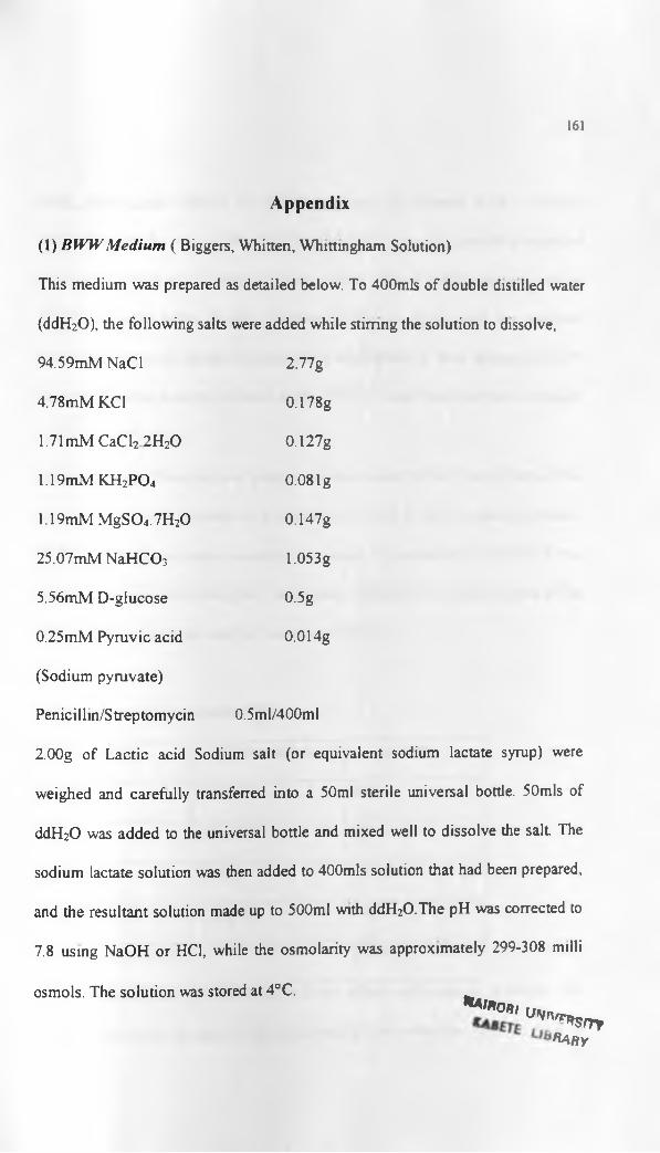
161
A ppend ix
(1) BW W M edium ( Biggers, Whitten, Whittingham Solution)
This medium was prepared as detailed below. To 400mls of double distilled water
(ddH20), the following salts were added while stirring the solution to dissolve,
94.59mM NaCl 2.77 g
4.78mM KC1 0.178g
1.71 mM CaCl2 2H20 0.127g
1.19mM KH2P 0 4 0.08 lg
1.19mM M gS04.7H20 0.147g
25.07mMNaHCO3 1.053g
5.56mM D-glucose 0.5g
0.25mM Pyruvic acid 0.014g
(Sodium pyruvate)
Penicillin/Streptomycin 0.5ml/400ml
2.00g of Lactic acid Sodium salt (or equivalent sodium lactate syrup) were
weighed and carefully transferred into a 50ml sterile universal bottle. 50mls of
ddH20 was added to the universal bottle and mixed well to dissolve the salt The
sodium lactate solution was then added to 400mls solution that had been prepared,
and the resultant solution made up to 500ml with ddH2O.The pH was corrected to
7.8 using NaOH or HC1, while the osmolarity was approximately 299-308 milli
osmols. The solution was stored at 4°C.**t*on,
u'<n'r*sm,Ra r y

162
Night before use: 200mls of BWW medium was filtered with a 0 45pm
filter(Millipore) for use. 20ml of the filtered BWW was aliquoted in a universal
bottle and labeled as a Control. A further 20mls filtered BWW was aliquoted in
another universal bottle. 20 mis of filtered BWW was mixed well in a universal
bottle with 20mls of paraffin oil and left to equilibrate. A 5mls aliquot of BWW
was also made in another universal bottle. All the media was incubated overnight
at 37°C and 5% C 02
Morning of use: Three drops of phenol red were added to the Control bottle. This
was then kept in the incubator as a measure of the BWW acidity (pink indicated a
pH 7.2-7 4, and goes yellow as acidity increases). The actual pH of the BWW was
measured and recorded using the 5 ml aliquot 0.3% BSA was added to one of the
20ml aliquots and it was used for Swim-up of sperm.
(2) Phosphate buffered saline (PBS)
Chemical Concentration Amount
NaCl 137mM 80g
KC1 2.7mM 2g
Na2H P04 lOmM 14.4g
k h 2p o 4 1.76mM 2.4g
Double distilled H20 1 litre
A X10 stock solution of Phosphate buffered saline was prepared as above. The
solution was stirred at room temperature until all the salts dissolved. To make a

163
working solution, a 1:10 dilution was made using double distilled water This was
aliquoted in 500ml bottles and sterilized by autoclaving.
(3) SDS Equilibration Solution/Buffer
This was constituted as follows while stirring the solution,
Chemical Ingredients Amount Final Concentration
1.5M Tris-HCl pH 8.8 3.35mls 50mM
Urea 36.035g 6M
87%v/v Glycerol 34.5mls 30% v/v
SDS 2.0g 2%w/v
Double distilled 1 OOmls
25ml aliquots of the solution were made and frozen at -20°C.
Prior to use the aliquots were thawed to room temperature and a few grains of
bromophenol blue added before equilibrating Immobilized pH gradient stops
(IPG’s) for the second dimension SDS PAGE.
(4) CAPS 3-[cyclohexylamino/-l-propanesulfonic acid buffer for protein
transfer
(ai Preparation of Stock Solution of CAPS buffer {1 OX CAPS (lOOmM, pH=ll):
22.13 g. of CAPS was dissolved in 900mls of distilled water. Titration with 2M
NaOH (about 20mls) made the pH 11. Distilled water was added to make a total
volume of 1 liter. The stock solution was stored at 4°C.

(b) Electroblotting buffer (IX stock buffer in 10% methanol):
This was prepared by making a 1:1:8 dilution of 10X CAPS stock buffer:
methanol: distilled water respectively.

165
REFERENCES
Adeoya-Osiguwa, S.A. and Fraser, L.R (1996) Evidence for Ca2* -dependent
ATPase activity, stimulated by decapacitation factor and calmodulin in mouse
sperm. Molecular Reproduction and Development 44(1 ):111-120.
Adoyo, P.A., Lea, LA., Richardson, R.T., Widgren, E.E. and O’Rand,
M.G. (1997) Sequence and characterization of the sperm protein Sp 17 from the
baboon. Molecular Reproduction and Development 47:66-71.
Aebersold, R. and Mann, M. (2003) Mass spectrometry-based proteomics.
Nature 422:198-207.
Aitken, R.J. (2001) Fertility: Immunological Control. In: Encyclopedia o f Life
Sciences, pp. 1-6, Nature Publishing Group.
Akama, K., Terao, K., Tanaka, Y., Noguchi, A., Yonezawa, N., Nakano,
M. and Tobita, T. (1994) Purification and characterization of a novel acrosin-
like enzyme from boar cauda epididymal sperm. Journal ot Biochemistry
(Tokyo). 116(2):464-470.
Al-Eisa, N„ Ojoo, R O. and Moore, H. D. M. (2001) Characterisation of
Human Sperm Antigens Exposed After the Acrosome Reaction That May
Participate In Gamete Fusion. In: Andrology in the 21st Century; Proceedings
o f the VJIth International Congress o f Andrology (eds. B. Robaire, H.
Chemes and C.R. Morales), pp.89-94, Medimond Inc.
Allen, C.A. and Green, D.P. (1995) Monoclonal antibodies which recognize
equatorial segment epitopes presented de novo following the A2j 187-induced
acrosome reaction of guinea pig sperm. Journal ot Cell Science 108. 767-777

166
Almeida, E.A.C., Houvila, A.P.J., Sutherland, A.E., Stephens, L.E.,
Calarco, P.G., Shaw, L.M., Mercurio, A.M., Sonnenberg, A., PrimakofT.,
P., Myles, D.G. and White, J.M. (1995). Mouse egg integrin a601 functions
as a sperm receptor. Cell 81:1095-1104.
Anderson, N.L. and Anderson, N.G. (1978) Analytical techniques for cell
fractions XXII. Two-dimensional analysis of serum and tissue proteins: multiple
gradient -slab gel electrophoresis. Analytical Biochemistry 85:331-340.
Anderson, D.J., Johnson, P.M., Alexander, N.J., Jones, W.R. and Griffin,
P.D. (1987) Monoclonal antibodies to human trophoblast and sperm antigens:
report of two WHO-sponsored workshops, June 30th 1986, Toronto, Canada.
Journal of Reproductive Immunology 10:231-257.
Arienti, G., Carlini, E., Saccardi, C. and Palmerini, C.A. (2004) Role of
human prostasomes in the activation of spermatozoa. Journal of Cellular and
Molecular Medicine 8(l):77-84.
Arts, E.G., Kuiken, J., Jager, S. and Hoekstra, D (1993) Fusion of artificial
membranes with mammalian spermatozoa. Specific involvement of the
equatorial segment after acrosome reaction. European Journal of Biochemistry
217:1001-1009.
Auer, J., Senechal, H., Desvaux, F.X., Albert, M. and De Almeida, M.
(2000) Isolation and characterization of two sperm membrane proteins
recognized by sperm- associated antibodies in infertile men Molecular
Reproduction and Development 57:393-405

167
Austin, C.R. (1951) Observations on the penetration of sperm into the
mammalian egg. Australian Journal of Scientific Research Series B 4:581-596.
Austin, C.R. (1961) The Mammalian Egg, Thomas, Springfield, Illinois
Baba, T., Azuma, S., Kashiwabara, S. and Toyoda, Y. (1994) Sperm from
mice carrying a targeted mutation of the acrosin gene can penetrate the oocyte
zona pellucida and effect fertilization. The Journal of Biological Chemistry
269:31845-31849.
Bagavant, H.,Yurewicz, E.C., Sacco, A.G., Talwar, G.P. and Gupta, S.K.
(1993a) Delineation of epitopes on porcine zona pellucida relevant for binding
of sperm to oocyte using monoclonal antibodies. Journal of Reproductive
Immunology 23:265-279.
Bagavant, H., Yurewicz, E.C., Sacco, A.G., Talwar, G.P. and Gupta, S.K
(1993b). Block in porcine gamete interaction by polyclonal antibodies to a pig
ZP3B fragment having partial sequence homology to human ZP3. Journal of
Reproductive Immunology 25:277-283.
Bagavant, H., Thillai-Koothan, P., Sharma, M.G., Talwar, G.P. and
Gupta, S.K. (1994) Antifertility effects of porcine zona pellucida-3
immunization using permissible adjuvants in female bonnet monkeys (Macaca
radiata)'. reversibility, effect on follicular development and hormonal profiles.
Journal of Reproduction and Fertility 102(1): 17-25.

168
Baldi, E., Luconi, iM., Bonaccorsi, L., Muratori, M. and Forti, G. (2000)
Intracellular events and signaling pathways involved in sperm acquisition of
fertilizing capacity and acrosome reaction. Frontiers in Bioscience 5:el 10-123
Bamezai, A.K., Mahi-Brown, C.A. and Talwar, G.P (1988) Inhibition of
penetration o f canine zonae pellucidae by homologous spermatozoa in vitro
using monoclonal antibodies raised against porcine zonae Journal of
Reproductive Immunology 13(l):85-95.
Bangham, A.D. (1961) Electrophoretic characteristics of ram and rabbit
spermatozoa. Proceedings of the Royal Society of London [Biol] 155:292-305.
Barber, M.R. and Fayrer-Hosken, R.A. (2000) Possible mechanisms of
mammalian immunocontraception. Journal of Reproductive Immunology
46:103-24.
Barros, C. and Yanagimachi, R. (1972) Polyspermy - preventing mechanisms
in the golden hamster egg. The Journal of Experimental Zoology 180 251-265
Baskin, M. (1932) Temporary sterilization by injection of human spermatozoa:
a preliminary report. American Journal of Obstetrics and Gynaecology 24:892-
897.
Bearer, E.L. and Friend, D.S. (1980) Anionic lipid domains: Correlation and
functional topography in a mammalian cell membrane. Proceedings ot the
National Academy of Sciences of the United States of .America 7 6601-6605.
Bearer, E.L. and Friend, D.S. (1990) Morphology of mammalian sperm
membranes during differentiation, maturation and capacitation. Journal of
Electron Microscopy Technique 16 281-29
169
Beaton, S., ten Have J., Cleary, A. and Bradley, M.P (1995) Cloning and
partial characterization of the cDNA encoding the fox sperm protein FSA-Acr I
with similarities to the SP-10 antigen. Molecular Reproduction and
Development 40:242-252.
Bedford, J.M. (1994) The contraceptive potential of fertilization: A
physiological perspective. Human Reproduction 9:842-858.
Bedford, J.M . (1998) Mammalian fertilization misread? Sperm penetration of
the eutherian zona pellucida is unlikely to be a lytic event. Biology of
Reproduction 59:1275-1287.
Bedford, J.M., Moore, H.D.M. and Franklin, L.E. (1979) Significance of the
equatorial segment of the acrosome of the spermatozoon in eutherian mammals.
Experimental Cell Research 119:119-126.
Bellve, A.R., Chandrika, R., Martinova, Y.S. and Barth, A.H. (1992) The
perinuclear matrix as a structural element of the mouse sperm nucleus Biology
of Reproduction 47:451-465.
Berkelman, T., Stenstedt, T., Bjellqvist, B., Laird, N., Olsson, L, Stochaj,
W. and Westermeier, R. (1998) 2-D electrophoresis: Using immobilized pH
gradients, Principles and methods, Amersham Pharmacia Biotech Inc.
Berruti, G. and Martegani, E. (1989) Identification of proteins cross-reactive
to phosphotyrosine antibodies and of a tyrosine kinase activity in boar
spermatozoa. Journal of Cell Science 93(4):667-674.

170
Bjellquist, B., EK, K., Righetti, P.G., Gianazza, E., Gorg, A., Westermeier,
R. and Postel, W. (1982) Isoelectric focusing on immobilized pH gradients:
principle, methodology and some applications. Journal of Biochemical and
Biophysical Methods 6: 317-339.
Bleil, J.D. and Wassarman, P.M. (1980) Mammalian sperm-egg interaction:
identification o f a glycoprotein in mouse egg zonae pellucidae possessing
receptor activity for sperm. Cell 20:873-882.
Bleil, J.D. and Wassarmann, P.M. (1990) Identification of a ZP3-binding
protein on acrosome-intact mouse sperm by photoaffinity crosslinking. .
Proceedings of the National Academy of Sciences of the United States of
America 87:5563-5567.
Bleil, J.D., Greve, J.M. and Wassarman, P.M. (1988) Identification of a
secondary sperm receptor in the mouse egg zona pellucida: role in maintenance
of binding of acrosome-reacted sperm to eggs. Developmental Biology
128:376-385.
Blobel, C.P., Wolfesberg, T.G., Turck, C.W., Myles, D.G., PrimakofT, P.
and White, J.M. (1992) A potential fusion peptide and an integrin ligand
domain in a protein active in sperm-egg fusion. Nature 356 248-252.
Bohring, C., Krause, E., Habermann, B. and Krause, W. (2001) Isolation
and identification of sperm membrane antigens recognized by antisperm
antibodies and their possible role in immunological infertility disease. Molecular
Human Reproduction 7(2): 113-118.

Bookbinder, L.H., Cheng, A. and Bleil, J.D. (1995) Tissue and species-
specific expression of sp56, a mouse sperm fertilization protein. Science
269:86-89.
171
Breed, W.G. (1997) Evolution of the spermatozoon in australasian rodents.
Australian Journal of Zoology 45:459-478.
Brewis, I.A. (1999) The potential importance of proteomics to research in
reproduction. Human Reproduction 14(12): 2927-2929.
Brewis, I.A. and Moore, H.D. (1997) Molecular mechanisms of gamete
recognition and fusion at fertilization. Human Reproduction 12: 156-165.
Brewis, I.A. and Wong, C.H. (1999) Gamete recognition: sperm proteins that
interact with the egg zona pellucida. Reviews of Reproduction 4:135-142.
Brewis, I.A., Barrat, C.L.R, Hornby, D.P. and Moore, H.D.M. (1995) The
signal transduction pathway of the acrosome reaction in response to
recombinant human ZP3 involves phosphorylation of a receptor kinase and a
rapid influx o f Ca“ into human spermatozoa. Journal of Reproduction and
Fertility Abstract Series 15: 6.
Bronson, R.A. and Fusi, F. (1990) Evidence that an Arg-Gly-Asp adhesion
sequence plays a role in mammalian fertilization. Biology ot Reproduction
43:1019-1025.

172
Brown, J.L. (1979) A comparison of the turnover of amino-terminally
acetylated and non-acetylated mouse L-cell proteins. The Journal of Biological
Chemistry 254:1447-1449.
Brown, J.L. and Roberts, W.K. (1976) Evidence that approximately eighty
percent of the soluble proteins from Ehrlich ascites cells are amino-terminally
acetylated. The Journal of Biological Chemistry 251:1009-1014.
Buffone, M.G., Doncel, G.F., Marin Briggiler, C.L, Vazquez-Levin, M.H.
and Calamera, J.C. (2004) Human sperm subpopulations: relationship
between functional quality and protein tyrosine phosphorylation Human
Reproduction 19(1): 139-146.
Burks, D.J, Carballada, R., Moore, H.D.M. and Saling, P.M. (1995).
Interaction o f a tyrosine kinase from human sperm with the zona pellucida at
fertilization. Science 269: 83 - 86.
Calvete, J.J., Sanz, L., Dostalova, Z. and Topfer-Peterson, E. (1993a)
Characteriztion of AWN-1 glycosylated isoforms helps define the zona
pellucida and serine-proteinase inhibitor-binding region on boar spermadhesins.
FEBS Letters 334:37-40.
Calvete, J.J., Solis, D., Sanz, L., Diaz-Maurino, T. and Topfer-Petersen E.
(1994) Glycosylated Boar spermadhesins AWN-1 isoforms: Biological origin,
structural characterization by lectin mapping, localization of O-glycosylation
sites and effect of glycosylation on ligand binding. Biological Chemistry Hoppe-
Seylar 375:667-673.

173
Calvete, J.J., Solis, D., Sanz, L., Diaz-Maurino, T., Shafer, W., Mann, K.
and Topfer-Petersen, E. (1993b) Characterization of two glycosylated boar
spermadhesins. European Journal o f Biochemistry 218:719-725.
Camaioni, A., Salustri, A., Yanagishita, M. and Hascall, V.C. (1996)
Proteins and proteoglycans in the extracellular matrix of mouse cumulus cell-
oocyte complexes. Archives of Biochemistry and Biophysics 325:190-198
Campbell, E., Takahashi, Y., Abramovitz, M., Peretz, M. and Listowsky, I.
(1990) A distinct human testis and brain p-class glutathione S-transferase
Molecular cloning and characterization of a form present even in individuals
lacking hepatic type p isoenzymes. The Journal of Biological Chemistry
265(16) 9188-9193.
Caron, C.P. and Saling, P.M. (1991) Sperm antigens and immunological
interference of fertilization. In. Elements o f Mammalian Fertilization, (ed. PM.
Wassarman), pp. 147-176, CRC Press, Boca Raton, Florida.
Carrera, A., Moos, J., Ning, X.P., Gerton, G.L., Tesarik, J., Kopf, G.S.
and Moss, S.B. (1996) Regulation of protein tyrosine phosphorylation in
human sperm by a calcium/calmodulin-dependent mechanism: identification of
A kinase anchor proteins as major substrates for tyrosine phosphorylation.
Developmental Biology 180(l):284-296.
Chang, M.C. (1951) Fertilizing capacity of spermatozoa deposited within the
fallopian tubes. Nature 168:997-998.

174
Chang, Y.S., Wang, S.C., Tsao, C.C. and Huang, F.L. (1996) Molecular
cloning structural analysis and expression of carp ZP3 gene. Molecular
Reproduction and Development 44:295-304.
Chapman, N.R. and Barrat, C.L.R. (1996) The role of carbohydrates in
sperm-ZP3 adhesion. Molecular Human Reproduction 2:767-774.
Charbonneau, H. (1993) Strategies for obtaining partial amino acid sequence
data from small quantities (<500pmol) of pure or partially purified protein In: A
practical guide to protein and peptide purification for microsequencing, 2nd
edition (ed. P.T. Matsudaira), pp. 15-41, Academic Press, Inc., San Diego,
California.
Chen, J., Litscher, E.S. and Wassarman, P.M. (1998) Inactivation of the
mouse sperm receptor, mZP3, by site directed mutagenesis of individual serine
residues located at the combining site for sperm. Proceedings of the National
Academy of Sciences of the United States of America 95:6193-6197
Chen, M.S., Tung, K.S.K., Coonrod, S.A., Takahashi, Y., Bigler, D.,
Chang, A., Yamashita, Y., Kincade, P.W., Herr, J.C. and White, J.M.
(1999) Role o f the integrin-associated protein CD-9 in binding between sperm
ADAM 2 and the egg integrin a6pl: implications for murine fertilization.
Proceedings of the National Academy of Sciences of the Lnited States ot
America 96(21): 11830-11835.

175
Cheng, A., Le, T., Palacios, M., Bookbinder, L.H., Wassarman, P.M.
Suzuki, F. and Bleil, J.D. (1994) Sperm-egg recognition in the mouse:
Characterization of sp56, a sperm protein having specific affinity for ZP3. The
Journal of Cell Biology 125:867-878.
Chevallet, M., Procaccio, V. and Rabilloud, T. (1997) A non-radioactive
double detection method for the assignment of spots in two-dimensional blots.
Analytical Biochemistry 251:69-72.
Cho, C., Bunch, D.O., Faure, J-E., Goulding, E.H., Eddy, E.M.,
PrimakofT, P. and Myles, D.G. (1998) Fertilization defects in sperm from mice
lacking fertilin p. Science 281(18): 1857-1859.
Chowdhury, A.K. and Steinberger, E. (1976) A study of germ cell
morphology and duration of the spermatogenic cycle in the baboon Papio
anubis. The Anatomical Record 186:155-170.
Cohen, D.J., Ellerman, D.A. and Cuasnicu, P.S. (2000) Mammalian sperm-
egg fusion:evidence that epididymal protein DE plays a role in mouse gamete
fusion. Biology of Reproduction 63(2):462-468.
Coonrod, S.A., Westusin, M.E. and Herr, J.C. (1996) Inhibition of bovine
fertilization invitro by antibodies to SP-10. Journal of Reproduction and
Fertility 107:287-297.
Coutu, L., Des Rosiers, P. and Sullivan, R. (1996) Purification of P26h. a
hamster sperm protein. Biochemistry and Cell Biology 74(2):227-2131.

176
Crosby, J.A., Jones, R., Barros, C. and Carvallo, P. (1998) Characterization
of the functional domains of boar acrosin involved in non-enzymatic binding to
homologous zona pellucida glycoproteins. Molecular Reproduction and
Development 49(4):426-434.
Cross, N.L. (1998) Role of cholesterol in sperm capacitation Biology of
Reproduction 59:7-11.
Cuasnicu, P.S., Conesa, D. and Rochwerger, L. (1990) Potential
contraceptive use of an epididymal protein that participates in fertilization. In:
Gamete Interaction: Prospects for Immunocontraception. (eds. N.J. Alexander,
D. Griffin, J.M. Spieler and G.M.H. Waites), pp. 143-153, Wiley-Liss, New
York.
Cuasnicu, P.S., Ellerman, D.A., Cohen, D.J., Busso, D., Morgenfeld, M.M.
and Da Ros, V.G. (2001) Molecular mechanisms involved in mammalian
gamete fusion. Archives of Medical Research 32(6):614-618.
De’almeida, M. and Voisin, G.A. (1977) Human and guinea-pig cross-
reacting spermatozoa autoantigens and autoantibodies. Clinical and
Experimental Immunology 28:163-172.
Dean, J. (2004) Reassessing the molecular biology of sperm-egg recognition
with mouse genetics. BioEssays 26:29-38.
De Jonge, C. (2005) Biological basis of human capacitation. Human
Reproduction Update 11(3):205-214.

177
de Krester, D.M. and Kerr, J.B. (1994) The cytology of the testis In: The
Physiology o f Reproduction 2nd edition, (eds. E. Knobil and J D Neill), pp
1177-1290, Raven Press, NewYork.
de Lamirande, E., Leclerc, P. and Gagnon, C. (1997) Capacitation as a
regulatory event that primes spermatozoa for the acrosome reaction and
fertilization. Molecular Human Reproduction 3(3): 175-194.
Demott, R.P. and Suarez, S.S. (1992) Hyperactivated sperm progress in the
mouse oviduct. Biology of Reproduction 46:779-785.
De Vries, K.J., Wiedmer, T., Sims, P.J. and Gadeila, B.M. (2003) Caspase-
independent exposure of aminophospholipids and tyrosine phosphorylation in
bicarbonate responsive human sperm cells. Biology of Reproduction 68:2122-
2134
Diekman, A.B. (2003) Glycoconjugates in sperm function and gamete
interactions: how much sugar does it take to sweet-talk the egg? Cellular and
Molecular Life Sciences 60: 298-308.
Diekman, A.B. and Goldberg, E. (1995) Sperm antigens: where are the
private specificities? In: Immunology o f Human Reproduction (eds. M. Kurpisz
and N. Fernandez), pp. 1-22, BIOS Scientific Publishers Limited, Oxford, UK.

178
Diekman, A.B., Norton, E.J., Westbrook, V.A., Klotz, K.L., Naaby-
Hansen, S. and Herr, J.C. (2000) Anti-sperm antibodies from infertile patients
and their cognate sperm antigens: a review. Identity between SAGA-1, H6-3C4
antigen and CD52. American Journal of Reproductive Immunology 43:134-143
Diekman, A.B., Westbrook-Case, V.A., Naaby-Hansen, S., Klotz, K.L.
Flickinger, C.J. and Herr, J.C. (1997) Biochemical characterization of sperm
agglutination antigen-1, a human sperm surface antigen implicated in gamete
interactions. Biology of Reproduction 57:1136-1144.
Dove, A.Q. (1999) Proteomics: translating genomics into products. Nature
Biotechnology 17:233-236.
Dunbar, B.S. and W'olgemuth, D.J. (1984) Structure and function of the
mammalian zona pellucida, a unique extracellular matrix. Molecular and Cellular
Biology 3:77-111.
Dunbar, B.S., Lo, C.L., Powell, J. and Stevens, V.C. (1989) Use of a
synthetic peptide adjuvant for the immunisation of baboons with denatured and
deglycosylated pig zona pellucida glycoproteins. Fertility and Sterility 52:311-
318.
East, I.J., Gulyas, B.J. and Dean, J. (1985) Monoclonal antibodies to the
murine zona pellucida protein with sperm receptor activity: effects on
fertilization and early development. Developmental Biology 109:268-273

179
Eberspaecher, U., Becker, A., Bringmann, P., van der Menve, L. and
Donner, P. (2001) Immunohistochemical localization of zona pellucida proteins
ZPA, ZPB and ZPC in human, cynomolgous monkey and mouse ovaries. Cell
and Tissue Research 303:277-287.
Eddy, E.M. and O’Brien, D.A. (1994) The Spermatozoon. In: The
Physiology o f Reproduction (eds. E. Knobil and J.D Neill), pp 29-77, Raven
Press, NY. Eddy, E.M., Vernon, R.B., Muller, C.H., Hahnel, A.C. and
Fenderson, B.A. (1985) Immunodissection of sperm surface modifications
during epididymal maturation. American Journal of Anatomy 174:225-237
Ellerman, D.A., Brantua, V.S., iMartinez, S.P., Cohen, D.J., Conesa, D.
and Cuasnicu, P.S. (1998) Potential contraceptive use of epididymal proteins:
immunization of male rats with epididymal protein DE inhibits sperm fusion
bility. Biology of Reproduction 59:1029-1036.
Ellerman, D.A., Da Ros, V.G., Cohen, D.J., Busso, D., Morgenfeld, M.M.
and Cuasnicu, P.S. (2002) Expression and structure-function analysis of DE, a
sperm cysteine-rich secretory protein that mediates gamete fusion. Biology of
Reproduction 67 (4): 1225-1231.
Evans, J.P. (2001) Fertilin and other ADAMS as integrin ligands: insights into
cell adhesion and fertilization. Bioessays 23:628-639.
180
Evans, J.P., Hardy, D.M. and Storey, B.T. (2001) Sperm-Egg Interactions:
Sperm-Egg Binding in Mammals. In: Encyclopedia o f Life Sciences, pp 1-9,
Nature Publishing Group.
Evans, S.C., Youakim, A. and Shur, B. (1995) Biological consequences of
targeting beta 1,4, galactosyltransferase to two different subcellular
compartments. Bioessays 17:261-268.
Fanarraga, M.L., Parraga, M., Aloria, K., del Mazo, J., Avila, J. and
Zabala, J.C. (1999) Regulated expression of p 14 (cofactor A) during
spermatogenesis. Cell Motility and the Cytoskeleton 43(3):243-254.
Fawcett, D.W. (1975) The mammalian spermatozoon. Developmental Biology
44:394-436.
Fawcett, D.W. (1981) The Cell, 2nd edition, pp.588-598, W B. Saunders Co.
Ltd., Philadelphia.
Fawcett, D.W. and Phillips, D.M. (1969) The fine structure and development
of the neck region of the mammalian spermatozoon. The Anatomical Record
165:153-184.
Fayrer-Hosken, R.A., Bertschinger, H.J., Kirkpatrick, J.F., Grobler, D.,
Lamberski, N., Honneyman, G. and llrich, T. (1999) Contraceptive
potential of the porcine zona pellucida vaccine in the .African elephant
(Loxodonta africana). Theriogenology 52(5) 835-846.

181
Fenichel, P. and Durand-Clement M. (1998) Role of integrins during
mammalian fertilization in mammals. Human Reproduction 13(4):31 -34
Fenner, G.P., Johnson, L.A., Hruschka, W.R. and Bolt, D.J. (1992) Two-
dimensional electrophoresis and densitometric analysis of solubilized bovine
sperm plasma membrane proteins detected by silver staining and
radioiodination. Archives of Andrology 29( 1 ):21 -32.
Feuchter, F.A., Vernon, R.B. and Eddy, E.M. (1981) .Analysis of the sperm
surface with monoclonal antibodies: topographically restricted antigens
appearing in the epididymis. Biology of Reproduction 24:1099-1110.
Fichmann, J. and Westermeier, R. (1999) 2-D protein gel electrophoresis:
An overview. In: Methods in Molecular Biology Vol. 112, 2-D proteome
analysis protocols (ed. A.J. Link), pp. 1 - 7, Humana Press Inc., Totowa, NJ
Figeys, D., Me Broom, L.D. and Moran, M.F. (2001) Mass spectrometry for
the study of protein-protein interactions. Methods 24(3):230-239.
Flechon, J.E. (1974) Freeze-fracturing of rabbit spermatozoa. Journal de
Microscopie (Paris) 19(l):59-64.
Flesch, F.M. and Gadella, B.M. (2000) Dynamics of the mammalian sperm
plasma membrane in the process of fertilization. Biochimica et Biophysica Acta
1469:197-235.

182
Flickinger, C.J., Rao, J., Bush, L.A., Sherman, N.E., Oko, R.J., Jayes,
F.C. and Herr, J.C. (2001) Outer dense fiber proteins are dominant
postobstruction autoantigens in adult Lewis rats. Biology of Reproduction
64(5): 1451-1459.
Florman, H.iM. and First, N.L. (1988) The regulation acrosomal exocytosis.
1. Sperm capacitation is required for the induction of acrosome reactions by the
bovine zona pellucida in vitro. Developmental Biology 128:453-463.
Florman, H.M. and Wassarman P.M. (1985) O-linked oligosaccharides of
mouse egg. ZP3 accounts for its sperm receptor activity. Cell 41:313-324
Fontaine, J.M., Rest, J.S., Welsh, M.J. and Benndorf, R. (2003) The sperm
outer dense fiber protein is the 10th member of the superfamily of mammalian
small stress proteins. Cell Stress and Chaperones 8(l):62-69.
Foster, J.A. and Herr, J.C. (1992) Interactions of human sperm acrosomal
protein SP-10 with the acrosomal membranes. Biology of Reproduction 46:981-
990.
Foster, J.A., Klotz, K.L., Flickinger, C.J., Thomas, T.S., Wright, R.M.,
Castillo, J.R. and Herr, J.C. (1994) Human SP-10: acrosomal distribution,
processing and fate after the acrosome reaction. Biology of Reproduction
53:462-471.
Fouquet, J.P. and Kann, M.L. (1994) The cytoskeleton of mammalian
spermatozoa. Biology of the Cell 81(2):89-93

183
Fraser, L.R. (1982) p-Aminobenzamidine, an acrosin inhibitor, inhibits mouse
sperm penetration of the zona pellucida but not the acrosome reaction. Journal
of Reproduction and Fertility 65(1): 185-194.
Fraser, L.R., Adeoya-Osiguwa, S.A. and Baxendale, R.W. (2003) First
messenger regulation of capacitation via G protein-coupled mechanisms: a tale
of serendipity and discovery. Molecular Human Reproduction 9:739-748.
Frayne, J. and Hall, L. (1998) The gene for human tMDC I sperm surface
protein is non-functional: implications for its proposed role in mammalian
sperm-egg recognition. The Biochemical Journal 334(1): 171-176.
Frayne, J. and Hall, L. (1999) The potential use of sperm antigens as targets
for immunocontraception; past, present and future. Journal of Reproductive
Immunology 43( 1): 1 -33.
Freemerman, A.J., Wright, R.M., Flickinger, C.J. and Herr, J.C. (1993)
Cloning and sequencing of baboon and cynomolgous monkey intraacrosomal
protein SP-10: Homology with human SP-10 and mouse sperm antigen (MSA-
63). Molecular Reproduction and Development 34:140-148.
Freemerman, A.J., Wright, R.M., Flickinger, C.J. and Herr, J.C. (1994)
Tissue specificity of the acrosomal protein SP-10: a contraceptive vaccine
candidate molecule. Biology of Reproduction 50(3):615-621
Friend, D.S. and Fawcett, D.W. (1974) Membrane differentiations in freeze
fractured mammalian sperm. The Journal of Cell Biology 63 641-664.

184
Fujiki, Y., Hubbard, A.L., Fowler, S. and Lazarow, P.B. (1982) Isolation of
intracellular membranes by means of sodium carbonate treatment: Application
to endoplasmic reticulum. The Journal of Cell Biology 93 97-102.
Fusi, F.M., Vignalli, M., Gailit, J. and Bronson, R.A. (1993) Mammalian
oocytes exhibit specific recognition of the RGD (Arg-Gly-Asp) tripeptide and
express oolemmal integrins. Molecular Reproduction and Development 36:212-
219.
Goldberg, E. (1977) Isozymes in testes and spermatozoa. In: Isozymes:
Current topics in biological and medical research, vol. I (eds. M. Ratazzi, J.
Scandalios and G. Whitt), pp.79-124, Alan R Liss, New York.
Goldberg, E., Vandeburg, J.L., Mahoney, M.C. and Doncel, G.F. (2001)
Immune response of male baboons to testis-specific LDH-C4. Contraception
64(2):93-98.
Gorg, A. and Weiss, W. (1999) Analytical LPG-Dalt In: Methods in Molecular
Biology, Vol. 112: 2-D Proteome Analysis Protocols. (ed. A.J. Link), pp 189-
195, Humana Press Inc., Totowa, NJ.
Gorg, A., Postel, W. and Gunther, S. (1988) The current state of two-
dimensional electrophoresis with immobilized pH gradients. Electrophoresis
9:531-546.
Gorg, A., Obermaler, C., Boguth, G., Harder, A., Scheibe, B., Wildbruber,
R. and Weiss, W. (2000) The current state of two-dimensional electrophoresis
with immobilized pH gradients. Electrophoresis 21:1037-105 j

185
Graham, C.E. (1981) Reproductive biology of great apes In: Comparative and
Biomedical perspectives (ed. C.E.Graham) pp.407-427 Acad Press Inc
NewYork.
Graves, P.R. and Haystead, T.A.J. (2002) Molecular Biologist's Guide to
Proteomics. Microbiology and Molecular Biology Reviews 66(l):39-63.
Greve, J.M. and Wassarman, P.M. (1985) Mouse egg extracellular coat is a
matrix of interconnected filaments possessing a structural repeat. Journal of
Molecular Biology 181:253-264.
Griffin, P.R., Furer-Jonscher, K., Hood, L.E. and Yates, J.R (1992).
Analysis of proteins by mass spectrometry In: Techniques in Protein Chemistry
III, pp.467-476, Academic Press Inc.
Gromov, P.S., Celis, J.E., Hansen, C., Tommerup, N., Gromova, I. and
Madsen, P. (1998) Human rablla: transcription, chromosome mapping and
effect on the expression levels of host GTP-binding proteins. FEBS Letters
429(3):359-364.
Gulyas, B.J., Gwatkin, R.B.L. and Yuan, L.C. (1983) Active immunisation
of cynomologous monkeys (Macaca fascularis) with porcine zonae pellucidae.
Gamete Research 4:299-307.
Gupta, G.S. and Chaturvedi, G. (2000) Regulation of immune functions of
sperm-specific LDH and its differences with somatic isozyme in primary and
secondary lymphocyte cultures. .American Journal of Reproductive Immunology
44(5): 160-169.

186
Gwatkin, R.B., Williams, D.T. and Carlo, D.J. (1977) Immunization of mice
with heat-solubilized hamster zonae: production of anti-zona antibody and
inhibition of fertility. Fertility and Sterility 28(8): 871-877.
Haden, N.P., Hickox, J.R , Whisnant, C.S. and Hardy, D.M. (2000)
Systematic characterization of sperm-specific membrane proteins in swine
Biology of Reproduction 63(6): 1839-1847.
Hall, J.C. and Tubbs, C.E. (1997) Purification and characterization of protein
D/E a putative sperm-binding protein involved in fertilization. Preparative
Biochemistry and Biotechnology 27(4):239-251.
Hamatani, T., Tanabe, K., Kamei, K., Sakai, N., Yamamoto, Y. and
Yoshimura, Y. (2000) A monoclonal antibody to human SP-10 inhibits in vitro
the binding o f human sperm to hamster oolemma but not to human zona
pellucida. Biology of Reproduction 62(5): 1201-1208.
Hansen, K.B. (1972) Immunofluorescent studies on mammalian spermatozoa
Antigenic relationship between human, bull and rabbit spermatozoa. Journal of
Reproduction and Fertility 29:19-28.
Hao, Z., Wolkowicz, M.J., Shetty, J., Klotz, K., Bolling, L., Sen, B.,
Westbrook, V.A., Coonrod, S„ Flickinger, C.J. and Herr, J.C. (2002)
SAMP32, a testis-specific, isoantigenic sperm acrosomal membrane-associated
protein. Biology of Reproduction 66(3): 73 5-744.

187
Harris, J.D., Hibler, D.W., Fontenot, G.K., Hsu, K.T., Yurewicz, E.C. and
Sacco, A.G. (1994) Cloning and characterization of zona pellucida genes and
cDNAs from a variety of mammalian species, the ZPA, ZPB and ZPC gene
families. DNA Sequence 4(6):361-93.
Hasegawa, A., Koyama, K., Inoue, M., Takemura, T. and Isojima, S.
(1992) Antifertility effect of active immunization with ZP4 glycoprotein family
of porcine zona pellucida in hamsters. Journal of Reproductive Immunology
22(2): 197-210.
Hayashi, M., Fujimoto, S., Takano, H., Ushiki, T., Abe, K., Ishikura, H.,
Yoshida, M.C., Kirchoff, C., Ishibashi, T., and Kasahara, M. (1996)
Characterization of a human glycoprotein with the potential role in sperm-egg
fusion: cDNA cloning, immunohistochemical localization and chromosomal
assignment of the gene (AEGL1). Genomics 32:367-374.
Hayes, J.D. and Pulford, D.J. (1995) The glutathione S-transferase supergene
family: regulation of GST and the contribution of the isoenzymes to cancer
chemoprotection and drug resistance. Critical Reviews in Biochemistry and
Molecular Biology 30(6):445-600.
Hemachand, T., Gopalakrishnan, B., Salunke, D.M., Totey, S.M. and
Shaha, C. (2002) Sperm plasma-membrane-associated glutathione S-
transferases as gamete recognition molecules. Journal of Cell Science 115:205_>-
2065.
Hemler, M.E. (1998) Integrin associated proteins. Current Opinion in Cell
Biology 10(5):578-585.
T2K?"***
188
Herr, J.C., Flickinger, C.J., Homyk, iM., Klotz, K. and John, E. (1990a)
Biochemical and morphological characterization of the intra-acrosomal antigen
SP-10 from human sperm. Biology of Reproduction 42:181-193
Herr, J.C., Wright, R.M., John, E., Foster, J., Kays, T. and Flickinger,
C.J. (1990b) Identification of human acrosomal antigen SP-10 in primates and
pigs. Biology o f Reproduction 42:377-382.
Herr, J.C., Klotz, K., Shannon, J., Wright, R.M. and Flickinger, C.J.
(1992) Purification and microsequencing of the intra-acrosomal protein SP-10.
Evidence that SP-10 heterogeneity results from endoproteolvtic processes
Biology of Reproduction 47:11 -20
Hinsch, E., Boehm, J.G., Groeger, S., Mueller-Schloesser, F. and Hinsch,
K.D. (2003) Identification of cvtokeratins in bovine sperm outer dense fiber
fractions. Reproduction in Domestic Animals 38(2): 155-160.
Ho, H-C. and Suarez, S.S. (2001) Hyperactivation of mammalian
spermatozoa: function and regulation. Reproduction 122(4) 519-526.
Ho, H.C. and Suarez, S.S. (2003) Characterization of the intracellular calcium
store at the base of the sperm flagellum that regulates hvperactivated motility
Biology of Reproduction 68(5): 1590-1596
Hogervorst, F., Kuikman, L, von dem Borne. A .E . and Sonnenberg, A.
(1990) Cloning and sequence analysis of P-4 cDNA. An integrin subunit that
contains a unique 118 kDa cytoplasmic domain. The ciMBO Journal 9 765- - 0

189
Holt, W.V. (1984) Membrane heterogeneity in the mammalian spermatozoon
International Review of Cytology 87:159-194.
Honda, A., Siruntawineti, J. and Baba, T. (2002a) Role of acrosomal matrix
proteases in sperm-zona pellucida interactions. Human Reproduction Update
8(5):405-412.
Honda, A., Yamagata, K., Sugiura, S., Watanabe, K. and Baba, T. (2002b)
A Mouse Serine Protease TESP5 Is Selectively Included into Lipid Rafts of
Sperm Membrane Presumably as a Glycosylphosphatidylinositol-anchored
Protein. The Journal of Biological Chemistry 277:16976-16984.
Howes, L. and Jones, R. (2002) Interactions between zona pellucida
glycoproteins and sperm procrosin/acrosin during fertilization. Journal of
Reproductive Immunology 53:181-192.
Hunnicutt, G.R., Primakoff, P. and Myles, D.G. (1996) Sperm surface
protein PH-20 is bifunctional: one activity is a hyaluronidase and a second,
distinct activity is required in secondary sperm-zona binding. Biology of
Reproduction 55:80-86.
Hunter, R.H.F. (2002) Vital aspects of fallopian tube physiology in pigs.
Reproduction in Domestic Animals 37:186-190
Isahakia, M. (1988). Characterization of baboon testicular antigen using
monoclonal antisperm antibodies. Biology of Reproduction 39 889 -899

190
Isahakia, M. (1989) Monoclonal antibody localization secreted by the
epididymis of the baboon (Papio cynocephalus). Journal of Reproduction and
Fertility 86: 51 - 58.
Isahakia, M. and Alexander, N.J. (1984). Interspecies cross-reactivity of
monoclonal antibodies directed against human sperm antigens. Biology of
Reproduction 30: 1015 - 1026.
Isahakia, M.A. and Bambra, C.S. (1990) Primate models for research in
reproduction. In: Gamete Interaction: Prospects o f Immunocontraception:
Proceedings o f a CONRAD - WHO International Workshop, (eds. N.J
Alexander, D. Griffin, J.M. Spieler and G.M.H. Waites), pp. 487 - 500, Wiley
Liss Inc., NY.
Jansen, R.P.S. (1980) Cyclic changes in the human fallopian tube isthmus and
their functional importance. American Journal of Obstetrics and Gynaecology
136:292-308.
Johnson, M.H. and Everitt, B.J. (1995) Ovarian Function In: Essential
Reproduction, pp 60-78, Blackwell Science Ltd., Oxford.
Jury, J.A., Frayne, J. and Hall, L. (1997) The human fertilin gene is non
functional: implications for its proposed role in fertilization. The Biochemical
Journal 321:577-581.

191
Jury, J.A., Frayne, J. and Hall, L. (1998) Sequence analysis of a variety of
primate fertilin genes: evidence for non-functional genes in the gorilla and man
Molecular Reproduction and Development 51:92-97.
Kaji, K., Oda, S., Shikano, T., Ohnuki, T., Uematsu, Y., Sakagami, J.,
Tada, N., Miyazaki, S. and Kudo, A. (2000). The gamete fusion process is
defective in eggs of CD-9 deficient mice. Nature Genetics 24:279-282.
Kaushal, G.P. and Shah, S.V. (2000) The new kids on the block: ADAMTS,
potentially multifunctional metalloproteinases of the ADAM family. The Journal
of Clinical Investigation 105(10): 1335-1337.
Kidd, L.C., Woodson, K., Taylor, P.R., Albanes, D., Virtamo, J. and
Tangrea, J.A. (2003) Polymorphisms in glutathione-S-transferase genes (GST-
Ml, GST-T1 and GST-P1) and susceptibility to prostate cancer among male
smokers of the ATBC cancer prevention study. European Journal of Cancer
Prevention 12(4):317-320.
Kierszenbaum, A.L. (2001) Spermatid manchette: plugging proteins to zero
into the sperm tail. Molecular Reproduction and Development 59(4):347-349
Kim, K.S., Cha, M.C. and Gerton, G.L. (2001) Mouse sperm protein sp56 is
a component of the acrosomal matrix. Biology of Reproduction 64(l):36-43
Kim, Y.H., de Kretser, D.M., Temple-Smith, P.D., Hearn, M.T.W. and
McFarlane, J.R. (1997) Isolation and characterization of human and rabbit
sperm tail fibrous sheath. Molecular Human Reproduction 3(4):307-313

192
Kirkman-Brown, J.C., Bray, C., Stewart, P.M., Barratt, C.L.R. and
Publicover, S.J. (2000) Biphasic elevation of [Ca2*] in individual human
spermatozoa exposed to progesterone. Developmental Biology 222:326-335.
Kirkman-Brown, J.C., Punt, E.L., Barratt, C.L. and Publicover, S.J.
(2002) Zona pellucida and progesterone-induced Ca2 signaling and acrosome
reaction in human spermatozoa. Journal of Andrology 23:306-315.
Klinefelter, G.R., Welch, J.E., Perreault, S.D., Moore, H.D., Zucker, R.M.,
Suarez, J.D., Roberts, N.L., Bobseine, K. and JefTay, S. (2002) Localization
of the sperm protein SP22 and inhibition of fertility in vivo and in vitro. Journal
of Andrology 23:48-63.
Klose, J. (1975) Protein mapping by combined isoelectric focusing and
electrophoresis o f mouse tissues. Humangenetik. 26: 231-243.
Koehler, J.K. (1981) Lectins as probes of the spermatozoon surface. Archives
of Andrology 6:197-217.
Kohler, G. and Milstein, C. (1975) Continuous cultures of fused cells
secreting antibody of predefined specificity. Nature 256:495-497.
Kong, M., Richardson, RT., Widgren, E.E. and O'Rand, M.G. (1995)
Sequence and localization of mouse sperm autoantigenic protein, sp 17. Biology
of Reproduction 53: 579-590.

193
Kritsas, J.J., Schopperle, W.M., DeWolf, W.C. and Morgentaler, A. (1992)
Rapid high resolution two-dimensional electrophoresis of human sperm protein
Electrophoresis 13(7):445-449.
Kuo, R.C., Baxter, G.T., Thompson, S.H., Strieker, S.H., Patton, C.,
Bonaventura, J. and Epel, D. (2000) NO is necessary and sufficient for egg
activation at fertilization. Nature 406:633-636.
Kurth, B.E., Weston, C., Reddi, P.P., Bryant, D., Bhattacharya. R.,
Flickinger, C.J. and Herr, J.C. (1997a) Oviductal antibody response to a
defined recombinant sperm antigen in macaques. Biology of Reproduction
57(5):981-989.
Kurth, B.E., Bryant, D., Naaby-Hansen, S., Reddi, P.P., Weston, C.,
Foley, P., Bhattacharya, R., Flickinger, C.J. and Herr, J.C. (1997b)
Immunological response in the primate oviduct to a defined recombinant sperm
immunogen. Journal of Reproductive Immunology 35(2): 13 5-150.
Laemmli, U.K. (1970) Cleavage of structural proteins during the assembly ot
the head of the bacteriophage T4. Nature 277:680-685.
Lathrop, W.F., Carmichael, E.P., Myles, D.G. and Primakoff, P. (1990)
cDNA cloning reveals the structure of a sperm surface protein, PH-20, involved
in sperm-egg adhesion and the wide distribution of its gene among mammals.
The Journal o f Cell Biology 111:2939-2949.

194
Lea, I. A., Richardson, R.T., Widgren, E.E., and O’Rand, M.G. (1996)
Cloning and sequencing of cDNA’s encoding the human sperm protein Sp 17
Biochimica et Biophysica Acta 1307:263-266.
Lea, I.A., Kurth, B. and O’Rand, M.G. (1998a) Immune response to
immunization with sperm antigens in the macaque oviduct. Biology of
Reproduction 58(3):794-800.
Lea, I.A., van Lierop, M.J., Widgren, E.E., Grootenhuis, A., Wen, Y., van
Duin, M. and O’Rand, iVLG. (1998b) A chimeric sperm peptide induces
antibodies and strain specific reversible infertility in mice. Biology of
Reproduction 59(3):527-536.
Lea, I.A, Widgren, E.E. and O’Rand, M.G. (2004) Association of sperm
protein 17 with A-kinase anchoring protein 3 in flagella. Reproductive Biology
and Endocrinology 2:57.
Leclerc, P., de Lamirande, E. and Gagnon, C. (1997) Regulation of protein-
tyrosine phosphorylation and human sperm capacitation by reactive oxygen
derivatives. Free Radical Biology and Medicine 22(4):643-656.
Lee, S.H. and Ahuja, K.K. (1987) An investigation using lectins of
glycocomponents of mouse spermatozoa during capacitation and sperm-zona
binding. Journal of Reproduction and Fertility 80(l):65-74.
Lee, V.H. and Dunbar, B.S. (1993). Developmental expression of rabbit
55kDa zona pellucida protein and messenger RNA in ovarian follicles.
Developmental Biology 155:371-382.

195
LeGendre, N., Mansfield, M., Weiss, A. and Matsudaira, P. (1993)
Purification of proteins and peptides by SDS-PAGE In: A practical guide to
protein and peptide purification fo r microsequencing 2nd Edition (ed P T
Matsudaira), pp. 71-101, Academic Press Inc., San Diego, California.
Leyton, L., Leguen, P., Bunch, D. and Saling, P.M. (1992) Regulation of
mouse gamete interaction by a sperm tyrosine kinase Proceedings of the
National Academy of Sciences of the United States of America 89:11692-11695
Lin, Y., Kimmel, L.H., Myles, D.G. and PrimakofT, P. (1993) Molecular
cloning of the human and monkey sperm surface protein PH-20. Proceedings of
the National Academy of Sciences o f the United States of America 90:10071-
10075.
Lin, Y., Mahan, K., Lathrop, W.F., Myles, D.G. and PrimakofT, P. (1994)
A hyaluronidase activity of the sperm plasma membrane protein PH-20 enables
sperm to penetrate the cumulus cell layer surrounding the egg. The Journal of
Cell Biology 125:1157-1163.
Lindemann, C.B., Kanous, K.S. and Gardner, T.K. (1991) The
interrelationships of calcium and cAMP mediated effects on reactivated
mammalian sperm models. In: Comparative spermatology: 20 years after (eds.
B. Baccetti), Raven press, New York.
Linder, B. and Heinlein, U.A. (1997) Decreased in vitro fertilization
efficiencies in the presence of specific cyritestin peptides. Development. Growth
and Differentiation 39(2):243-247.

196
Liskin, L., Pile, J.M. and Quillan, W.F. (1983) Vasectomy: safe and simple
Population Reports Series D 4:61-100.
Liu, D.Y. and Baker, H.W. (1993) Inhibition of acrosin activity with a trypsin
inhibitor blocks human sperm penetration of the zona pellucida Biology of
Reproduction 48:340-348.
Liu, M.S., Aebersold, R., Fann, C.H. and Lee, C.Y. (1992) Molecular and
developmental studies of a sperm acrosome antigen recognized by HS-63
monoclonal antibody. Biology of Reproduction 46(5):937-48.
Liu, M.S., Yang, Y., Pan, J., Liu, H.W., Menge, A.C. and Lee, C.Y.G.
(1989) Purification of an intraacrosomal antigen recognized by a monoclonal
antibody and infertility effects of isoimmune serum. International Journal of
Andrology 12:451-457.
Lopez, M.L. and de Souza, W. (1991) Distribution of filipin-sterol complexes
in the plasma membrane of stallion spermatozoa during the epididymal
maturation process. Molecular Reproduction and Development 28:158-168
Lopez, M.L., de Souza, W. and Bustos-Obregon, E. (1987) Cytochemical
analysis of the anionic sites on the membrane of the stallion spermatozoa during
epididymal transit. Gamete Research 18:319-332.
Loscalzo, J. (2004) Ozone-from environmental pollutant to atherogenic
determinant. The New England Journal of Medicine 350(8):834-835
197
Lu, Q. and Shur, B.D. (1997) Sperm from pi,4-galactosyltransferase-null
mice are refractory to ZP3-induced acrosome reactions and penetrate the zona
pellucida poorly. Development 124(20):4121-4131.
Luconi, M., Muratori, M., Maggi, M., Pecchioli, P., Peri, A., Mancini, M.,
Filimberti, E., Forti, G. and Baldi, E. (2000) Uteroglobin and
transglutaminase modulate human sperm functions. Journal of Andrology
21:676-688.
Lyons, C.E., Payette, K.L., Price, J.L. and Huang, RX.C. (1993)
Expression and structural analysis of a teleost homolog of a mammalian zona
pellucida. The Journal of Biological Chemistry 261:21351-21358
Mack, S.R , Zaneveld, L.J., Peterson, R.N., Hunt, W. and Russell, L.D.
(1987) Characterization of human sperm plasma membrane: glycolipids and
polypeptides. The Journal of Experimental Zoology 243(2):339-346.
Macko, V. and Stegemann, H. (1969) Mapping of potato proteins by
combined electrofocusing and electrophoresis. Identification of varieties
Hoppe- Seyler’s Zeitschrift fur Physiologische Chemie 350: 917-919.
Mahi-Brown, C.A. (1996) Primate response to immunization with a
homologous zona-pellucida peptide. Journal of Reproduction and Fertility
Suppliment 50:165-174.

198
Mahi-Brown, C.A., Yanagimachi, R., Hoffman, J.C. and Huang, T.T. Jr
(1985) Fertility control in the bitch by active immunization with porcine zonae
pellucidae: use o f different adjuvants and patterns of estradiol and progesterone
levels in estrous cycles. Biology of Reproduction 32(4):761-772.
Manandhar, G. and Toshimori, K. (2001) Exposure of sperm head equatorin
after acrosome reaction and its fate after fertilization in mice. Biology of
Reproduction 65:1425-1436.
Mandal, A., Naaby-Hansen, S., Wolkowicz, M.J., Klotz, K., Shetty, J.,
Retief, J.D., Coonrod, S.A., Kinter, M., Sherman, N., Cesar, F., Flickinger,
C.J. and Herr, J.C. (1999) FSP95, a testis-specific 95-kilodalton fibrous
sheath antigen that undergoes tyrosine phosphorylation in capacitated human
spermatozoa. Biology of Reproduction 61(5): 1184-1197
Martin, I., Epand, R.M. and Ruysschaert, J.M. (1998) Structural properties
of putative fusion peptide of fertilin, a protein active in sperm-egg fusion upon
interaction with the lipid bilayer. Biochemistry 37:17030-17039.
Mckeon, F.D., Kirschner, M.W. and Caput, D. (1986) Homologies in both
primary and secondary structure between nuclear envelope and intermediate
filamaent proteins. Nature 319:463-468.

199
Mdaughiin, E.A., Frayne, J., Bloomberg, G., and Hall, L. (2001) Do fertilin
P and cyritestin play a major role in mammalian sperm-oolemma interactions9 A
critical re-evaluation of the use of peptide mimics in identifying specific oocyte
recognition proteins. Molecular Human Reproduction 7(4):313-317.
Mcleskey, S.B., Dowds C., Carballada R., White, R.R. and Saling P .\l.
(1998). Molecules involved in mammalian sperm-egg interaction. International
Review of Cytology 177:57-113.
Menge, A.C. (1971) Effects of isoimmunization and isoantisera against seminal
antigens on fertility process in female rabbits. Biology of Reproduction
4(2): 137-44.
Menge, A.C. and Fuller, B. (1975) Testis antigens of man and some other
primates. Fertility and Sterility 26:473-479.
Millar, S.E., Chamow, S.M., Baur, A.W., Oliver, C., Robey, F. and Dean,
J. (1989) Vaccination with a synthetic zona pellucida peptide produces long
term contraception in female mice. Science 246(4932):935-938.
Miller, B.J., Georges-Labouesse, E., PrimakofT, P and Myles, D.G. (2000)
Normal fertilization occurs with eggs lacking the integrin a6 31 and is C 09-
dependent. The Journal of Cell Biology 149(6): 1289-1295.
Miller D.J., Maceck M.B. and Shur B.D. (1992a) Complementarity between
sperm surface (31,4 galactosyltransferase and egg coat ZPj mediates sperm-egg
binding. Nature 357: 589-593.

200
Miller, D., Brough, S. and Al-Harbi, O. (1992b) Characterization and cellular
distribution of human spermatozoal heat shock proteins. Human Reproduction
7(5):637-645.
Mochida, K., Tres, L.L. and Kierszenbaum, A.L. (1998) Isolation of the rat
spermatid manchette and its perinuclear ring. Developmental Biology 200 46-
56.
Mochida, K., Tres, L.L. and Kierszenbaum, A.L. (1999) Structural and
biochemical features of fractionated spermatid manchettes and sperm axonemes
of the azh/azh mutant mouse. Molecular Reproduction and Development
52:434-444.
Mochida, K., Rivkin, E., Gil, M. and Kierszenbaum, A.L. (2000) Keratin 9
is a component of the perinuclear ring of the manchette of rat spermatids.
Developmental Biology 227(2):510-519.
Moller, C.C. and Wassarman, P.M. (1989) Characterization of a proteinase
that cleaves zona pellucida glycoprotein ZP2 following activation of mouse
eggs. Developmental Biology 132.103-112.
Molloy, M.P. (2000) Two-dimensional electrophoresis of membrane proteins
using immobilized pH gradients. Analytical Biochemistry 280: 1-10.
Molloy, M.P., Herbert, B.R, Slade, M.B., Rabilloud, T., Nouwens, A.S.,
Williams, K.L. and Gooley, A.A. (2000) Proteomic analysis of Escherichia
coli outer membrane. European Journal of Biochemistry 267: 2871-2881

201
Moore, H.D.M. and Bedford, J.M. (1978) Ultrastmcture of the equatorial
segment of hamster spermatozoa during penetration of oocytes. Journal of
Ultrastructural Research 62:110-117.
Moore, H.D.M. and Bedford, J.M. (1983) The interaction of mammalian
gametes in the female sperm/egg interactions in vivo. In: Mechanism anil
control o f animal fertilization (ed. J.F. Hartmann), pp.453-497, Academic
press, New York.
Moore, H.D.M., Hartman, T.D., Bye, A.P., Lutjen, P., De Witt, M., and
Trounson, A.O. (1987) Monoclonal antibody against a sperm antigen Mr
95,000 inhibits attachment of human spermatozoa to the zona pellucida Journal
of Reproductive Immunology 11:157-166.
Moore, H.D.M., Smith, C.A. and Moore, A. (1990) Morphological
Development and Function of the Mammalian Sperm Acrosome Explored with
18.6 Monoclonal Antibody In: Gamete Interaction: Prospects for
Immunocontraception , pp. 53-61, Wiley-Liss Inc.
Moore, H.D.M., Clayton, R., Barrat, C.L.R. and Hornby, D.P. (1995)
Induction of the acrosomal reaction in capacitated human spermatozoa with
recombinant human ZP3 is associated with a 95kDa phosphotyrosine epitope
and is inhibited with a specific monoclonal antibody. Journal of Reproduction
and Fertility Abstract Series 15:7.

202
Mortillo, S. and Wassarman, P.M. (1991) Differential binding of gold-labeled
zona pellucida glycoproteins mZP2 and mZP3 to mouse sperm membrane
compartments. Development 113:141-149.
Mortimer, S.T., Swan, M.A. and Mortimer, D. (1998) Effect of seminal
plasma on capacitation and hyperactivation in human spermatozoa Human
Reproduction 13(8):2139-46.
Naaby-Hansen, S. (1990) Electrophoretic map of acidic and neutral human
spermatozoal proteins. Journal of Reproductive Immunology 17:167-185.
Naaby-Hansen, S., Flickinger, C.J. and Herr, J.C. (1997) Two-dimensional
gel electrophoretic analysis of vectorially labeled surface proteins of human
spermatozoa. Biology of Reproduction 56:771-787
Naz, R.K. (1996a) Application of sperm antigens in immunocontraception.
Frontiers in Bioscience l:e87-95.
Naz, R.K. (1996b) Protein tyrosine phosphorylation and signal transduction
during capacitation-acrosome reaction and zona pellucida binding in human
sperm. Archives of Andrology 37:47-55.
Naz, R.K. (1999) Vaccine for contraception targeting sperm. Immunological
Reviews 171:193-202.
203
Naz, R.K. and Ahmad, K. (1994). Molecular identities of human sperm
proteins that bind human zona pellucida mature of sperm- zona interaction,
tyrosine kinase activity and involvement of FA-1. Molecular Reproduction and
Development 39:397-408.
Naz, R.K. and Menge, A.C. (1994) Antisperm antibodies: origin, regulation,
sperm reactivity in human infertility. Fertility and Sterility 61(6): 1001-1013
Naz, R.K. and Zhu, X. (1998) Recombinant fertilization antigen-1 causes a
contraceptive effect in actively immunized mice. Biology of Reproduction
59(5): 1095-1100.
Naz, R.K., Ahmad, K. and Kumar, R (1991) Role of membrane
phosphotyrosine proteins in human spermatozoal function. Journal of Cell
Science 99(1): 157-165.
Naz, R.K., Brazil, C. and Overstreet, J.W. (1992) Effects of antibodies to
sperm surface fertilization antigen-1 on human zona pellucida interaction
Fertility and Sterility 57(6): 1304-1310.
Nevo, A.C., Michaeli, I. and Schindler, H. (1961) Electrophoretic properties
of bull and rabbit spermatozoa. Experimental Cell Research 23 :69-83.
Noland, T.D., Olson, G.E. and Barberg, D.L. (1984) Protein phosphorylation
of plasma membranes from bovine epididymal spermatozoa. Biology of
Reproduction 31:185-194.

204
Noor, M.M. and Moore, H.D. (1999) Monoclonal antibody that recognizes an
epitope of the sperm equatorial region and specifically inhibits sperm-oolemma
fusion but not binding. Journal of Reproduction and Fertility 115 :215-224
Nottola, S.A., Macchiarelli, G., Familiari, G., Stallone, T., Sathananthan,
A.H. and Motta, P.M. (1998) Egg-sperm interactions in humans:
ultrastructural aspects. Italian Journal of Anatomy and Embryology 103(1):85-
101.
O’Farell, P.H. (1975) High resolution two-dimensional electrophoresis of
proteins. The Journal of Biological Chemistry 250: 4007-4021.
O’Hern, P.A., Bambra, C.S., Isahakia. M. and Goldberg, E. (1995)
Reversible immunocontraception in female baboons immunized with a synthetic
epitope of sperm-specific lactate dehydrogenase. Biology of Reproduction
52:331-339
Ohmura, K., Kohno, N., Kobayashi, Y., Yamagata, K., Sato, S.,
Kashiwabara, S. and Baba, T. (1999) A homologue of pancreatic trypsin is
localized in the acrosome of mammalian sperm and is released during acrosome
aeaction. The Journal of Biological Chemistry 274:29426-29432.
Okada, M., Matsumoto, K-I., Niki, T., Taira T., Iguchi-Ariga, S.M.M. and
Ariga, H. (2002) DJ-1, a target protein for an endocrine disrupter, participates
in the fertilization in mice. Biological and Pharmaceutical Bulletin 25.85j -856

205
Oko, R., Aul, R.B., Wu, A. and Sutovsky, P. (2001) The sperm head
cytoskeleton. In: Andrology in the 21st Century, Proceedings o f the With
International Congress on Andrology (eds. B. Robaire, H. Chemes and C M
Morales), pp.37-51, Medimond Inc., Montreal, Quebec, Canada.
O'Rand, M.G. (1981) Inhibition of fertility and sperm-zona binding by
antiserum to the rabbit sperm membrane autoantigen RSA-1. Biology of
Reproduction 25(3):621-628.
O’Rand, M.G. (1988) Sperm-egg recognition and barriers to interspecies
fertilization. Gamete Research 19:315-328.
O'Rand, M.G., Irons, G.P. and Porter, J.P. (1984) Monoclonal antibodies to
rabbit sperm autoantigens. I. Inhibition of in vitro fertilization and localization
on the egg. Biology of Reproduction 30(3): 721-729.
O’Rand, M.G., Widgren, E.E. and Fisher, S.J. (1988) Characterization of
the rabbit sperm membrane autoantigen, RSA, as a lectin-like zona binding
protein. Developmental Biology 129( 1 ):231 -40.
Olson, G.E. and Winfrey, V.P. (1985) Substructure of a cytoskeletal complex
associated with the hamster sperm acrosome. Journal of Lltrastructural
Research 92:167-179.

206
Olson, G.E., Winfrey, V.P., Garbers, D.L. and Noland, T.D. (1985)
Isolation and characterization of a macromolecular complex associated with the
outer acrosomal membrane of bovine spermatozoa. Biology of Reproduction
33:761-779.
Parrington, J., Swann, K. and Shevchenko, V.I. (1996) Calcium oscillations
in mammalian eggs triggered by a soluble sperm protein. Nature 379:364-368
Paterson, M. and Aitken, R.J. (1990) Development of vaccines targeting the
zona pellucida. Current Opinion in Immunology 2(5):743-747.
Pecheur, E.I., Sainte-iMarie, J., Bienven e.A. and Hoekstra, D. (1999)
Peptides and membrane fusion: Towards an understanding of the molecular
mechanism of protein-induced fusion. The Journal of Membrane Biology 167:1-
17.
Peterson, R.N., Russell, L.D., Hunt, W., Bundman, D. and Freund, M.
(1983) Characterization of boar sperm plasma membranes by two-dimensional
PAGE and isolation of specific groups of polypeptides by anion exchange
chromatography and lectin affinity chromatography. Journal of Andrology
4(1):71-81.
Phelps, B.M., PrimakofT, P., Koppel, D.E., Low, M.G. and Myles, D.G.
(1988) Restricted lateral diffusion of PH-20 a Pi-anchored sperm membrane
protein. Science 240:1780-1782.

207
Phillips, D.M. (1970) Ultrastructure of spermatozoa of the woolly opossum
Caluromysphilander. Journal of Ultrastructural Research 33:381-397.
Phillips, D.M. and Shalgi, R. (1980) Surface architecture of the mouse and
hamster zona pellucida and oocyte. Journal of Ultrastructural Research 72 1-12
Prasad, S.Y., Wilkins, B., Skinner, S.M. and Dunbar, B.S. (1996)
Evaluating zona pellucida structure and function using antibodies to rabbit
55kDa ZP protein expressed in baculovirus expression system. Molecular
Reproduction and Development 43:519-529
Primakoff, P. and Myles, D.G. (1983) A map of the guinea pig sperm surface
constructed with monoclonal antibodies. Developmental Biology 98(2):417-
428.
Primakoff, P. and Myles, D.G. (2000) The ADAM gene family surface
proteins with adhesion and protease activity. Trends in Genetics 16(2) 83-87
Primakoff, P. and Myles, D.G. (2002) Penetration, Adhesion, and Fusion in
Mammalian Sperm-Egg Interaction. Science 296:2183-2185.
Primakoff, P., Hyatt, H. and Myles, D.G. (1985) A role tor the migrating
sperm surface antigen PH-20 in the guinea pig sperm binding to the egg zona
pellucida. The Journal of Cell Biology 101:2239-2244.

208
Primakoff P., Hyatt H. and Tredic-KJine J. (1987) Identification and
purification of a sperm surface protein with a potential role in sperm-egg
membrane fusion. The Journal of Cell Biology 104:141-149.
PrimakofT, P., Cowan, A., Hyatt, H., Tredick-Kline, J., and Myles D.G.
(1988) Purification of the guinea pig sperm PH-20 antigen and detection of a
site specific endoproteolytic activity in sperm preparations that cleaves PH-20
into two disulphide-linked fragments. Biology of Reproduction 38:921-934.
Rabilloud, T. (1996) Solubilization of proteins for electrophoretic analyses.
Electrophoresis 17:813-829.
Rabilloud, T. (1998) Use of thiourea to increase the solubility of membrane
proteins in two-dimensional electrophoresis. Electrophoresis 19: 758-760.
Ramarao, C.S., Myles, D.G., White, J.M. and PrimakofT, P. (1996) Initial,
evaluation of fertilin as an immunocontraceptive antigen and molecular cloning
of the cynomolgous monkey fertilin beta subunit. Molecular Reproduction and
Development 43(1):70-75.
Rankin, T., Familari, M., Lee, E., Ginsberg, A., Dwyer, N.,
Blanchettemackie, J., Drago, J., Westphal, H., and Dean, J. (1996) Mice
homozygous for an insertional mutation in the ZP3 gene lack a zona pellucida
and are infertile. Development 122:2903-2910.
209
Reddi, P.P., Naaby-Hansen, S., Aguolnik, L, Tsai, J.Y., Silver, L.M.,
Flickinger, C.J. and Herr, J.C. (1995) Complementary deoxyribonucleic acid
cloning and characterization of mSP-10: the mouse homologue of human
acrosomal protein SP-10. Biology of Reproduction 53 :873-881
Richardson, R.T. and O'Rand, M.G. (1996) Site-directed Mutagenesis of
Rabbit Proacrosin. Identification of residues involved in zona pellucida binding
The Journal of Biological Chemistry 271:24069-24074.
Richardson, R.T., Yamasaki, N. and O’ Rand, M.G. (1994) Sequence of the
rabbit zona pellucida binding protein and localization during the acrosome
reaction. Developmental Biology 165:688-701.
Rogers, M.A., Winter, H., Wolf, C., Heck, M. and Schweizer, J. (1998)
Characterization of a 190-kilobase pair domain of human type I hair keratin
genes. The Journal of Biological Chemistry 273(41 ):26683-26691
Rosiere, T.K. and Wassarman, P.M. (1992). Identification of a region of
mouse zona pellucida glycoprotein mZP3 that possesses sperm receptor
activity. Developmental Biology 154.309-317.
Rubinstein, E., Le Naour, F., Lagaudriere-Gesbert, C., Billard, M.,
Conjeaud, H. and Boucheix, C. (1996) CD9, CD63, CD81, and CD8_ are
components of a surface tetraspan network connected to HLA-DR and \ LA
integrins. European Journal of Immunology 26(11 ):265 7-2665

210
Ruoslahti, E. (1996a) Integrin signalling and matrix assembly. Tumour Biology
17:117-124.
Ruoslahti, E. (1996b) RGD and other recognition sequences for integrins.
Annual Review of Cell and Developmental Biology 12:697-715.
Russell, L.D. (1980) Sertoli-germ cell interactions: A review. Gamete Research
3:179-202.
Russell, L.D. (1984) Spermiation - the sperm release process: Ultrastructural
observations and unresolved problems In: Ultrastructure o f reproduction (eds.
J. Van Vlerkom and PM Motta), pp. 46-66, Martinus Nijhoff, The Hague.
Russell, L., Petersen, R.N. and Freund, M. (1979) On the presence of bridges
on the inner acrosomal membranes of boar spermatozoa. The Anatomical
Record 198:449-459.
Russell, L.D., Peterson, R.N., Russell, T.A. and Hunt, W. (1983) An
electrophoretic map of boar sperm plasma membrane polypeptides and
localization and fractionation of specific polypeptide subclasses. Biology ot
Reproduction 28:393-413.
Sabeur, K., Cherr, G.N., Yudin, A.L, PrimakofT, P., Li, M-VY. and
Overstreet, J.W. (1997) The PH-20 protein in human spermatozoa. Journal of
Andrology. 18:151-158.
Sacco, A.G., Subramanian, M.G., Yurewicz, E.C., DeMayo, F.J. and
Dukelow, W.R. (1983) Heteroimmunization of squirrel monkeys (Saimiri
sciureus) with a purified porcine zona antigen (PPZA). immune response and
biologic activity of antiserum. Fertility and Sterility 39(3) 350-358

211
Saling, P. (1991). How the egg regulates sperm function during gamete
interaction: facts and fantasies. Biology of Reproduction 44: 246-251.
Saling, P.M., Irons, G. and Waibel, R. (1985) Mouse sperm antigens that
participate in fertilization. I. Inhibition of sperm fusion with the egg plasma
membrane using monoclonal antibodies. Biology of Reproduction 33(2):515-26
Saling, P.M., Waibel, R. and Lakoski, K.A. (1986) Immunological
identification of sperm antigens that participate in fertilization. Advances in
Experimental Medicine and Biology 207:95-111.
Santoni, V., Molloy, M. and Rabilloud, T. (2000) Membrane proteins and
proteomics: Un amour impossible? Electrophoresis 21:1054-1070.
Sarge, K.D. (1995) Male germ cell-specific alteration in temperature set point
of the cellular stress response. The Journal of Biological Chemistry
270(32): 18745-18748.
Sathananthan, A.H., Ng, S.C., Edirisinghe, R., Ratnam, S. S., and Wong,
P. C. (1986) Sperm-oocyte interaction in the human during polyspermic
fertilization in vitro. Gamete Research 15:317-326.
Sengoku, K., Tamate, K., Horikawa, M., Takaoka, V., Ishikawa, M and
Dukelow, W.R. (1995) Plasma membrane block to polyspermy in human
oocytes and pre-implantation embryos. Journal of Reproduction and Fertilitv
105:85-90.

212
Shalgi, R. and Phillips, D. (1980) Mechanics of sperm entry in cycling
hamsters. Journal of Ultrastructural Research. 71:154-161.
Shaper, N.L., Wright, W.W. and Shaper, J.H. (1990) Murine (3 1,4-
galactosyltransferase:Both the amount and the structure of the mRNA are
regulated during spermatogenesis. Proceedings of the National Academy of
Sciences of the United States of .America 87:791-795.
Shetty, J., Naaby-Hansen, S., Shibahara, H., Bronson, R., Flickinger, C.J.
and Herr, J.C. (1999) Human Sperm Proteome: Immunodominant Sperm
Surface Antigens Identified with Sera from Infertile Men and Women. Biology
of Reproduction 61:61-69.
Shetty, J., Wolkowicz, M.J., Digilio, L.C., Klotz, K.L., Jayes, F.L.,
Diekman, A.B., Westbrook, V.A., Farris, E.M., Hao, Z., Coonrod, S.A.,
Flickinger, C.J. and Herr, J.C. (2003) SAMP 14, a novel, acrosomal
membrane-associated, glycosylphosphatidylinositol-anchored member of the Ly-
6/Urokinase-type plasminogen activator receptor superfamily with a role in
Sperm-Egg interaction. The Journal of Biological Chemistry 278:30506-30515.
Shivers, C.A. and Dunbar, B.S. (1977) Autoantibodies to zona pellucida: a
possible cause for infertility in women. Science 197:1082-1084.
Shur, B.D. and Hall, N.G. (1982) A role for mouse sperm surface
galactosyltransferase in sperm binding to the egg zona pellucida The Journal ot
Cell Biology 95:574-579.

213
Si, Y. and Okuno, iM. (1999) Role of tyrosine phosphorylation of flagellar
proteins in hamster sperm hyperactivation. Biology of Reproduction 61(1)240-
246.
Snell, W.J. and White, J.M. (1996) The molecules of mammalian fertilization
Cell 85:629-637.
Srinivasan, J., Tinge, S., Wright, R., Herr, J.C. and Curtiss, R.1II. (1995)
Oral immunization with attenuated Salmonella expressing human sperm antigen
induces antibodies in serum and the reproductive tract. Biology of
Reproduction 53:462-571.
Steen, H. and Mann, M. (2004) The ABCS (and XYZ'S) of peptide
sequencing. Nature Reviews.Molecular and Cell Biology 5:699-711.
Storey, B.T. (1995) Interactions between gametes leading to fertilization: the
sperm’s eye view. Reproduction, Fertility and Development 7(4):927-942.
Suarez, S.S. and Ho H.C. (2003) Hyperactivation of mammalian sperm.
Cellular and Molecular Biology (Noisy-le-Grand) 49(3):351-356.
Suarez, S.S., Dai, X.B., Demott, R.P., Redfern, K. and Mirando, M.A.
(1992) Movement characteristics of boar sperm obtained from the oviduct or
hyperactivated in vitro. Journal of Andrology 13:75-80.
Suarez, S.S., Brockman, K. and Lefebvre, R. (1997) Distribution of mucus
and sperm in bovine oviducts after artificial insemination. Biology of
Reproduction 56:447-453.

214
Sutovsky, P., Manandhar, G., McCauley, T.C., Caamano, J.N., Sutovsky,
M., Thompson, W.E. and Day, B.N. (2004) Proteasomal interference
prevents zona pellucida penetration and fertilization in mammals. Biology of
Reproduction 71(5): 1625-1637.
Sutovsky, P. McCauley, T.C. Sutovsky, M and Day, B.N. (2003) Early
degradation of paternal mitochondria in domestic pig (Sus scrofa) is prevented
by selective proteasomal inhibitors lactacystin and MG 13 2. Biology of
Reproduction 68(5): 1793-1800.
Swann, K. and Parrington, J. (1999) Mechanism of Ca: release at
fertilization in mammals. The Journal of Experimental Zoology 285(3):267-275.
Swann, K., Saunder, C.M., Rogers, N.T. and Lai F.A. (2006) PLC£ (zeta):
A sperm protein that triggers Ca2+ oscillations and egg activation in mammals.
Seminars in Cell and Developmental Biology 17:264-273.
Tachibana, I. and Hemler, M.E. (1999) Role of transmembrane 4 superfamily
(TM4SF) proteins CD9 and CD81 in muscle cell fusion and myotube
maintenance. The Journal of Cell Biology 146(4):893-904.
Taggart, D.A., O'Brien, H.P. and Moore, H.D. (1993) Ultrastructural
characteristics of in vivo and in vitro fertilization in the grey short-tailed
opossum, Monodelphis domestic. The Anatomical Record 237( 1):21 -37.
iiatrobf u^rvFRsrrrKABETE UbftARY

215
Takahashi, Y., Bigler, D., Ito, Y. and White, J.M. (2001) Sequence-specific
interaction between the disintegrin domain of mouse ADAM3 and murine eugs:
Role of pi integrin-associated proteins CD9, CD81 and CD98 Molecular
Biology of the Cell 12:809-820.
Takano, H., Yanagimachi, R. and Urch, U.A. (1993) Evidence that acrosin
activity is important for the development of fusibility of mammalian
spermatozoa with the oolemma: inhibitor studies using the golden hamster
Zygote 1:79-91.
Talbot, P., Shur, B.D. and Myles, D.G. (2003) Cell Adhesion and
Fertilization: Steps in Oocyte Transport, Sperm-Zona Pellucida Interactions,
and Sperm-Egg Fusion. Biology of Reproduction 68:1-9.
Telkka, A., Fawcett, D.W. and Christensen, A.K. (1961) Further
observations on the structure of the mammalian sperm tail. The .Anatomical
Record 141:231-246.
Tesarik, J., Pilka, L., Drahorad, J., Cechova, D. and Veselsky, L. (1988)
The role of cumulus cell-secreted proteins in the development of human sperm
fertilizing ability: implication in IVF. Human Reproduction 3(1): 129-132.
Thaler, C.D. and Cardullo, R.A. (1996) The initial molecular interaction
between mouse sperm and the zona pellucida is a complex binding event. The
Journal of Biological Chemistry 271:23289-23297.

216
Tipler, C.P., Hutchon, S.P., Hendil, K.5 Tiinuks, K«, Fishel, S. and VIjycr,
R.J (1997) Purification and characterization of 26S proteasomes from human
and mouse spermatozoa. Molecular Human Reproduction 3(12): 1053-1060
Topfer-Petersen, E. (1996) Molecular mechanism of fertilization in the pig.
Reproduction in Domestic Animals 31:93-100.
Topfer-Petersen, E. (1999) Carbohydrate-based interactions on the route of a
spermatozoon to fertilization. Human Reproduction Update 5:314-329.
Topfer-Petersen, E., Calvette, J.J., Sanz, L. and Sinowatz, F. (1995)
Carbohydrate and heparin-binding proteins in mammalian fertilization.
Andrologia 27:303-324.
Topfer-Petersen, E., Petroukina, A.M. and Ekhlasi-Hundrieser, M. (2000)
Oocyte-sperm interactions. Animal Reproduction Science 60-61:653-62
Toshimori, K. (1998) Maturation of mammalian spermatozoa: modifications ot
the acrosome and plasma membrane leading to fertilization. Cell and Tissue
Research 293:177-187.
Toshimori, K. (2001) Sperm-egg fusion in mammals. In: Andrology m the 21st
Century, Proceedings o f the VIIth International C ongress on Andrology (eds
B. Robaire, H. Chemes and C M. Morales), pp.247-257, Medimond Inc.,
Montreal, Quebec, Canada.

Toshimori, K, Tanii, L, Araid, , and ^ c (I#B)
antigen recogmzed by the monoclone annbody MS9 ^
pathway to the equatorial segment of sperm head dunng spem ao*** d
and Tissue Research 270:459-468
Toshimori, K„ Saxena, D.K., Tanii, I. and Yoshmaga, K. i - s , v MN .
antigenic molecule, equatorin is required for successful sperm • •
mice. Biology o f Reproduction 59:22-29
Towbin, H., Staehelin, T.T. and Gordon, J. (1979) Electr
of proteins from polyacrilamide gels to nitrocellulose sheets Pr ccduc at :
some applications. Proceedings of the National Academv of Sciences t " ■
United States o f America 76:3450-3454.
Tsubamoto, H., Hasegawa, A., Inoue, M., Yamasaki, N. and Ko>anu. K
(1996) Binding of recombinant pig zona pellucida protetn l(zpl) to aaoMflW-
reacted spermatozoa. Journal of Reproduction and Fertility -
^ in vivo and m vitro fertilization ofTsunoda, Y. and Chang, M.C. (1976) vivo
, ,A„r treatment with anti-hamster ovary antiserumhamster, rat and mouse eggs after
The Journal of Experimental Zoology 195(3) 409-415
_ . /2001) Mammalian sperm molecules thatTulsiani, D.R. and Abou-Haila, A. <200 U
k female genital tract and cuupotentially important in interaction with
vestments. Zygote 9(1):51-69.

218
Tung, K.S.K., Han, L-BP. and Evan, A.P. (1979) Differentiation autoantigen
of testicular cells and spermatozoa in the guinea pig. Developmental Biology
68:224-238.
Tung, K.S.K., PrimakofT, P., Woolman-Gamer, L. and Myles, D.G. (1997)
Mechanism of infertility in male guinea pigs immunized with sperm PH-20.
Biology of Reproduction 56.1133-1141.
Udenfriend, S. and Kodukula, K. (1995) How glycosylphosphatidylinositol-
anchored membrane proteins are made. Annual Review of Biochemistry 64 563-
591.
Urner, F., Leppens-Luisier, G. and Sakkas, D. (2001) Protein tyrosine
phosphorylation in sperm during gamete interaction in the mouse: the influence
of glucose. Biology of Reproduction 64(5): 1350-1357
Villaroya, S. and Scholler, R. (1986) Regional heterogeneity of human
spermatozoa detected with monoclonal antibodies. Journal of Reproduction and
Fertility 76:435-447.
Villasante, A., Wang, D., Dobner, P., Dolph, P., Lewis, S.A. and Cowan,
N.J. (1986) Six mouse alpha-tubulin mRNAs encode five distinct isotypes:
testis-specific expression of two sister genes. Molecular and Cellular Biology
6(7):2409-2419.

219
Visconti, P.E., Muschietti, J.P., Flawia, M.M. and Tezon, J.G. (1990)
Bicarbonate dependence of cAMP accumulation induced by phorbol esters in
hamster spermatozoa. Biochimica et Biophysica Acta 1054:231-236
Visconti, P.E., Ning, X., Fornes, M.W., Alvarez, J.G., Stein, P., Connors,
S.A. and Kopf, G.S. (1999). Cholesterol efflux mediated signal transduction in
mammalian sperm: cholesterol release signals an increase in protein tyrosine
phosphorylation during mouse sperm capacitation. Developmental Biology
214:429-443.
Wango, E.O. (1990) Non-human primates as models for research in human
reproduction: A review. Discovery and Innovation 2:33-36.
Warrent, R.W. and Kim, S-H. (1978) a-Helix-double helix interaction shown
in the structure of a protamine-transfer RNA complex and a nucleoprotamine
model. Nature 271:130-135.
Wassarman, P.M. (1988) Zona pellucida glycoproteins. Annual Review of
Biochemistry 108:1-17.
Wassarman, P.M. (1990) Profile of a mammalian sperm receptor.
Development 108:1-17.
Wassarman, P.M. (1999) Fertilization in Animals. Developmental Genetics
25:83-86.

220
Wassarman, P.M. (2005) Contribution of mouse egg zona pellucida
glycoproteins to gamete recognition during fertilization. Journal of Cellular
Physiology 204:388-391.
Wassarman, P.M. and Albertini, D.F. (1994) The mammalian ovum In The
Physiology o f Reproduction 2nd edition, (eds. E. Knobil and J.D Neil), pp 79-
122, Raven press, New York.
Welch, J.E. and O'Rand, M.G. (1985) Identification and distribution of actin
in spermatogenic cells and spermatozoa of the rabbit. Developmental Biology
109(2):411-417.
Welch, J.E., Barbee, R.R., Roberts, N.L., Suarez, J.D. and Klinefelter,
G.R. (1998) SP22: a novel fertility protein from a highly conserved gene family
Journal of Andrology 19:385-393.
Wilkins, M.R., Gasteiger, E., Sanchez, J.C., Bairoch, A. and Hochstrasser,
D.F. (1998) Two-dimensional gel electrophoresis for proteome projects: the
effects of protein hydrophobicity and copy number. Electrophoresis 19 1501-
1505.
Wojcik, C., Benchaib, M, Lomage, J., Czyba, J.C. and Guerin, J.F. (2000)
Proteasomes in human spermatozoa. International Journal of Andrology
23:169-177.
Wolf, D.P. (1978) The block to sperm penetration in zonal-free mouse eggs.
Developmental Biology 64: 1-10.

221
Wolfe, C.A., Cladera, J., Ladha, S., Senior, S., Jones, fL and O’Shea, P.
(1999) Membrane interactions of the putative fusion peptide (MFa P) from
fertilin a , the mouse sperm protein complex involved in fertilization Molecular
Membrane Biology 16:257-263.
Wolkowicz, M.J., Shetty, J., Westbrook, A., Klotz, K., Jayes, F., .Vlandal,
A., Flickinger, C.J. and Herr, J.C. (2003) Equatorial segment protein defines
a discrete acrosomal subcompartment persisting throughout acrosomal
biogenesis. Biology of Reproduction 69:735-745.
World Health Organization (1999) WHO laboratory manual for examination
o f human semen and sperm - cervical mucus interaction. - 4th edition,
Cambridge University Press, Cambridge.
Wright, R.M., John, E., Klotz, K., Flickinger, C.J. and Herr, J.C. (1990)
Cloning and sequencing of cDNAs coding for the human intra-acrosomal
antigen SP-10. Biology of Reproduction 42:693-701.
Xu, C., Rigney, D.R. and Anderson, D.J. (1994) Two-dimensional
electrophoretic profile of human sperm membrane proteins. Journal of
Andrology 15:595-602.
Xu, W. and Hamilton, D.W. (1996) Identification of the rat epididymis-
secreted 4E9 antigen as protein E: further biochemical characterization of the
highly homologous epididymal secretory proteins D and E. Molecular
Reproduction and Development 43(3):347-357
222
Yamagata, K., Nakanishi, T., Ikawa, Mm Yamaguchi, R , Moss, S.B. and
Oka be, M. (2002) Sperm from the calmegin-deficient mouse have normal
abilities for binding and fusion to the egg plasma membrane. Developmental
Biology 250(2):348-357.
Yanagimachi, R. (1969) In vitro capacitation of hamster spermatozoa by
follicular fluid. Journal of Reproduction and Fertility 18 275-286
Yanagimachi, R. (1988) Sperm-egg fusion. In: Current Topics in Membranes
and Transport, (eds. N. Duzgunes and F. Bronner), pp 3-43, Academic Press,
Orlando, Florida.
Yanagimachi, R (1994) Mammalian fertilization. In: The Physiology o f
Reproduction, (eds. E. Knobil and J.D Neil), pp. 189-317, Raven Press, New
York.
Yanagimachi, R , Noda, Y.D., Fujimoto, M. and Nicolson, G. (1972) The
distribution of negative surface charges on mammalian spermatozoa. The
American Journal of Anatomy 135:497-520.
Yanagimachi, R , Miyashiro, L.R and Yanagimachi, H. (1980) Reversible
inhibition of sperm-egg fusion in the hamster by low pH. Development, Growth
and Differentiation 22:281-288.
Yonezawa, N., Aoki, H., Hatanaka, Y. and Nakano, M. (1995) Involvement
of N-linked carbohydrate of pig zona pellucida in sperm-egg binding European
Journal of Biochemistry 233:35-41.
223
Yoshinaga, K., Saxena, D.K., Oh-oka, T., Tanii, I. and Toshimori, K.
(2001) Inhibition of mouse fertilization in vivo by intraoviductal injection of an
anti-equatorin monoclonal antibody. Reproduction 122 649-655.
Youakim, A., Hathaway, H.J., Miller, D.J., Gong, X. and Shur, B.D.
(1994) Overexpressing sperm surface beta 1,4-galactosyltransferase in
transgenic mice affects multiple aspects of sperm-egg interactions. The Journal
of Cell Biology 126(6): 1573-1583.
Yuan, R., Primakoff, P. and Myles, D.G. (1997) A role for the disintegrin
domain of cyritestin, a sperm surface protein belonging to the ADAM family in
mouse sperm-egg plasma membrane adhesion, and fusion. The Journal of Cell
Biology 137:105-112.
Yurewicz, E.C., Pack, B.A.,Armant, D.R- and Sacco, A.G. (1993) Porcine
zona pellucida ZP3a glycoprotein mediates binding ot the biotin-labelled Mr
55,000 family (ZP3) to boar sperm membrane vesicles. Molecular Reproduction
and Development 36:382-389.
Zhu, X., Bansal, N.P. and Evans, J.P. (2000) Identification of key functional
amino acids o f the mouse fertilin beta (ADAM2) disintegrin loop tor cell-cell
adhesion during fertilization. The Journal ot Biological Chemistry
275(11):7677-7683.

224
Ziemba, H., Bialy, L.P., Fracki, SM Bablok, L. and Wojcik, C. (2002)
Proteasome localization and ultrastructure of spermatozoa from patients with
varicocoele-immunoelectron microscopic study. Folia Histochemica et
Cytobiologica 40(2): 169-170.
Zuccotti, M., Yanagimachi, R. and Yanagimachi, H. (1991) The ability of
hamster oolemma to fuse with spermatozoa: its acquisition during oogenesis
and loss after fertilization. Development 112:143-152.
©URO**’ 'HJSITTKAMUl w i u HABY