Genotypic and phenotypic diversity of Ralstonia pickettii and

Comparative Genotypic and Phenotypic Analysis of CronobacterSpecies Cultured from Four Powdered Infant Formula ProductionFacilities: Indication of Pathoadaptation along the Food Chain
Qiongqiong Yan,a Juan Wang,a Jayanthi Gangiredla,b Yu Cao,a Marta Martins,a Gopal R. Gopinath,b Roger Stephan,c Keith Lampel,b
Ben D. Tall,b Séamus Fanninga
UCD Centre for Food Safety, WHO Collaborating Centre for Research, Reference & Training on Cronobacter, School of Public Health, Physiotherapy & Population Science,University College Dublin, Belfield, Dublin, Irelanda; U.S. Food and Drug Administration, Center for Food Safety and Applied Nutrition, OARSA, Silver Spring, Maryland,USAb; Institute for Food Safety and Hygiene, Vetsuisse Faculty, University of Zurich, Zurich, Switzerlandc
Cronobacter species are opportunistic pathogens commonly found in the environment. Among the seven Cronobacter species,Cronobacter sakazakii sequence type 4 (ST-4) is predominantly associated with recorded cases of infantile meningitis. This studyreports on a 26-month powdered infant formula (PIF) surveillance program in four production facilities located in distinct geo-graphic regions. The objective was to identify the ST(s) in PIF production environments and to investigate the phenotypic fea-tures that support their survival. Of all 168 Cronobacter isolates, 133 were recovered from a PIF production environment, 31were of clinical origin, and 4 were laboratory type strains. Sequence type 1 (n � 84 isolates; 63.9%) was the dominant type in PIFproduction environments. The majority of these isolates clustered with an indistinguishable pulsotype and persisted for at leastan 18-month period. Moreover, DNA microarray results identified two phylogenetic lineages among ST-4 strains tested. There-after, the ST-1 and -4 isolates were phenotypically compared. Differences were noted based on the phenotypes expressed by theseisolates. The ST-1 PIF isolates produced stronger biofilms at both 28°C and 37°C, while the ST-4 clinical isolates exhibitedgreater swimming activity and increased binding to Congo red dye. Given the fact that PIF is a low-moisture environment andthat the clinical environment provides for an interaction between the pathogen and its host, these differences may be consistentwith a form of pathoadaptation. These findings help to extend our current understanding of the epidemiology and ecology ofCronobacter species in PIF production environments.
Cronobacter (formerly known as Enterobacter sakazakii) was ac-cepted as a new bacterial genus in 2007 (1). It consists of seven
species, including C. sakazakii, C. malonaticus, C. turicensis, C.muytjensii, C. dublinensis, C. universalis, and C. condimenti (2, 3).The epidemiological link between Cronobacter infection in neo-nates and contaminated powdered infant formula (PIF) has beenpreviously established (4, 5), with C. sakazakii sequence type 4(ST-4) being linked to cases of meningitis (6). Outbreaks havebeen associated with contaminated food products and the pres-ence of this bacterium in PIF production environments (5, 7).
In order to rapidly and accurately characterize Cronobacterspecies in PIF and its associated environments, several molecular-based protocols have been developed, which include direct targetgene detection and subtyping methods (8–20). Pulsed-field gelelectrophoresis (PFGE) is an accepted method for tracking iso-lates across the food chain, and this approach is generally consid-ered suitable for epidemiological studies (12, 21–28). A multilocussequence typing (MLST) scheme for Cronobacter species was de-veloped, which focuses on single nucleotide polymorphisms asso-ciated with seven housekeeping genes (including atpD, fusA, glnS,gltB, gyrB, infB, and pps) and identifies their associated alleles (29).This protocol has been used to describe some of the diversity re-lated to the genus (6, 29–31). Both PFGE and MLST have beenwidely applied to study the genomic diversity of Cronobacter iso-lated from manufacturing facilities, commercial PIF, and fol-low-up formula, as well as clinical isolates (20, 32, 33). These re-ports highlight the dominance of C. sakazakii and the importanceof the ST-4 clonal complex as the etiological agent in meningitiscases. However, there is a lack of data comparing the phenotypes
of isolates within these clusters. Cruz-Cordova et al. (34) investi-gated the role of flagella from C. sakazakii ST-1 and -4. No signif-icant difference was observed during proinflammatory cytokineactivation in macrophages when ST-1 and -4 strains were com-pared.
This study reports on a 26-month PIF surveillance program infour production facilities geographically located in distinct re-gions. The study was designed to describe the genotypes and phe-notypes of the Cronobacter species recovered (summarized in Fig.S1 in the supplemental material). The objective was to character-ize the ST(s) in these PIF manufacturing environments and toinvestigate the phenotypes of the isolates.
Received 3 February 2015 Accepted 16 April 2015
Accepted manuscript posted online 24 April 2015
Citation Yan Q, Wang J, Gangiredla J, Cao Y, Martins M, Gopinath GR, Stephan R,Lampel K, Tall BD, Fanning S. 2015. Comparative genotypic and phenotypicanalysis of Cronobacter species cultured from four powdered infant formulaproduction facilities: indication of pathoadaptation along the food chain. ApplEnviron Microbiol 81:4388 – 4402. doi:10.1128/AEM.00359-15.
Editor: J. Björkroth
Address correspondence to Séamus Fanning, [email protected].
Supplemental material for this article may be found at http://dx.doi.org/10.1128/AEM.00359-15.
Copyright © 2015, American Society for Microbiology. All Rights Reserved.
doi:10.1128/AEM.00359-15
4388 aem.asm.org July 2015 Volume 81 Number 13Applied and Environmental Microbiology
on April 8, 2018 by guest
http://aem.asm
.org/D
ownloaded from

MATERIALS AND METHODSBacterial isolates. A total of 133 Cronobacter isolates were cultured fromfinished products (FP), semifinished products (BP), and environmentalswabs (Env) at PIF facilities in four different geographical regions betweenApril 2011 and May 2013. Thirty-one clinical isolates and four laboratorytype strains were included for comparison (see Table S1 in the supplemen-tal material). All bacteria were grown on tryptone soy agar (TSA; OxoidLtd., Basingstoke, United Kingdom) at 37°C overnight and stored at�80°C on cryo-beads (Technical Service Consultants Ltd., Lancashire,United Kingdom).
Purification of DNA and PCR amplification of target genes. Tem-plate DNA was extracted by using a simple boiling procedure (whichyielded approximately 50 ng from a 50-�l PCR mixture). Real-time PCRwas used to confirm the bacterial genus (9, 35). The species were identifiedusing rpoB as the gene target for the PCR amplification, as describedpreviously (13, 17). Serotyping was conducted using PCR amplification oftargeted genes within the species, as originally described (11, 15, 16, 19).All amplicons were analyzed in 1% [wt/vol] agarose gels in 1� Tris-bo-rate-EDTA buffer (TBE; Sigma-Aldrich, Gillingham, United Kingdom)that were stained with SYBR Safe (Life Technologies, CA, USA), visual-ized, and photographed with a Kodak Gel Logic 1500 imaging system(Carestream Health, Inc., NY, USA).
PFGE subtyping of Cronobacter. PFGE analysis was performed on all168 bacterial isolates (Fig. 1; see also Table S1 in the supplemental mate-rial). A modified version of the standard PulseNet protocol for Cronobac-ter (36) was used. Briefly, pulsed-field certified agarose (Bio-Rad, Hercu-les, CA) was selected for preparation of agarose plugs. Each plug was lysedwith 0.1 mg/ml proteinase K at 54°C for 90 min, followed by two 10-minwashes with 18 M� water and a further three 15-min washes in Tris-HCl(pH 8.0)-EDTA [TE] buffer. The restriction digestion was carried out at37°C for 3 h using 50 U XbaI. Treated plugs were cast into a 1% (wt/vol)agarose gel and electrophoresed in 0.5� TBE buffer with a CHEF MapperXA system (Bio-Rad, Hercules, CA). The running conditions used were asfollows: initial switch time, 2.16 s; final switch time, 63.8 s; voltage. 6 V;angle of 120°; run time of 20 h at a constant temperature of 14°C. Theresulting gel was then stained with 0.01% (wt/vol) SYBR Safe stainingbuffer for 30 min and destained with 500 ml 18 M� water for a further 30min. Tiff images were then acquired and uploaded to BioNumerics ver-sion 7.1 (Applied Maths, Saint-Martens-Latem, Belgium) for analysis us-ing the DICE coefficient and unweighted pair group method with arith-metic mean (UPGMA). Both the optimization and band-matchingtolerance were 1.0%. When comparing the DNA fingerprint patterns, acutoff value of 90% similarity was applied.
MLST characterization of Cronobacter. MLST analysis was carriedout on all 168 strains (strain information is listed in Fig. 1; see also TableS1 in the supplemental material). All PCRs mixtures included Taq DNApolymerase with ThermoPol buffer (New England BioLabs Inc., USA)and were assembled into mixtures following the manufacturer’s instruc-tions. Primers for all seven housekeeping genes were those used previously(29). Amplicons were dispatched for commercial Sanger sequencing(MWG Eurofins, Ebersberg, Germany). The nucleotide sequence tracefiles were uploaded to BioNumerics version 7.1 (Applied Maths, Saint-Martens-Latem, Belgium) and mapped against information on the MLSTCronobacter website (http://pubmlst.org/cronobacter/) for assignment ofeach allele, ST profile, and clonal cluster. A minimum spanning tree wasgenerated to analyze the relatedness of all isolates studied.
DNA microarray analysis. A custom-designed multigenome DNAmicroarray was developed by the U.S. FDA for the identification andcharacterization of Cronobacter species (37). This array contains over21,402 unique genes, representing the pan-genomes of all seven currentlyrecognized Cronobacter species. The probe development and optimiza-tion have been described in detail by Tall et al. (37). For this study, 59representatives of the 168 isolates were selected, based on their PFGEclusters and ST(s) along with 14 isolates representing other Cronobacterspecies and nearest neighbors, such as Salmonella enterica serovar Typhi-
murium, Klebsiella pneumoniae, Citrobacter freundii, Siccibacter turicensis,Franconibacter helveticus, and Franconibacter pulveris, which were used ascontrols for this custom-designed pan-genome microarray (Table 1).
Briefly, genomic DNA was purified from all of these bacterial isolates,fragmented by DNase I, and labeled as reported previously (37, 38). Hy-bridization was performed according to the Affymetrix GeneChip expres-sion analysis technical manual for the 49-format array. Following hybrid-ization, washing and staining procedures were carried out on anAffymetrix FS-450 fluidics station (Affymetrix, CA, USA) using themini_prok2v1_450 fluidics script. Reagents for washing and staining wereprepared according to the GeneChip expression analysis technical man-ual. Arrays were then scanned using an Affymetrix GeneChip scanner3000 running AGCC software (Affymetrix, CA, USA). For each gene rep-resented on the microarray, probe set intensities were summarized usingthe Robust multiarray averaging (RMA) method (Bioconductor Affypackage and Affymetric power tools) and compared across all strains in-vestigated. If the same gene in different strains had an RMA intensitydifference greater than 8-fold (log2 � 3), this gene was considered to be“different” between the two isolates. Thereafter, the absence/presencegene calls similar to binary nucleotide calls for each isolate were generatedinto Fasta-formatted files, which were then directly uploaded to theMEGA5 software package. Phylogenetic trees were generated using themaximum likelihood method as described by Jackson et al. (38).
Bacterial motility assays. Swim and swarm assays were performedwith all 168 isolates as described previously (39). Briefly, agar plates werefreshly prepared using Luria-Bertani (LB) broth (Becton Dickinson, MD,USA) supplemented with 0.3% (wt/vol) agar (Sigma-Aldrich, Gilling-ham, United Kingdom) for the swim assay or with 0.6% (wt/vol) agaralong with 0.5% (wt/vol) glucose for the swarm assay. Overnight cultureswere stabbed into the center of the swim plates and spotted onto theswarm plates, and these plates were subsequently incubated at 37°C for 8and 24 h, respectively. The diameter of the colony growth on each platewas measured using a standard ruler, recorded, and imaged using a KodakGel Logic 1500 imaging system (Carestream, Dublin, Ireland) and aNikon D3100 camera (Nikon, Japan). Salmonella enterica serovar Typhi-murium DT104 13348, a strain that expresses a high-motility phenotype,was included as the positive control for these assays. All assays were per-formed in duplicate.
Biofilm formation assay. Biofilm formation in standard 96-well mi-crotiter plates for all 168 isolates was performed using minimal mediumM9 (6 g/liter Na2HPO4, 3 g/liter KH2PO4, 0.5 g/liter NaCl, 1 g/literNH4Cl, 2 mM MgSO4, 0.1% glucose, and 0.1 mM CaCl2). After overnightgrowth in TSB medium, a 1:100 dilution was prepared, and 200 �l of cellsuspension was inoculated in each of the 96-well microtiter plate wells.These inoculated plates were then incubated at either 28 or 37°C for 72 h.A crystal violet (CV) staining assay was carried out, which comprisedthree brief washes with 200 �l of phosphate-buffered saline (PBS) solu-tion, followed by a 20-min fixation step with 200 �l methanol. Plates wereallowed to air dry for 15 min. After the latter step, all plates were thenstained with 200 �l 0.4% (wt/vol) CV for a period of 15 min and washedwith 200 �l PBS, followed by air drying for another 15 min. The formedbiofilm was then dissolved with 200 �l of 33% (vol/vol) acetic acid for 30min. The biofilm formed was then measured at an optical density of 570nm in a microtiter plate reader (Tecan, Männedorf, Switzerland) andanalyzed as described previously (39). Salmonella Typhimurium ATCC14028, a strong biofilm-forming strain, was selected as the positive controlfor the biofilm formation assays. These biofilm assays were performed intriplicate that included biological duplicates.
Detection of the Congo red dye binding. The colony morphology ofCronobacter species on Congo red agar was examined for the binding ofthe Congo red dye as described previously (40). Congo red agar plateswere prepared using LB agar without salt as a base and supplemented with40 �g/ml Congo red dye (Sigma-Aldrich, Gillingham, United Kingdom)as well as 20 �g/ml of Coomassie brilliant blue (ThermoFisher Scientific,Waltham, MA). Three microliters of an overnight culture was inoculated
A Form of Pathoadaptation among Cronobacter Isolates
July 2015 Volume 81 Number 13 aem.asm.org 4389Applied and Environmental Microbiology
on April 8, 2018 by guest
http://aem.asm
.org/D
ownloaded from

FIG 1 Dendrogram showing the 14 clusters of the 168 strains, denoted clusters C1 to C14, generated using BioNumerics v7.1. The similarity cutoff value was90%. Both the optimization and tolerance values were 1.0%.
Yan et al.
4390 aem.asm.org July 2015 Volume 81 Number 13Applied and Environmental Microbiology
on April 8, 2018 by guest
http://aem.asm
.org/D
ownloaded from

FIG 1 continued
A Form of Pathoadaptation among Cronobacter Isolates
July 2015 Volume 81 Number 13 aem.asm.org 4391Applied and Environmental Microbiology
on April 8, 2018 by guest
http://aem.asm
.org/D
ownloaded from

TABLE 1 Pan-genome DNA microarray analysis of a subset of isolates selected from the PFGE and MLST studies as well as other isolatesrepresenting other species of Cronobacter and their nearest neighbors
Isolate group and ID Species Serotype MLST ST no. ST clonal complex
Cronobacter isolates selected fromPFGE and MLST assays
206N Cronobacter sakazakii O:2 4 ST-4207N Cronobacter sakazakii O:2 4 ST-4208N Cronobacter sakazakii O:2 4 ST-4255N Cronobacter sakazakii O:2 4 ST-4306N Cronobacter sakazakii O:2 4 ST-4CQ2 Cronobacter sakazakii O:2 4 ST-4CQ3 Cronobacter sakazakii O:2 4 ST-4CQ4 Cronobacter sakazakii O:2 4 ST-4CQ5 Cronobacter sakazakii O:2 4 ST-4CQ6 Cronobacter sakazakii O:2 4 ST-4CQ13 Cronobacter sakazakii O:2 4 ST-4CQ14 Cronobacter sakazakii O:2 4 ST-4CQ15 Cronobacter sakazakii O:2 4 ST-4CQ17 Cronobacter sakazakii O:2 4 ST-4CQ19 Cronobacter sakazakii O:2 4 ST-4CQ24 Cronobacter sakazakii O:4 1 ST-1CQ25 Cronobacter sakazakii O:4 1 ST-1CQ26 Cronobacter sakazakii O:4 1 ST-1CQ31 Cronobacter sakazakii O:2 4 ST-4CQ32 Cronobacter sakazakii O:1 1 ST-1CQ34 Cronobacter sakazakii O:1 1 ST-1CQ35 Cronobacter sakazakii O:1 1 ST-1CQ36 Cronobacter sakazakii O:1 1 ST-1CQ37 Cronobacter sakazakii O:3 1 ST-1CQ38 Cronobacter sakazakii O:1 1 ST-1CQ40 Cronobacter sakazakii O:1 1 ST-1CQ42 Cronobacter sakazakii O:1 1 ST-1CQ43 Cronobacter sakazakii O:1 1 ST-1CQ44 Cronobacter sakazakii O:1 1 ST-1CQ45 Cronobacter sakazakii O:1 1 ST-1CQ46 Cronobacter sakazakii O:1 1 ST-1CQ61 Cronobacter sakazakii O:1 1 ST-1CQ68 Cronobacter sakazakii O:1 57 ST-1CQ75 Cronobacter sakazakii O:3 8 ST-8CQ83 Cronobacter sakazakii O:4 6 UnknownCQ92 Cronobacter sakazakii O:4 6 UnknownCQ99 Cronobacter sakazakii O:4 6 UnknownCQ111 Cronobacter sakazakii O:3 4 ST-4CQ112 Cronobacter sakazakii O:4 1 ST-1CQ114 Cronobacter sakazakii O:2 31 UnknownCQ115 Cronobacter sakazakii O:2 31 UnknownCQ116 Cronobacter sakazakii O:2 31 UnknownCQ121 Cronobacter sakazakii O:3 4 ST-4CQ122 Cronobacter sakazakii O:3 4 ST-4CQ123 Cronobacter sakazakii O:3 4 ST-4CQ124 Cronobacter sakazakii O:3 4 ST-4CQ126 Cronobacter sakazakii O:3 4 ST-4CQ127 Cronobacter sakazakii O:3 4 ST-4CQ128 Cronobacter sakazakii O:3 4 ST-4E654 Cronobacter sakazakii O:1 1 ST-1E657 Cronobacter sakazakii O:1 1 ST-1E755 Cronobacter sakazakii O:4 8 ST-8E758 Cronobacter sakazakii O:4 8 ST-8E760 Cronobacter sakazakii O:2 264 UnknownE788 Cronobacter sakazakii O:3 4 ST-4ATCC BAA-894 Cronobacter sakazakii O:1 1 ST-1CQ41 Cronobacter malonaticus O:2 129 ST-129CQ62 Cronobacter malonaticus O:2 129 ST-129ATCC 51329 Cronobacter muytjensii O:2 81 ST-81
(Continued on following page)
Yan et al.
4392 aem.asm.org July 2015 Volume 81 Number 13Applied and Environmental Microbiology
on April 8, 2018 by guest
http://aem.asm
.org/D
ownloaded from

in the center of the Congo red plates and plates were incubated at 28°C for72 h. The morphology of each colony was photographed using a NikonD3100 camera (Nikon, Japan). Salmonella Typhimurium ATCC 14028was included as the positive control. The experiment was performed induplicate.
Examination of cellulose production. The production of cellulose byCronobacter species was examined on calcofluor agar plates as describedpreviously (40). A concentration of 50 �g/ml fluorescent brightener 28(Sigma-Aldrich, Gillingham, United Kingdom) was added into the LBagar without salt. Three microliters of an overnight culture grown in TSBmedium was inoculated into the center of the plate, and plates were incu-bated at 28°C for 72 h. The morphology and color of each colony werephotographed under a 366-nm UV light using a Nikon D3100 camera(Nikon, Japan). Binding of any cellulose produced by the bacterial isolatewas observed based on the presence of a blue colony under UV light.Salmonella Typhimurium ATCC 14028, a strong biofilm-forming strain,was included as the positive control. Each isolate was tested in duplicate.
Statistical analysis. All data were analyzed using Microsoft Excel 2010and IBM SPSS statistics version 20 unless indicated specifically. The Stu-dent t test was performed with the null hypothesis to understand thestatistical significance among isolates of different groups. Bivariate corre-lation analysis was carried out using a Spearman’s rho coefficient testamong different phenotype traits in various groups. Correlation was con-sidered significant at the level of 0.01 and 0.05.
RESULTSCronobacter species and serotypes identified in the PIF produc-tion sites. Using species-specific PCR amplification of the rpoBgene, as described by Stoop et al. (13) and Lehner et al. (17), C.sakazakii was identified among 126 (94.7%) of the 133 isolatescultured from PIF production sites, thus constituting the domi-nant species (Table 2). Seven isolates (5.3%) were identified as C.malonaticus. No other Cronobacter species were cultured from ei-ther PIF or its production environments in this study. Serotypes ofall PIF isolates were determined by using PCR assays as describedby Mullane et al. (11) and Jarvis et al. (15, 19) (Fig. 1). Of therecovered 126 Cronobacter sakazakii isolates, the following sero-types were identified (Table 2): C. sakazakii O:1 (71 isolates,53.4%), C. sakazakii O:2 (24 isolates, 18.0%), C. sakazakii O:3 (19isolates, 14.3%), and C. sakazakii O:4 (15 isolates, 9.0%). Four
isolates were identified as C. malonaticus O:1 (3.0%), and threeothers were identified as C. malonaticus O:2 (2.2%) (Table 2).
PFGE subtyping analysis. Following molecular subtyping byPFGE, and based on the analysis of the pulsotypes obtained, 14clusters (with similarities above 90%) were identified comprising133 PIF isolates along with 35 clinical and type strains, which wereincluded for comparison (Fig. 1). Four clustered pulsotypes (de-noted as C1, C5, C6, and C14) were identified in one of the fourfacilities analyzed. The pulsotype designated cluster C1 consistedof 70 isolates cultured from facility A, and all of these were iden-tified as C. sakazakii serotype O:1 (Fig. 1). These isolates wererecovered from finished product (denoted as FP) or the produc-tion environments (denoted as Env), and these were isolated onthree separate occasions in December 2011, May through Septem-ber 2012, and again in May 2013. Cluster C5 included four isolatesof C. malonaticus serotype O:1, which were cultured on three oc-casions in June, July, and September 2012 from either base powderor the environment of facility A (Fig. 1). Cluster C6 consisted offour isolates cultured from the environment of facility A in April2012 and which were identified as C. sakazakii serotype O:2 (Fig. 1).
TABLE 1 (Continued)
Isolate group and ID Species Serotype MLST ST no. ST clonal complex
Other Cronobacter speciesincluded as internal controls
CI825 Cronobacter malonaticus O:2 7 ST-7464 Cronobacter dublinensis O:1 79CFS237 Cronobacter dublinensis O:1 106E515 Cronobacter dublinensis O:2 80 ST-80z3032 Cronobacter turicensis O:1 19 ST-24797-2 Cronobacter universalis O:1 54LMG26250 Cronobacter condimenti Unknown 98
Nearest neighbors used ascontrols
STM Salmonella enterica serovarTyphimurium
214 Klebsiella pneumoniae508 Siccibacter turicensisz1159 Franconibacter helveticusz513 Franconibacter helveticus1160 Franconibacter pulveris601 Franconibacter pulveris
TABLE 2 Species and serotype distribution of all 168 Cronobacterisolates studieda
Species andserotype
No. of isolates culturedfrom PIF/PIF facilities
No. of clinicalstrains
No. of ATCCtype strains
C. sakazakii O:1 71 6 2C. sakazakii O:2 24 14 1C. sakazakii O:3 19 3 0C. sakazakii O:4 12 0 0C. malonaticus O:1 4 3 0C. malonaticus O:2 3 5 0C. muytjensii O:2 0 0 1
Total 133 31 4a The isolates studied included 133 PIF isolates cultured from raw materials, swabs fromthe production environment, semifinished products, or finished products in PIFproduction facilities, and also 31 clinical isolates and 4 laboratory type strains.
A Form of Pathoadaptation among Cronobacter Isolates
July 2015 Volume 81 Number 13 aem.asm.org 4393Applied and Environmental Microbiology
on April 8, 2018 by guest
http://aem.asm
.org/D
ownloaded from

Cluster C14 included two C. malonaticus serotype O:2 isolateswhich were recovered from the environment of facility A in July2012 (Fig. 1).
Of note, four clusters (including C4, C7, C9, and C10) wererecovered from facilities in different geographical regions. ClusterC4 contained 12 C. sakazakii isolates, which were cultured fromeither finished product, base powder, or the environment duringApril, July, August, October, and December 2011 in three produc-tion locations (facilities A, C, and D) (Fig. 1). All were identified asC. sakazakii serotype O:4. Similarly, cluster C7 included 18 C.sakazakii isolates of serotype O:2, and these were cultured frombase powder, finished product, or the environments in three of thefacilities (facilities B, C, and D) (Fig. 1) during June, July, andOctober 2011 and July and October 2012. Cluster C9 included 10C. sakazakii of serotype O:3. These were cultured from two facil-ities (facilities A and B) (Fig. 1) on a number of occasions duringApril and December 2011 and January through March, June, andJuly 2012. Finally, cluster C10 consisted of eight C. sakazakii sero-type O:3 isolates, which were isolated in June, July, and September2012 from base powder, finished product, or environments at fa-cilities A and B (Fig. 1).
Cronobacter isolates of clinical origin clustered independentlyinto six groups, which included cluster C2 (2 isolates) and C3 (2isolates) of C. sakazakii serotype O:1, C8 (10 isolates) of C. saka-zakii serotype O:2, C11 (3 isolates) of C. malonaticus serotype O:1,C12 (3 isolates) of C. malonaticus serotype O:2, and C13 (2 iso-lates) of C. sakazakii serotype O:4. In addition, five PIF isolates(including one C. malonaticus and four C. sakazakii isolates) andnine clinical isolates, along with four type strains, did not groupinto any of the previously described clusters.
MLST profiling. MLST subtyping was performed with all 133PIF isolates, along with the 31 clinical isolates and four type strains(Fig. 1). Sequence trace files were uploaded to BioNumerics v7.1to aid with the generation of a minimum spanning tree (Fig. 2).The cross-link among isolates of PIF origin is highlighted in theorange circle. Of the 133 PIF isolates, ST-1 (n � 84 isolates, 63.2%)was identified as the dominant sequence type, followed by 35 iso-lates (26.3%) that were identified as ST-4 (Fig. 2a). Other STsidentified included ST-6 (3.0%), ST-8 (0.8%), ST-31 (3.0%),
ST-57 (0.8%), ST-103 (0.8%), ST-129 (1.5%), and a single un-known type (0.8%) (Sequence information for the unknown typeis described in Text S1 in the supplemental material). When theseSTs were grouped by bacterial species, C. sakazakii was identifiedamong ST-1, -4, -6, -8, -31, -57, and -103, while C. malonaticuswas represented by ST-6 and -129 (Fig. 2a). A single unknown STwas identified as C. malonaticus serotype O:2. Additionally, the 31clinical strains studied consisted of 23 C. sakazakii and 8 C. ma-lonaticus isolates. ST-4 was the dominant type among these clini-cal isolates, comprising 45.2% of these. Lastly, five ST-7 C. ma-lonaticus isolates and five ST-8 C. sakazakii isolates togethercomprised 32.2% of the collection.
Phylogenetic analysis of Cronobacter species based on a cus-tom-designed multigenome DNA microarray. Microarray anal-ysis aptly distinguished the seven Cronobacter species from oneanother and from non-Cronobacter species, which were used ascontrols for this custom-designed pan-genome microarray.Within each species, the isolates grouped into various distinctsubclusters based on their pan-genomic diversity (Fig. 3). In ad-dition, the microarray analysis separated C. sakazakii isolates intosix clusters, and the strains clearly segregated according to se-quence type. Interestingly the microarray also placed 25 ST-4 iso-lates into two distinct subclusters. Microarray gene differencesnoted between the two lineages are shown in Tables S2 and S3 inthe supplemental material. Strains from lineage 1 differed fromthose in lineage 2 by 24 genes, of which 7 were phage related. Incontrast, isolates within lineage 2 differed from those in lineage 1across 71 genes, of which 17 were associated with the pESA3-harbored type 6 secretion system (T6SS) gene cluster (41).
To better understand this diversity, PCR analysis of these 25isolates was carried out using primers to detect plasmid pESA3and the presence or absence of four regions within the T6SS genecluster, as described by Franco et al. (41). Table S4 in the supple-mental material summarizes the results of this PCR analysis. All 25isolates were PCR positive for the single plasmid IncFIB incom-patibility group replication protein gene repA (ESA_pESA3; loca-tion 115 to 588). Further PCR analysis of the T6SS in the 25 iso-lates revealed that most of the isolates representing each lineagepossessed the 5= end of the T6SS gene cluster. However, none of
FIG 2 MLST distributions of 133 PIF and 31 clinical isolates. (a) PIF isolates; (b) all 164 PIF and clinical isolates studied. Distinct sequence types are shown indifferent colors.
Yan et al.
4394 aem.asm.org July 2015 Volume 81 Number 13Applied and Environmental Microbiology
on April 8, 2018 by guest
http://aem.asm
.org/D
ownloaded from
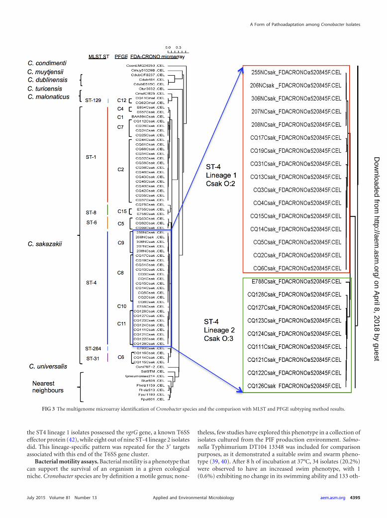
the ST4 lineage 1 isolates possessed the vgrG gene, a known T6SSeffector protein (42), while eight out of nine ST-4 lineage 2 isolatesdid. This lineage-specific pattern was repeated for the 3= targetsassociated with this end of the T6SS gene cluster.
Bacterial motility assays. Bacterial motility is a phenotype thatcan support the survival of an organism in a given ecologicalniche. Cronobacter species are by definition a motile genus; none-
theless, few studies have explored this phenotype in a collection ofisolates cultured from the PIF production environment. Salmo-nella Typhimurium DT104 13348 was included for comparisonpurposes, as it demonstrated a suitable swim and swarm pheno-type (39, 40). After 8 h of incubation at 37°C, 34 isolates (20.2%)were observed to have an increased swim phenotype, with 1(0.6%) exhibiting no change in its swimming ability and 133 oth-
FIG 3 The multigenome microarray identification of Cronobacter species and the comparison with MLST and PFGE subtyping method results.
A Form of Pathoadaptation among Cronobacter Isolates
July 2015 Volume 81 Number 13 aem.asm.org 4395Applied and Environmental Microbiology
on April 8, 2018 by guest
http://aem.asm
.org/D
ownloaded from

ers (79.2%) displaying reduced swim activity compared to thereference strain (Fig. 4; see also Table S1 in the supplemental ma-terial). By extending the incubation time to a total of 24 h at 37°C,25 isolates (14.9%) were observed to have reduced swim activity,with 143 of the remaining isolates (85.1%) able to spread acrossthe entire plate, indicating that these isolates possessed the sameswim activities as the reference. The reference strain SalmonellaTyphimurium DT104 13348 demonstrated good swarming activ-ity; however, in comparison with the reference, all Cronobacterisolates exhibited a reduced swarming activity at both 8 and 24 hwhen incubated at 37°C (see Table S1).
Biofilm formation under defined substrate growth condi-tions. The ability to form a biofilm under laboratory-defined con-ditions for all Cronobacter isolates was studied. This was investi-gated by incubating bacterial cultures in M9 minimal medium at28 and 37°C in standard microtiter plates (see Table S1 in thesupplemental material). Salmonella Typhimurium ATCC 14028,a strong biofilm-forming strain (39), was included as the positivecontrol. When incubated at 28°C, the formation of a strong bio-film was observed with 115 isolates (68.5%), whereas 35 isolates(20.8%) were defined as moderate biofilm formers and 18 isolates(10.7%) demonstrated weak biofilm formation. Similarly, whenexposed to a temperature of 37°C, 119 isolates (70.8%) formedstrong biofilms, with 32 isolates (19.1%) producing moderate bio-films and 17 isolates (10.1%) forming weak biofilms. Interest-ingly, temperature-dependent biofilm formation was observedamong 36 isolates. Fifteen isolates produced strong biofilms at28°C and formed moderate biofilms when incubated at 37°C.Twenty strong biofilm formers at 37°C produced moderate orweak biofilms at 28°C, while one moderate biofilm former at 37°Cshowed weak biofilm formation when incubated at 28°C.
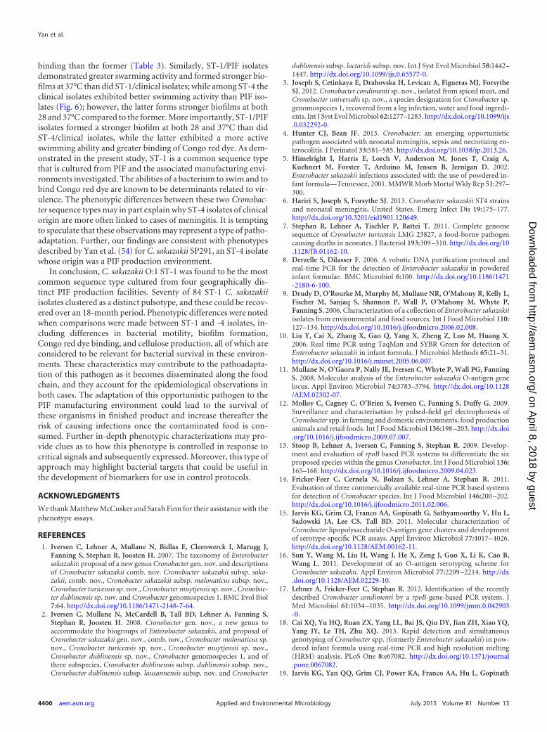
Morphotypes of Cronobacter species. All Cronobacter isolateswere incubated separately on LB agar plates supplemented witheither Congo red or calcofluor. The colony color and morphologywere recorded as an indication of the binding of the Congo red dye(Fig. 5) and cellulose production (see Table S1 in the supplemen-tal material). Salmonella Typhimurium ATCC 14028 was selectedas the reference strain (40). Four morphotypes were noted, asshown in Fig. 5, and included red, dry, and rough (RDAR) (Fig.5a), brown, dry, and rough (BDAR) (Fig. 5b), red and smooth(RAS) (Fig. 5c), and brown and smooth (BAS) (Fig. 5d) types. Thereference strain was characterized as RDAR (Fig. 5a). The most
common morphotype among the Cronobacter species studied wasBAS, as noted for 95 isolates (56.5%), followed by 43 isolates(25.6%) that were identified as BDAR, 21 isolates (12.5%) with theRAS morphotype, and 9 isolates (5.4%) with the RDAR morpho-type. Only isolates defined by either the RDAR or BDAR morpho-types were considered positive for the Congo red dye bindingassay, and these accounted for 31.0% of the tested isolates. Isolatesthat exhibited a RAS morphotype (12.5%) were considered toexhibit reduced binding of the Congo red dye, while those with theBAS morphotype (56.5%) did not show any binding of the Congored dye.
Production of cellulose was detected through monitoring thefluorescent signal observed at 366 nm under UV light. The refer-ence strain generated a strong fluorescent signal. Most Cronobac-ter isolates (118 isolates, 70.2%) showed weak cellulose produc-tion, while 50 isolates (29.8%) were negative for this phenotype(see Table S1 in the supplemental material).
Comparative analysis of selected phenotypes expressed byCronobacter ST-1 and -4 when recovered from various sources.In order to determine whether or not the origin of a Cronobacterisolate would reflect its phenotype, we sought to evaluate a num-ber of correlations among the isolates in the various groups, asshown in Table 3. When all ST-1 isolates were compared againstthose of ST-4 regardless of origins, significant differences in theability to swim (when measured for 8 and 24 h) and to bind Congored dye (P � 0.000) were noted (Table 3). Analysis of these phe-notypic comparisons showed that STs had significant negativecorrelations with swim activities at both 8 h (r � �0.414, P �0.000) and 24 h (r � �0.527, P � 0.000), as well as with the abilityto form biofilms under laboratory-defined conditions at 37°C(r � �0.353, P � 0.000) (Table 3). In contrast, correlationsthat were significantly positive were noted for biofilm forma-tion at 28°C (r � 0.173, P � 0.044) and Congo red dye binding(r � 0.310, P � 0.000) (Table 3). Similar correlations wereobserved between ST-1/PIF and ST-4/PIF isolates; however, asignificant difference was only observed in the ability to swarmafter 24 h of incubation between ST-1/clinical and ST-4/clini-cal isolates (Table 3).
When the bacterial isolates defined as ST-1/PIF and ST-1/clin-ical were compared, a significantly different phenotype (P �0.000) was observed for cellulose production. The origin of anisolate (PIF or clinical, as shown here) demonstrated a significant
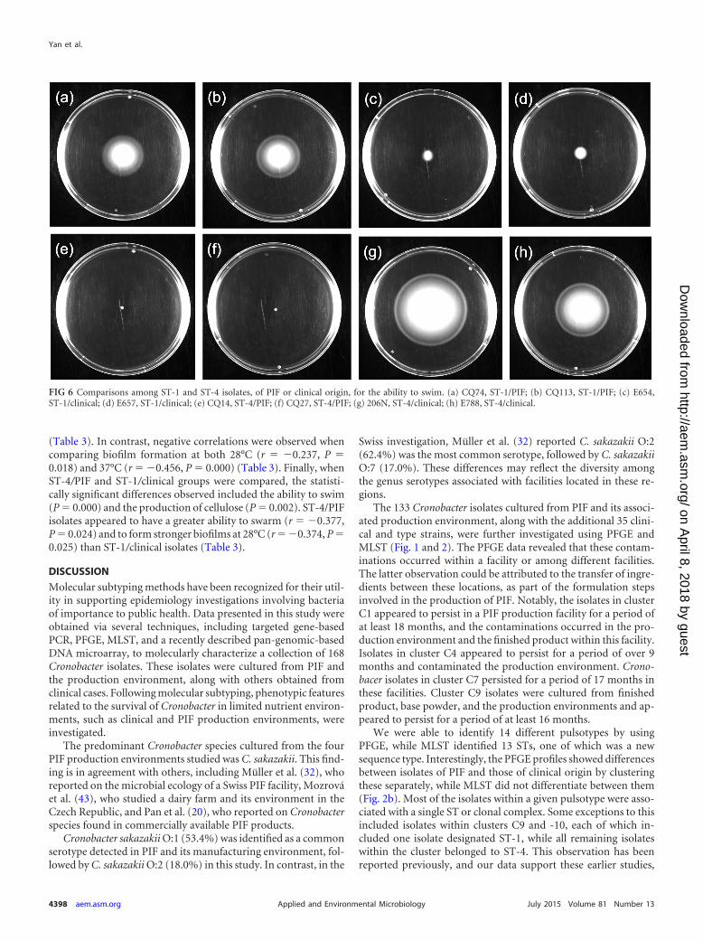
FIG 4 Swimming ability of Cronobacter isolates compared to that of the reference strain S. Typhimurium DT104 13348. The reference strain showed an averageswimming activity of 60 mm after 8 h of incubation at 37°C. (a) E657, C. sakazakii serotype O:1, clinical origin; (b) CQ8, C. sakazakii serotype O:4, PIF origin;(c) ATCC BAA-894, C. sakazakii serotype O:1, PIF origin; (d) 206N, C. sakazakii serotype O:2, clinical origin.
Yan et al.
4396 aem.asm.org July 2015 Volume 81 Number 13Applied and Environmental Microbiology
on April 8, 2018 by guest
http://aem.asm
.org/D
ownloaded from

negative correlation with the ability to swarm after 24 h of incu-bation (r � �0.232, P � 0.023) and to form a biofilm at 37°C (r ��0.222, P � 0.038) (Table 3). Similarly, when ST-4/PIF and ST-4/clinical were compared, phenotypic differences were observedin the ability to swim (when measured at both 8 and 24 h; P �0.000) and to form a biofilm at both 28°C (P � 0.000) and 37°C(P � 0.009). In this case, there was a significantly positive correla-tion with the ability to swim after incubation for 8 h (r � 0.717,P � 0.000) or 24 h (r � 0.480, P � 0.001) and a negative ability to
form biofilms at both 28°C (r � �0.592, P � 0.000) and 37°C (r ��0.510, P � 0.000) (Table 3).
When ST-1/PIF isolates were compared against the recognizedST-4/clinical isolates, statistically significant differences in biofilmformation at both 28°C (P � 0.001) and 37°C (P � 0.000), as wellas the Congo red dye binding (P � 0.014) were noted. Further-more, some of these results correlated positively between the twogroups, specifically in the case of their swim phenotype (r � 0.199,P � 0.047) and the Congo red dye binding (r � 0.212, P � 0.034)
FIG 5 Four types of cell morphology observed with Congo red dye binding. (a) red, dry, and rough (RDAR); (b) brown, dry, and rough (BDAR); (c) red andsmooth (RAS); (d) brown and smooth (BAS).
TABLE 3 Bivariate correlation analysis results for all phenotypic traits investigateda
Comparison
Swimmingb Swarmingb Biofilm formation atc:Celluloseproductiond
Congo red dyebindinge8 h 24 h 8 h 24 h 28°C 37°C
Overall ST-1 vs ST-4 �0.414** �0.527** 0.015 0.125 0.173* �0.353** 0.048 0.310*ST-1/PIF vs ST-4/PIF �0.651** �0.667** 0.055 0.106 0.346** �0.231** 0.030 0.292**ST-1/clinical vs ST-4/clinical 0.000 0.000 �0.415 0.562* 0.451 0.041 �0.182 0.162ST-1/PIF vs ST-1/clinical �0.009 0.000 0.147 �0.232* �0.195 �0.222* 0.099 0.061ST-4/PIF vs ST-4/clinical 0.717** 0.480** �0.181 0.180 �0.592** �0.510** 0.053 �0.016ST-1/PIF vs ST-4/clinical 0.199* 0.000 �0.056 0.102 �0.237* �0.456** 0.067 0.212*ST-4/PIF vs ST-1/clinical 0.258 0.226 0.269 �0.377* �0.374* �0.245 0.140 �0.137a The phenotypic traits investigated in the various isolate groups were analyzed on the basis of STs and sample origins by using Spearman’s rho coefficient test (data shown are theresulting r values). Statistical significance is indicated by asterisks: **, P � 0.01; *, P � 0.05.b In the motility assays (including swimming and swarming tests), the diameter of each colony was measured after incubation and normalized against that of the reference strain.c Optical densities (at 570 nm) for each isolate were normalized to that of the reference strain.d Cellulose production, when lacking (negative), was assigned a score of 1, while weak production was assigned a score of 2.e Congo red dye binding results were interpreted as follows: the RAS group was assigned a score of 1; the BAS group score was 2 (both RAS and BAS results were considerednegative for Congo red dye binding); the RDAR group was assigned a score of 3; the BDAR group received a score of 4 (both the RDAR and BDAR groups were considered positivefor Congo red dye binding).
A Form of Pathoadaptation among Cronobacter Isolates
July 2015 Volume 81 Number 13 aem.asm.org 4397Applied and Environmental Microbiology
on April 8, 2018 by guest
http://aem.asm
.org/D
ownloaded from
(Table 3). In contrast, negative correlations were observed whencomparing biofilm formation at both 28°C (r � �0.237, P �0.018) and 37°C (r � �0.456, P � 0.000) (Table 3). Finally, whenST-4/PIF and ST-1/clinical groups were compared, the statisti-cally significant differences observed included the ability to swim(P � 0.000) and the production of cellulose (P � 0.002). ST-4/PIFisolates appeared to have a greater ability to swarm (r � �0.377,P � 0.024) and to form stronger biofilms at 28°C (r � �0.374, P �0.025) than ST-1/clinical isolates (Table 3).
DISCUSSION
Molecular subtyping methods have been recognized for their util-ity in supporting epidemiology investigations involving bacteriaof importance to public health. Data presented in this study wereobtained via several techniques, including targeted gene-basedPCR, PFGE, MLST, and a recently described pan-genomic-basedDNA microarray, to molecularly characterize a collection of 168Cronobacter isolates. These isolates were cultured from PIF andthe production environment, along with others obtained fromclinical cases. Following molecular subtyping, phenotypic featuresrelated to the survival of Cronobacter in limited nutrient environ-ments, such as clinical and PIF production environments, wereinvestigated.
The predominant Cronobacter species cultured from the fourPIF production environments studied was C. sakazakii. This find-ing is in agreement with others, including Müller et al. (32), whoreported on the microbial ecology of a Swiss PIF facility, Mozrováet al. (43), who studied a dairy farm and its environment in theCzech Republic, and Pan et al. (20), who reported on Cronobacterspecies found in commercially available PIF products.
Cronobacter sakazakii O:1 (53.4%) was identified as a commonserotype detected in PIF and its manufacturing environment, fol-lowed by C. sakazakii O:2 (18.0%) in this study. In contrast, in the
Swiss investigation, Müller et al. (32) reported C. sakazakii O:2(62.4%) was the most common serotype, followed by C. sakazakiiO:7 (17.0%). These differences may reflect the diversity amongthe genus serotypes associated with facilities located in these re-gions.
The 133 Cronobacter isolates cultured from PIF and its associ-ated production environment, along with the additional 35 clini-cal and type strains, were further investigated using PFGE andMLST (Fig. 1 and 2). The PFGE data revealed that these contam-inations occurred within a facility or among different facilities.The latter observation could be attributed to the transfer of ingre-dients between these locations, as part of the formulation stepsinvolved in the production of PIF. Notably, the isolates in clusterC1 appeared to persist in a PIF production facility for a period ofat least 18 months, and the contaminations occurred in the pro-duction environment and the finished product within this facility.Isolates in cluster C4 appeared to persist for a period of over 9months and contaminated the production environment. Crono-bacer isolates in cluster C7 persisted for a period of 17 months inthese facilities. Cluster C9 isolates were cultured from finishedproduct, base powder, and the production environments and ap-peared to persist for a period of at least 16 months.
We were able to identify 14 different pulsotypes by usingPFGE, while MLST identified 13 STs, one of which was a newsequence type. Interestingly, the PFGE profiles showed differencesbetween isolates of PIF and those of clinical origin by clusteringthese separately, while MLST did not differentiate between them(Fig. 2b). Most of the isolates within a given pulsotype were asso-ciated with a single ST or clonal complex. Some exceptions to thisincluded isolates within clusters C9 and -10, each of which in-cluded one isolate designated ST-1, while all remaining isolateswithin the cluster belonged to ST-4. This observation has beenreported previously, and our data support these earlier studies,
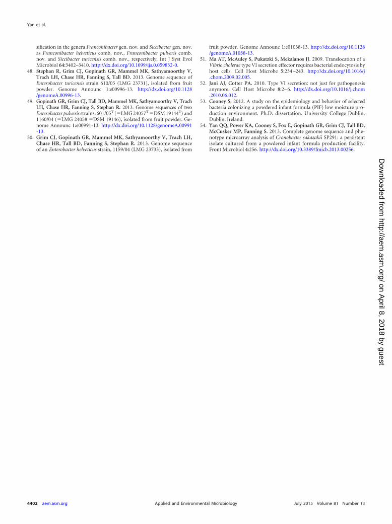
FIG 6 Comparisons among ST-1 and ST-4 isolates, of PIF or clinical origin, for the ability to swim. (a) CQ74, ST-1/PIF; (b) CQ113, ST-1/PIF; (c) E654,ST-1/clinical; (d) E657, ST-1/clinical; (e) CQ14, ST-4/PIF; (f) CQ27, ST-4/PIF; (g) 206N, ST-4/clinical; (h) E788, ST-4/clinical.
Yan et al.
4398 aem.asm.org July 2015 Volume 81 Number 13Applied and Environmental Microbiology
on April 8, 2018 by guest
http://aem.asm
.org/D
ownloaded from

which stated that combining both PFGE and MLST as a subtypingapproach would improve accuracy (20).
Two earlier studies have been reported that entailed microar-ray-based protocols to investigate the genomic diversity withinthe genus Cronobacter. Healy et al. (44) used a microarray designplatform based on 276 open reading frames, which were selectedfrom C. sakazakii ATCC BAA-894, to determine the gene differ-ences among five of the six Cronobacter species initially describedby Iversen et al. (2). Kucerova et al. (45) constructed a 387,000-probe oligonucleotide microarray covering the whole genome ofC. sakazakii ATCC BAA-894, in an effort to identify the pan-ge-nome of Cronobacter using five of the seven recognized species.
The microarray approach reported here was developed for themolecular characterization of Cronobacter from foods, primarilyto address source attribution in trace-back investigations and toinvestigate the genomic diversity and evolutionary history ofCronobacter species. Fifty-nine isolates selected from this studywere compared directly with five nearest neighbors and seven typestrains (Fig. 3). The microarray was able to accurately assess eachstrain’s identity and could differentiate Cronobacter species fromthe nearest neighbors. Furthermore, the microarray results sup-ported the rpoB-based identities of Cronobacter species as de-scribed by Stoop et al. (13) and Lehner et al. (17). The results alsoconcurred with recently published studies that have discussed thephylogenetic divergence of the genus from the most recent com-mon ancestral species into two major clusters, one consisting of C.dublinensis and C. muytjensii, and the other comprised of C. saka-zakii, C. malonaticus, C. universalis, and C. turicensis as postulatedby Grim et al. (46). C. condimenti was a distant outlier of these twoclusters. Of note, these results also offer a more in-depth analysisto the recent proposal to include Enterobacter pulveris, E. helveti-cus, and E. turicensis as members of this genus (36), and the resultssupport their reclassification as proposed by Stephan et al. (47).These data agree with the genome sequence information reportedpreviously (48–50).
In addition, our microarray data suggest the existence of someevolving lineages at the nucleotide level for C. sakazakii comparedwith other members of the genus. This is consistent with the factthat C. sakazakii accounts for approximately 84.8% of the isolatesstudied using this protocol. As an example, ST-4 strains separatedinto two clades comprising distinct lineages, which differed in thepresence or absence of genes associated with the pESA3-harboredT6SS. As is known for C. sakazakii ATCC BAA-894, the T6SS is arecently characterized protein secretion system that consists of 16ORFs (ESA_pESA3p 05491 to -5506) (41). T6SSs have been stud-ied primarily in the context of pathogenic bacterium-host inter-actions (51). Recent data suggest, however, that these versatileprotein secretion systems may also function to promote commen-sal or mutualistic relationships between bacteria and eukaryotes,or to mediate cooperative or competitive interactions betweenbacteria (52). One hypothesis is that T6SSs may be involved inoverall cell fitness, promulgating ecology-driven selection pro-cesses as cells interact with other cells present within their envi-ronment. Our data showed the presence of a repA gene in all of theisolates studied, which signified that these isolates possess thepESA3 common virulence plasmid. The vgrG (valine-glycine re-peat G protein gene) codes for a T6SS effector protein and is asingle-copy gene in this cluster. The VgrG protein also has relatedsequences that are distributed on the chromosome, but most ofthese are not associated with any other T6SS gene cluster (41).
Furthermore, the chromosomal T6SS genes in C. sakazakii ATCCBAA-894 do not share significant homology at the nucleotide levelwith the pESA3 T6SS gene locus (45). These results suggest thatthis region of the virulence plasmid, in these strains, may be in“genetic flux,” with either insertions or deletions most likely oc-curring in the 3= region of the gene locus. These observationsfurther support the perceived changes in gene content measuredvia PCR positive and negative controls with relevance for this re-gion, as described by Franco et al. (41). However, the reasons forthese changes remain unknown.
Microarray analysis showed that within the C. sakazakii clus-ter, six STs were identified, while 11 PFGE pulsotypes could alsobe grouped (Fig. 3). Interestingly, the ST-4 isolates, which weredivided into two subclusters denoted lineage 1 and lineage 2, alsodifferentiated according to serotypes: C. sakazakii O:2 and C.sakazakii O:3, respectively. Previous results reported by Hariri etal. (6) suggested that ST-4 strains form a distinct cluster withrelated STs, such as ST-110, -107, and -108; this cluster has beendefined as the ST-4 clonal complex. The finding of two closelyrelated lineages among ST-4 strains further defines and improvesthe phylogenetic resolution of this important meningitis-causinggroup.
Phenotype correlation analysis showed significant differences(P � 0.01) with respect to swim phenotype exhibited after a 24-hincubation period, along with an ability to swarm at both 8 and 24h. Isolates in lineage 1 demonstrated an increased ability to swarmat both 8 h (r � �0.650, P � 0.000) and 24 h (r � �0.442, P �0.027), while isolates in lineage 2 had better swimming ability at 24h (r � 0.579, P � 0.002) (data not shown).
Previously, MLST studies highlighted the importance of ST-4isolates linked to serious cases of meningitis (6). The same ST hasalso been identified among nonclinical isolates, including thosecultured from PIF and follow-up formula (20, 30, 32). An exampleof the latter is C. sakazakii SP291, which was included in this studybut did not cluster with any of the 14 pulsotypes. This isolate wasoriginally cultured from an environmental sample obtained fromfacility B and is historically known to persist in PIF productionenvironments for a period of at least 30 months (53). In this study,ST-1 is identified as the most frequent sequence type recovered,following the screening of all four PIF production sites from dif-ferent geographical regions (Fig. 2a), a feature reported previouslyby Pan et al. (20). Furthermore, an ST-1 isolate, C. sakazakiiATCC BAA-894, was cultured from a PIF source and had a pul-sotype profile that matched that of a clinical isolate. This isolatewas linked to the death of an infant who had consumed a portionof a contaminated batch of PIF in Tennessee, USA, in 2001 (5).These observations raise questions as to the nature of the pheno-typic differences between ST-1 and ST-4, which may in part ac-count for the dominance of each ST in different niche settings.
The phenotypes related to the survival of ST-1 and ST-4 iso-lates have not been compared previously, and moreover, such acomparison is warranted, based on the findings of this study. Phe-notypic experiments designed to compare both sequence typeswere performed on 168 isolates. These experiments included com-parisons between cell motility (swimming and swarming), biofilmformation, Congo red dye binding, and cellulose production (seeTable S1 in the supplemental material). In general, Cronobacterisolates of ST-1 exhibited a greater ability to swim and to formbiofilms at 37°C compared to ST-4 isolates, while the latter formeda stronger biofilm at 28°C and exhibited greater Congo red dye
A Form of Pathoadaptation among Cronobacter Isolates
July 2015 Volume 81 Number 13 aem.asm.org 4399Applied and Environmental Microbiology
on April 8, 2018 by guest
http://aem.asm
.org/D
ownloaded from
binding than the former (Table 3). Similarly, ST-1/PIF isolatesdemonstrated greater swarming activity and formed stronger bio-films at 37°C than did ST-1/clinical isolates; while among ST-4 theclinical isolates exhibited better swimming activity than PIF iso-lates (Fig. 6); however, the latter forms stronger biofilms at both28 and 37°C compared to the former. More importantly, ST-1/PIFisolates formed a stronger biofilm at both 28 and 37°C than didST-4/clinical isolates, while the latter exhibited a more activeswimming ability and greater binding of Congo red dye. As dem-onstrated in the present study, ST-1 is a common sequence typethat is cultured from PIF and the associated manufacturing envi-ronments investigated. The abilities of a bacterium to swim and tobind Congo red dye are known to be determinants related to vir-ulence. The phenotypic differences between these two Cronobac-ter sequence types may in part explain why ST-4 isolates of clinicalorigin are more often linked to cases of meningitis. It is temptingto speculate that these observations may represent a type of patho-adaptation. Further, our findings are consistent with phenotypesdescribed by Yan et al. (54) for C. sakazakii SP291, an ST-4 isolatewhose origin was a PIF production environment.
In conclusion, C. sakazakii O:1 ST-1 was found to be the mostcommon sequence type cultured from four geographically dis-tinct PIF production facilities. Seventy of 84 ST-1 C. sakazakiiisolates clustered as a distinct pulsotype, and these could be recov-ered over an 18-month period. Phenotypic differences were notedwhen comparisons were made between ST-1 and -4 isolates, in-cluding differences in bacterial motility, biofilm formation,Congo red dye binding, and cellulose production, all of which areconsidered to be relevant for bacterial survival in these environ-ments. These characteristics may contribute to the pathoadapta-tion of this pathogen as it becomes disseminated along the foodchain, and they account for the epidemiological observations inboth cases. The adaptation of this opportunistic pathogen to thePIF manufacturing environment could lead to the survival ofthese organisms in finished product and increase thereafter therisk of causing infections once the contaminated food is con-sumed. Further in-depth phenotypic characterizations may pro-vide clues as to how this phenotype is controlled in response tocritical signals and subsequently expressed. Moreover, this type ofapproach may highlight bacterial targets that could be useful inthe development of biomarkers for use in control protocols.
ACKNOWLEDGMENTS
We thank Matthew McCusker and Sarah Finn for their assistance with thephenotype assays.
REFERENCES1. Iversen C, Lehner A, Mullane N, Bidlas E, Cleenwerck I, Marugg J,
Fanning S, Stephan R, Joosten H. 2007. The taxonomy of Enterobactersakazakii: proposal of a new genus Cronobacter gen. nov. and descriptionsof Cronobacter sakazakii comb. nov. Cronobacter sakazakii subsp. saka-zakii, comb. nov., Cronobacter sakazakii subsp. malonaticus subsp. nov.,Cronobacter turicensis sp. nov., Cronobacter muytjensii sp. nov., Cronobac-ter dublinensis sp. nov. and Cronobacter genomospecies 1. BMC Evol Biol7:64. http://dx.doi.org/10.1186/1471-2148-7-64.
2. Iversen C, Mullane N, McCardell B, Tall BD, Lehner A, Fanning S,Stephan R, Joosten H. 2008. Cronobacter gen. nov., a new genus toaccommodate the biogroups of Enterobacter sakazakii, and proposal ofCronobacter sakazakii gen. nov., comb. nov., Cronobacter malonaticus sp.nov., Cronobacter turicensis sp. nov., Cronobacter muytjensii sp. nov.,Cronobacter dublinensis sp. nov., Cronobacter genomospecies 1, and ofthree subspecies, Cronobacter dublinensis subsp. dublinensis subsp. nov.,Cronobacter dublinensis subsp. lausannensis subsp. nov. and Cronobacter
dublinensis subsp. lactaridi subsp. nov. Int J Syst Evol Microbiol 58:1442–1447. http://dx.doi.org/10.1099/ijs.0.65577-0.
3. Joseph S, Cetinkaya E, Drahovska H, Levican A, Figueras MJ, ForsytheSJ. 2012. Cronobacter condimenti sp. nov., isolated from spiced meat, andCronobacter universalis sp. nov., a species designation for Cronobacter sp.genomospecies 1, recovered from a leg infection, water and food ingredi-ents. Int J Syst Evol Microbiol 62:1277–1283. http://dx.doi.org/10.1099/ijs.0.032292-0.
4. Hunter CJ, Bean JF. 2013. Cronobacter: an emerging opportunisticpathogen associated with neonatal meningitis, sepsis and necrotizing en-terocolitis. J Perinatol 33:581–585. http://dx.doi.org/10.1038/jp.2013.26.
5. Himelright I, Harris E, Lorch V, Anderson M, Jones T, Craig A,Kuehnert M, Forster T, Arduino M, Jensen B, Jernigan D. 2002.Enterobacter sakazakii infections associated with the use of powdered in-fant formula—Tennessee, 2001. MMWR Morb Mortal Wkly Rep 51:297–300.
6. Hariri S, Joseph S, Forsythe SJ. 2013. Cronobacter sakazakii ST4 strainsand neonatal meningitis, United States. Emerg Infect Dis 19:175–177.http://dx.doi.org/10.3201/eid1901.120649.
7. Stephan R, Lehner A, Tischler P, Rattei T. 2011. Complete genomesequence of Cronobacter turicensis LMG 23827, a food-borne pathogencausing deaths in neonates. J Bacteriol 193:309 –310. http://dx.doi.org/10.1128/JB.01162-10.
8. Derzelle S, Dilasser F. 2006. A robotic DNA purification protocol andreal-time PCR for the detection of Enterobacter sakazakii in powderedinfant formulae. BMC Microbiol 6:100. http://dx.doi.org/10.1186/1471-2180-6-100.
9. Drudy D, O’Rourke M, Murphy M, Mullane NR, O’Mahony R, Kelly L,Fischer M, Sanjaq S, Shannon P, Wall P, O’Mahony M, Whyte P,Fanning S. 2006. Characterization of a collection of Enterobacter sakazakiiisolates from environmental and food sources. Int J Food Microbiol 110:127–134. http://dx.doi.org/10.1016/j.ijfoodmicro.2006.02.008.
10. Liu Y, Cai X, Zhang X, Gao Q, Yang X, Zheng Z, Luo M, Huang X.2006. Real time PCR using TaqMan and SYBR Green for detection ofEnterobacter sakazakii in infant formula. J Microbiol Methods 65:21–31.http://dx.doi.org/10.1016/j.mimet.2005.06.007.
11. Mullane N, O’Gaora P, Nally JE, Iversen C, Whyte P, Wall PG, FanningS. 2008. Molecular analysis of the Enterobacter sakazakii O-antigen genelocus. Appl Environ Microbiol 74:3783–3794. http://dx.doi.org/10.1128/AEM.02302-07.
12. Molloy C, Cagney C, O’Brien S, Iversen C, Fanning S, Duffy G. 2009.Surveillance and characterisation by pulsed-field gel electrophoresis ofCronobacter spp. in farming and domestic environments, food productionanimals and retail foods. Int J Food Microbiol 136:198 –203. http://dx.doi.org/10.1016/j.ijfoodmicro.2009.07.007.
13. Stoop B, Lehner A, Iversen C, Fanning S, Stephan R. 2009. Develop-ment and evaluation of rpoB based PCR systems to differentiate the sixproposed species within the genus Cronobacter. Int J Food Microbiol 136:165–168. http://dx.doi.org/10.1016/j.ijfoodmicro.2009.04.023.
14. Fricker-Feer C, Cernela N, Bolzan S, Lehner A, Stephan R. 2011.Evaluation of three commercially available real-time PCR based systemsfor detection of Cronobacter species. Int J Food Microbiol 146:200 –202.http://dx.doi.org/10.1016/j.ijfoodmicro.2011.02.006.
15. Jarvis KG, Grim CJ, Franco AA, Gopinath G, Sathyamoorthy V, Hu L,Sadowski JA, Lee CS, Tall BD. 2011. Molecular characterization ofCronobacter lipopolysaccharide O-antigen gene clusters and developmentof serotype-specific PCR assays. Appl Environ Microbiol 77:4017– 4026.http://dx.doi.org/10.1128/AEM.00162-11.
16. Sun Y, Wang M, Liu H, Wang J, He X, Zeng J, Guo X, Li K, Cao B,Wang L. 2011. Development of an O-antigen serotyping scheme forCronobacter sakazakii. Appl Environ Microbiol 77:2209 –2214. http://dx.doi.org/10.1128/AEM.02229-10.
17. Lehner A, Fricker-Feer C, Stephan R. 2012. Identification of the recentlydescribed Cronobacter condimenti by a rpoB-gene-based PCR system. JMed Microbiol 61:1034 –1035. http://dx.doi.org/10.1099/jmm.0.042903-0.
18. Cai XQ, Yu HQ, Ruan ZX, Yang LL, Bai JS, Qiu DY, Jian ZH, Xiao YQ,Yang JY, Le TH, Zhu XQ. 2013. Rapid detection and simultaneousgenotyping of Cronobacter spp. (formerly Enterobacter sakazakii) in pow-dered infant formula using real-time PCR and high resolution melting(HRM) analysis. PLoS One 8:e67082. http://dx.doi.org/10.1371/journal.pone.0067082.
19. Jarvis KG, Yan QQ, Grim CJ, Power KA, Franco AA, Hu L, Gopinath
Yan et al.
4400 aem.asm.org July 2015 Volume 81 Number 13Applied and Environmental Microbiology
on April 8, 2018 by guest
http://aem.asm
.org/D
ownloaded from

G, Sathyamoorthy V, Kotewicz ML, Kothary MH, Lee C, Sadowski J,Fanning S, Tall BD. 2013. Identification and characterization of five newmolecular serogroups of Cronobacter spp. Foodborne Pathog Dis 10:343–352. http://dx.doi.org/10.1089/fpd.2012.1344.
20. Pan Z, Cui J, Lyu G, Du X, Qin L, Guo Y, Xu B, Li W, Cui Z, Zhao C.2014. Isolation and molecular typing of Cronobacter spp. in commercialpowdered infant formula and follow-up formula. Foodborne Pathog Dis11:456 – 461. http://dx.doi.org/10.1089/fpd.2013.1691.
21. Mullane NR, Whyte P, Wall PG, Quinn T, Fanning S. 2007. Applicationof pulsed-field gel electrophoresis to characterise and trace the prevalenceof Enterobacter sakazakii in an infant formula processing facility. Int JFood Microbiol 116:73– 81. http://dx.doi.org/10.1016/j.ijfoodmicro.2006.12.036.
22. Mullane N, Healy B, Meade J, Whyte P, Wall PG, Fanning S. 2008.Dissemination of Cronobacter spp. (Enterobacter sakazakii) in a powderedmilk protein manufacturing facility. Appl Environ Microbiol 74:5913–5917. http://dx.doi.org/10.1128/AEM.00745-08.
23. El-Sharoud WM, O’Brien S, Negredo C, Iversen C, Fanning S, Healy B.2009. Characterization of Cronobacter recovered from dried milk and re-lated products. BMC Microbiol 9:24. http://dx.doi.org/10.1186/1471-2180-9-24.
24. Terragno R, Salve A, Pichel M, Epszteyn S, Brengi S, Binsztein N. 2009.Characterization and subtyping of Cronobacter spp. from imported pow-dered infant formulae in Argentina. Int J Food Microbiol 136:193–197.http://dx.doi.org/10.1016/j.ijfoodmicro.2009.10.013.
25. Craven HM, McAuley CM, Duffy LL, Fegan N. 2010. Distribution,prevalence and persistence of Cronobacter (Enterobacter sakazakii) in thenonprocessing and processing environments of five milk powder facto-ries. J Appl Microbiol 109:1044 –1052. http://dx.doi.org/10.1111/j.1365-2672.2010.04733.x.
26. Miled-Bennour R, Ells TC, Pagotto FJ, Farber JM, Kerouanton A,Meheut T, Colin P, Joosten H, Leclercq A, Besse NG. 2010. Genotypicand phenotypic characterisation of a collection of Cronobacter (Enterobac-ter sakazakii) isolates. Int J Food Microbiol 139:116 –125. http://dx.doi.org/10.1016/j.ijfoodmicro.2010.01.045.
27. Brengi SP, O’Brien SB, Pichel M, Iversen C, Arduino M, Binsztein N,Jensen B, Pagotto F, Ribot EM, Stephan R, Cernela N, Cooper K,Fanning S. 2012. Development and validation of a PulseNet standardizedprotocol for subtyping isolates of Cronobacter species. Foodborne PathogDis 9:861– 867. http://dx.doi.org/10.1089/fpd.2012.1161.
28. Yan QQ, Fanning S. 2015. Pulsed-field gel electrophoresis (PFGE) forpathogenic Cronobacter species, p 55– 69. In Jordan K, Marion D (ed),Pulsed field gel electrophoresis: methods and protocols. Springer Science,New York, NY.
29. Joseph S, Sonbol H, Hariri S, Desai P, McClelland M, Forsythe SJ. 2012.Diversity of the Cronobacter genus as revealed by multilocus sequencetyping. J Clin Microbiol 50:3031–3039. http://dx.doi.org/10.1128/JCM.00905-12.
30. Baldwin A, Loughlin M, Caubilla-Barron J, Kucerova E, Manning G,Dowson C, Forsythe S. 2009. Multilocus sequence typing of Cronobactersakazakii and Cronobacter malonaticus reveals stable clonal structures withclinical significance which do not correlate with biotypes. BMC Microbiol9:223. http://dx.doi.org/10.1186/1471-2180-9-223.
31. Joseph S, Forsythe SJ. 2012. Insights into the emergent bacterial pathogenCronobacter spp., generated by multilocus sequence typing and analysis.Front Microbiol 3:397. http://dx.doi.org/10.3389/fmicb.2012.00397.
32. Müller A, Stephan R, Fricker-Feer C, Lehner A. 2013. Genetic diversityof Cronobacter sakazakii isolates collected from a Swiss infant formulaproduction facility. J Food Prot 76:883– 887. http://dx.doi.org/10.4315/0362-028X.JFP-12-521.
33. Gicová A, Oriešková M, Oslanecová L, Drahovská H, Kaclíková E. 2014.Identification and characterization of Cronobacter strains isolated frompowdered infant foods. Lett Appl Microbiol 58:242–247. http://dx.doi.org/10.1111/lam.12179.
34. Cruz-Cordova A, Rocha-Ramirez LM, Ochoa SA, Gonzalez-Pedrajo B,Espinosa N, Eslava C, Hernandez-Chinas U, Mendoza-Hernandez G,Rodriguez-Leviz A, Valencia-Mayoral P, Sadowinski-Pine S, Hernandez-Castro R, Estrada-Garcia I, Munoz-Hernandez O, Rosas I, Xicohtencatl-Cortes J. 2012. Flagella from five Cronobacter species induce pro-inflammatory cytokines in macrophage derivatives from human monocytes.PLoS One 7:e52091. http://dx.doi.org/10.1371/journal.pone.0052091.
35. Seo KH, Brackett RE. 2005. Rapid, specific detection of Enterobacter
sakazakii in infant formula using a real-time PCR assay. J Food Prot 68:59 – 63.
36. Brady C, Cleenwerck I, Venter S, Coutinho T, De Vos P. 2013. Taxo-nomic evaluation of the genus Enterobacter based on multilocus sequenceanalysis (MLSA): proposal to reclassify E. nimipressuralis and E. amnige-nus into Lelliottia gen. nov. as Lelliottia nimipressuralis comb. nov. andLelliottia amnigena comb. nov., respectively, E. gergoviae and E. pyrinusinto Pluralibacter gen. nov. as Pluralibacter gergoviae comb. nov. and Plu-ralibacter pyrinus comb. nov., respectively, E. cowanii, E. radicincitans, E.oryzae and E. arachidis into Kosakonia gen. nov. as Kosakonia cowaniicomb. nov., Kosakonia radicincitans comb. nov., Kosakonia oryzae comb.nov. and Kosakonia arachidis comb. nov., respectively, and E. turicensis, E.helveticus and E. pulveris into Cronobacter as Cronobacter zurichensis nom.nov., Cronobacter helveticus comb. nov. and Cronobacter pulveris comb.nov., respectively, and emended description of the genera Enterobacterand Cronobacter. Syst Appl Microbiol 36:309 –319. http://dx.doi.org/10.1016/j.syapm.2013.03.005.
37. Tall BD, Gangiredla J, Gopinath GR, Yan Q, Chase HR, Lee B, HwangS, Trach L, Park E, Yoo Y, Chung T, Jackson SA, Patel IR, Sathyamoor-thy V, Pava-Ripoll M, Kotewicz ML, Carter L, Iversen C, Pagotto F,Stephan R, Lehner A, Fanning S, Grim CJ. 2015. Development of acustom-designed, pan genomic DNA microarray to characterize strain-level diversity among Cronobacter spp. Front Pediatr 3:1–11. http://dx.doi.org/10.3389/fped.2015.00036.
38. Jackson SA, Patel IR, Barnaba T, LeClerc JE, Cebula TA. 2011. Inves-tigation the global genomic diversity of Escherichia coli using a multi-genome DNA microarray platform with novel gene prediction strategies.BMC Genomics 12:349. http://dx.doi.org/10.1186/1471-2164-12-349.
39. Martins M, McCusker MP, McCabe EM, O’Leary D, Duffy G,Fanning S. 2013. Evidence of metabolic switching and implications forfood safety from the phenome(s) of Salmonella enterica serovar Typhi-murium DT104 cultured at selected points across the pork productionfood chain. Appl Environ Microbiol 79:5437–5449. http://dx.doi.org/10.1128/AEM.01041-13.
40. Finn S, Hinton JC, McClure P, Amezquita A, Martins M, Fanning S.2013. Phenotypic characterization of Salmonella isolated from food pro-duction environments associated with low-water activity foods. J FoodProt 76:1488 –1499. http://dx.doi.org/10.4315/0362-028X.JFP-13-088.
41. Franco AA, Kothary MH, Gopinath G, Jarvis KG, Grim CJ, Hu L, DattaAR, McCardell BA, Tall BD. 2011. Cpa, the outer membrane protease ofCronobacter sakazakii, activates plasminogen and mediates resistance toserum bactericidal activity. Infect Immun 79:1578 –1587. http://dx.doi.org/10.1128/IAI.01165-10.
42. Hachani A, Allsopp LP, Oduko Y, Filloux A. 2014. The VgrG proteinsare “a la carte” delivery systems for bacterial type VI effectors. J Biol Chem289:17872–17884. http://dx.doi.org/10.1074/jbc.M114.563429.
43. Mozrová V, Bøenová N, Mrázek J, Lukešová D, Marounek M. 2014.Surveillance and characterisation of Cronobacter spp. in Czech retail foodand environmental samples. Folia Microbiol (Praha) 59:63–38. http://dx.doi.org/10.1007/s12223-013-0266-2.
44. Healy B, Huynh S, Mullane N, O’Brien S, Iversen C, Lehner A, StephanR, Parker CT, Fanning S. 2009. Microarray-based comparative genomicindexing of the Cronobacter genus (Enterobacter sakazakii). Int J FoodMicrobiol 136:159 –164. http://dx.doi.org/10.1016/j.ijfoodmicro.2009.07.008.
45. Kucerova E, Clifton SW, Xia XQ, Long F, Porwollik S, Fulton L,Fronick C, Minx P, Kyung K, Warren W, Fulton R, Feng DY, WollamA, Shah N, Bhonagiri V, Nash WE, Pepin KH, Wilson RK, McClellandM, Forsythe SJ. 2010. Genome sequence of Cronobacter sakazakii BAA-894 and comparative genomic hybridization analysis with other Crono-bacter species. PLoS One 5:e9556. http://dx.doi.org/10.1371/journal.pone.0009556.
46. Grim CJ, Kotewicz ML, Power K, Pagotto F, Gopinath G, Mammel MK,Jarvis KG, Yan QQ, Kothary MH, Franco AA, Patel IR, Jackson SA, HuL, Sathyamoorthy V, Iversen C, Lehner A, Stephan R, Farber JM,Fanning S, Tall BD. 2013. Pan genome analysis of the emerging food-borne pathogen Cronobacter spp. suggests a species-level bidirectional di-vergence driven by niche adaption. BMC Genomics 14:366. http://dx.doi.org/10.1186/1471-2164-14-366.
47. Stephan R, Grim CJ, Gopinath GR, Mammel MK, Sathyamoorthy V,Trach LH, Chase HR, Fanning S, Tall BD. 2014. Re-examination of thetaxonomic status of Enterobacter helveticus, Enterobacter pulveris, and En-terobacter turicensis as members of the genus Cronobacter and their reclas-
A Form of Pathoadaptation among Cronobacter Isolates
July 2015 Volume 81 Number 13 aem.asm.org 4401Applied and Environmental Microbiology
on April 8, 2018 by guest
http://aem.asm
.org/D
ownloaded from
sification in the genera Franconibacter gen. nov. and Siccibacter gen. nov.as Franconibacter helveticus comb. nov., Franconibacter pulveris comb.nov. and Siccibacter turicensis comb. nov., respectively. Int J Syst EvolMicrobiol 64:3402–3410. http://dx.doi.org/10.1099/ijs.0.059832-0.
48. Stephan R, Grim CJ, Gopinath GR, Mammel MK, Sathyamoorthy V,Trach LH, Chase HR, Fanning S, Tall BD. 2013. Genome sequence ofEnterobacter turicensis strain 610/05 (LMG 23731), isolated from fruitpowder. Genome Announc 1:e00996-13. http://dx.doi.org/10.1128/genomeA.00996-13.
49. Gopinath GR, Grim CJ, Tall BD, Mammel MK, Sathyamoorthy V, TrachLH, Chase HR, Fanning S, Stephan R. 2013. Genome sequences of twoEnterobacter pulveris strains, 601/05T (�LMG 24057T �DSM 19144T) and1160/04 (�LMG 24058 �DSM 19146), isolated from fruit powder. Ge-nome Announc 1:e00991-13. http://dx.doi.org/10.1128/genomeA.00991-13.
50. Grim CJ, Gopinath GR, Mammel MK, Sathyamoorthy V, Trach LH,Chase HR, Tall BD, Fanning S, Stephan R. 2013. Genome sequenceof an Enterobacter helveticus strain, 1159/04 (LMG 23733), isolated from
fruit powder. Genome Announc 1:e01038-13. http://dx.doi.org/10.1128/genomeA.01038-13.
51. Ma AT, McAuley S, Pukatzki S, Mekalanos JJ. 2009. Translocation of aVibrio cholerae type VI secretion effector requires bacterial endocytosis byhost cells. Cell Host Microbe 5:234 –243. http://dx.doi.org/10.1016/j.chom.2009.02.005.
52. Jani AJ, Cotter PA. 2010. Type VI secretion: not just for pathogenesisanymore. Cell Host Microbe 8:2– 6. http://dx.doi.org/10.1016/j.chom.2010.06.012.
53. Cooney S. 2012. A study on the epidemiology and behavior of selectedbacteria colonizing a powdered infant formula (PIF) low moisture pro-duction environment. Ph.D. dissertation. University College Dublin,Dublin, Ireland.
54. Yan QQ, Power KA, Cooney S, Fox E, Gopinath GR, Grim CJ, Tall BD,McCusker MP, Fanning S. 2013. Complete genome sequence and phe-notype microarray analysis of Cronobacter sakazakii SP291: a persistentisolate cultured from a powdered infant formula production facility.Front Microbiol 4:256. http://dx.doi.org/10.3389/fmicb.2013.00256.
Yan et al.
4402 aem.asm.org July 2015 Volume 81 Number 13Applied and Environmental Microbiology
on April 8, 2018 by guest
http://aem.asm
.org/D
ownloaded from