“COMPARATIVE EVALUATION OF THE APICAL SEALING ABILITY …
54
“COMPARATIVE EVALUATION OF THE APICAL SEALING ABILITY OF MATCHED TAPER SINGLE CONE TECHNIQUE WITH OTHER OBTURATION TECHNIQUES” - AN IN VITRO STUDY. Dissertation submitted to: THE TAMILNADU DR.M.G.R MEDICAL UNIVERSITY In partial fulfillment for the degree of MASTER OF DENTAL SURGERY BRANCH - IV CONSERVATIVE DENTISTRY AND ENDODONTICS APRIL -2011
Transcript of “COMPARATIVE EVALUATION OF THE APICAL SEALING ABILITY …
“COMPARATIVE EVALUATION OF THE APICAL
SEALING ABILITY OF MATCHED TAPER SINGLE
CONE TECHNIQUE WITH OTHER OBTURATION
TECHNIQUES” - AN IN VITRO STUDY.
Dissertation submitted to:
THE TAMILNADU DR.M.G.R MEDICAL UNIVERSITY
In partial fulfillment for the degree of
MASTER OF DENTAL SURGERY
BRANCH - IV CONSERVATIVE DENTISTRY AND ENDODONTICS
APRIL -2011

CERTIFICATE
This is to certify that DR.YOUSEF RISHAL P.T, post graduate student
(2008-2011) in the Department Of Conservative Dentistry And Endodontics,
J.K.K.Nataraja Dental College, Komarapalayam , Namakkal District –
638183, Tamil Nadu. has done the dissertation titled
“COMPARATIVE EVALUATION OF THE APICAL SEALING ABILITY
OF MATCHED TAPER SINGLE CONE TECHNIQUE WITH OTHER
OBTURATION TECHNIQUES ” - AN IN VITRO STUDY. Under my direct
guidance and supervision in the partial fulfillment of the regulations laid down by
THE TAMIL NADU DR.M.G.R MEDICAL UNIVERSITY, CHENNAI, FOR
M.D.S BRANCH – IV CONSERVATIVE DENTISTRY AND ENDODONTICS
DEGREE EXAMINATION.
It has not been submitted (partial or full) for the award of any other degree
or diploma.
Dr.J.V.Karunakaran M.D.S, Dr.Siva Kumar,M.D.S, Professor& Head, Principal, Department of Conservative J.K.K.Nataraja Dental Dentistry & Endodontics, College Komarapalayam, Komarapalayam, Namakkal Dist – 638183, Namakkal Dist – 638183 Tamilnadu. Tamilnadu.
ACKNOWLEDGEMENT
I take this opportunity to sincerely thank my post graduate
teacher and my guide Dr. J.V.Karunakaran M.D.S, Professor and
HOD, Department of Conservative Dentistry & Endodontics, J.K.K
Nataraja Dental College, for his perseverance in motivating and
supporting me throughout my study period.
My sincere thanks to Dr. Sivakumar M.D.S, Professor and
Principal, Department of Oral Surgery,J.K.K Nataraja Dental College,
who had helped with his advice and immense support throughout my
postgraduate curriculum.
I would like to express my sincere gratitude to my professor,
Dr. N.S Mohan Kumar M.D.S, Professor, Department of Conservative
Dentistry & Endodontics ,J.K.K Nataraja Dental College, for his
valuable suggestions, support and encouragement throughout my post
graduate curriculum.
I extend my sincere thanks to Dr. S. Senthil Kumar M.D.S,
Reader, for his continuous guidance and constant encouragement
throughout my study period.
I thank Dr. Sunil Mankar M.D.S, Dr. Satyanarayanan M.D.S, Dr.
Chandrasekar M.D.S, Senior lecturers for their support, guidance and for
their constant encouragement throughout the completion of this work.
I am extremely thankful to Dr.Mira Mohanty, M.D. (Pathology)
Head of Dept. of Implant Biology, Sree Chittira Tirunnal Institute of
Biomedical research, Trivandrum who helped me with the
stereomicroscope facility and allowing me to perform the study under
her guidance.
I would like to thank Dr. Shabareeshwaran M.V.Sc (Pathology)
Dept. of Implant Biology, Sree Chittira Tirunnal Institute of Biomedical
research, Trivandrum, in helping me out in using the stereomicroscope
and for his valuable suggestions.
I am profoundly thankful to Dr P.Perumal, M.Pharm, Principal,
J.K.K Nataraja Pharmacy College for allowing me to use the research
facilities available in their college, and guiding me throughout the study.
My sincere thanks to M.Prasad, MSc, MBA, MPhil, PGDOR,
K.S.Rangasamy College Of Arts and Science for his guidance in
biostatistics.
I remain ever grateful to all my batch mates, colleagues and
friends for their eternal support.
Above all, I am thankful to God almighty, to have given me the
strength to choose the right path and to have given these wonderful
people in my life.

CONTENTS
S.NO INDEX PAGE.NO
1 INTRODUCTION 1
2 REVIEW OF LITERATURE 5
3 MATERIALS AND METHODS 23
4 RESULTS 31
5 DISCUSSION 36
6 SUMMARY 48
7 CONCLUSION 49
8 BIBILIOGRAPHY 50
Introduction
1
The primary objective of root canal therapy is disinfection of the root
canal space, to eliminate bacterial pathogens and to achieve a three
dimensional obturation of the root canal system with a bio-compatible filling
material achieving a hermetic seal, thereby preventing bacterial growth and
penetration of fluid and antigenic agents between the canal and periapical
tissues.
Microorganisms present inside the root canal remain active in the
dentinal tubules even after vigorous chemo-mechanical preparation. Apical
leakage continues to be a topic of great interest because in spite of advances in
endodontics, clinical failures still occur. Total apical seal is desirable to
prevent the remaining bacteria and endotoxins form reaching the root apex and
beyond. Apical leakage is considered to be a common cause for endodontic
therapy failure and is influenced by many variables such as filling techniques,
sealer properties etc. Leakage can also be from the coronal end of the root
canal restoration.
The most common cause of apparent failure in endodontically treated
teeth is apical percolation resulting in incomplete canal obturation. (John
Ingle, 1976)34.
The advent of Nickel – Titanium makes predictably more efficient
preparation of the root canal and makes apical cone fit a possibility in many
cases. (Buchanan et al 2001)9. They are far superior to stainless steel
instruments, more elastic and have good torsional fracture resistance.
(Clifford J Ruddle, 2005)12.
Introduction
2
Rotary Ni-Ti instruments improve the working safety, shorten working
times and cause fewer canal transportations.
Protaper (Dentsply Maillefer, Ballaigues, Switzerland) is a recent new
file design of rotary nickel titanium instruments and designed to provide
superior flexibility, efficiency and safety. The Protaper system features a
group of 6 instruments - SX shaper, 2 shaper files – S1, S2, and three finishing
files - F1, F2, F3.
Although numerous materials have been used for root canal obturation,
the most commonly used material is gutta-percha in conjunction with a sealer
(Carrotte.P et al 2004)15. The physical properties of gutta-percha have made it
widely popular and have made possible different obturation techniques.
The cold lateral condensation technique is still one of the most
frequently used technique. It’s ability to adapt to the internal surface of root
canal has been questioned. There are also other disadvantages in this technique
like voids, incomplete fusion of gutta-percha cones and lack of surface
adaptation (Tamer Tasdemir et al 2009)61.
Single cone obturation came to the fore in the 1960s with the
development of ISO standards for endodontic instruments and filling points.
After reaming a circular stop in the apical 2mm of the canal, a single gutta-
percha, sectional silver or titanium point was selected to fit with “tug-back” to
demonstrate inlay-like snugness of fit. The cone was then cemented in place
with thin uniform layer of traditional sealer at least at the apical portion of the
canal. Concerns about the single cone technique were compounded by the
Introduction
3
realization that canal transportation is common and that damaged roots are
difficult to seal. (John Whitworth, 2005)36.
Recently matched taper gutta-percha points for Protaper system
(Dentsply, Maillefer) have been introduced, for simple time efficient
obturation. In this system the canals prepared with the protaper instruments
are filled with the point that matches the size of the finisher file. The system
is simple and time efficient.
The matched taper single cone technique uses gutta-percha cones that
closely matches the geometry of the nickel-titanium instrumentation systems.
This technique minimizes the sealer component and ensures three dimensional
obturation of the root canal space without accessory cones and saves time
spent on lateral condensation. (Gordon M.P et al , 2005)27.
The thermoplasticized gutta-percha technique (Yee et al, 1977)26
introduced in the late seventies has since evolved into various types of
thermoplastic gutta-percha techniques. The thermoplasticized gutta-percha
injection system is used by many practitioners to back-fill the root canal
system after the downpack phase of the master cone. The high temperature
generated can be dissipated through the canal and periodontal ligament
(Timothy L. Sweatman et al 2001)63.
In this study we have used a high temperature Obtura-II system, which
has been experimentally superior to lateral condensation technique for
obturation of the root canals.

Introduction
4
The aim of the present study is to compare the apical sealing ability of
matched taper single cone technique of obturation, the cold lateral
condensation technique and the Obtura-II thermoplasticized gutta-percha
injection technique in teeth which have been prepared using Protaper
instruments. (Dentsply, Maillefer) under laboratory conditions.
The dye penetration method used for measuring leakage uses India ink
which was done under high vacuum conditions to eliminate the entrapped air
within the canals. The apical seal was then evaluated after clearing the
specimens and the samples observed under a Binocular stereomicroscope with
a digital measurement software and the results analyzed.

Review of Literature
5
Green & David in 196028 found that in 50% of posterior teeth the
apical foramen was on the root surface and not on the anatomic apex.
John Ingle in 197634 describes 58.66% of failures of root canal therapy
could be attributed to incomplete obturation and 9.6% were attributed to root
perforations. He stresses the importance of the knowledge of root canal
morphology and achievement of a hermetic seal for successful outcome of
endodontic therapy.
Fulton.S.Yee et al in 197726 first applied the technique of
thermoplasticized gutta-percha injection in vitro and found that the injection
moulding technique leads to a seal comparable to that of conventional
approach and suggested that this technique held great promise for in vivo use.
Mahmoud Torabinejad et al in 197844 compared thermoplasticized
gutta- percha with lateral condensation, warm gutta-percha with vertical
condensation and chloropercha respectively and found that the injection
moulding technique resulted in the observation of root canal system which
was comparable to other conventional approaches.
Don Robertson et al in 198021 described a simple and inexpensive
technique for in vitro examination of endodontically treated or untreated root
canal systems which clears the teeth and renders the canal contents visible.
J. Marlin et al in 198132 performed a preliminary study on the clinical
use of thermoplasticized gutta-percha for root canal system obturation and
found that this method shows promise because the success rate seems

Review of Literature
6
comparable to the rate achieved with conventional gutta-percha obturation
procedures.
David A Allison et al in 198117 in their study of the influence of master
cone adaptation on the quality of the apical seal, showed that the microleakage
of the canals obturated with lateral condensation technique using gutta-percha
and sealer was directly proportional, to the shape of the prepared canal. There
was little or no microleakage if the canal had sufficient taper to permit
spreader penetration to within 1mm of the prepared length. The techniques
evaluated were standardized or step-back instrumentation techniques.
Yee RDJ in 198455 studied the effect of canal preparation on the formation
and leakage characteristics of apical dentin plug and found that
1. Dentin plugs occurred during instrumentation and can produce a seal
that resist penetration by the isotope
2. Formation of this seal in a dentin plug is not reliable, even after
instrumentation with five file series.
3. Leakage resistance varied according to the density of the plug
4. The proportion of hard to soft tissue components of each plug
increases with increased instrumentation and irrigation.
Andrew Michanowicz et al in 19842 investigated the apical sealing
properties of a low temperature thermoplasticized gutta-percha with or without
sealer and compared it with the lateral condensation technique with sealer and
found that the low temperature gutta-percha creates a low seal especially if
used in conjunction with a sealer. Several advantages of low temperature
injection gutta-percha technique is that it creates a good seal, especially if used
Review of Literature
7
in conjunction with a sealer. Radiographic observations showed that a uniform
mass was achieved when low temperature gutta-percha was used.
Mahmoud. E Eldeeb et al in 198543 evaluated the sealing ability of
injection moulded thermoplasticized gutta-percha with and without sealer and
compared it with laterally condensed gutta-percha with sealer and found that
leakage increase significantly when the injection technique was used without
sealer and overfilling occurred 75% of time with vertical condensation of
thermoplasticized gutta-percha. There was no significant difference in leakage
between the lateral condensation and injection techniques providing sealer was
used.
Kimberly Swanson et al in 198738 evaluated coronal microleakage
overtime when the obturation material was exposed to fluids in his study of
evaluation of microleakage in endodontically treated teeth. They observe that
coronal microleakage following exposure of root canal obturation material to
oral fluids caused considerable dye penetration while the group not exposed to
saliva exhibited no dye penetration. No significant differences were found
between the groups exposed to artificial saliva. They concluded that a
significant amount of coronal microleakage was evident after 3 days of
exposure to artificial saliva.
Budd et al in 199113 in their study compared the quality of obturation
of high and low thermoplasticized gutta-percha injection technique with
standard lateral condensation. Statistical analysis of the results indicated both
the technique were significantly better than lateral condensation. There was no
significant difference between either of the obturation techniques. Although

Review of Literature
8
High and low temperature thermoplasticized gutta-percha techniques produced
superior obturations within the parameters of the in vitro study, further
investigation is needed to evaluate an acceptable clinical technique which
allows these materials to be predictably condensed and confined within the
root canal.
Scott. S. Dickson et al in 199359 evaluated leakage with or without
vacuum with two gutta-percha filled techniques and found there was no
significant difference between vacuumed and non-vacuumed for degree of ink
penetration.
Dilek M. Dalat et al in 199420 compared the apical leakage in root
canals obturated with various gutta-percha techniques using a dye vacuum
tracing method and found that there was no statistically significant difference
between the gutta-percha obturation methods. He also observed that
radiographically detected voids resulted in an increased failure rate. This study
concentrated in detecting microlumina in obturated root canal spaces in the
apical region using this dye tracer technique.
P. Portman et al in 199451 evaluated a new vacuum obturation
technique and compared it with lateral condensation technique and found that
obturation with the new vacuum technique resulted in significantly less
leakage than lateral condensation and is also less time consuming.
John Masters et al in 199535 compared dye leakage pattern of prepared
root canal with that of glass tubes with or without vacuuming. The results
showed that filled and unfilled, prepared root canals leaked significantly more
than their glass tube counterparts and vacuuming may not be necessary in dye

Review of Literature
9
leakage studies in filled root canals. The findings of this study questioned the
importance of the entrapped air dye leakage studies in obturated root canals.
The use of vacuum may not be necessary for dye leakage studies in filled root
canals.
Norman Weller et al in 199750 compared the ability of three types of
Thermafil obturators, Obtura II Thermoplasticized gutta-percha injection
technique and the lateral condensation technique to obturate a standardized
root canal. Obtura-II technique demonstrated the best in adaptation to canal
walls. No root canal sealer was used in this study. However a sealer is always
indicated in any obturation technique, to reduce the clinical microleakage into
the root canal system.
Zmener O et al in 199771 evaluated the sealing properties of a new
epoxy resin based root canal sealer (AH-plus) in vitro for apical leakage and
compared it with AH-26 and found that neither material produced an effective
apical seal and that most leakage occurred between the wall of the root canal
and the sealer. He also observed that failures observed in both materials were
unlikely the result of problems inherent in the sealers or observation technique
per se. Other factors such as the presence of smear layer, entrapped air at the
interface, accessory canals, fins, or oval shaped canals that are difficult to
prepare or fill may also be responsible for failure.
Larz. S.W. Spangberg et al in 199841 studied the influence of entrapped
air on the accuracy of leakage studies using dye penetration methods. A new
approach to study dye penetration was developed in which the entrapped air
was evacuated before the dye was introduced. This was compared with a

Review of Literature
10
passive dye infusion technique. And the results suggests that the sample
should be evacuated prior to dye introduction inorder to demonstrate the full
extent of voids. This study also found that passive soaking in dye solutions
does not reveal void and therefore is unreliable.
A. Katz et al in 199837 – In their in vitro study to assess the root apex
position on leakage to dye under reduced pressure (560mm Hg) found that the
tooth positioning has a significant effect on linear dye penetration or reduced
pressure and emphasized the need for standardized factors that influence
penetration when assessing leakage study methodology.
Blaire T. Johnson et al in 19996 in their in vitro study compared the
dye leakage in canals backfilled with single increment and canals backfilled
with multiple increment with Obtura-II with two different sealers. The results
of the study suggests that it may be clinically acceptable to backfill canals
upto 10mm in a single increment using sealer and the Obtura-II gutta-percha
system.
Kytridou.V. et al in 199940 in this in vitro study, evaluated the
adaptation and short and long term sealability of two different thermoplastic
technique, a core carrier technique – Thermafil, and a warm vertical
continuous wave of compaction technique – system B. They concluded that
Thermafil demonstrated the more filling material extrusion beyond the apex
and significantly more long – term apical leakage. They concluded that

Review of Literature
11
1. Both Thermafil and system B obturation techniques demonstrated
acceptable root canal fillings in the coronal, middle and apical thirds
2. There was significantly more filling material extrusion beyond the
apex with the Thermafil technique.
3. The 67 day Thermafil group showed significantly more leakage than
the 67 day system B group. No difference in leakage was noted in
the 10 day and 24 hr groups of teeth obturated with the two
techniques.
4. Both thermoplastic obturation techniques demonstrated good
adaptation of gutta-percha and sealer to the canal irregularities.
Von Fraunhaufer et al in 200067 in the study on the effect of root canal
preparation on microleakage in an in vitro setting observed that smear layer
removal is beneficial to root canal sealing. Obturation with thermoplasticized
gutta-percha provides a superior seal whilst canal instrumentation with engine-
driven NiTi files reduces the extent of microleakage in root canals.
Rajeshwari et al in 200053 performed an in vitro evaluation of apical
microleakage and compared Thermafil and Obtura with cold lateral
condensation using a fluid filtration system and found that cold lateral
condensation had the most apical leakage followed by Obtura and Thermafil
presented the least.
Lyroudia et al in 200034 deviced a new method for analysis of apical
microleakage and they found that this 3-dimensional reconstruction method
proved to be a useful tool. India ink was used as a passive stain in this study.
This method enables the accurate evaluation of total volume of microleakage

Review of Literature
12
from different viewpoints. This method used high magnification microscopic
images of cross sections thus allowing histological study of microleakage.
Yuichi Kimura et al in 200169 studied the effect of laser preparation on
the root canals and found that root canal preparation by laser does not affect
the apical leakage after obturation compared with leakage in canals prepared
using conventional method. Laser irradiation allows successful endodontic
treatment because of imparted acid resistance, sterilization of the contaminated
root walls, haemostatic effect, and the removal of debris and smear layer from
the root canals.
E.G. Kontakiotis et al in 200139 studied the penetration of dye in dry
and water filled gaps along root canal filling and found that methylene blue
dye penetrated along the root canal more easily in dry gaps than in water-filled
gaps.
Timothy L. Sweatman et al in 200163 evaluated the radicular
temperature associated with thermoplasticized guta-percha injection and found
that at no time did the system B, Obtura-II or ultrasonic delivery of warm
gutta-percha exceed an increase of 100C at any thermocouple level at the
external root surface.
Ralph M.P. Gilhooly et al in 200154 evaluated and compared the
sealability of lateral condensation gutta-percha technique with a single gutta-
percha cone technique and concluded thermomechanically condensed gutta-
percha used in conjunction with a single gutta-percha cone had poor
radiographic quality than laterally condensed gutta-percha.
Review of Literature
13
Aneet S. Bhal et al in 20013 compared the quality of seal in canals
prepared in a standardized manner and obturated with 0.06 and 0.02 gutta-
percha cones using lateral condensation. The difference between the two
groups was not significant. They also observed that most posterior teeth had
complex anatomy with curves, isthmuses, cul-de-sacs and fins. Further studies
are needed to determine whether filling these more complex teeth with
matched gutta-percha cone will result in an acceptable seal. They also found
that the depth of spreader penetration does not seem to affect the coronal
microleakage.
Ludovic Pommel et al in 200142 compared in vitro the apical sealing
ability of System B with other techniques and found that the samples obturated
with the single-cone technique showed the highest leakage.
CobanKara et al in 200225 in their quantitative evaluation of apical
leakage of four sealers found that root fillings with Roeko sealer in
combination with cold lateral condensation technique showed better sealing
when compared with other sealers (Ketac-endo, AH-plus and Sultan)
Bousetta et al in 20037 did an in vitro evaluation of apical
microleakage and concluded that the Herofill soft core system was a reliable
obturation system in the apical portion and compared favourably with other
gutta-percha filling techniques. In this study, a softcore thermoplasticized
gutta-percha obturation system produced a better seal than conventional lateral
condensation obturation system.

Review of Literature
14
Tani Ishii et al in 200362 evaluated clinical and radiographic healing of
236 root canal treatments, in 131 cases obturated with Obtura II system. They
reported a 96% success rate with the Obtura II system and root filling excess
had no impact on the healing process of periapical lesions. The treatment
outcome for roots with apical periodontitis was not dependent on the level of
root filling in relation to root apex. Post operative sizes of the periapical
lesions were smaller in the over filled cases after 12 months.
P.R. Cathro et al in 200310 compared the proportion of gutta-pecha,
sealer and voids in fillings of simulated root canals using two warm gutta-
percha techniques and concluded that the Microseal technique produced
heterogeneous filling and the system B / Obtura II produced a homogenous
obturation of gutta-percha with minimal sealer with no voids at all levels.
Wu et al in 200347 in their study on the fluid movement along the
coronal two thirds of gutta-percha root fillings placed by three different
techniques found that the coronal two-thirds of the warm vertical compaction
technique did not prevent fluid movement when Roeko automix sealer was
used.
Wilson & Baumgartner et al in 20038 compared the initial penetration
depth of fine Ni-Ti and fine stainless steel spreaders during lateral compaction
of 0.02 or 0.04 taper master gutta-percha cones, and evaluated the effect of
increasing canal curvature on penetration depth and found no significant
difference in both the groups for varying canal curvatures.

Review of Literature
15
E.Schafer Vlassis et al in 200458 compared the cleaning effectiveness
and shaping ability of Protaper and RaCe Ni-Ti rotary instruments during the
preparation of curved root canals in an in vitro setting and observed that under
the conditions of the study, RaCe instruments resulted in relatively good
cleaning and maintained the original curvature significantly better than
Protaper.
Schoop et al in 20049 compared four different lasers within the root
canal and achieved an apical seal in in vitro conditions. This would make the
apex impermeable to bacteria and their toxins. However, there was a very high
temperature rise associated with the use of lasers though a satisfactory seal
was achieved.
Clifford J.Ruddle et al in 200512 in his description of Protaper
instruments suggest that it is safe for use by both experienced and
inexperienced users. The Protaper instruments provide unique geometrics that
when sequenced and used correctly offers extra ordinary flexibility, efficiency,
safety and simplicity. The Protaper sequence is always the same regardless of
the tooth or anatomical configuration of the canal being treated. The Protaper
instruments relocate the canal orifices away from the furcation, produced a
centered preparation and contact a significant portion of the internal canal
walls.
John Whitworth in 200536 in his review of root canal filling methods
observes that laboratory evidence suggests comparable cross sectional area
occupied by gutta percha using single matched - taper cones with lateral
condensation and in significantly less time under in vitro conditions.

Review of Literature
16
But clinical trial data are as such unavailable. He also observes that cold
lateral condensation is probably the most commonly taught and practiced
filling technique world wide and is regarded as the bench mark against which
others must be evaluated.
Markus Haapasalo et al in 200545 states that cleaning and shaping of
the root canal is the single most important factor in the prevention and
treatment of endodontic diseases, and the effects of instrumentation and
irrigation on intracanal infection have been a focus of increased activity in
endodontics. Although sterility of the rootcanal can occasionally be achieved
by instrumentation and irrigation with antibacterial solutions, the protocols
used today cannot predictably provide sterile canals. As none of the elements
of endodontic therapy (Host defence system, systemic antibiotic therapy,
instrumentation and irrigation, intracanal medicaments, permanent root filling,
and coronal restoration) can alone guarantee complete disinfection.
Gordon M.P.J et al in 200527 compared the area occupied by gutta
percha, sealer or voids in standardized 0.06 taper, prepared, simulated curved
canals and in mesio-buccal canals of extracted maxillary first molars, filled
with single 0.06 gutta-percha and sealer or lateral condensation of multiple
0.02 gutta-percha and sealer. They found that the 0.06 taper single cone
technique was comparable to lateral condensation in the amount of gutta-
percha occupying the prepared 0.06 tapered canal and that the 0.06 single cone
technique was faster.
Review of Literature
17
Gregory T Engel et al in 200529 studied to determine if a final rinse
with either 70% isopropyl alcohol or peridex (chlorhexidine) in instrumented,
smear free canals, would affect apical microleakage or Rothes 801 sealer
penetration into dentinal tubules. He found that these tensioactive agents may
alter dentinal wettability allowing increased sealer penetration into dentinal
tubules which enhances the apical seal and better entombing of remaining
bacteria. No significant difference was found compared to NaOCl. The clinical
significance of this study is that both agents can be safely used as a final rinse
in non-surgical root canal therapy without adversely affecting the
microleakage.
Allen Aptekar et al in 20061 in their study of comparative analysis of
microleakage, compared Resilon /Epiphany and gutta-percha and concluded
that the R/E system provides a better option than gutta-percha which creates a
chemical bond with the internal tooth structure or entire root area that is
maintained over time compared to gutta-percha. He also observed that R/E has
the potential to replace gutta-percha in the setting.
Denusa Moreira et al in 200619 in the review of methodologies for
assessment of apical leakage, recommended the standardization of various
methodologies used for assessing leakage.
Shemesh.H et al in 200660 in their longitudinal ex vivo study of apical
leakage with or without smear layer found that glucose penetration model is a
sensitive method to detect leakage along root fillings and canals filled with
Resilon had more glucose penetration than gutta-percha and AH – 26, while

Review of Literature
18
No statistical difference was found between the Resilon and gutta-percha
filled teeth in the fluid transportation model either at 1 or 8 weeks.
Min-Kai et al in 200647 evaluated the leakage of single cone fillings
with Roeko-RSA sealer and concluded that in wide and straight canals single
cone fillings with Roeko-RSA sealer prevented fluid – transport for 1 year.
Johannes Mente et al in 200733 in their in vitro study on leakage
associated with three root filling techniques in large and extremely large root
canals found that ultrasonic lateral condensation might be a valuable root
filling technique in such teeth, in that it appears to afford better apical seal and
can be achieved using the cold lateral condensation technique. In addition,
injury to periradicular tissues caused by ultrasonically generated heat can be
avoided if activation time does not exceed 15 seconds.
Prashanth B.R et al in 200752 evaluated the efficacy of 4 different
technique to obturate the root canal prepared to a constant taper of 0.06. 40
extracted human maxillary central incisors were prepared using K3 NiTi
rotary system to a constant taper of 0.06 upto size 40 and were obturated with
4 different techniques. Teeth were horizontally sectioned at 2 & 4mm from the
apical foramen and percentage of gutta filled area (PGFA) was calculated.
They concluded that quality of fillings was compromised in irregularly shaped
canals. Thermo plasticized gutta-percha techniques could be the material of
choice to achieve a 3 dimensional obturation.
David Sonntag et al in 200718 examined the results of rotary root canal
preparation with NiTi systems - the K3, Protaper and Mtwo and found no

Review of Literature
19
significant difference in the preparation length, transportation or taper. All the
three system achieved good preparation results.
Ugur Inal et al in 200766 compared the apical sealing ability of three
different root canal obturation techniques and found that under the conditions
of the study the thermoplastic gutta-percha technique (Thermafil and system
B) created a better apical seal than cold lateral condensation. The dye
penetration and electrochemical technique ranked the 3 obturation technique
in the same order and gave similar results.
Mercedes Perez Heredia et al in 200746 compared the apical sealing
ability in mesiobuccal canals of extracted molars obturated with low-
temperature thermoplasticized gutta-percha or cold lateral condensation
techniques using a 0.06 or a 0.02 mm/mm tapered gutta-percha master cone.
They also evaluated the depth of spreader penetration in root canals using a
0.06 or a 0.02 mm/mm tapered gutta-percha master cone. They concluded that
the Ultrafil 3D system and cold lateral condensation techniques with 0.06 or
0.02 tapered master cones were equally effective in the apical sealing of
curved canals. The spreader penetrated deeper using a 0.02mm/mm tapered
gutta-percha master cone.
Chris Yelton et al in 200711 measured the ability of Thermoplasticized
injectable gutta-percha delivery system to fill up prepared root canal to
working length and replicate intracanal defects as a function of root canal
preparation with varying lateral dimensions.. Within the limitations of this in
vitro study, the results suggests a 0.4mm apical gauge preparation might yield

Review of Literature
20
better intracanal effect and working length replication when using
Thermoplasticized injectable gutta-percha in the clinical situations.
Monticelli et al in 200748 in their in vitro study evaluated the sealing
efficiency of three root filling systems nanely warm vertical compaction with
gutta-percha/AH Plus, single cone technique with active G.P, single cone
technique with gutta flow and found that warm vertical compaction technique
using thermoplasticized gutta-percha and AH Plus to be most effective.
Bhavana Gaikwad et al 20085 evaluated the coronal and apical sealing
ability of gutta-percha root filling used with either Mineral trioxide Aggregate
(MTA), calcium hydroxide based sealers, and zinc-oxide eugenol as sealer. It
was concluded that the single cone technique with MTA can provide favorable
coronal and apical seal.
Ugur Inan et al in 200965 in their study of apical sealing ability of
matched - taper single-cone obturation technique found that the apical sealing
ability of this technique was comparable to that of Thermafil and lateral
condensation. Since the study was done in straight canals, he recommends the
use and evaluation of this technique in posterior teeth which have curved
canals and complex anatomy.
Zeleha Yilmaz et al in 200970 in their study on the micro leakage
evaluation of roots filled with different obturation techniques and sealers
found that single-cone Protaper gutta-percha and lateral compaction
techniques showed a similar sealing effect, though other studies on root filled
with the single cone method have shown different results. The tapered single
cone may be used with a sealer that adapts well to the dentinal walls.

Review of Literature
21
Tamer Tasdemir et al in 200961 in their in vitro study compared the
sealing ability of three filling techniques - the tapered single-cone techniques,
lateral condensation, or warm vertical compaction in extracted mandibular
premolar teeth prepared with either Protaper or Mtwo rotary systems. AH-plus
was used as the sealer. They concluded that filling with lateral condensation,
single-cone or warm vertical compaction in canals prepared with Protaper or
Mtwo rotary instruments showed similar levels of sealing efficacy.
Toshiko – Ozawa et al in 200964 evaluated techniques for obturating
oval-shaped root canals. They compared 3 obturation techniques namely
1. Protaper canal preparation with single cone obturation with matching
gutta-percha point,
2. Protaper preparation plus thermoplastic obturation (Thermafil)
3. Profile 0.06 taper and matching master cone with lateral condensation.
They found that in single straight root canals prepared with Protaper
instruments, obturation with a single Protaper gutta percha point is as effective
as lateral condensation, laterally compacted Protaper gutta-percha and
combination of system B and thermoplasticized gutta-percha. The leakage was
increased in all obturation techniques within a 3 month period.
Mahera Fani et al 200949 studied the microleakage of four obturation
techniques –lateral condensation, lateral condensation of protaper gutta-
percha, single protaper gutta-percha and warm vertical condensation over a 3
month period and found no statistically significant difference between the four
groups. However the leakage was increased in all obturation techniques over a
period of 3 months.

Review of Literature
22
Bahareh Dadresanfar et al in 201049, in his in vitro study compared the
sealing ability of lateral condensation technique and the Beefill system after
canal preparation with the Mtwo rotary system. They concluded that the mean
dye leakage in the Beefill thermoplasticized injection group was less than the
lateral condensation group but there was no significant difference between the
experimental groups on the parametric independent test.

Materials and methods
23
ARMAMENTARIUM
PREPARATION OF SAMPLES
1. Normal saline (Nirlife Health Care, Nirma Products, India)
2. 3% hydrogen peroxide(Nice chemicals Pvt Ltd, India)
3. 3% sodium hypochlorite(Nice chemicals Pvt Ltd, India)
4. No. #8 and #10 K-file (K-endo, VDW,GmBH,Munich,Germany)
5. Water cooled diamond saw.
6. Distilled water.
ROOTCANAL PREPARATION
1. Protaper system (Dentsply Maillefer, Ballaigues, Switzerland)
2. X-smart (Dentsply Maillefer,Ballaigues,Switzerland)
3. 3% sodium hypochlorite(Nice chemicals Pvt Ltd, India)
4. 17% EDTA solution(Nice chemicals Pvt Ltd, India)
5. Distilled water
6. Digital ultrasonic cleaner.
OBTURATION
1. Matched taper single cone (gutta-percha - ProTaper, size –F3).
2. .02 taper gutta-percha cone (Dentsply Maillefer, Ballaigues,
Switzerland)
3. Thermoplasticized gutta-percha injection system (Obtura-II,
Spartan, Fenton, U.S.A)
Materials and methods
24
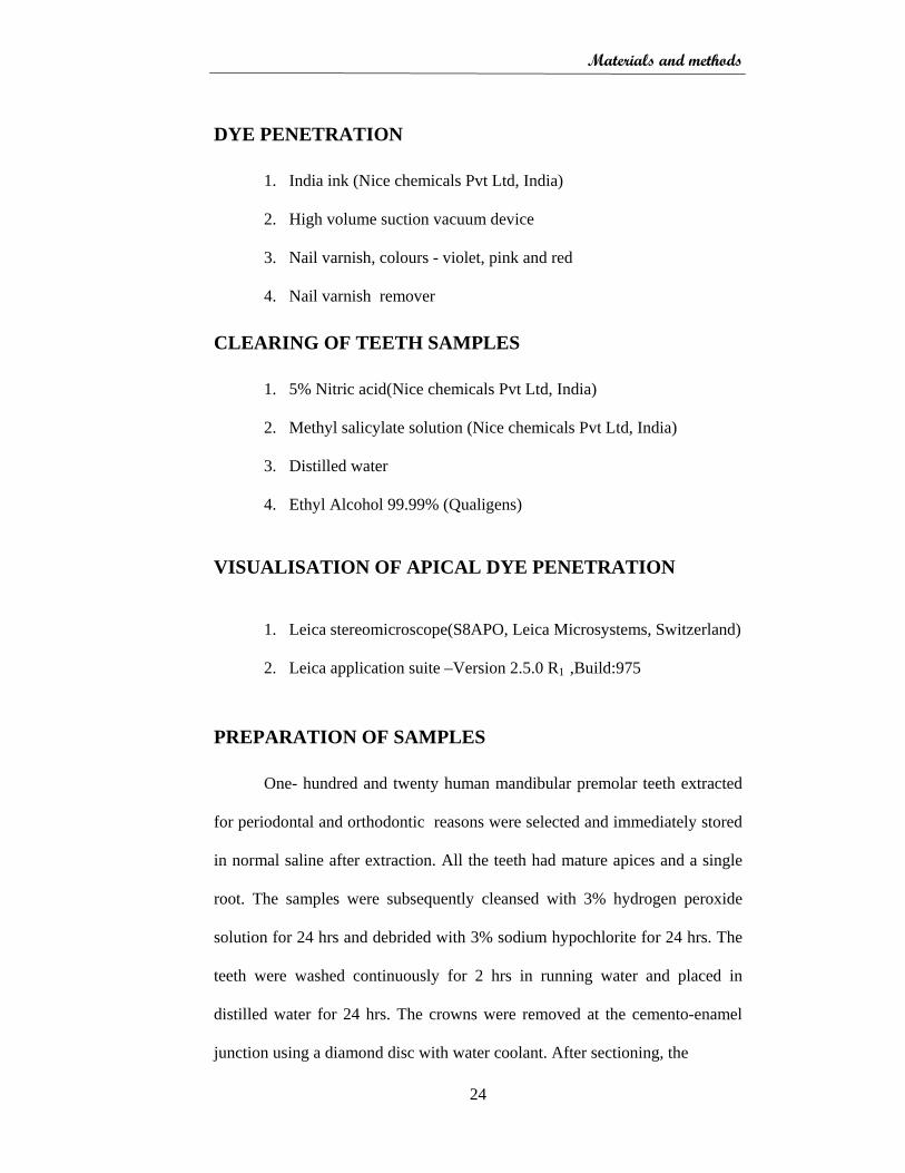
DYE PENETRATION
1. India ink (Nice chemicals Pvt Ltd, India)
2. High volume suction vacuum device
3. Nail varnish, colours - violet, pink and red
4. Nail varnish remover
CLEARING OF TEETH SAMPLES
1. 5% Nitric acid(Nice chemicals Pvt Ltd, India)
2. Methyl salicylate solution (Nice chemicals Pvt Ltd, India)
3. Distilled water
4. Ethyl Alcohol 99.99% (Qualigens)
VISUALISATION OF APICAL DYE PENETRATION
1. Leica stereomicroscope(S8APO, Leica Microsystems, Switzerland)
2. Leica application suite –Version 2.5.0 R1 ,Build:975
PREPARATION OF SAMPLES
One- hundred and twenty human mandibular premolar teeth extracted
for periodontal and orthodontic reasons were selected and immediately stored
in normal saline after extraction. All the teeth had mature apices and a single
root. The samples were subsequently cleansed with 3% hydrogen peroxide
solution for 24 hrs and debrided with 3% sodium hypochlorite for 24 hrs. The
teeth were washed continuously for 2 hrs in running water and placed in
distilled water for 24 hrs. The crowns were removed at the cemento-enamel
junction using a diamond disc with water coolant. After sectioning, the

Materials and methods
25
specimens were washed with water and stored in 3% sodium hypochlorite for
24 hrs, and then were placed in distilled water for further processing.
Assessment of the roots were done and a size #8 k-file (K-endo,
VDW,GmBH,Munich,Germany) was inserted into the canal to assess the
patency of the canal and establish the glide path. Samples with non-single
canal configurations were discarded and a total of seventy specimens were
singled out for the study .Working length was determined by inserting a size
#10 K-file (Dentsply Maillefer) until it was visible at the apical foramen. The
working length of each canal was calculated to be 1 mm short of that position.
ROOTCANAL PREPARATION
All teeth were prepared using NiTi rotary instruments of the Protaper
system to size F3. A total of 6 instruments were used with the X-Smart Device
(Dentsply Maillefer) with a gear reduction of 16:1; the speed of rotation was
maintained at 250 rpm. A new set of Protaper instruments were used for every
5 teeth. 2 ml of 3% sodium hypochlorite solution was used for irrigation
between each file size. Protaper files were used in the following sequence,
according to the manufacturer’s recommendations:
1. The rootcanal was filled with 3% sodium hypochlorite solution
and the S1 file was used to enlarge the coronal two-thirds of the
canal.
2. The canal was re-irrigated and the SX file was inserted into the
canal until it encountered light resistance. Shaping with the SX
Materials and methods
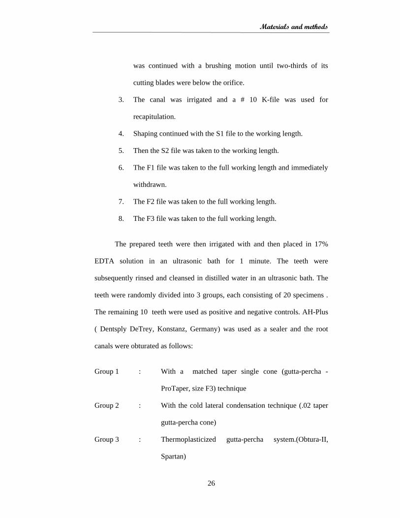
26
was continued with a brushing motion until two-thirds of its
cutting blades were below the orifice.
3. The canal was irrigated and a # 10 K-file was used for
recapitulation.
4. Shaping continued with the S1 file to the working length.
5. Then the S2 file was taken to the working length.
6. The F1 file was taken to the full working length and immediately
withdrawn.
7. The F2 file was taken to the full working length.
8. The F3 file was taken to the full working length.
The prepared teeth were then irrigated with and then placed in 17%
EDTA solution in an ultrasonic bath for 1 minute. The teeth were
subsequently rinsed and cleansed in distilled water in an ultrasonic bath. The
teeth were randomly divided into 3 groups, each consisting of 20 specimens .
The remaining 10 teeth were used as positive and negative controls. AH-Plus
( Dentsply DeTrey, Konstanz, Germany) was used as a sealer and the root
canals were obturated as follows:
Group 1 : With a matched taper single cone (gutta-percha -
ProTaper, size F3) technique
Group 2 : With the cold lateral condensation technique (.02 taper
gutta-percha cone)
Group 3 : Thermoplasticized gutta-percha system.(Obtura-II,
Spartan)

Materials and methods
27
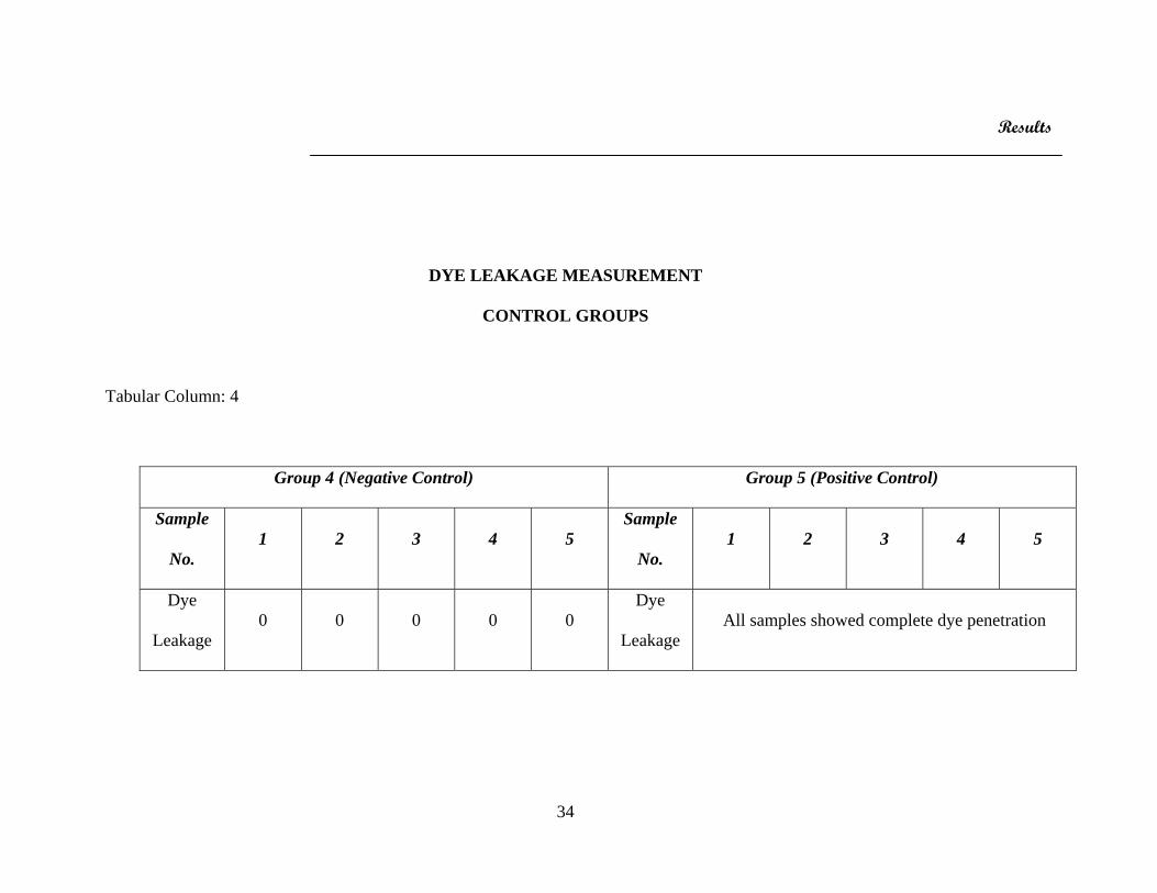
Group 4 : The negative control group was obturated with either of
the above techniques and all the root surfaces were
covered with 3 layers of nail varnish, with sufficient dry
time to ensure no leakage.
Group 5 : The positive control group in which the rootcanals were
left unobturated.
OBTURATION TECHNIQUES
Matched Taper Single Cone Obturation
The 20 specimens in group-1 were obturated with a size F3 gutta-
percha cone (Dentsply Maillefer) at the working length. First the canal was
dried with paper points (Dentsply Maillefer) and AH-Plus (Dentsply DeTrey
Konstantz, Germany)sealer was applied to the root canal walls with a size 25
reamer rotated anti-clockwise. Then the gutta-percha cone (Protaper F3 size)
cone was lightly coated with the sealer and placed into the canal to the
working length.
Cold Lateral Condensation Obturation
The 20 specimens in group-2 were obturated with size #30 .02 taper
gutta-percha cone (Dentsply Maillefer) at the working length. First the canal
was dried, and AH - Plus (Dentsply, DeTrey, Konstantz, Germany) was
applied to the canal walls. The master cone was coated with the sealer and
placed into the canal at the working length. Lateral condensation was done
with finger spreaders and accessory cones. Lateral condensation was

Materials and methods
28
completed until the spreader could no longer penetrate more than 3mm into
the canal.
Obtura II -Thermoplasticized Guttapercha System
The 20 specimens in-group-3 were obturated with the Obtura-II heated
gutta-percha system. The canals were coated with very little quantity of AH-
Plus sealer, because excessive sealer causes pooling. The gutta-percha pellets
(Regular flow gutta-percha) were loaded into the obtura handpiece and the
temperature control on the unit was adjusted to 190 degrees. The 25 gauge-
injection needle was placed within 3 to 5 mm of the working length and the
canal was totally filled as the needle was withdrawn. Finger pluggers were
used to vertically condense the filling.
After Obturation , the excess gutta-percha was removed with a heated
instrument and the cavities sealed with composite resin after the application of
a bonding agent(Adper single bond 2, 3M ESPE, St. Paul, U.S.A). The teeth
were stored at 37 degrees for 24 hours in 100% humidity to allow the
complete setting of the sealer. In the experimental groups, all root surfaces
except the apical 3 mm were covered with 3 coats of nail varnish. For each of
the experimental group a different colour nail varnish was used. Specimens
selected for positive (n = 5) and negative (n = 5) control groups were
instrumented in the same way as the specimens for the experimental groups,
but the root canals were not obturated in the positive control group to allow
100% leakage. The teeth in the negative control group were obturated with
either the lateral condensation, single cone or obtura techniques, and the
Materials and methods
29
whole root surface were covered with 3 layers of nail varnish to ensure
there was no leakage.
DYE PENETRATION TECHNIQUE
The samples in each group was immersed in an India ink container and
placed in a specially designed vacuum chamber connected to a high suction
vacuum device ( 760 mm Hg ) and the vacuum was maintained for 30
minutes. After removing the specimens from the chamber, they were allowed
to remain in the dye solution for 24 hrs. They were subsequently washed in
water and the nail varnish coating was removed using a nail varnish remover
and were then subjected to a clearing technique for visualization of the apical
leakage.
CLEARING TECHNIQUE FOR THE VISUALISATION OF
THE ROOTCANAL FILLING
The samples of individual experimental groups were stored in separate
containers and decalcified with 5% nitric acid at room temperature. The nitric
acid solution was changed in every 6 hrs and agitated with hand in every 2 hrs.
After completion of the procedure, the samples were rinsed in distilled water,
dehydrated and placed in methyl salicylate solution, which rendered the
samples transparent.
The samples were then observed, photographed and sent for analysis
using a stereomicroscope ( Leica stereomicroscope S8APO, Leica
Microsystems, Switzerland ) at 10X magnification , and the depth of dye
penetration in each sample was measured digitally using an inbuilt software

Materials and methods
30
( Leica Application Suite –Version 2.5.0 R1 , Build:975 ), and the data was
recorded and stored in a computer for further analysis.
Apical leakage was measured as the distance from the anatomic apex
to the maximum extent of dye penetration in a coronal direction.
Observer reliability was assessed by re-measuring 10 specimens from
each group on a separate occasion without the knowledge of the first readings.
The results were subsequently analyzed.

Results
31
DYE LEAKAGE MEASUREMENT
MATCHED TAPER SINGLE CONE TECHNIQUE (GROUP – 1)
Tabular Column: 1
Sample
No.
1 2 3 4 5 6 7 8 9 10 11 12 13 14 15 16 17 18 19 20
Dye
Leakage
(mm) 5.20 3.84 2.78 3.19 4.07 2.68 2.52 2.99 1.93 3.40 2.39 1.72 3.22 1.83 2.45 1.60 1.80 3.02 3.21 3.11
Results
32
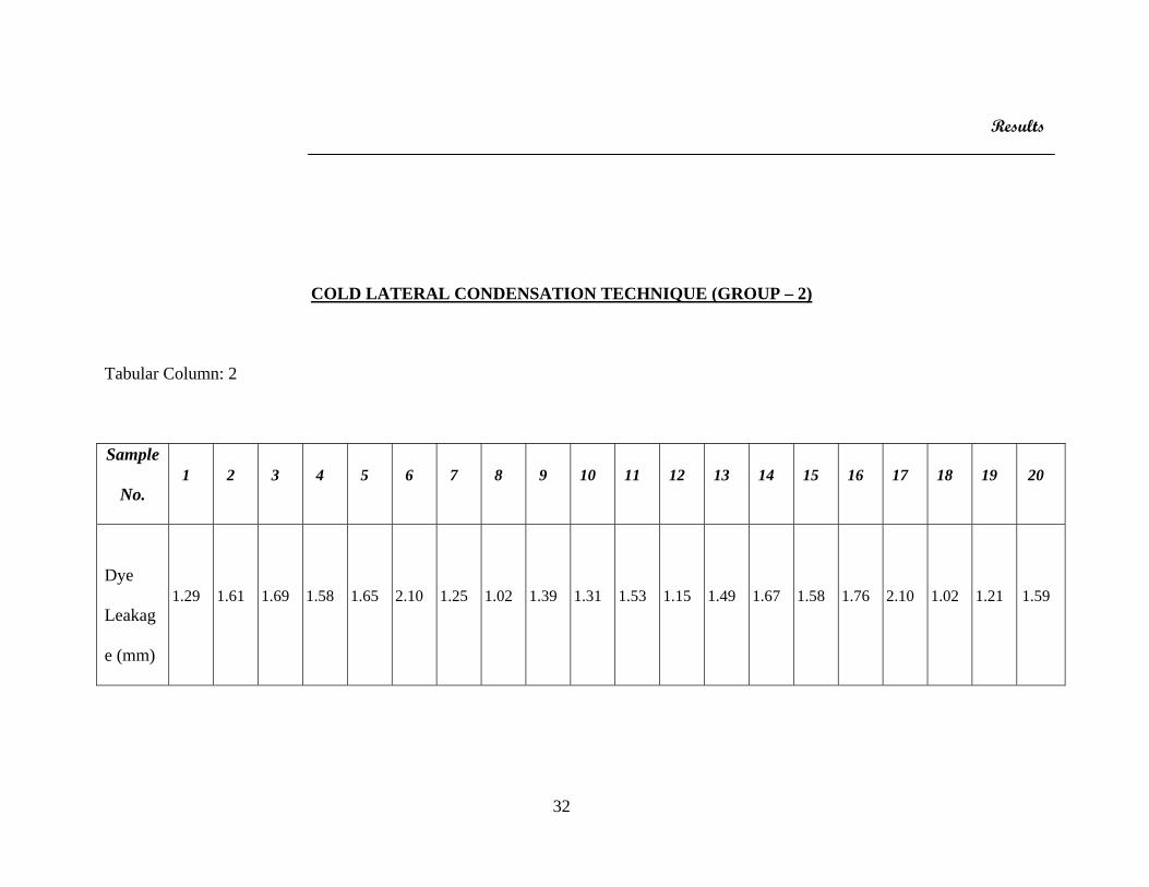
COLD LATERAL CONDENSATION TECHNIQUE (GROUP – 2)
Tabular Column: 2
Sample
No. 1 2 3 4 5 6 7 8 9 10 11 12 13 14 15 16 17 18 19 20
Dye
Leakag
e (mm)
1.29 1.61 1.69 1.58 1.65 2.10 1.25 1.02 1.39 1.31 1.53 1.15 1.49 1.67 1.58 1.76 2.10 1.02 1.21 1.59
Results
33
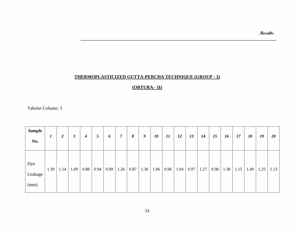
THERMOPLASTICIZED GUTTA-PERCHA TECHNIQUE (GROUP - 3)
(OBTURA - II)
Tabular Column: 3
Sample
No. 1 2 3 4 5 6 7 8 9 10 11 12 13 14 15 16 17 18 19 20
Dye
Leakage
(mm)
1.39 1.14 1.09 0.88 0.94 0.89 1.26 0.87 1.36 1.06 0.96 1.04 0.97 1.27 0.98 1.38 1.15 1.40 1.25 1.13
Results
34
DYE LEAKAGE MEASUREMENT
CONTROL GROUPS
Tabular Column: 4
Group 4 (Negative Control) Group 5 (Positive Control)
Sample
No. 1 2 3 4 5
Sample
No. 1 2 3 4 5
Dye
Leakage 0 0 0 0 0
Dye
Leakage All samples showed complete dye penetration

Results
35

Discussion
36
The primary objective of rootcanal therapy is to obturate the debrided
canals with a biocompatible filling material, inorder to eliminate the source of
infection from the residual organic material or from apical percolation. The
most common cause of apparent failure in endodontically treated teeth is
apical percolation resulting from incomplete canal obturation (John Ingle,
1976)34.
The successful outcome of endodontic treatment is dependant on the
efficient cleaning, shaping and 3-dimensional obturation of the root canal
system, with the aim of achieving a hermetic seal. The apical 3rd of the root
presents a unique anatomical configuration in that the majority of the
ramifications and accessory canals are more likely to be found in this region.
The incidence of accessory canals in the apical one-third has been reported to
be as high as 73.5% (Vertucci F.J 1984)68.
Micro organisms present inside root canals may remain viable in
dentinal tubules even after efficient chemo - mechanical preparation of the
canals. Thus achieving a perfect apical seal is vital to prevent the remaining
bacteria and their endotoxins from reaching the apex and the periradicular
tissues.
Apical leakage is considered to be a common cause for failure of
endodontic therapy. It is influenced by various factors such as the anatomy of
the apex, the chemo - mechanical preparation of the apical one-third, the
presence or absence of smear layer, different types of obturation techniques,
the selection of a sealer, the properties of sealers and the ability of the sealer to
bond to dentin as well as gutta-percha. (John Whitworth 2005)36.

Discussion
37
Although apical seal of root canal system has received a lot of
attention, achieving a good coronal seal is equally important.
Coronal leakage can also be responsible for contamination of the canal
with microorganisms and is most often the result of the loss of the temporary
filling or inadequate endodontic filling or crown sealing.
Endodontic sealers play a very important role preventing
microleakage. They seal the interface between gutta-percha and dentinal walls.
Leakage may however occur at the interfaces between the sealer and dentin,
sealer and gutta-percha, or the spaces in the sealer itself. Therefore, the quality
of the rootcanal filling to a large extent depends upon the sealing capacity or
the efficiency of sealers (Cobankara et al 2002)25
Ideally, the development of sealers should focus on materials that
1. Penetrate the patent dentinal tubules.
2. Bind closely to both the organic and inorganic phases of dentin
3. Neutralize or destroy microorganisms and their products.
4. Predictably induce a regenerative cement response on the apical
foramen.
5. Strengthen the root system
6. Biocompatible (Guttmann JL, Witherspoon DE, 2002)30
A method often used to create an apical stop or matrix for obtaining a
biological apical seal supports the placement of dentinal chips or artificial
barriers ie. Calcium hydroxide, demineralised dentin, lyophilised bone,
tricalcium phosphate, hydroxyapatite, collagen, MTA etc before canal

Discussion
38
obturation. This is not a new technique and favourable results were obtained
60 years ago using dentin chips . These barriers in addition to creating
biological apical seal may help to confine the irrigating solutions within the
canal. However, they may or may not enhance the seal of the canal apically
(Yee RDJ, et al 1984)55.
Clinical studies evaluating endodontic failures have reported that
incomplete obturation in the apical region was the principle cause of
microleakage. These studies agree that evaluation of apical leakage of
particles or solutions between the root canal filling and the rootcanal walls is a
proper method to establish the quality of endodontic obturation although
achieving a hermetic seal at the apex is one of the primary objective of
rootcanal therapy. The issue of the apical termination of the filling material
often arises as considerable variants occurs at the apical portion of the canals.
The cemento-dentinal junction is a histologic position, not a clinical position
in the root canal system. It is not always found at the apical constriction. The
distance from the apical foramen to the constriction depends on various factors
such as increased cemental deposition or radicular resorption. The position of
the foramen and the cemento-dentinal junction is highly variable and can exist
anywhere from the direct radiographic apex upto 3mm or more coronal to the
radiographic apex depending upon the morphology. Long term evaluation
studies favour and support obturation within the confines of the root canal
system in all cases, in order to attempt to prevent further challenge to the
already compromised and challenged periradicular tissues.

Discussion
39
A number of in vitro tests have been used to test the efficacy of
endodontic filling techniques and materials.
The most common method for evaluation of microleakage has been the
linear measurement of tracer dye penetration. Eg. Methylene blue, Eosin, India
ink, Radioisotopes , fungi and even bacteria. They have also been measured by
longitudinal sections, cross sections or decalcification and clearing of the root
structure. Other methods that have been tried for evaluating apical leakage are
the vacuum technique, the fluid filtration or transportation technique, the
electrochemical technique, the dye extraction technique, the bacterial and
toxin infiltration method, capillary flow pyrometry and glucose penetration
technique. All the described methods have their limitations. The largest
limitations are their low reproducibility and lack of standardization (De
Bruyne et al 2005)23.When using dyes, the particle size, the pH value and
chemical reactivity affect the degree of penetration. India ink particles with a
diameter smaller than 3 μm have been widely used as it is unlikely that
bacterial invasion would occur in spaces inside the canal where this dye is
unable to penetrate. It has been reported that the weight and size of India ink
molecules are smaller than bacterial molecules found in the root canal (Denusa
Moreira et al 2006)19.
One of the major considerations in the dye penetration studies are the
air entrapped in voids along the root canal filling which may hinder the fluid
movement. Use of a vacuum while exposing the specimen to the dye and
maintenance of this vacuum for a sufficient period of time removes air
entrapped within the voids and helps penetration of the dye.

Discussion
40
Passive soaking in dye solutions does not reveal the entire voids
present and therefore is unreliable . When used along with vacuum, consistent
results were found for dye penetration (Spangberg et al 1989)41.
Though various methodologies are available for assessing leakage,
there is a real lack of technique standardization even when the same
methodology is used (Denusa Moreira et al 2006)19.
In the present study India ink was used in conjunction with a specially
designed vacuum chamber connected to a high volume vacuum pump. The
level of the vacuum was maintained at 760mm Hg for 30 minutes.
Cleaning and shaping of the root canal has been the single most
important factor for preventing and treatment of endodontic diseases as this
removes all the etiological factors associated with endodontic diseases
(Haapasalo M , 2005)45.
In the present study, in all the sample groups canal preparation was
done using the Protaper system (Dentsply, Maillefer). These are NiTi
instruments which represents a new generation of instruments for shaping
canals. Each of the Protaper instrument has changing percentage of taper over
the length of its cutting blades. They have convex triangular section, a
changing helical angle and pitch over their cutting blades and a non-cutting,
modified guiding tip. The Protaper system is comprised of three shaping and
three finishing instruments. Canal preparation is improved when instruments
pass through the access opening, effortlessly slide down smooth axial walls
and are easily inserted into the orifice. The potential to consistently shape
canals and clean root canal systems is significantly enhanced when the coronal

Discussion
41
two - thirds of the canal is first pre-enlarged followed by preparing its apical
one-third.
The Protaper system features six instruments namely SX shaper, two
shaping files (S1and S2) and 3 finishing files (F1, F2, F3). The principles in
the use of these instruments are
1. Creation of a glide path.
2. Use of S1 and S2 in sequence to the working length.
3. No pressure is applied during instrumentation and they are passively
allowed to follow the glide path.
4. To optimize safety and efficiency, it is used with a brush like motion to
a laterally and selectively cut dentin on the outstroke. A brush like
cutting action creates a lateral space which will facilitate larger ,
stronger and more active cutting blades on the shaping instrument to
safely and progressively more deeper into the canal.
After every instrument, recapitulation with a #10 size k-file is done.
When the coronal one-third of the canal is shaped, the attention is shifted
to the apical - third procedures. Now a decision has to be made whether a
hand or rotary preparation of apical third is to be done. The Protaper
sequence is always the same regardless of the tooth or anatomical
configuration of the canals.
Although several techniques have been used for 3-dimensional
obturation of the root canal system using gutta-percha, cold lateral
condensation is still one of the most frequently used techniques. Filling of

Discussion
42
rootcanals traditionally uses .02 taper standard gutta-percha cones followed by
accessory standard gutta-percha cones, after lateral condensation of gutta-
percha with spreaders, in conjunction with a rootcanal sealer.
Gutta-percha cones are now manufactured to match the taper of the
canals prepared with 0.04 or 0.06 rotary instruments. Large taper gutta-percha
can be used with warm vertical compaction techniques or cold lateral
compaction technique. (Wilson and Baumgartner et al 2003)8.
With the NiTi rotary preparation of the root canals and use of the
sealer, these cones may provide 3-dimensional obturation of the root canals
over its entire length without the requirement of accessory cones or time spent
on lateral condensation (Himbrough et al 2002)31.
Use of excessive forces during lateral compaction can result in root
fractures. Some clinicians have proposed the use of NiTi spreaders in curved
canals. Stainless steel spreaders have the own advantage in that they are more
stiff without buckling, a common problem with NiTi spreaders.
When the single cone obturation technique is filled at room
temperature and used in conjunction with a sealer, the thickness of the sealer
varies depending on the adaptation of the single cone to the canal wall. The
volume of the sealer required is larger than the volume of sealer necessary to
complete a compaction technique. It has been reported that single cone filled
roots have more than half the canal space filled with sealer (Wu M K et al
2006)60.

Discussion
43
When the sealer dissolves after some time, the single cone fillings may
have larger voids than lateral or vertical filling. The single cone is no longer
included in current endodontic text books. Porosities in large volumes of
sealer, setting contraction and dissolution of the sealer are the main
disadvantages of this technique.
More recently gutta-percha cones for Protaper have been introduced
for simple time efficient obturation. In this system, the rootcanals are
prepared with Protaper instruments and filled with a point that fits the size of
the finisher file. The manufacturer claims that the Protaper gutta-percha
points perfectly fit canals that have been prepared with the Protaper files.
The matched taper single cone technique in this study was compared
with a cold lateral condensation technique and a thermoplasticized – gutta-
percha injection technique to evaluate the apical seal in an in vitro setting.
The thermoplasticized gutta percha injection technique has been
developed which helps flow of gutta-percha efficiently into the ramifications
of the rootcanal space. They are used in conjunction with a sealer. Both high
temperature ( 2000C ) and low temperature ( 700C ) are available. Obtura-II
system has been found to be significantly superior to lateral condensation
methods and has demonstrated the best adaptation to the 3-dimensional
rootcanal system (Budd CS et al 1991)13.
Obtura-II system is also commonly used for Backfilling and it has been
suggested that it might be clinically acceptable to backfill canals upto 10mm
in a single increment using sealer and Obtura-II system. The high temperature
Discussion
44
generated in the root canal can be dissipated through the root surface and the
periodontal ligament. It is generally accepted that a temperature rise of
approximately 100C above normal body temperature is most critical. Studies
have proved that the maximum temperature change on the external root
surface with the Obtura-II system is 6.2oC (Timothy L Sweatman et al
2001)63. This rise in temperature produced by the system on the external
surface of the root was well less than 100C which is potentially harmless to the
surrounding periodontal ligament .
Apical extrusion of gutta-percha has been a common problem with
injection technique. The advent of NiTi makes predictably centered
preparation more realistic than ever in curved canals, and may make accurate
apical cone fit a possibility in many cases. Contemporary advertising on
ergonomic, matched file and cone systems may serve to promote single cone
cementation techniques. Laboratory evidence in fact suggests comparable
cross-sectional area of canal occupied by gutta-percha using single matched
taper cones compared with lateral condensation, and in significantly less time
,but clinical trial data is unavailable (Gordon et al 2005)27.
With the widespread use of NiTi instruments it has brought about the
adaptation of single cone technique to the NiTi system, where the tapers of the
gutta-percha cone are matched to the tapers of the last instrument used.
The most important objectives of endodontic therapy are total
debridement of the pulp space, development of a fluid tight seal at the apical
foramen and total obturation of the root canal. Therefore leakage tests are a
relevant way to evaluate the apical seal.
Discussion
45
In this study, the sealer was lightly coated within the canal using a 25
size reamer rotated anti-clockwise and the cones were also lightly coated with
the sealer before placement to the working length.
In the present study we have used an AH- plus sealer because of its
low solubility (Schafer et al 2003)24. Ideally sealers should have very low
solubility because, leaching of components from the root canal filling would
have undesirable biological effects on the surrounding tissues. Degradation of
the sealer also may result in percolation at the sealer / dentin or sealer / gutta-
percha interface (Dag Orstavik et al 2001)16.
Dimensional stability has been introduced as a requirement in the Draft
International Standard (DIS) for root sealing materials. The requirements for
compliance with the standard have been set at a linear expansion of not more
than 0.1%, shrinkage of not more than 1% (Draft International Standard –
DIS)22.
Among the three groups tested for apical leakage in this study, the
Group-3 (thermoplasticized gutta-percha injection technique-obtura-II)
showed the least amount of apical penetration of the dye with a mean value of
1.223 and was statistically significant when compared with other groups
(p<0.05).
The results showed that Group-1 (matched taper single cone technique)
showed the highest values for the dye penetration under vacuum with a mean
value of 2.8475.
Discussion
46
In the single cone technique the volume of the sealer required is larger
than the volume required to complete a compaction technique. The larger
volume of sealer used is inherently prone to more changes than the small
volume used for compaction techniques.
Some studies on the comparison of the matched taper single cone
technique with the lateral condensation technique have found no significant
difference in the sealing ability and the cross-sectional area of gutta-percha
was comparable (Mahera Fani et al 2009)49. The matched taper single cone
technique was also faster.
The same sealer AH-Plus was used for all the three techniques. It is
very important to avoid void formation while loading the sealer into the canal.
The matched taper single cone technique matches the taper of gutta-percha
cones to the taper of the last instrument used to prepare the canal, and has been
advocated for obturation of curved canals.
This technique has got certain inherent advantages like safe coronal
extrusion of cement with minimal apical extrusion, more uniform gutta-
percha, lesser obturation time, elimination of lateral stresses during obturation,
avoids potential damage of radicular tissues due to increase in temperature, no
obturation material shrinkage and comparatively lower cost.
In a study of comparison of two single cone techniques (Activ GP and
Gutta-flow) with warm vertical compaction technique, the single cone
techniques did not ensure a durable apical seal. (Monticelli et al 2007)48.
Discussion
47
The Group-2 (cold lateral condensation technique) had higher apical
leakage than Group-3 (thermoplasticized gutta-percha injection technique –
obtura-II) with a mean of 1.4995.
There was significant difference between the three groups when
statistically analysed (p<0.05).
The teeth used in this in vitro study had straight canals. The posterior
teeth have curved canals with complex anatomical presentation, which might
pose greater challenges to these techniques. The cross-sectional shape of the
canals also contribute to the long-term efficacy of the seal achieved. In the
single cone and cold lateral condensation techniques, the comparatively larger
amount of sealer used can possibly cause more shrinkage and apical leakage in
the long term.
In the matched taper single cone technique, the use of a matched taper
gutta-percha for cold obturation relies on the original canal shape and ability
to create a tapered circular preparation. A smaller diameter canal would more
suit this technique. Oval or longer diameter canals would necessitate excessive
preparation for this technique to be effective.
A slight deviation from the manufacturer’s recommeneded technique
of use could result in the mis-match between the canal geometry and the size
of the last instrument used thereby leading to an ill-fitting protaper gutta-
percha cone. Therefore the operator must ensure an optimum match between
the geometry of the gutta-percha cone and the rotary instrument used.

summary
48
Seventy single-rooted mandibular premolar teeth were selected,
cleaned and stored in normal saline solution. They were sectioned at the level
of the cementoenamel junction and the rootcanal preparation was done as per
the manufacturer’s recommendations. The teeth were then divided into five
groups, two of which were used as positive and negative control groups. The
other three groups were obturated with matched taper single cone technique,
cold lateral condensation technique and thermoplasticized gutta-percha
injection technique (obtura-II) respectively. The teeth were then subjected to a
dye penetration technique using high vacuum and left in the dye solution for
24 hrs. They were then cleaned and subjected to a technique for clearing the
roots. Subsequently the dye penetration at the apex was measured in all the
three groups using a binocular stereomicroscope and analysed using a Leica
application suite. The results were tabulated and statistically analysed.

Conclusion
49
Matched taper single cone obturation technique was evaluated for the
apical seal in comparison with the cold lateral condensation technique and
thermoplasticized gutta-percha injection technique(obtura-II) of obturation and
the following conclusions were made.
• The thermoplasticized gutta-percha injection technique of obturation,
obtura-II (Group-3) showed the least amount of apical percolation of
dye and was better than other two techniques in the apical sealing
ability.
• There was statistically significant difference between all the three
groups (p<0.05).
• Matched-taper single-cone technique of obturation (Group-1) showed
the highest amount of apical percolation of the dye.
• The cold lateral condensation method of obturation (Group-2) was also
efficient in terms of apical sealing ability though it could not match
Group-3,( p<0.05).
The matched taper single cone technique needs to be further
studied and evaluated with regards to the limitations of this technique.