Comparative Evaluation of Sealing Ability of Three Newer ... · Comparative Evaluation of Sealing...
12
Comparative Evaluation of Sealing Ability….Bhandi S H et al ORIGINAL RESEARCH Journal of International Oral Health. Jan-Feb 2013; 5(1):54-65 [ 54 ] Comparative Evaluation of Sealing Ability of Three Newer Root Canal Obturating Materials Guttaflow, Resilon and Thermafil: An In Vitro Study Shilpa H Bhandi 1 , Subhash T S 2 1 Assistant Professor, Department of Conservative Dentistry and Endodontics, M S Ramaiah Dental College and Hospital, Bangalore, India; 2 Professor and HOD, Department of Dentistry MMC & RI, K. R. Hospital, Mysore, Karnataka, India. Introduction According to Ingle the most common causes of endodontic failure is incomplete obturation. He reported that 59% endodontic failure were due to leakage in the canal seal. 1 According to Mannocci et al 1999 microleakage between root canal filling and root canal walls may adversely affect the results of root canal treatment. Therefore, complete obturation of the root canal with an inert filing material and creation of an apical seal have been proposed as goals for successful endodontic treatment by Nguyen in 1984. 2 The three main functions of obturation are : i) To entomb any bacteria remaining in the root canal system. ii) To stop the influx of periapical tissue derived fluid from reentering the root canal iii) To prevent coronal leakage of bacteria. Although gutta-percha has many desirable properties it does not always bond to the internal tooth structure resulting in the absence of complete seal. This produces a ABSTRACT Introduction: Microleakage continues to be a main reason for failure of root canal treatment where the challenge has been to achieve an adequate seal between the internal structure and the main obturating material. The objective of this study is to compare the sealing ability of 3 newer obturating materials GuttaFlow, Resilon/Epiphany system (RES) and Thermafil, using silver nitrate dye and observing under stereomicroscope. Methodology: Thirty single rooted teeth were divided into following groups. Group I : GuttaFlow ;Group II : Resilon /Epiphany sealer Group III : Thermafil with AH-Plus sealer. Teeth were decoronated and instrumented with profile rotary system and obturated with specified materials. Apical seal was determined by dye penetration method using silver nitrate. Then the specimens were transversely sectioned at each mm till 3 mm from the apex. Dye leakage was determined using stereomicroscope. Statistical analysis of the results was performed using Kruskall-Wallis test. Results: The results showed that Group II i.e., Resilon with Epiphany sealer showed the least amount of microleakage when compared to Group I i.e., GuttaFlow and Group III i.e., Thermafil with AH-plus sealer. Conclusion: Based on the results of this study it can be concluded that RES had higher sealing ability followed by Thermafil and GuttaFlow in vitro but further studies have to be carried out to make a direct correlation between these results and invivo situation. Key words: Apical microleakage, Silver nitrate dye, Profile rotary system, Stereomicroscope. How to cite this article: Bhandi S H, Subhash T S. Comparative Evaluation of Sealing Ability of Three Newer Root Canal Obturating Materials Guttaflow, Resilon and Thermafil: An In Vitro Study. J Int Oral Health 2013; 5(1):54-65. Source of Support: Nil Conflict of Interest: None Declared Received: 13 th November 2012 Reviewed: 10 th December 2012 Accepted: 29 th December 2012 Address for Correspondence: Dr Shilpa H Bhandi, Assistant Professor, Department of Conservative Dentistry and Endodontics, M S Ramaiah Dental College and Hospital, Bangalore. Mobile: 9742095997, e-mail: [email protected]
Transcript of Comparative Evaluation of Sealing Ability of Three Newer ... · Comparative Evaluation of Sealing...
Comparative Evaluation of Sealing Ability….Bhandi S H et al
ORIGINAL RESEARCH
Journal of International Oral Health. Jan-Feb 2013; 5(1):54-65 [ 54 ]
Comparative Evaluation of Sealing Ability of Three Newer Root
Canal Obturating Materials Guttaflow, Resilon and Thermafil: An In
Vitro Study Shilpa H Bhandi1, Subhash T S2
1Assistant Professor, Department of Conservative Dentistry and Endodontics, M S Ramaiah Dental College and Hospital, Bangalore, India;
2Professor and HOD, Department of Dentistry MMC & RI, K. R. Hospital, Mysore, Karnataka, India.
Introduction
According to Ingle the most common causes of
endodontic failure is incomplete obturation. He
reported that 59% endodontic failure were due to
leakage in the canal seal.1 According to Mannocci et al
1999 microleakage between root canal filling and root
canal walls may adversely affect the results of root
canal treatment. Therefore, complete obturation of the
root canal with an inert filing material and creation of
an apical seal have been proposed as goals for
successful endodontic treatment by Nguyen in
1984.2
The three main functions of obturation are : i)
To entomb any bacteria remaining in the root
canal system. ii) To stop the influx of
periapical tissue derived fluid from reentering
the root canal iii) To prevent coronal leakage
of bacteria. Although gutta-percha has many
desirable properties it does not always bond
to the internal tooth structure resulting in the
absence of complete seal. This produces a
ABSTRACT
Introduction: Microleakage continues to be a main reason for failure of root canal treatment where the challenge
has been to achieve an adequate seal between the internal structure and the main obturating material. The
objective of this study is to compare the sealing ability of 3 newer obturating materials GuttaFlow,
Resilon/Epiphany system (RES) and Thermafil, using silver nitrate dye and observing under stereomicroscope.
Methodology: Thirty single rooted teeth were divided into following groups. Group I : GuttaFlow ;Group II :
Resilon /Epiphany sealer Group III : Thermafil with AH-Plus sealer. Teeth were decoronated and instrumented
with profile rotary system and obturated with specified materials. Apical seal was determined by dye penetration
method using silver nitrate. Then the specimens were transversely sectioned at each mm till 3 mm from the apex.
Dye leakage was determined using stereomicroscope. Statistical analysis of the results was performed using
Kruskall-Wallis test.
Results: The results showed that Group II i.e., Resilon with Epiphany sealer showed the least amount of
microleakage when compared to Group I i.e., GuttaFlow and Group III i.e., Thermafil with AH-plus sealer.
Conclusion: Based on the results of this study it can be concluded that RES had higher sealing ability followed by
Thermafil and GuttaFlow in vitro but further studies have to be carried out to make a direct correlation between
these results and invivo situation.
Key words: Apical microleakage, Silver nitrate dye, Profile rotary system, Stereomicroscope.
How to cite this article: Bhandi S H, Subhash T S. Comparative Evaluation of Sealing Ability of Three Newer Root
Canal Obturating Materials Guttaflow, Resilon and Thermafil: An In Vitro Study. J Int Oral Health 2013; 5(1):54-65.
Source of Support: Nil Conflict of Interest: None Declared Received: 13th November 2012 Reviewed: 10th December 2012 Accepted: 29th December 2012
Address for Correspondence: Dr Shilpa H Bhandi, Assistant Professor, Department of Conservative Dentistry
and Endodontics, M S Ramaiah Dental College and Hospital, Bangalore. Mobile: 9742095997, e-mail:
Comparative Evaluation of Sealing Ability….Bhandi S H et al
ORIGINAL RESEARCH
Journal of International Oral Health. Jan-Feb 2013; 5(1):54-65 [ 55 ]
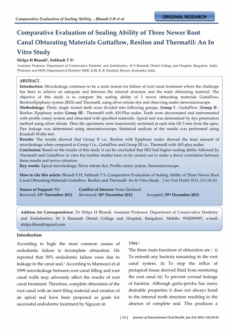
Fig. 1: Instruments used
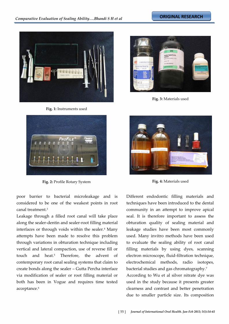
Fig. 2: Profile Rotary System
Fig. 3: Materials used
Fig. 4: Materials used
poor barrier to bacterial microleakage and is
considered to be one of the weakest points in root
canal treatment.3
Leakage through a filled root canal will take place
along the sealer-dentin and sealer-root filling material
interfaces or through voids within the sealer.4 Many
attempts have been made to resolve this problem
through variations in obturation technique including
vertical and lateral compaction, use of reverse fill or
touch and heat.5 Therefore, the advent of
contemporary root canal sealing systems that claim to
create bonds along the sealer – Gutta Percha interface
via modification of sealer or root filling material or
both has been in Vogue and requires time tested
acceptance.6
Different endodontic filling materials and
techniques have been introduced to the dental
community in an attempt to improve apical
seal. It is therefore important to assess the
obturation quality of sealing material and
leakage studies have been most commonly
used. Many invitro methods have been used
to evaluate the sealing ability of root canal
filling materials by using dyes, scanning
electron microscope, fluid-filtration technique,
electrochemical methods, radio isotopes,
bacterial studies and gas chromatography.7
According to Wu et al silver nitrate dye was
used in the study because it presents greater
clearness and contrast and better penetration
due to smaller particle size. Its composition
Comparative Evaluation of Sealing Ability….Bhandi S H et al
ORIGINAL RESEARCH
Journal of International Oral Health. Jan-Feb 2013; 5(1):54-65 [ 56 ]
being Assay (AgNO3) – 99.8% minimum, chloride (Cl)
– 0.001% Maximum, Sulphate (SO4) 0.02% maximum,
Lead (Pb) – 0.002% maximum, Iron (Fe) – 0.001%
maximum.6
A plethora of studies have indicated that
microleakage whether from an apical or coronal
direction adversely affects the success of root canal
treatment. Many anatomical parameters and clinical
considerations influence microleakage during the
course of non surgical root canal treatment including
root morphology, canal anatomy, patient cooperation,
operator skill in preparation and obstruction of the
canal and root canal sealing and filling material. The
development and maintenance of a seal of the root
system is considered to be a major prerequisite in
success in root canal treatment. Therefore the
evaluation of the quality of the root canal filling
material using a variety of leakage tests to some
degree is a relevant concept.8
Methodology
Thirty freshly extracted single rooted human teeth
were selected for study on the basis of the following
criteria, no root caries, resorption or fracture; mature,
fully formed apices, single rooted and single canal,
were stored in normal saline. Clinical crowns were
sectioned at the cemento- enamel-junction with a low-
speed diamond disc under continuous water spray.
Working length was established 1mm short of the
apex. Instrumentation was performed with a crown
down technique using profile Ni-Ti rotary instrument
system according to specific set of instruments. All
canals were prepared to ISO size 40, 0.06 taper. Canal
patency was maintained by passing with an ISO size
15. K-file. Which was extended 1mm beyond the apex
to maintain apical patency. The canal was irrigated
between each instrument with 5.25% NaOCl and 17%
EDTA alternatively. After completion of
instrumentation, the root canals were dried with
paper points and teeth were divided in to 3 groups of
10 teeth each randomly.
Group – I: Teeth obturated with GuttaFlow.
After the completion of the instrumentation,
master cone was selected and the cone was
coated with GuttaFlow and it was placed in
the prepared canal, and rest of the root canal
space was back filled with GuttaFlow.
Group – II : Warm vertical compaction of
Resilon with Epiphany sealer.
Epiphany self-etching primer was introduced
into the root canal with a micro-brush and
excess primer was removed with paper points.
A non-standardized Resilon master cone was
tried in to within 1mm of working length.
Epiphany sealer was then placed in to the root
canal using a lentulo spiral, and the Resilon
root canal filling material was down packed
using the continuous wave condensation
technique. (System B) at a reduced
temperature of 1500 and a power setting of 10
as recommended by the manufacturers.
Backfilling was performed with Obtura-II
using 23 gauge needle tips at a temperature of
1400C. After backfilling, the coronal surface of
the root filling was light-cured for 40sec. LED
light curing unit was used to polymerize the
surface of the dual-cured methacrylate sealer.
Group-III : Thermafil Obturation Technique.
A size verification carrier No.40 reaching to
the working length with no resistance or
twisting was selected. AH plus was taken as a
root canal sealer, and was mixed according to
manufacture instructions. The sealer was
coated to root canal walls, and the Thermafil
obturators were heated in Thermaprep oven
according to manufacturer instructions. The
appropriate size of obturator was removed
from the carrier wheel with care taken not to
allow contact with the oven components. The
oven opening was closed after each removal.
Firm apical pressure was used to insert the
Thermafil obturator to the previously
Comparative Evaluation of Sealing Ability….Bhandi S H et al
ORIGINAL RESEARCH
Journal of International Oral Health. Jan-Feb 2013; 5(1):54-65 [ 57 ]
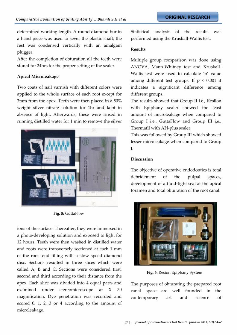
Fig. 5: GuttaFlow
determined working length. A round diamond bur in
a hand piece was used to sever the plastic shaft; the
rest was condensed vertically with an amalgam
plugger.
After the completion of obturation all the teeth were
stored for 24hrs for the proper setting of the sealer.
Apical Microleakage
Two coats of nail varnish with different colors were
applied to the whole surface of each root except for
3mm from the apex. Teeth were then placed in a 50%
weight silver nitrate solution for 1hr and kept in
absence of light. Afterwards, these were rinsed in
running distilled water for 1 min to remove the silver
ions of the surface. Thereafter, they were immersed in
a photo-developing solution and exposed to light for
12 hours. Teeth were then washed in distilled water
and roots were transversely sectioned at each 1 mm
of the root- end filling with a slow speed diamond
disc. Sections resulted in three slices which were
called A, B and C. Sections were considered first,
second and third according to their distance from the
apex. Each slice was divided into 4 equal parts and
examined under stereomicroscope at X 30
magnification. Dye penetration was recorded and
scored 0, 1, 2, 3 or 4 according to the amount of
microleakage.
Statistical analysis of the results was
performed using the Kruskall-Wallis test.
Results
Multiple group comparison was done using
ANOVA, Mann-Whitney test and Kruskall-
Wallis test were used to calculate ‘p’ value
among different test groups. If p < 0.001 it
indicates a significant difference among
different groups.
The results showed that Group II i.e., Resilon
with Epiphany sealer showed the least
amount of microleakage when compared to
Group I i.e., GuttaFlow and Group III i.e.,
Thermafil with AH-plus sealer.
This was followed by Group III which showed
lesser microleakage when compared to Group
I.
Discussion
The objective of operative endodontics is total
debridement of the pulpal spaces,
development of a fluid-tight seal at the apical
foramen and total obturation of the root canal.
The purposes of obturating the prepared root
canal space are well founded in the
contemporary art and science of
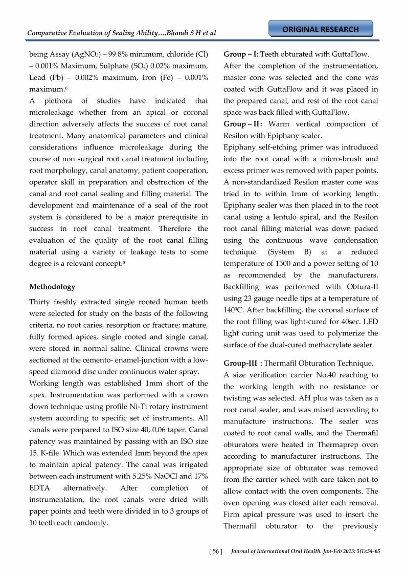
Fig. 6: Resion Epiphany System
Comparative Evaluation of Sealing Ability….Bhandi S H et al
ORIGINAL RESEARCH
Journal of International Oral Health. Jan-Feb 2013; 5(1):54-65 [ 58 ]
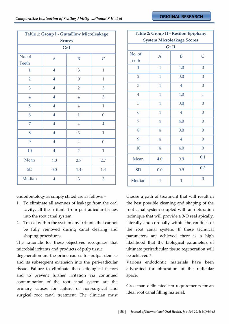
Table 1: Group I - GuttaFlow Microleakage
Scores
Gr I
No. of
Teeth A B C
1 4 3 1
2 4 0 1
3 4 2 3
4 4 4 3
5 4 4 1
6 4 1 0
7 4 4 4
8 4 3 1
9 4 4 0
10 4 2 1
Mean 4.0 2.7 2.7
SD 0.0 1.4 1.4
Median 4 3 3
Table 2: Group II - Resilon Epiphany
System Microleakage Scores
Gr II
No. of
Teeth A B C
1 4 4.0 0
2 4 0.0 0
3 4 4 0
4 4 4.0 1
5 4 0.0 0
6 4 4 0
7 4 4.0 0
8 4 0.0 0
9 4 4 0
10 4 4.0 0
Mean 4.0 0.9 0.1
SD 0.0 0.9 0.3
Median 4 1 0
endodontology as simply stated are as follows –
1. To eliminate all avenues of leakage from the oral
cavity, all the irritants from periradicular tissues
into the root canal system.
2. To seal within the system any irritants that cannot
be fully removed during canal clearing and
shaping procedures
The rationale for these objectives recognizes that
microbial irritants and products of pulp tissue
degeneration are the prime causes for pulpal demise
and its subsequent extension into the peri-radicular
tissue. Failure to eliminate these etiological factors
and to prevent further irritation via continued
contamination of the root canal system are the
primary causes for failure of non-surgical and
surgical root canal treatment. The clinician must
choose a path of treatment that will result in
the best possible cleaning and shaping of the
root canal system coupled with an obturation
technique that will provide a 3-D seal apically,
laterally and coronally within the confines of
the root canal system. If these technical
parameters are achieved there is a high
likelihood that the biological parameters of
ultimate periradicular tissue regeneration will
be achieved.9
Various endodontic materials have been
advocated for obturation of the radicular
space.
Grossman delineated ten requirements for an
ideal root canal filling material.
Comparative Evaluation of Sealing Ability….Bhandi S H et al
ORIGINAL RESEARCH
Journal of International Oral Health. Jan-Feb 2013; 5(1):54-65 [ 59 ]
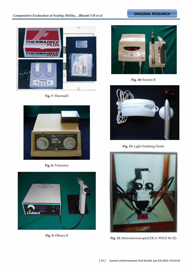
Fig. 7: Thermafil
Fig. 8: Triturator
Fig. 9: Obtura II
Fig. 10: System B
Fig. 11: Light Emitting Diode
Fig. 12: Stereomicroscope(LEICA WILD M-32)
Comparative Evaluation of Sealing Ability….Bhandi S H et al
ORIGINAL RESEARCH
Journal of International Oral Health. Jan-Feb 2013; 5(1):54-65 [ 60 ]
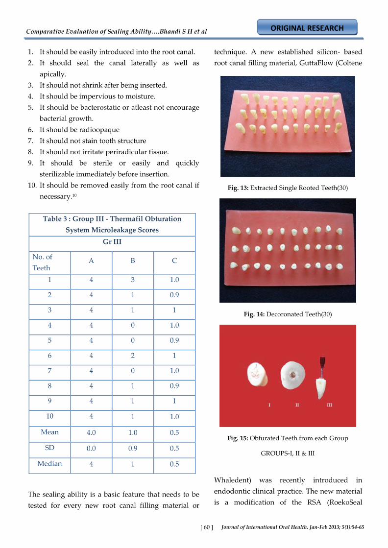
Table 3 : Group III - Thermafil Obturation
System Microleakage Scores
Gr III
No. of
Teeth A B C
1 4 3 1.0
2 4 1 0.9
3 4 1 1
4 4 0 1.0
5 4 0 0.9
6 4 2 1
7 4 0 1.0
8 4 1 0.9
9 4 1 1
10 4 1 1.0
Mean 4.0 1.0 0.5
SD 0.0 0.9 0.5
Median 4 1 0.5
Fig. 13: Extracted Single Rooted Teeth(30)
Fig. 14: Decoronated Teeth(30)
Fig. 15: Obturated Teeth from each Group
GROUPS-I, II & III
1. It should be easily introduced into the root canal.
2. It should seal the canal laterally as well as
apically.
3. It should not shrink after being inserted.
4. It should be impervious to moisture.
5. It should be bacterostatic or atleast not encourage
bacterial growth.
6. It should be radioopaque
7. It should not stain tooth structure
8. It should not irritate periradicular tissue.
9. It should be sterile or easily and quickly
sterilizable immediately before insertion.
10. It should be removed easily from the root canal if
necessary.10
The sealing ability is a basic feature that needs to be
tested for every new root canal filling material or
technique. A new established silicon- based
root canal filling material, GuttaFlow (Coltene
Whaledent) was recently introduced in
endodontic clinical practice. The new material
is a modification of the RSA (RoekoSeal
Comparative Evaluation of Sealing Ability….Bhandi S H et al
ORIGINAL RESEARCH
Journal of International Oral Health. Jan-Feb 2013; 5(1):54-65 [ 61 ]
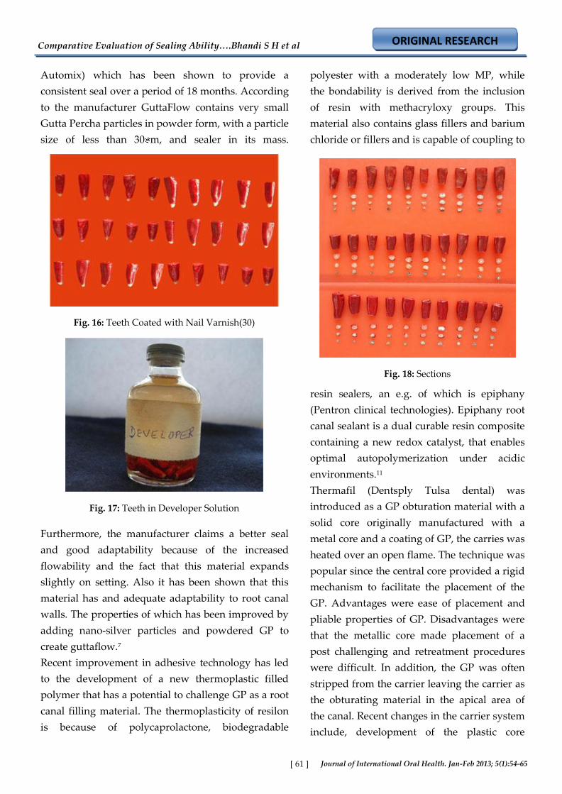
Fig. 16: Teeth Coated with Nail Varnish(30)
Fig. 17: Teeth in Developer Solution
Fig. 18: Sections
Automix) which has been shown to provide a
consistent seal over a period of 18 months. According
to the manufacturer GuttaFlow contains very small
Gutta Percha particles in powder form, with a particle
size of less than 30 m, and sealer in its mass.
Furthermore, the manufacturer claims a better seal
and good adaptability because of the increased
flowability and the fact that this material expands
slightly on setting. Also it has been shown that this
material has and adequate adaptability to root canal
walls. The properties of which has been improved by
adding nano-silver particles and powdered GP to
create guttaflow.7
Recent improvement in adhesive technology has led
to the development of a new thermoplastic filled
polymer that has a potential to challenge GP as a root
canal filling material. The thermoplasticity of resilon
is because of polycaprolactone, biodegradable
polyester with a moderately low MP, while
the bondability is derived from the inclusion
of resin with methacryloxy groups. This
material also contains glass fillers and barium
chloride or fillers and is capable of coupling to
resin sealers, an e.g. of which is epiphany
(Pentron clinical technologies). Epiphany root
canal sealant is a dual curable resin composite
containing a new redox catalyst, that enables
optimal autopolymerization under acidic
environments.11
Thermafil (Dentsply Tulsa dental) was
introduced as a GP obturation material with a
solid core originally manufactured with a
metal core and a coating of GP, the carries was
heated over an open flame. The technique was
popular since the central core provided a rigid
mechanism to facilitate the placement of the
GP. Advantages were ease of placement and
pliable properties of GP. Disadvantages were
that the metallic core made placement of a
post challenging and retreatment procedures
were difficult. In addition, the GP was often
stripped from the carrier leaving the carrier as
the obturating material in the apical area of
the canal. Recent changes in the carrier system
include, development of the plastic core
Comparative Evaluation of Sealing Ability….Bhandi S H et al
ORIGINAL RESEARCH
Journal of International Oral Health. Jan-Feb 2013; 5(1):54-65 [ 62 ]
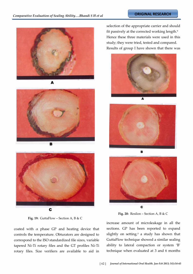
Fig. 19: GuttaFlow – Section A, B & C
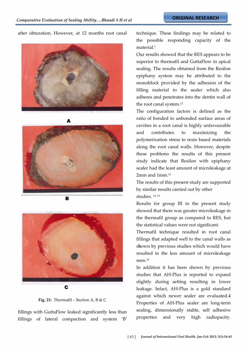
Fig. 20: Resilon – Section A, B & C
coated with α phase GP and heating device that
controls the temperature. Obturators are designed to
correspond to the ISO standardized file sizes, variable
tapered Ni-Ti rotary files and the GT profiles Ni-Ti
rotary files. Size verifiers are available to aid in
selection of the appropriate carrier and should
fit passively at the corrected working length.9
Hence these three materials were used in this
study; they were tried, tested and compared.
Results of group I have shown that there was
increase amount of microleakage in all the
sections. GP has been reported to expand
slightly on setting.4 a study has shown that
GuttaFlow technique showed a similar sealing
ability to lateral compaction or system ‘B’
technique when evaluated at 3 and 6 months
Comparative Evaluation of Sealing Ability….Bhandi S H et al
ORIGINAL RESEARCH
Journal of International Oral Health. Jan-Feb 2013; 5(1):54-65 [ 63 ]
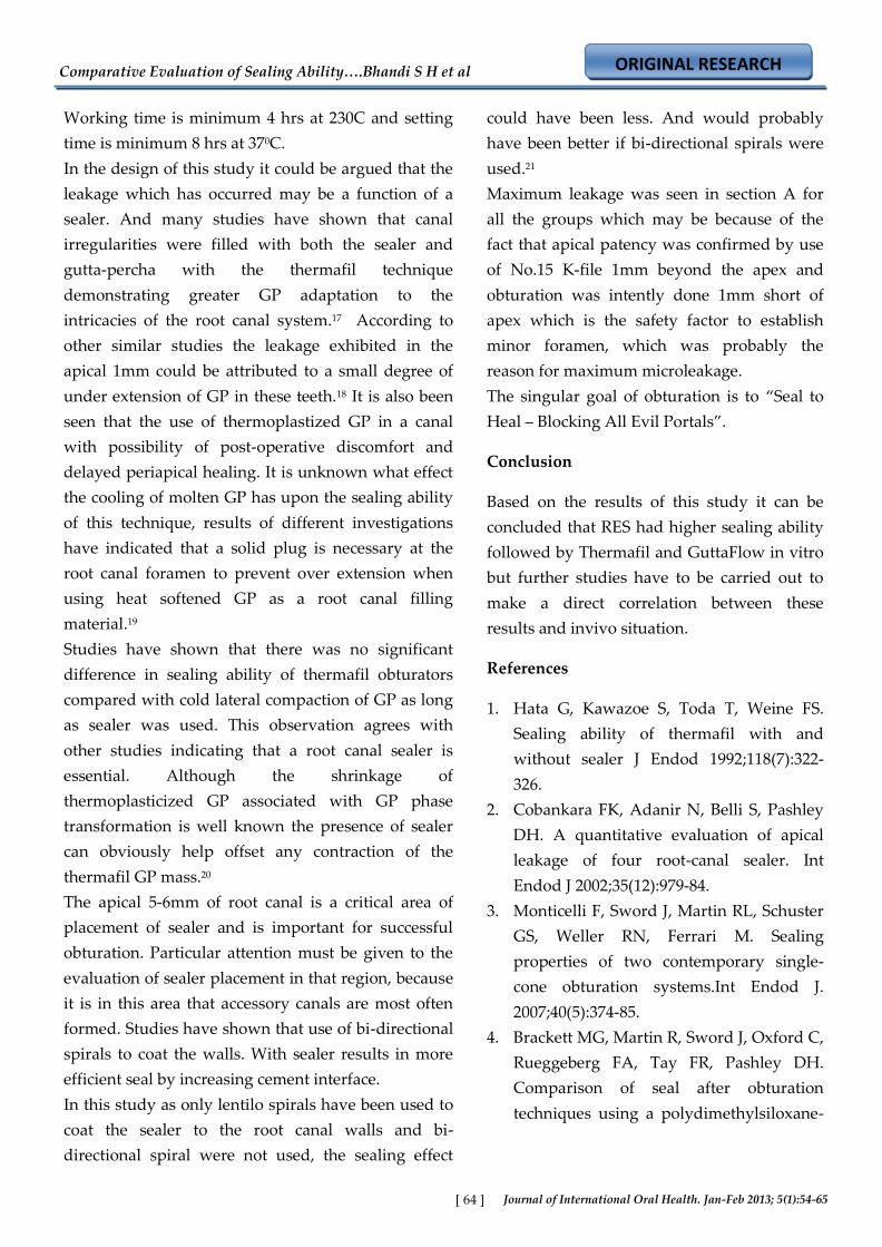
Fig. 21: Thermafil – Section A, B & C
after obturation. However, at 12 months root canal
fillings with GuttaFlow leaked significantly less than
fillings of lateral compaction and system ‘B’
technique. These findings may be related to
the possible responding capacity of the
material.7
Our results showed that the RES appears to be
superior to thermafil and GuttaFlow in apical
sealing. The results obtained from the Resilon
epiphany system may be attributed to the
monoblock provided by the adhesion of the
filling material to the sealer which also
adheres and penetrates into the dentin wall of
the root canal system.12
The configuration factors is defined as the
ratio of bonded to unbonded surface areas of
cavities in a root canal is highly unfavourable
and contributes to maximizing the
polymerization stress to resin based materials
along the root canal walls. However, despite
these problems the results of this present
study indicate that Resilon with epiphany
sealer had the least amount of microleakage at
2mm and 1mm.13
The results of this present study are supported
by similar results carried out by other
studies. 14, 15
Results for group III in the present study
showed that there was greater microleakage in
the thermafil group as compared to RES, but
the statistical values were not significant.
Thermafil technique resulted in root canal
fillings that adapted well to the canal walls as
shown by previous studies which would have
resulted in the less amount of microleakage
seen.16
In addition it has been shown by previous
studies that AH-Plus is reported to expand
slightly during setting resulting in lower
leakage. Infact, AH-Plus is a gold standard
against which newer sealer are evaluated.4
Properties of AH-Plus sealer are long-term
sealing, dimensionally stable, self adhesive
properties and very high radiopacity.
Comparative Evaluation of Sealing Ability….Bhandi S H et al
ORIGINAL RESEARCH
Journal of International Oral Health. Jan-Feb 2013; 5(1):54-65 [ 64 ]
Working time is minimum 4 hrs at 230C and setting
time is minimum 8 hrs at 370C.
In the design of this study it could be argued that the
leakage which has occurred may be a function of a
sealer. And many studies have shown that canal
irregularities were filled with both the sealer and
gutta-percha with the thermafil technique
demonstrating greater GP adaptation to the
intricacies of the root canal system.17 According to
other similar studies the leakage exhibited in the
apical 1mm could be attributed to a small degree of
under extension of GP in these teeth.18 It is also been
seen that the use of thermoplastized GP in a canal
with possibility of post-operative discomfort and
delayed periapical healing. It is unknown what effect
the cooling of molten GP has upon the sealing ability
of this technique, results of different investigations
have indicated that a solid plug is necessary at the
root canal foramen to prevent over extension when
using heat softened GP as a root canal filling
material.19
Studies have shown that there was no significant
difference in sealing ability of thermafil obturators
compared with cold lateral compaction of GP as long
as sealer was used. This observation agrees with
other studies indicating that a root canal sealer is
essential. Although the shrinkage of
thermoplasticized GP associated with GP phase
transformation is well known the presence of sealer
can obviously help offset any contraction of the
thermafil GP mass.20
The apical 5-6mm of root canal is a critical area of
placement of sealer and is important for successful
obturation. Particular attention must be given to the
evaluation of sealer placement in that region, because
it is in this area that accessory canals are most often
formed. Studies have shown that use of bi-directional
spirals to coat the walls. With sealer results in more
efficient seal by increasing cement interface.
In this study as only lentilo spirals have been used to
coat the sealer to the root canal walls and bi-
directional spiral were not used, the sealing effect
could have been less. And would probably
have been better if bi-directional spirals were
used.21
Maximum leakage was seen in section A for
all the groups which may be because of the
fact that apical patency was confirmed by use
of No.15 K-file 1mm beyond the apex and
obturation was intently done 1mm short of
apex which is the safety factor to establish
minor foramen, which was probably the
reason for maximum microleakage.
The singular goal of obturation is to ‚Seal to
Heal – Blocking All Evil Portals‛.
Conclusion
Based on the results of this study it can be
concluded that RES had higher sealing ability
followed by Thermafil and GuttaFlow in vitro
but further studies have to be carried out to
make a direct correlation between these
results and invivo situation.
References
1. Hata G, Kawazoe S, Toda T, Weine FS.
Sealing ability of thermafil with and
without sealer J Endod 1992;118(7):322-
326.
2. Cobankara FK, Adanir N, Belli S, Pashley
DH. A quantitative evaluation of apical
leakage of four root-canal sealer. Int
Endod J 2002;35(12):979-84.
3. Monticelli F, Sword J, Martin RL, Schuster
GS, Weller RN, Ferrari M. Sealing
properties of two contemporary single-
cone obturation systems.Int Endod J.
2007;40(5):374-85.
4. Brackett MG, Martin R, Sword J, Oxford C,
Rueggeberg FA, Tay FR, Pashley DH.
Comparison of seal after obturation
techniques using a polydimethylsiloxane-
Comparative Evaluation of Sealing Ability….Bhandi S H et al
ORIGINAL RESEARCH
Journal of International Oral Health. Jan-Feb 2013; 5(1):54-65 [ 65 ]
based root canal sealer. J Endod 2006;32(12):1188-
90.
5. Williams C, Loushine RJ, Weller RN, Pashley DH,
Tay FR. A comparison of cohesive strength and
stiffness of resilon and gutta-percha. J
Endod2006;32(6):553-5.
6. Xavier CB, Weismann R, De Oliveira MG,
Demarco FF, Pozza DH. Root-end filling
materials: Apical microleakage and marginal
adaptation. J Endod 2005;31(7):539-42.
7. Kontakiotis EG, Tzanetakis GN, Loizides AL. A
12-month longitudinal in vitro leakage study on a
new silicon-based root canal filling material
(Gutta-Flow). Oral Surg Oral Med Oral Pathol
Oral Radiol Endod 2007;103(6):854-9.
8. Leonard JE, Gutmann JL, Guo IY. Apical and
coronal seal of roots obturated with a dentine
bonding agent and resin. Int Endod J;1996
Mar;29(2):76-83.
9. Cohen S. Pathways of the pulp. In: Obturation of
the cleaned and shaped root canal system. Cohen
S, Hargreaves KM edt. 9th edn. Chapter 10,
Mosby 2006:358-99.
10. Ingle J. Endodontics. In: Obturation of the
Radicular space. Ingle J, Bakland LK.. 5th ED, part
I, Chapter 11, B.C. Decker, Canada. 2002:571-668.
11. Tay FR, Loushine RJ, Weller RN, Kimbrough WF,
Pashley DH, Mak YF. Ultrastructural evaluation
of the apical seal in roots filled with a
polycaprolactone-based root canal filling material.
J Endod; 2005;31(7):514-9.
12. Bodrumlu E, Tunga U. Apical Leakage of resilon
obturation material.J Contemp Dent Pract;
2006;7(4):45-52.
13. Onay EO, Ungor M, Orucoglu H. An in vitro
evaluation of the apical sealing ability of a new
resin-based root canal obturation system.J
Endod;2006;32(10):976-8.
14. Shipper G, Orstavik D, Teixeira FB, Trope M. An
evaluation of microbial leakage in roots filled
with a thermoplastic synthetic polymer-based
root canal filling material (Resilon). J
Endod 2004;30(5):342-7.
15. Aptekar A, Ginnan K. Comparative
analysis of microleakage and seal for 2
obturation materials: Resilon / Epiphany
and gutta-percha. J Can Dent Assoc;
2006;72(3):245.
16. Pathomvanich S, Edmunds DH. The
sealing ability of thermafil obturators
assessed by four different microleakage
techniques. Int Endod J 1996;29(5):327-34.
17. Gutmann JL, Saunders WP, Saunders EM,
Nguyen L. An assessment of the plastic
thermafil obturation technique part 2
Material adaptation and sealability. Int
Endod J 1993;26(3):179-83.
18. Veis AA, Molyvdas A, Lambrianidis TP,
Beltes PG. In vitro evaluation of apical
leakage of root canal fillings after in situ
obturation with thermoplasticized and
laterally condensed gutta-percha. Int
Endod J 1994;27(4):213-7.
19. Ritchie GM, Anderson DM, Sakumura JS.
Apical extrusion of thermoplasticized
gutta-percha used as a root canal filling. J
Endod; 1988;14(3):128-32.
20. Schafer E, Priv D, Olthoff G. Effect of three
different sealers on the sealing ability of
both thermafil obturators and cold
laterally compacted gutta-percha. J Endod
2002;28(9):638-42.
21. Chandak M. Seal to Heal – Blocking all
Evil Portals. Dentistry Today 2007; 2(3):6-
7.