COMPARATIVE EVALUATION OF MARGINAL AND INTERNAL …
141
COMPARATIVE EVALUATION OF MARGINAL AND INTERNAL ADAPTATION OF CLASS II ZIRCONIA CERAMIC INLAYS Vs FELDSPATHIC CERAMIC INLAYS WITH AND WITHOUT A RESIN BASE AND DIFFERENT INTERFACE TREATMENTS - AN IN VITRO SCANNING ELECTRON MICROSCOPIC STUDY Dissertation submitted to THE TAMILNADU Dr. M.G.R. MEDICAL UNIVERSITY In partial fulfillment for the Degree of MASTER OF DENTAL SURGERY BRANCH IV CONSERVATIVE DENTISTRY AND ENDODONTICS APRIL 2017
Transcript of COMPARATIVE EVALUATION OF MARGINAL AND INTERNAL …

COMPARATIVE EVALUATION OF MARGINAL AND
INTERNAL ADAPTATION OF CLASS II ZIRCONIA
CERAMIC INLAYS Vs FELDSPATHIC CERAMIC INLAYS
WITH AND WITHOUT A RESIN BASE AND DIFFERENT
INTERFACE TREATMENTS - AN IN VITRO SCANNING
ELECTRON MICROSCOPIC STUDY
Dissertation submitted to
THE TAMILNADU Dr. M.G.R. MEDICAL UNIVERSITY
In partial fulfillment for the Degree of
MASTER OF DENTAL SURGERY
BRANCH IV
CONSERVATIVE DENTISTRY AND ENDODONTICS
APRIL 2017

ACKNOWLEDGEMENT
My sincere heartfelt pranams to my post graduate teacher and
my GURU, my guide Dr. M. Rajasekaran, M.D.S., Professor,
Department of Conservative Dentistry and Endodontics, Ragas Dental
College and Hospital, he has been my sole support and the main reason
to complete my dissertation successfully. His encouragement, guidance
and his faith in me has groomed me into being what I am today. He has
been caring and walked along with me to take me across these years. I
will be ever indebted to my GURU and thank him from the bottom of my
heart. I am truly blessed to be under the guidance of my GURU.
I extend my sincere thanks and gratitude to, Dr. R. Anil Kumar,
M.D.S., Professor, HOD, Department of Conservative Dentistry and
Endodontics, Ragas Dental College and Hospital. His belief in my
capabilities and constant word of encouragement has been of immense
support for me. His guidance has helped throughout these years and
hope to continue under his guidance always.
My sincere thanks to Dr. R. Indira, M.D.S., Professor and
Dr. S. Ramachandran, M.D.S. Professor, whose blessings and
guidance has helped me throughout my post graduate curriculum.
My sincere thanks to Dr. P. Shankar, M.D.S., Professor, Ragas
Dental College and Hospital, for his guidance, care and constant
encouragement which has majorly helped me through these years. I
revere sir for his belief in me and his everlasting support.
My deepest heartfelt thanks to Dr. C. S. Karumaran, M.D.S.,
Professor, Ragas Dental College and Hospital, for his everlasting
support in my post-graduation. I am indeed blessed to be under the
supervision and assistance of sir and ever thankful to him for always
backing me whenever I needed his support. I pray to always get his
blessings.
I solemnly thank Dr. Veni Ashok, M.D.S., Associate Professor,
for the trust he has in me. I adore the incessant support, guidance and
care sir has shown me and his belief in me at all times has helped me
strive hard. I pray to always get his blessings and guidance in
everything.
I am grateful to Dr. Shankar Narayan, M.D.S., Reader, for the
relentless care and support he has shown me. He has all the time been
encouraging and appreciative of all my endeavours. Ever thankful for
such care and concern sir has shown me and pray to always shower the
same on me.

I express my sincere thanks to Dr. S.M. Venkatesan, M.D.S,
Reader, for his guidance and support all throughout my post-
graduation. I acknowledge and express my gratitude to Dr. Sabari,
M.D.S., Senior lecturer, whose support was immense and his guidance
in all ways has been huge. I am thankful to Dr. Arrvind Vikram, M.D.S.
and Dr. B. Venkatesh, M.D.S., Senior lecturers for the abundant
support and encouragement they have given me.
I also wish to thank Dr. N.S. Azhagarasan, M.D.S., Principal
and the management of Ragas Dental College and Hospital, Chennai
for their help and support and also all the office staff for their help.
My sincere thanks to Dr. Ravanan, Ph.D for his guidance in
statistics. Special thanks to Dr. R. Ajay Rakkesh, Ph.D National Center
for Nanosciences and Nanotechnology – University of Madras for his
assistance during my Scanning Electron Microscopic evaluation.
My sincere thanks to Mr. K. Thavamani and Miss. R. Sudha for
their guidance and support in DTP and Binding works.
Lastly, I bow down to my Mentors Late. Dr. Premila
Gnanapragasam M.D.S. and Dr. G. Emmanuel Solomon Sathish
M.D.S., who have been my greatest inspiration. They not only imparted

knowledge in academics but guided me to become a better person in my
life. I will be ever obliged to them and need their blessings always.
My pillars of strength during my post-graduate curriculum were
my seniors Dr. M. Manivannan, Dr. Purushotham Mohankumar,
Dr. Sandeep N and my friends Dr. Nithyalakshmi. J and Dr. Amit
Kumar. S without whom I could have never achieved anything during
my post-graduation. I thank all my batch mates and juniors and special
mention to Dr. Rathna piriyanga .R.S and Dr. Pavan Kumar.V for all
their help and support.
God Saibaba couldn’t come in person in this world to shower his
blessings but he sent me my Maternal GRANDPARENTS, my
FATHER AND MOTHER without who I would have never even seen
this world. I miss My Father Late Dr. N. SAICHANDHRAN M.B.B.S,
D. Ortho., M.S. Ortho., to whom I owe all my success and growth. My
MOTHER Mrs. Kalyani Saichandran is the lady behind my existence
today. Even after the loss of her husband, she has not given up on
herself and lives only for her children. I adore her and will ever be
indebted to my parents.
My brothers Mr. Swaroop Saichandran and Mr. Subhash
Saichandran and their family are my only support. They have been like

my father in protecting and guiding me all my life. It is because of them
that I am able to continue my post-graduation after the loss of our
father. Words can’t explain the love I bore for them and I am blessed to
have such a family.

LIST OF ABBREVIATIONS
S.NO ABBREVIATIONS EXPANSION
1. CAD/CAM
Computer aided designing/computer aided
machining
2. Y-TZP
Yttria tetragonal zirconia polycrystalline
ceramics
3. SEM Scanning electron microscope
4. y-PSZ
yttrium-Oxide Partially Stabilised Zirconia
(Ceramill Amann Girrbach)
5. Post hoc Tukey HSD test Post hoc Tukey Honestly significant
difference test
6. p <0.001** Significant at 1 level (Highly Significant)
7. RCT Resin Coating Technique
8. MDP 10-methacryloyloxydecyl dihydrogen phosphate

CONTENTS
S. NO. INDEX PAGE.NO
1. INTRODUCTION 1
2. AIM AND OBJECTIVES 6
3. REVIEW OF LITERATURE 7
4. MATERIALS AND METHODS 29
5. RESULTS 39
6. DISCUSSION 43
7. SUMMARY 61
8. CONCLUSION 63
9. BIBLIOGRAPHY 65
10. ANNEXURE -

LIST OF TABLES
S.NO. TITLE
Table 1 SEM VALUES OF MARGINAL ADAPTATION MEASURED IN
OCCLUSAL AND PROXIMAL AREAS
Table 2 SEM VALUES OF INTERNAL ADAPTATION MEASURED IN
OCCLUSAL, AXIAL AND CERVICAL DENTIN AREAS
Table 3
THE MEAN AND STANDARD DEVIATION COMPARISON OF
MARGINAL ADAPTATION IN THE OCCLUSAL AREA
BETWEEN ALL THE FOUR GROUPS BY ONE-WAY ANOVA
Table 4
PAIRWISE COMPARISON OF MARGINAL ADAPTATION IN
THE OCCLUSAL AREA BETWEEN ALL THE FOUR GROUPS
BY POST HOC TESTS - TUKEY HSD
Table 5
THE MEAN AND STANDARD DEVIATION COMPARISON OF
MARGINAL ADAPTATION IN THE PROXIMAL AREA
BETWEEN ALL THE FOUR GROUPS BY ONE-WAY ANOVA
Table 6
PAIRWISE COMPARISON OF MARGINAL ADAPTATION IN
THE PROXIMAL AREA BETWEEN ALL THE FOUR GROUPS
BY POST HOC TESTS - TUKEY HSD
Table 7
THE MEAN AND STANDARD DEVIATION COMPARISON OF
INTERNAL ADAPTATION IN THE OCCLUSAL DENTIN AREA
WITH ALL THE GROUPS USING ONE-WAY ANOVA
Table 8
PAIRWISE COMPARISON OF INTERNAL ADAPTATION IN
THE OCCLUSAL DENTIN AREA WITH ALL THE GROUPS
USING POST HOC TESTS – TUKEY HSD
Table 9
THE MEAN AND STANDARD DEVIATION COMPARISON OF
INTERNAL ADAPTATION IN THE AXIAL DENTIN AREA
WITH ALL FOUR GROUPS USING ONE-WAY ANOVA

Table 10
PAIRWISE COMPARISON OF INTERNAL ADAPTATION IN
THE AXIAL DENTIN AREA WITH ALL THE FOUR GROUPS
USING POST HOC TESTS – TUKEY HSD TESTS
Table 11
MEAN AND STANDARD DEVIATION COMPARISON OF
INTERNAL ADAPTATION IN THE CERVICAL DENTIN AREA
WITH ALL THE GROUPS USING ONE-WAY ANOVA
Table 12
PAIRWISE COMPARISON OF INTERNAL ADAPTATION IN
THE CERVICAL AREA WITH ALL THE GROUPS USING POST
HOC TESTS – TUKEY HSD TESTS

LIST OF GRAPHS
S.NO. TITLE
Graph 1 SHOWS THE MEAN COMPARISON OF MARGINAL
ADAPTATION IN THE OCCLUSAL AREA WITH ALL THE
FOUR GROUPS USING ONE-WAY ANOVA ANALYSIS
Graph 2 SHOWS THE MEAN COMPARISON OF THE MARGINAL
ADAPTATION IN THE PROXIMAL AREA WITH ALL THE
FOUR GROUPS USING ONE-WAY ANOVA ANALYSIS
Graph 3
SHOWS THE MEAN COMPARISON OF INTERNAL
ADAPTATION IN THE OCCLUSAL DENTIN AREA WITH
ALL THE FOUR GROUPS USING ONE-WAY ANOVA
ANALYSIS
Graph 4
SHOWS THE MEAN COMPARISON OF THE INTERNAL
ADAPTATION IN THE AXIAL DENTIN AREA WITH ALL
THE FOUR GROUPS USING ONE-WAY ANOVA ANALYSIS
Graph 5
SHOWS THE MEAN COMPARISON OF THE INTERNAL
ADAPTATION IN THE CERVICAL DENTIN AREA WITH
ALL THE FOUR GROUPS USING ONE-WAY ANOVA
ANALYSIS

LIST OF FIGURES
S.NO TITLE
Figure 1 Teeth specimen
Figure 2 Materials used for mounting and embedding samples
Figure 3 Embedded samples - Group IA
Figure 4 Embedded samples - Group 1B
Figure 5 Embedded samples - Group 2A
Figure 6 Embedded samples - Group 2B
Figure 7 Armamentarium
Figure 8a Inlay cavity preparation
Figure 8b
Roundening of axio-pulpal line angle with gingival margin
trimmer
Figure 9 Tetric-N-Flow used as Base
Figure 10 Applicaton of Base
Figure 11 Materials used for impression
Figure 12 Impression making for Group 2
Figure 13 Scanner Map400
Figure 14 Ceramill machine

Figure 15 VITA Ceramic Furnace
Figure 16 Materials for luting
Figure 17 Prepared Cavity
Figure 18 Application of Etchant
Figure 19 Application of Bonding agent
Figure 20 Curing
Figure 21 5% Hydrofluoric acid used for Group 2
Figure 22 Application of Silane coupling agent for group 1 and 2
Figure 23 Luting cement applied
Figure 24 Luted inlay
Figure 25 Mechanical loading
Figure 26 Sectioning of specimen
Figure 27 Steam cleaner
Figure 28 Steam cleaning
Figure 29 Ultrasonic cleaning
Figure 30 Gold sputtering machine
Figure 31 Gold sputtered specimen

Figure 32 Scanning Electron Microscope
Figure 33 SEM image of group 1A – Zirconia with base
Figure 34 SEM image of group 1B – Zirconia without base
Figure 35 SEM image of group 2A – Feldspathic ceramic with base
Figure 36 SEM image of group 2B – Feldspathic ceramic without base

Introduction

Introduction
1
INTRODUCTION
A dental restoration is done to restore function, integrity and regain the
structural loss of tooth tissues and to bring back the normal shape, appearance,
aesthetics and to hinder the progression of dental caries preventing its spread
to the dental pulp.
The decision-making process of what to restore with majorly; depends
on the lesion size, aetiology, aesthetics, occlusion, endodontic and periodontal
considerations, number of teeth affected, patient compliance, habits,
preferences and the dentist’s own competence and underlying beliefs about
restorative treatment.61
Direct restorations can be done in traditional Class I,
II, III, IV, V situations and where single step quick setting restorations are
required. But in case of large cavities and/or failed direct restorations with
multiple missing cusps; anterior teeth with large interproximal cavities
involving incisal edges requiring replacement; large rehabilitation cases
requiring the recreation of multiple occlusal surfaces; an indirect restoration is
the best treatment of choice. The materials available for indirect restorations
were Gold, noble metals, Porcelain fused to metal and all ceramics.43
Amalgam and cast gold restorations have been the gold standard for
posterior proximal restorations due to their durability and long term success in
clinical studies. An aesthetic alternative has always been preferred by the
patients and the emergence of composites and ceramics were a boon.31

Introduction
2
Post-operative sensitivity and polymerization shrinkage were the main
problems faced in direct composites or indirect composite restorations used in
large class II proximal restorations.64
Indirect Class II inlay restorations fabricated with Ceramics are a
definite alternative to posterior metal restorations. They are mainly used in
compromised posterior teeth where the buccal and lingual walls are intact
which strengthen and conserve tooth structure by mechanical bonding to the
tooth.31
Improved aesthetics of ceramic materials, bonding techniques and
accessibility of newer technology has reinstated the use of Ceramic inlays.10
Today, material choices for posterior ceramic inlays include use of a
higher-strength ceramic material, or alternatively a high-strength ceramic core
material which may be veneered with a more translucent aesthetic veneer. The
development of higher strength ceramics was required with modern
technology and in 1970 Francois Duret pioneered the use of computer aided
design/ computer aided milling (CAD/CAM) in dentistry. This allowed the
inlays to be machined from pre-fired ceramic blocks in the dental office.31
The newer addition of material which are available as blocks for
milling or hot pressing are the Yttria Tetragonal Zirconia Polycrystalline –
based monolithic ceramics (Y-TZP) and the feldspathic porcelains reinforced
with leucite or lithium disilicate with higher strength, fracture resistance and
better aesthetics.50

Introduction
3
Any indirect restoration requires a cement for the prepared teeth to
retain them. This cement can largely influence the performance of the
restoration as a whole.71
Two broad categories of available cements are water
based cements and resin-based cements. The choice depends on the type of
material selected for the indirect restoration and the clinical requirements,
such as setting characteristics, film thickness, setting rates and adhesion to the
underlying tooth. There are a few non-resin cements that can be used with all
ceramic restorations, but they may reduce the overall strength of the
restoration owing to their lack of adhesion to the ceramic and the tooth. All-
ceramic restorations rely on technique-sensitive resin-based cements and
adhesives to hold them in place and to seal the tooth against leakage.71
An indirect inlay restoration has a key advantage of precision and
control over the final morphology and occlusion of the restoration which
indeed requires a tapered preparation design and sometimes increased tooth
tissue loss.49
A dentin seal is necessary in such indirect restorations during the
temporary phase which can be facilitated using a flowable composite.54
The
flowable composite as a base not only ensures a dentin seal but also helps in
blocking out undercuts, reducing microleakage, even to reduce sensitivity
during re-exposure and cleaning of dentin surface after temporary restoration
removal. It also helps as a stress absorber between the ceramic inlay
restoration and tooth interface.54

Introduction
4
This interface between the resinous base and luting composite or
between luting composite and inlay brings about the micro-mechanical
retention and the copolymerisation, which is key in adhesion of the
restoration. A pre-treatment of the cavity along with the base and the under
surface of the inlay restoration following the removal of the temporary cement
and before luting the indirect restoration either using pumice, soft air abrasion
or sandblasting helps in cleaning and chemo-mechanical activation of the
composite base which in turn helps in improving the micro-mechanical
retention and adhesion of the restoration.54
Despite the misconception that inadequacies of fit of ceramic inlays
can be compensated by the presence of composite luting cement at the
margins of a restoration, it has been shown that an accurately fitting
restoration is vital for long-term success in a clinical situation. The marginal
and internal gap sizes usually influence the longevity, wear, discolouration,
leakage, degradation of the luting agent and the ability of the restoration to
withstand loading.10
Various methods to measure the gap between the restoration and the
tooth are the non-invasive and invasive techniques. Groten et al reported, that
the accuracy of Scanning Electron Microscope (SEM) was better in providing
more appropriate and realistic observations than a light microscope.47

Introduction
5
The high range of magnification and depth of focus makes SEM an
useful tool for studying both the adaptation of restorations to cavity margins
and the surface characteristics of the restorations.55
It is useful in identifying
the location of marginal defects, whether on the inlay-cement interface, the
cement-tooth interface or within the cement layer itself. This degree of
precision is not possible using clinical examination alone.10
The more accurately the casting fits the prepared tooth, the more
difficult it is for cement to escape from the inner surface of the restoration and
the surface of the prepared tooth. The adverse effects of viscous luting
cements, variations in marginal designs, magnitudes of seating force, cements
and different seating aid materials may complicate restoration seating during
cementation.16
Hence the aim of this study was to compare and evaluate the marginal
and internal adaptation of Class II Zirconia ceramic inlays and Feldspathic
ceramic inlays with and without a resin base and different interface treatments
with Scanning Electron Microscope SEM.

Aim and Objectives

Aim and Objectives
6
AIM AND OBJECTIVES
AIM OF THE STUDY:
The aim of this in vitro study is to evaluate and compare the
marginal adaptation and internal adaptation of Class II Zirconia ceramic
inlays to Feldspathic ceramic inlays with and without a resin base and
different interface treatments with SEM.
OBJECTIVE OF THE STUDY:
This in vitro study compares
1. The marginal adaptation of the zirconia ceramic inlays with and
without base and feldspathic ceramic inlays with and without base
in the
occlusal junction between the restoration and tooth interface and
proximal cervical area junction between the restoration and the
cervical dentin.
2. The evaluation of internal adaptation was done along
the pulpal floor and distal wall line angle [occlusal dentin], along
the pulpal floor and axial wall line angle [axial dentin] and along
the axial wall and gingival seat line angle [cervical dentin] between
the zirconia ceramic inlays with and without base and feldspathic
ceramic inlays with and without base.

Review of Literature

Review of Literature
7
REVIEW OF LITERATURE
Matty F Abate et al (1989)1 evaluated the marginal fit of four ceramic
crown systems 1) metal ceramic crowns with metal margins 2) metal ceramic
crowns with a porcelain facial margins, 3) cerestore crowns 4) dicor crowns.
Measurements of the marginal adaptation were recorded from the facial and
lingual margins by using a video enhanced microscope with digital micrometer
and image intensification in a high-resolution television screen. Results indicate
that all 4 crown systems yielded comparable and acceptable marginal fit.
Blair K.F et al (1993)11
studied the microleakage associated with several
luting agents for ceramic inlays. One hundred twenty Class V inlays (occlusal
margins in enamel and gingival margins in dentin) were luted in extracted teeth
using zinc phosphate cement, two resin cements without a bonding agent, and two
resin cements with three dentin bonding agents. This study suggests that the use
of a dentin bonding agent with a resin cement will reduce microleakage in cast
glass-ceramic restorations.
Sjogren et al (1995)60
the marginal and internal fit of four different types
of ceramic inlays cerec, celay, empress, and vita in-ceram spinell was determined
after they had been luted on extracted premolars. There was no statistically
significant difference either in the proximal fit or in the gingiva-proximal fit

Review of Literature
8
between the four inlay systems studied, with the exception of the cerec inlays
made for preparations with sharp proximal boxes, which had wider marginal gaps.
The best internal fit was recorded for the celay inlays, whereas there was no
significant difference in the internal fit between the other systems. For the cerec
inlays the u-shaped proximal box shaping improved the marginal accuracy all
around the restoration.
Bergman M.A et al (1999)10
reviewed the clinical performance of
ceramic inlays. Ceramic inlays perform better when compared with aesthetic
intracoronal restorations. However, their high cost and extreme technique
sensitivity would appear to restrict their use to certain limited clinical situations.
Addi simon et al (2002)59
determined the fit of ceramic inlays
manufactured using CAD/CAM-system (Denzir) and of two types of laboratory
made heat pressed ceramics (IPS Empress and Opc). Extracted human premolars
were prepared to receive mesio-occluso-distal (MOD) ceramic inlays, for which
10 Denzir, 10 IPS Empress, and 10 Opc inlays were fabricated. The Denzir
restorations were produced by the manufacturer of the CAD/CAM-system, and
the IPS Empress and Opc by student Dental technicians. Before luting the internal
fit on the die stone models and on the premolars was determined using replicas.
After luting on the premolars with a resin composite the marginal and internal fit
were measured. The values were analysed and the results showed that after luting

Review of Literature
9
there were no significant differences between IPS Empress and Denzir, whereas
the marginal gap width was significantly wider for Opc than for IPS Empress and
Denzir. The internal fit was significantly wider for Opc than for IPS Empress,
whereas there were no significant differences between IPS Empress and Denzir or
between Opc and Denzir.
Mou et al (2002)46
evaluated the influence of different convergence
angles and tooth preparation heights on the internal adaptation of cerec crowns.
Tooth preparations were made on typodont teeth with different combinations of
convergence angles and occlusal-cervical heights: group I = 20° angle, 6 mm
height; group II = 20° angle, 4 mm height; group III = 12° angle, 6 mm height;
and group IV = 12° angle, 4 mm height. Three-way analysis of variance was used.
Cerec crowns with a 12° convergence angle demonstrated the best internal fit.
The difference between the 2 convergence types was within the range of the
scanning error (25 μm) produced by the cerec camera. The study confirmed that
there was little difference in the internal fit of cerec crowns prepared with
convergence angles of 12° and 20.
Blatz M.B et al (2003)12
reviewed resin ceramic bonding. The few
available studies on resin bonding to zirconium oxide ceramics suggest the use of
resin cements that contain special adhesive monomers. The rapidly increasing
popularity of all-ceramic systems requires further research.

Review of Literature
10
Dietschi D et al (2003)20
compared the marginal and internal adaptation
of class II fine hybrid composite inlays (Herculite, Kerr) made with or without
composite bases, having different physical properties. Freshly extracted human
molars were used for this study. The base extended up to the cervical margins on
both sides and was made from Revolution (Kerr), Tetric flow (Vivadent), Dyract
(Detrey-Dentsply) or Prodigy (Kerr), respectively. Before, during and after
mechanical loading (1 million cycles, with a force varying from 50 to 100 N), the
proximal margins of the inlay were assessed by scanning electron microscopy.
Experimental data were analysed using non-parametric tests. The final
percentages of marginal tooth fracture varied from 30.7% (no base) to 37.6%
(Dyract). In dentin, percentages of marginal opening varied from 9.2% (Tetric
Flow) to 30.1% (Prodigy), however, without significant difference between base
products. Mean values of opened internal interface with dentin varied from
11.06% (Tetric Flow) to 28.15% (Prodigy). The results regarding dentin
adaptation confirmed that the physical properties of a base can influence
composite inlay adaptation and that the medium-rigid flowable composite Tetric
Flow is a potential material to displace, in a coronal position,
Mota C.S et al (2003)45
studied the microleakage in ceramic inlays using
different resin cements with margins in enamel and cementum/dentine interface.
Dye leakage at the margins in enamel was statistically lower than at

Review of Literature
11
cementum/dentine interface. Relyx ARC performed better than resin cement and
composite restorations. Both material and substrate interface influenced
microleakage of the ceramic inlays.
Ausiello et al (2004)9 investigated the effect of differences in the resin-
cement elastic modulus on stress-transmission to ceramic or resin-based
composite inlay-restored class II mod cavities during vertical occlusal loading.
Three finite-element (fe) models of class II mod cavity restorations in an upper
premolar were produced. Model A represented a glass–ceramic inlay in
combination with an adhesive and a high young’s modulus resin-cement. Model B
represented the same glass–ceramic inlay in combination with the same adhesive
and a low young’s modulus resin-cement. Model C represented a heat-cured resin
composite inlay in combination with the same adhesive and the same low
Young’s modulus resin cement. Occlusal vertical loading of 400 n was simulated
on the fe models of the restored teeth. Ansys FE software was used to compute
the local von mises stresses. In the ceramic-inlay models, the greatest von mises
stress was observed on the lateral walls, vestibular and lingual, of the cavity.
Indirect resin-composite inlays performed better in terms of stress dissipation.
Glass–ceramic inlays transferred stresses to the dental walls and, depending on its
rigidity, to the resin-cement and the adhesive layers.
Review of Literature
12
Liu PR et al (2005)37
overviewed the development of various CAD/CAM
systems. Operational components, methodologies, and restorative materials used
with common CAD/CAM systems are discussed. Research data and clinical
studies are presented to substantiate the clinical performance of these systems.
The study concluded that CAD/CAM systems have dramatically enhanced
dentistry by providing high-quality restorations. The evolution of current systems
and the introduction of new systems demonstrate increasing user friendliness,
expanded capabilities, and improved quality, and range in complexity and
application. New materials also are more esthetic, wear more nearly like enamel,
and are strong enough for full crowns and bridges. Dental CAD/CAM technology
is successful today because of the vision of many great pioneers as Duret
concluded in his article in 1991, the systems will continue to improve in
versatility, accuracy, and cost effectiveness, and will be a part of routine dental
practice.
Karakaya S et al (2005)34
Investigated the internal adaptation of a
ceramic (ceramco II) and Two composite resin inlay materials (Surefil and 3M
Filtek Z 250TM
) using silicon replica technique as an Indicator. Forty-five
standard MOD cavities were prepared into brass moulds. Inlays were prepared
with indirect methods and replicas of the prepared cavities and inlays were
produced with a Polyvinyl siloxane material (Elite H-D). Two parallel slices

Review of Literature
13
(mesio-distally) were obtained from the replicas with a sharp blade. Thickness
between cavity and inlay was measured at seven points. The results showed that
in the surefil and ceramco II groups, the sizes of the contraction gaps at mesial
and distal gingival floors were greater than that of the occlusal marginal walls. In
comparison of gap formation at occlusal regions, while the 3M composite group
showed highest gap values, the ceramco II group revealed the lowest. At the
gingival floors, gap formation of ceramco II group was the highest. Neither group
showed any statistical difference between gap values of their self-occlusal and
gingival floors. In conclusion, the results showed that ceramic inlays did not
confer any big advantage for internal adaptation over the composite inlays.
Bortolotto et al (2007)13
evaluated the marginal adaptation of cerec
ceramic inlays, cerec composite inlays and direct composite restorations in
unbeveled proximal slot cavities under artificial aging conditions. Two groups of
each restoration type were prepared, one group with a self-etch adhesive, the other
group with H3PO4 enamel etching before the self-etch adhesive application.
Replicas were generated before and after long-term thermo-mechanical loading
and analyzed using SEM. The study showed results were statistically significant
difference before and after loading with respect to the percentages of “continuous
margins”, the direct composite filling with H3PO4 enamel etching giving the
lowest percentages of “continuous margins” after loading. The highest percentage
Review of Literature
14
was attained by composite inlays without H3PO4 enamel etching. These results
were not significantly different from ceramic inlays after stressing. The study
concluded that polymerization shrinkage is still one critical property of composite
restorative materials. The marginal adaptation of indirect adhesive proximal slot
restorations without enamel bevels both fabricated out of composite and ceramic
is better than that of directly placed.
Sadeghi M (2007)53
evaluated the influence of fluid composites as
gingival layer on microleakage of class II packable, microhybrid, and fiber-
reinforced composite restorations with the margins below the cemento-enamel
junction (CEJ). 45 sound premolars extracted for orthodontic reasons were
selected. Class II cavities were prepared on the mesial and distal aspects with the
gingival margin placed 1 mm below the CEJ, making 90 slot cavities. Teeth were
randomly assigned into 3 groups (n=15). In each group, one side of each tooth
was restored incrementally with respective packable, microhybrid, and fiber-
reinforced composites; whereas, on the other side, fluid composite was placed as a
1 mm thickness gingival increment before restoration with the same composites.
The teeth were stored for one week in distilled water at 37ºC, thermo-cycled
(5-55ºC, x 1500), and immersed in 0.5% basic fuchsine for 24 hours. Dye
penetration was evaluated using a stereomicroscope at 10X magnification. The

Review of Literature
15
data were analysed statistically showed that the fluid composite significantly
decreased the microleakage at gingival margins of Class II composite restorations.
Roland frankenberger et al (2008)23
evaluated the marginal integrity of
IPS empress inlays luted with different adhesives and cements before and after
thermo-mechanical loading (TML). Mod cavities with one proximal box beneath
the cemento-enamel junction were prepared in 72 extracted human third molars.
IPS empress inlays were luted with nine combinations of adhesive and luting
composite or self-etch cement alone: prime & bond (Nt) dual-cure + calibra (pc),
xp bond/sca+ calibra (xc), xp bond/sca light-cured + calibra (xl), syntac
+variolink ii (sv), multilink primer + multilink (ml), adhesse dc+variolink ii (av),
ed primer + panavia f 2.0 (ep), relyx unicem (ru), and maxcem (mc). Marginal
quality was analyzed under an SEM using epoxy resin replicas before and after
thermo-mechanical loading. The study showed that all systems involving the etch-
and-rinse approach resulted in significantly higher percentages of gap-free
margins in enamel than all other luting systems. Between the luting systems xc,
xl, sv, ml, av, ed,ep, and ru, no significant differences were computed. The study
showed that etch-and-rinse adhesives combined with conventional luting resin
composites reveal the best prognosis for adhesive luting of glass ceramic inlays.
Silva et al (2009)58
evaluated the performance of Ceramco inlays and
onlays over 40 months. The ceramic restorations did not show alterations that
Review of Literature
16
could result in their replacement, although there was a moderate failure in the
marginal adaptation.
Yüksel E et al (2011)67
studied the effects of both marginal fit and
cementing with different luting agents on the microleakage of all-ceramic crown
systems. Group1: CAD/CAM-fabricated ZrO2, Group 2: Heat-pressed lithium
disilicate, and Group 3: Cast Cr-Co copings as the control group. Marginal
discrepancy and cement type both had significant effects on microleakage. Lower
levels of microleakage were recorded with self-adhesive resin cement, while
CAD/CAM-fabricated ZrO2 copings showed smaller marginal discrepancy and
less microleakage in comparison to cast Cr-Co.
Medina AD et al (2012)42
evaluated the influence of material
combinations used in the resin coating technique (rct) on the marginal adaptation
of indirect restorations with gingival margins in enamel (em) and cement (cm).
Eighty third-molars were used. Two cavities were prepared in each tooth. The
cavities were distributed into 16 groups. The fillings were performed with the
sinfony-system (3M/ESPE). After 24 h, the teeth were submitted to thermocycling
(2,000 cycles, 5° to 55°c) and load-cycling (50,000 cycles, 50 n). Finally, the
caries-detector (kuraray) was applied to the restoration margins. Images from the
proximal margin were evaluated using the image-tool 3.0 software. The results
were submitted to ANOVA and Tukey’s test. The highest percentages of marginal

Review of Literature
17
gap on em or cm were found in the groups that did not use a liner. The article
concluded that the most appropriate rct combinations were the groups that used a
liner.
Rocca GT et al (2012)51
evaluated the influence of different composite
bases and surface treatments on marginal and internal adaptation of class II
indirect composite restorations, after simulated occlusal loading. Thirty-two class
II inlay cavities were prepared on human third molars, with margins located in
cementum. A 1-mm composite base extending up to the cervical margins was
applied on all dentin surfaces in the experimental groups. Impressions were made
and composite inlays fabricated. Tooth–restoration margins were analyzed by
SEM before and after loading. Internal adaptation was also evaluated after test
completion. No debonding occurred between the base and composite luting. A
significant, negative influence of cyclic loading was observed. The results of the
study supported the use of flowable or restorative composites as base/liner
underneath large class II restorations.
Colpani JT et al (2013)17
measured the marginal and internal adaptation
of different prosthetic crowns infrastructures (IS) and analysed two types of
methodologies (replica and weight technique) used to evaluate the adaptation of
indirect restorations. In this study, Ceramic IS were fabricated using CAD/CAM
technology and slip-casting technique, and metal IS were produced by casting

Review of Literature
18
(n = 10). For each experimental group, the adaptation was evaluated with the
replica (RT) and the weight technique (WT), using an impression material (low
viscosity silicon) to simulate the luting agent. Cross-sectional images of the
silicon replica were obtained and analysed with Image J software to measure the
low viscosity silicon layer thickness at pre-determined points. The results showed
that all IS evaluated showed clinically acceptable internal and marginal
adaptation. Metal IS showed the best adaptation, irrespective of the measuring
technique (RT and WT). The IS produced by CAD–CAM showed greater gap
values at the occlusal area than at other evaluated regions. The IS produced by the
dental laboratory technician showed similar gap values at all evaluated regions.
There is no correlation between RT and WT.
Hopp D Christa et al (2013)31
reviewed ceramic inlays in posterior teeth
which includes history of ceramic restorations, indications and contraindications.
It also discussed the potential for tooth wear, recommended preparation design
considerations, fabrication methods and material choices. The review concludes
with a section on luting considerations, and offers the clinician specific
recommendations for luting procedures.
Roland frankenberger et al (2013)24
evaluated the marginal quality and
resin–resin transition of milled CAD/CAM glass–ceramic inlays in deep proximal
cavities with and without 3-mm proximal box elevation (PBE) using resin
Review of Literature
19
composites before and after thermomechanical loading. The mod cavities with
one proximal box were prepared in 48 extracted human third molars. Proximal
boxes ending in dentin were elevated for 3 mm with different resin composites
(relyx unicem, g-cem, and maxcem elite as self-adhesive resin cements and
clearfil majesty posterior as restorative resin composite in one or three layers
bonded with adhesive) or left untreated. IPS empress CAD inlays were luted with
syntac and variolink. Marginal quality as well as the PBE–ceramic interface was
analyzed under an SEM using epoxy resin replicas before and after
thermomechanical loading. Bonding glass–ceramic directly to dentin showed the
highest amounts of gap-free margins in dentin. Bonded resin composite applied in
three layers achieved 84% gap-free margins in dentin; PBE with self-adhesive
resin cements exhibited significantly more gaps in dentin. The study concluded
that with a meticulous layering technique and bonded resin composite, PBE may
be an alternative to ceramic bonding to dentin. Self-adhesive resin cements seem
not suitable for this indication. Clinical relevance for deep proximal boxes ending
in dentin, a PBE may be an alternative to conventional techniques.
Zaruba M et al (2013)68
evaluated the effect of a proximal margin
elevation technique on marginal adaptation of ceramic inlays. Class II mod-
cavities were prepared in 40 human molars and randomly distributed to four
groups. In group EN (positive control) proximal margins were located in enamel,

Review of Literature
20
1 mm above the cemento-enamel junction, while 2 mm below in groups DE-1in,
DE-2in and DE. The groups DE-1in, DE-2in and DE simulated subgingival
location of the cervical margin. In group DE-1in one 3 mm and in group DE-2 in
two 1.5 mm composite layers (tetric) were placed for margin elevation of the
proximal cavities using Syntac classic as an adhesive. The proximal cavities of
group DE remained untreated and served as a negative control. In all groups,
ceramic inlays were adhesively inserted. Replicas were taken before and after
thermomechanical loading. Marginal integrity was evaluated with scanning
electron microscopy. Percentage of continuous margin was compared between
groups before and after cycling using ANOVA and Scheffé Post-hoc test. The
result showed that after thermomechanical loading, no significant differences
were observed between the different groups with respect to the interface
composite-inlay and tooth-composite with margins in dentin. Margin elevation
technique by placement of a composite filling in the proximal box before insertion
of a ceramic inlay results in marginal integrities not different from margins of
ceramic inlays placed in dentin.
Guess PC et al (2014)27
evaluate the marginal and internal fit of heat-
pressed and CAD/CAM fabricated all-ceramic onlays before and after luting as
well as after thermomechanical fatigue. Seventy-two caries-free, extracted human
mandibular molars were randomly divided into three groups (n = 24/group).
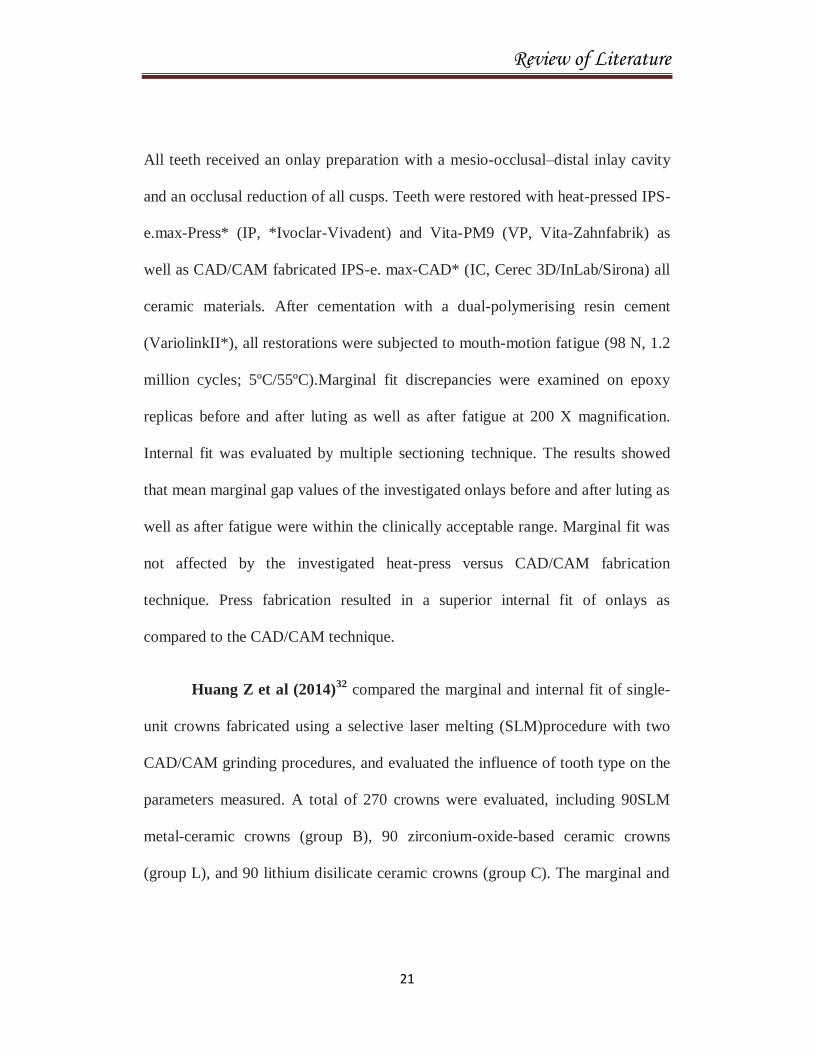
Review of Literature
21
All teeth received an onlay preparation with a mesio-occlusal–distal inlay cavity
and an occlusal reduction of all cusps. Teeth were restored with heat-pressed IPS-
e.max-Press* (IP, *Ivoclar-Vivadent) and Vita-PM9 (VP, Vita-Zahnfabrik) as
well as CAD/CAM fabricated IPS-e. max-CAD* (IC, Cerec 3D/InLab/Sirona) all
ceramic materials. After cementation with a dual-polymerising resin cement
(VariolinkII*), all restorations were subjected to mouth-motion fatigue (98 N, 1.2
million cycles; 5ºC/55ºC).Marginal fit discrepancies were examined on epoxy
replicas before and after luting as well as after fatigue at 200 X magnification.
Internal fit was evaluated by multiple sectioning technique. The results showed
that mean marginal gap values of the investigated onlays before and after luting as
well as after fatigue were within the clinically acceptable range. Marginal fit was
not affected by the investigated heat-press versus CAD/CAM fabrication
technique. Press fabrication resulted in a superior internal fit of onlays as
compared to the CAD/CAM technique.
Huang Z et al (2014)32
compared the marginal and internal fit of single-
unit crowns fabricated using a selective laser melting (SLM)procedure with two
CAD/CAM grinding procedures, and evaluated the influence of tooth type on the
parameters measured. A total of 270 crowns were evaluated, including 90SLM
metal-ceramic crowns (group B), 90 zirconium-oxide-based ceramic crowns
(group L), and 90 lithium disilicate ceramic crowns (group C). The marginal and

Review of Literature
22
internal gaps of the crowns were recorded using a replica technique with a
silicone indicator paste stabilized with a light-body silicone. The gap replica
specimens were sectioned buccolingually and mesiodistally and then examined
using a stereomicroscope at 30 X magnification. Ten reference points were
measured on each anterior and premolar specimen, and 20 reference points were
measured on each molar specimen. The results were statistically analysed and
concluded that SLM system demonstrated better marginal and internal fit
compared to the two CAD/CAM grinding systems examined. Tooth type did not
significantly influence the marginal or internal fit.
Zaruba M et al (2014)69
evaluated the effect of a minimally invasive mod
preparation on the marginal adaptation of ceramic and composite inlays with the
aim of saving sound dental substance. Class II mod cavities were prepared in50
extracted human molars and randomly allocated to five groups. In all groups, the
mesial proximal box margins were located in the dentin, 1 mm below the
cemento-enamel junction (CEJ), while the distal box margins were 1 mm above
the CEJ. In groups A and B, conventional standard preparations with a divergent
angle of = 6° were prepared. In groups C, D, and E, minimally invasive standard
preparations with a convergent angle of = 10° were prepared. In groups A and D,
composite inlays and, in groups B and C, ceramic inlays were fabricated and
adhesively inserted. In group E, a direct composite filling using the incremental

Review of Literature
23
technique was placed. Replicas were taken before and after thermomechanical.
Marginal integrity was evaluated by SEM. The percentage of continuous margins
in the different locations was compared between and within groups before and
after cycling, using ANOVA and Scheffé Post hoc test. Results showed that after
thermomechanical loading, no significant differences were observed between the
different groups with respect to the interface of luting composite-inlay.
Evanthia A et al (2015)5 evaluated the internal adaptation of pressed and
milled ceramic crowns made from digital impressions. Thirty polyvinyl siloxane
(PVS) impressions and 30 Lava COS impressions made of a prepared dentoform
tooth (master die) were fabricated. Thirty crowns were pressed in lithium
disilicate (IPS e. max Press), and 30 crowns were milled from lithium disilicate
blocks (IPS e. max CAD) (15/impression technique) with the E4D scanner and
milling engine. The master die and the intaglio of the crowns were digitized with
a 3-dimensional laser coordinate measurement machine. The digital master die
and intaglio of each crown were merged. The distance between the die and the
intaglio surface of the crown was measured at 3 standardized points. The results
revealed that the internal gap obtained from the Lava/press was significantly
greater than that obtained from the other groups (p<.001), while no significant
differences were found among PVS/press, PVS/CAD/CAM and
Lava/CAD/CAM.

Review of Literature
24
Durand LB et al (2015)21
determined the effect of cavity depth, ceramic
thickness and resin bases with different elastic modulus on von mises stress
patterns of ceramic inlays. 3D geometric models were developed and the
differences between the models were: depth of pulpal wall, ceramic thickness, and
presence of composite bases with different thickness and elastic modulus. A load
of 100 N was applied. The stress distribution pattern was analyzed with von mises
stress diagrams. The highest von mises stress value was found on models with 1-
mm-thick composite resin base and 1-mm-thick ceramic inlay. Intermediate
values occurred on models with 2-mm-thick composite resin base and 1-mmthick
ceramic inlay and 1-mm-thick composite resin base and 2-mm-thick ceramic
inlay. Lowest values were observed on models restored exclusively with ceramic
inlay. It was found that thicker inlays distribute stress more favorably and bases
with low elastic modulus increase stress concentrations on the internal surface of
the ceramic inlay. The increase of ceramic thickness tends to present more
favorable stress distribution, especially when bonded directly onto the cavity
without the use of supporting materials.
Guven Sedat et al (2015)28
examined the influence of two ceramic inlay
materials with different cavity designs on stresses in the inlay. Finite-element
analysis and three-dimensional modelling were used to examine the stress in
ceramic inlays resulting from a 250 N point load on occlusal surfaces. The

Review of Literature
25
adhesion properties and von mises stress values in the enamel, dentin, ceramic
materials and cement linings were simulated. Two ceramic inlay materials:
porcelain ceramic and zirconia ceramic, as well as two cavity corner designs:
rectangular and rounded, were evaluated. The obtained von mises stress results
indicated that the maximum and minimum forces were concentrated in the enamel
and dentin, respectively. The stress values in the dentin and inlay material were
similar in the porcelain ceramic and zirconia ceramic groups. However, in the
enamel, the stress values in the zirconia ceramic group were significantly lower
than those in the porcelain ceramic group. Additionally, cavities with rounded
corners were subject to significantly less stress compared to those with
rectangular corners. The study confirmed that, the zirconia ceramic inlay
demonstrated better performance under applied stress, based on the reduced stress
values in the tooth structure.
Irina ilgenstein et al (2015)33
investigated the influence of proximal box
elevation (PBE) with composite resin when applied to deep proximal defects in
root-filled molars with MOD cavities, which were subsequently restored with
CAD/CAM ceramic or composite restorations. Root canal treatment was
performed on 48 human mandibular molars. Standardized MOD cavities were
prepared with the distal box located 2 mm below the CEJ. The teeth were
randomly assigned to one of four experimental groups. In groups G1 and G2, the

Review of Literature
26
distal proximal box was elevated up to the level of the CEJ with composite resin
(PBE). No elevation was performed in the remaining two groups (G3, G4).
CAD/CAM restorations were fabricated with feldspathic ceramic in groups G1
and G3 or with resin nano-ceramic blocks in groups G2 and G4. Replicas were
taken before and after thermomechanical loading. Following TML, load was
applied until failure. Fracture analysis was performed under a stereomicroscope.
Marginal quality before and after TML was evaluated using scanning electron
microscopy. The results showed lower percentages of continuous margins in
groups G1–G3 compared with pre-TML assessments. For group G4-lav, the
marginal quality after TML was significantly better than in any other group. The
highest mean fracture value was recorded for group G4. No significant difference
was found for this value between the groups with PBE compared with the groups
without PBE, regardless of the material used. The specimens restored with
ceramic onlays exhibited fractures that were mainly restricted to the restoration
while, in teeth restored with composite onlays, the percentage of catastrophic
failures (fractures beyond bone level) was increased. The study concluded that
PBE had no impact on either the marginal integrity or the fracture behavior of
root canal-treated mandibular molars restored with feldspathic ceramic onlays.
Rocca et al (2015)52
presented an evidence-based update of clinical
protocols and procedures for cavity preparation and restoration selection for

Review of Literature
27
bonded inlays and onlays. In cases of severe bruxism or tooth fragilization,
CAD/CAM composite resins or pressed CAD/CAM lithium disilicate glass
ceramics are often recommended, although this choice relies mainly on scarce in
vitro research as there is still a lack of medium- to long-term clinical evidence.
The decision about whether or not to cover a cusp can only be made after a
multifactorial analysis, which includes cavity dimensions and the resulting tooth
biomechanical status, as well as occlusal and esthetic factors. The clinical Impact
of the modern treatment concepts such as – dual bonding (db)/immediate dentin
sealing (ids), cavity design optimization (cdo), and cervical margins relocation
(cmr) – should be followed. Despite the wide choice of restorative materials
(composite resin or ceramic) and techniques (classical or CAD/CAM), the cavity
for an indirect restoration should meet five objective criteria such as detailed
sharp margins, absence of undercuts, accessibility of subgingival margins,
absence of contact between the cavity and the adjacent teeth, and adequate inter-
occlusal space, before the impression.
Sandoval MJ et al (2015)54
evaluated the influence of different composite
bases and surface treatments on marginal and internal adaptation of class II cerec
CAD/CAM ceramic inlays, before and after simulated occlusal loading. Thirty-
two IPS empress cad class II inlays (MO or DO) were placed on third molars,
with margins 1 mm below the cementum-enamel junction (CEJ), following
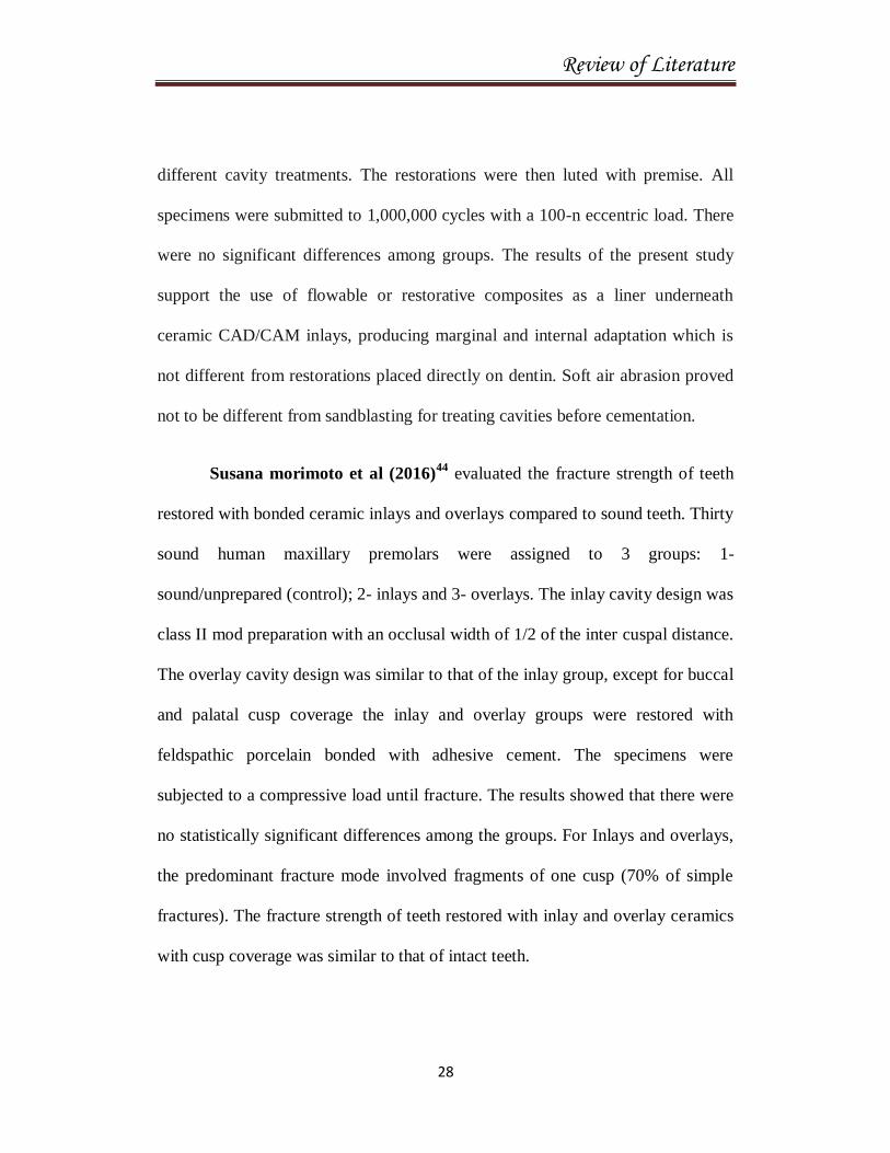
Review of Literature
28
different cavity treatments. The restorations were then luted with premise. All
specimens were submitted to 1,000,000 cycles with a 100-n eccentric load. There
were no significant differences among groups. The results of the present study
support the use of flowable or restorative composites as a liner underneath
ceramic CAD/CAM inlays, producing marginal and internal adaptation which is
not different from restorations placed directly on dentin. Soft air abrasion proved
not to be different from sandblasting for treating cavities before cementation.
Susana morimoto et al (2016)44
evaluated the fracture strength of teeth
restored with bonded ceramic inlays and overlays compared to sound teeth. Thirty
sound human maxillary premolars were assigned to 3 groups: 1-
sound/unprepared (control); 2- inlays and 3- overlays. The inlay cavity design was
class II mod preparation with an occlusal width of 1/2 of the inter cuspal distance.
The overlay cavity design was similar to that of the inlay group, except for buccal
and palatal cusp coverage the inlay and overlay groups were restored with
feldspathic porcelain bonded with adhesive cement. The specimens were
subjected to a compressive load until fracture. The results showed that there were
no statistically significant differences among the groups. For Inlays and overlays,
the predominant fracture mode involved fragments of one cusp (70% of simple
fractures). The fracture strength of teeth restored with inlay and overlay ceramics
with cusp coverage was similar to that of intact teeth.

Materials and Methods
Materials and Methods
29
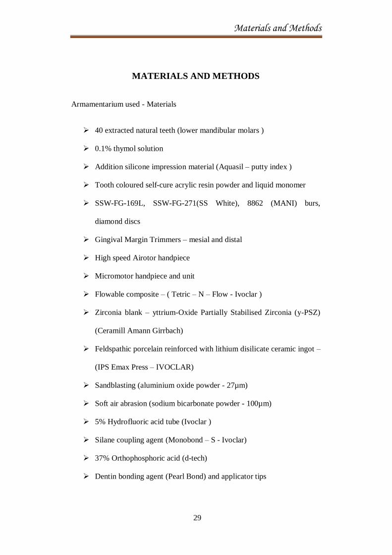
MATERIALS AND METHODS
Armamentarium used - Materials
40 extracted natural teeth (lower mandibular molars )
0.1% thymol solution
Addition silicone impression material (Aquasil – putty index )
Tooth coloured self-cure acrylic resin powder and liquid monomer
SSW-FG-169L, SSW-FG-271(SS White), 8862 (MANI) burs,
diamond discs
Gingival Margin Trimmers – mesial and distal
High speed Airotor handpiece
Micromotor handpiece and unit
Flowable composite – ( Tetric – N – Flow - Ivoclar )
Zirconia blank – yttrium-Oxide Partially Stabilised Zirconia (y-PSZ)
(Ceramill Amann Girrbach)
Feldspathic porcelain reinforced with lithium disilicate ceramic ingot –
(IPS Emax Press – IVOCLAR)
Sandblasting (aluminium oxide powder - 27µm)
Soft air abrasion (sodium bicarbonate powder - 100µm)
5% Hydrofluoric acid tube (Ivoclar )
Silane coupling agent (Monobond – S - Ivoclar)
37% Orthophosphoric acid (d-tech)
Dentin bonding agent (Pearl Bond) and applicator tips

Materials and Methods
30
Dual cure Resin cement – base and catalyst (Variolink – N – Ivoclar)
Dental surveyor -with 5 kg stone
Mixing pad and Agate spatula
Chip blower and cotton
Clear acrylic resin powder and monomer liquid
Light curing unit (3M ESPE)
Ultrasonic cleaner
Steam Cleaner
Scanning Electron Microscope along with gold sputtering machine
(Variable pressure – SEM - S – 3400 N – HITACHI)
INCLUSION CRITERIA
Extracted lower first and second mandibular molars with proper
coronal anatomy, all four walls of the teeth intact, complete root
formation, absence of dental caries
EXCLUSION CRITERIA
Teeth with attrition, loss of buccal or lingual walls, grossly
decayed, fractured teeth, abrasion, cracked teeth, lower mandibular
third molars

Materials and Methods
31
METHODOLOGY
40 extracted lower human mandibular molar teeth (figure 1) which
were extracted due to periodontal problems were selected and cleaned and
stored in 0.1% thymol solution at 4oC. All the teeth were then embedded using
tooth coloured self-curing acrylic resin (figure 2), using a putty index made
out of addition silicone impression material (figure 11).
TOOTH PREPARATION:
Tooth preparation was done to all the 40 samples.(figure 8a) The
cavity preparation was done based on the protocol given by ROCCA et al
2015 51,52
and Class II mesio – occlusal preparations (figure 17) were done in
all the 40 teeth with SSW-FG-169L, SSW-FG-271 (SS White) burs,
8862(Mani) diamond point (figure 7) and with the proximal margins 1mm
below the cemento-enamel junction and with a tapered proximal box 4mm in
width and 2 mm in depth at the bottom of the proximal box and with 5mm in
width and 3 mm in depth in the occlusal isthmus. All the walls had a taper of
about 10 degrees to 15 degrees of divergence.54
The axio-pulpal line angles
were rounded using gingival marginal trimmer (figure 8b). All line and point
angles, internal and external, were rounded to avoid stress concentrations in
the restoration and tooth, thereby reducing the potential for fractures.74

Materials and Methods
32
SAMPLE GROUPING:
The 40 teeth were randomly divided into two groups. The Group 1
had 20 teeth, which were further sub-divided into Group 1A (figure 3) and
Group 1B (figure 4) of 10 teeth samples each. The Group 1A got Zirconia
ceramic inlays [yttrium-Oxide Partially Stabilised Zirconia (y-PSZ) Ceramill
Amann Girrbach] with flowable composite base Tetric – N – Flow - Ivoclar)
(figure 9) whereas the other 10 teeth sample of Group 1B were made out of
Zirconia Ceramic inlay alone [y-PSZ] without a base. Similarly, Group 2 had
20 teeth, which were further sub-divided into Group 2A (figure 5) and Group
2B (figure 6) of 10 teeth samples each. The Group 2A were made of
Feldspathic ceramic inlays reinforced with lithium disilicate ceramic ingot
[IPS Emax Press – IVOCLAR] with a flowable composite base (Tetric – N –
Flow - Ivoclar) (figure 9) and the last 10 samples of the teeth of Group 2B
were made only with of Feldspathic ceramic inlays reinforced with lithium
disilicate ceramic ingots [IPS Emax Press – IVOCLAR] without a base.
PLACEMENT OF FLOWABLE COMPOSITE BASE:
Now the cavities of Group 1 A and 2 A which had to receive a
flowable composite base (Tetric – N – Flow – Ivoclar )20
(figure 9) underwent
etching only on the pulpal floor, axial wall and not on the gingival seat as the
base was not placed on the gingival seat area. Then, only the pulpal floor and
axial wall was etched with 37% phosphoric acid (d-tech) (figure 16) for about

Materials and Methods
33
20 seconds and was rinsed with water and dried using cotton pellets and chip
blower, while care was taken not to over dry the etched tooth surface. Now,
the denting bonding agent (pearl bond) (figure 16) was applied only on the
etched tooth surface with the help of an applicator tip and light cured (3M-
ESPE) (figure 20) for about 40 secs as per the manufacturer’s instructions.
Now the flowable composite base (Tetric – N – Flow – Ivoclar) of about 1mm
thickness (figure 10) is placed on the pulpal floor and the axial wall and light
cured (3M-ESPE). Care to be taken that the flowable composite base does not
cover the gingival seat area.77
The teeth in Group 1 B and 2 B did not have a base and served as a
control in both the groups.
FABRICATION OF INLAY:
Now all the prepared Group 1 (y-PSZ) samples were subjected to
direct optical scanning. The tooth surface to be scanned was coated with
titanium dioxide powder and the measurements for the zirconia inlay was
obtained by the scanner (figure 13) and the measurements was fed into the
CAD/CAM machine with the help of the software. The CAD/CAM machine
(figure 14) used the zirconia blanks (y-PSZ) to mill the ceramic inlay
according to the measurements scanned and the final product obtained.
The Group 2 [IPS Emax Press – IVOCLAR] samples were replicated
by making an impression (figure 12) using addition silicone impression
Materials and Methods
34
material (Aquasil) (figure 11) and a master cast obtained with die stone. Now
the Group 2 [IPS Emax Press – IVOCLAR] samples were manufactured by
the conventional layering technique by hot pressing using feldspathic ceramic
with the help of the master cast in a VITA ceramic furnace (figure 15).
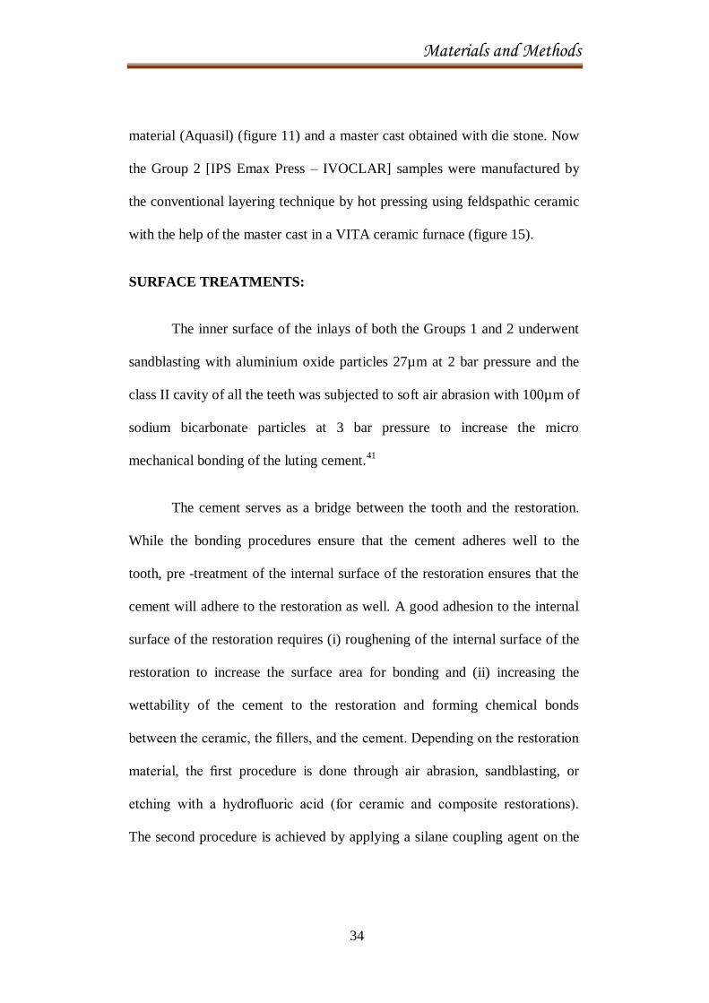
SURFACE TREATMENTS:
The inner surface of the inlays of both the Groups 1 and 2 underwent
sandblasting with aluminium oxide particles 27µm at 2 bar pressure and the
class II cavity of all the teeth was subjected to soft air abrasion with 100µm of
sodium bicarbonate particles at 3 bar pressure to increase the micro
mechanical bonding of the luting cement.41
The cement serves as a bridge between the tooth and the restoration.
While the bonding procedures ensure that the cement adheres well to the
tooth, pre -treatment of the internal surface of the restoration ensures that the
cement will adhere to the restoration as well. A good adhesion to the internal
surface of the restoration requires (i) roughening of the internal surface of the
restoration to increase the surface area for bonding and (ii) increasing the
wettability of the cement to the restoration and forming chemical bonds
between the ceramic, the fillers, and the cement. Depending on the restoration
material, the first procedure is done through air abrasion, sandblasting, or
etching with a hydrofluoric acid (for ceramic and composite restorations).
The second procedure is achieved by applying a silane coupling agent on the

Materials and Methods
35
etched porcelain or composite. The silane makes the ceramic chemically
adhere to the resin cement through covalent and hydrogen bonds (Horn 1983).
Silanating the internal surface of indirect restorations ensures that the fillers of
the luting composite react and adhere with the restoration (Calamia and
Simonsen 1985 ).77
LUTING OF INLAY WITH RESIN CEMENT WITH MECHANICAL
LOADING:
All the 40 teeth with class II cavity is then acid etched with 37%
phosphoric acid gel [d tech] (figure 18) for 20 secs and then rinsed with water
and dried with cotton pellets. Then dentin bonding agent [pearl bond]
(figure 19) was applied on the etched cavity surface with the help of an
applicator tip and light cured for 40 secs (figure 20). Thus, the tooth of all the
groups were prepared.
The Group 1 zirconia (y-PSZ) inlays were treated with silane coupling
agent (Monobond – S – Ivoclar) (figure 22) and left uncured until the dual
cure resin cement (Variolink – N – Ivoclar) (figure 16) base and catalyst paste
were dispensed on the mixing pad in equal quantity and mixed using an Agate
spatula and the cement was applied on the cavity surface (figure 23) and the
zirconia (y-PSZ) inlay placed on the cavity. Then the sample was placed in a
dental surveyor under 5 kg load simulating oral masticatory load (figure 25)

Materials and Methods
36
and the excess cement removed and light curing done for 20 seconds from all
the sides of the tooth.18,25
The inner surface of Group 2 feldspathic ceramic [IPS Emax Press –
IVOCLAR] inlays were now treated with 5% hydrofluoric acid (Ivoclar)
(figure 21) for 60 seconds and then rinsed with water and silane coupling
agent ( Monobond – S – Ivoclar ) (figure 22) applied and left uncured. The
dual cure resin cement (Variolink – N – Ivoclar) (figure 16) base and catalyst
paste dispensed on a mixing pad equally and mixed with an agate spatula and
loaded on the cavity (figure 23). The feldspathic ceramic [IPS Emax Press –
IVOCLAR] inlay was then placed on the cavity and subjected to 5 kg load
under a dental surveyor (figure 25) and excess cement was removed and light
cured for 20 secs from all the sides.18,25
PREPARATION OF SAMPLE FOR ANALYSIS:
All the 40 luted samples (figure 24) were left for 24 hours for
complete polymerisation and then all the samples were embedded in clear
acrylic resin (figure 2) used with the help of a putty index (addition silicone
impression material – Aquasil) (figure 11).
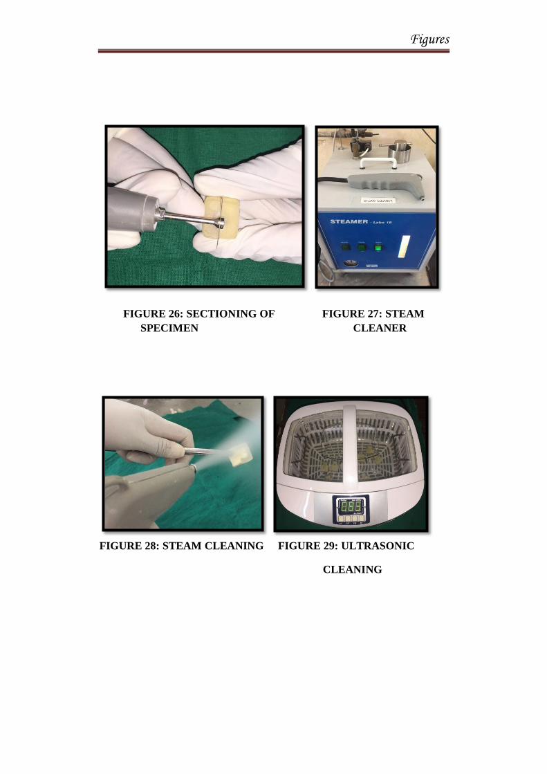
Now all the 40 samples were cut mesio – distally through and through
the entire resin sample using diamond discs (figure 26). One half of the cut
undamaged sample (lingual) was chosen for scanning electron microscopic
analysis.

Materials and Methods
37
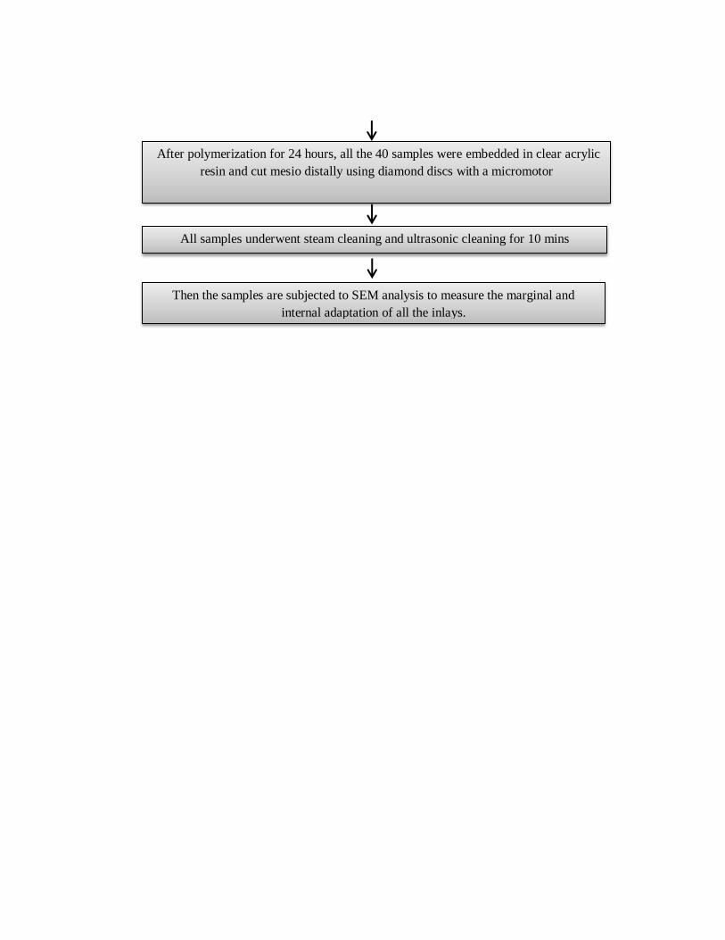
All the 40 samples were now subjected to cleaning (figure 28) in a
steam cleaner (figure 27) and further cleaned with distilled water in an
ultrasonic cleaner (figure 29) for 10 mins to ensure the cutting procedures
have not let any microscopic particles on the sample so that the SEM analysis
can be done without any hindrance and the samples placed in a sterile
container until SEM analysis was done.
SCANNING ELECTRON MICROSCOPIC ANALYSIS:
All the samples are now analysed using Scanning electron microscope
(figure 32) with magnifications 200 X and up to 500 X when required. All the
40 sliced samples of the four groups underwent gold sputtering (figure 31) in
the gold sputtering machine (figure 30) for about 15 seconds to make the
samples more electro-conductive underneath the SEM. The thickness of luting
cement was measured and expressed in µmicrons in various points to ensure
the marginal and internal adaptation. Then all samples were loaded in the
SEM machine one by one.
SEM evaluation of marginal adaptation of all the 40 samples were
evaluated at two points –
1. at the occlusal junction between the restoration and tooth interface
where the luting cement thickness was measured in µm and
2. at the proximal box-cervical area junction between the restoration and
the cervical dentin.

Materials and Methods
38
The internal adaptation of all the 40 sliced samples were evaluated by SEM at
three areas namely
1. Pulpal floor and distal wall line angle and along the pulpal floor
[occlusal dentin],
2. The pulpal floor and axial wall line angle and along the axial wall
[axial dentin].
3. The axial wall and gingival seat line angle and along the gingival seat
[cervical dentin].
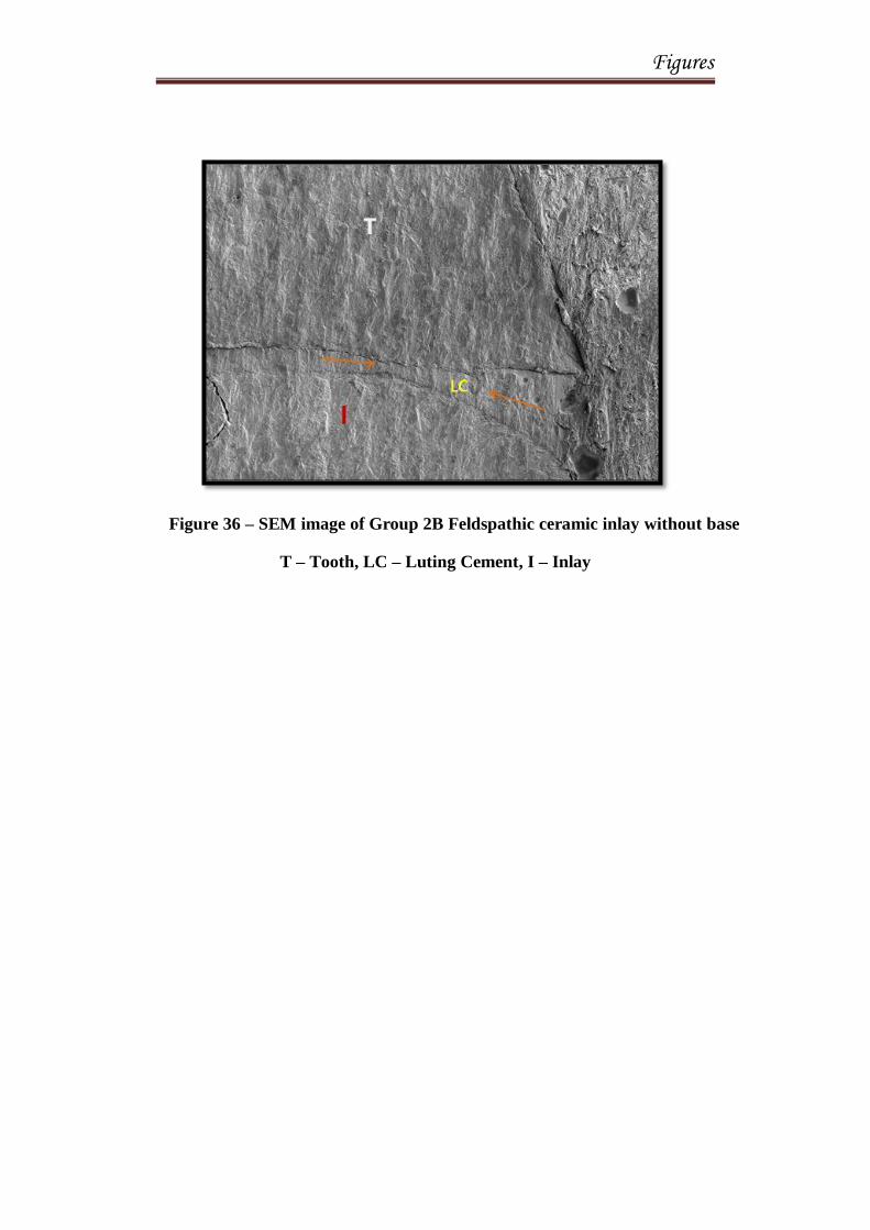
The thickness of the luting cement in all the areas were measured and
recorded between the restoration and tooth interface as in the Groups 1B
zirconia ceramic inlay without base (figure 34) and in Group 2B feldspathic
ceramic inlay without base (figure 36) whereas in the Group 1 A zirconia
ceramic inlay with base (figure 33) and Group 2 A feldspathic ceramic inlay
with base (figure 35) which consists of a flowable composite base, the
interface between the flowable composite base and the tooth was considered
as one whole interface and the amount of luting resin cement was measured
from the flowable composite base to the restoration interface. All the
measurements of the 40 sliced samples were then recorded and then tabulated.

METHODOLOGY- FLOWCHART
All the 40 inlay were luted with a dual cure resin cement under 5 kg mechanical
loading in a dental surveyor
All the cavities underwent soft air abrasion with 100µm of sodium bicarbonate
particles at 3 bar pressure. Then acid etching was done with 37% phosphoric acid for
20 secs and dentin bonding agent applied on the etched surface and cured for 40 secs
40 extracted mandibular molars were selected and embedded in acrylic resin. Class II
MO cavity preparation was done in all the 40 samples
Teeth were randomly divided into four groups containing 10 teeth in each group
Group 1A Zirconia
ceramic inlay with
flowable composite
base.
Group 1B Zirconia
ceramic inlay without
base
Group 2A Feldspathic
ceramic inlay with
flowable composite
base
Group 2B Feldspathic
ceramic inlay without
base
Flowable composite base applied on occlusal and axial dentin in groups 1A and 2A, the
groups 1B and 2B did not receive a base and served as control groups
Group 1 ceramic inlays were fabricated with zirconia by CAD/CAM, group 2 inlays were
fabricated with feldspathic ceramic by hot pressing
The inner surface of all 40 inlays underwent sandblasting with aluminium oxide
particles 27µm at 2 bar pressure
The inner surface of group1
Zirconia ceramic inlays
were treated with silane
coupling agent
The inner surface of group 2
feldspathic ceramic inlays
were first treated with 5%
hydrofluoric acid for 60 sec
and then with silane coupling
agent
After polymerization for 24 hours, all the 40 samples were embedded in clear acrylic
resin and cut mesio distally using diamond discs with a micromotor
All samples underwent steam cleaning and ultrasonic cleaning for 10 mins
Then the samples are subjected to SEM analysis to measure the marginal and
internal adaptation of all the inlays.

Figures

Figures
FIGURE 1: TEETH SPECIMEN
FIGURE 2: MATERIALS USED FOR MOUNTING AND
EMBEDDING SAMPLES

Figures
FIGURE 3: EMBEDDED SAMPLES - GROUP 1A
FIGURE 4: EMBEDDED SAMPLES - GROUP 1B
FIGURE 5: EMBEDDED SAMPLES - GROUP 2A

Figures
FIGURE 6: EMBEDDED SAMPLES - GROUP 2B
FIGURE 7: ARMAMENTARIUM

Figures
FIGURE 8 a: INLAY CAVITY
PREPARATION
FIGURE 9: TETRIC-N- FLOW USED AS BASE
FIGURE 10: APPLICATION OF BASE
FIGURE 8 b – ROUNDENING
OF PREPARATION OF
AXIO-PULPAL LINE ANGLE
WITH GMT

Figures
FIGURE 11: MATERIALS USED FOR IMPRESSION
FIGURE 12: IMPRESSION MAKING FOR GROUP 2
Figures
FIGURE 13: SCANNER MAP400
FIGURE 14: CERAMILL MACHINE

Figures
FIGURE 15: VITA CERAMIC FURNACE
FIGURE 16: MATERIALS USED FOR LUTING

Figures
FIGURE 17: PREPARED CAVITY
FIGURE 18: APPLICATION OF FIGURE 19: APPLICATION OF
ETCHANT BONDING AGENT
FIGURE 20: CURING

Figures
FIGURE 21: 5% HYDROFLUORIC FIGURE 22: APPLICATION OF
ACID USED FOR GROUP 2 SILANE COUPLING AGENT
FOR GROUP 1 AND 2
FIGURE 23: LUTING CEMENT FIGURE 24: LUTED INLAY
FIGURE 25: MECHANICAL LOADING
Figures
FIGURE 26: SECTIONING OF FIGURE 27: STEAM
SPECIMEN CLEANER
FIGURE 28: STEAM CLEANING FIGURE 29: ULTRASONIC
CLEANING

Figures
FIGURE 30: GOLD SPUTTERING MACHINE
FIGURE 31: GOLD SPUTTERED SPECIMENS

Figures
FIGURE 32: SCANNING ELETRON MICROSCOPE
Figure: 33 – SEM image of group 1A Zirconia ceramic inlay with base
T – Tooth, B – Base, LC – Luting Cement, I – Inlay

Figures
Figure 34 – SEM image of Group 1B Zirconia ceramic inlay without base
T – Tooth, LC – Luting Cement, I – Inlay
Figure 35 – SEM image of Group 2A Feldspathic ceramic inlay with base
T – Tooth, B – Base, LC – Luting Cement, I – Inlay
Figures
Figure 36 – SEM image of Group 2B Feldspathic ceramic inlay without base
T – Tooth, LC – Luting Cement, I – Inlay

Results

Results
39
RESULTS
In this study, Scanning Electron Microscope was used to evaluate the
marginal and internal adaptation of class II ceramic inlays fabricated with
zirconia and feldspathic ceramic. All the values were tabulated and Statistical
analysis was done using Software SPSS, Version 20.0. One-way analysis of
variance (ANOVA) is used to study the overall variance within groups. It is
the extension of the t-test done between groups to the situation in which more
than two groups are compared simultaneously. However, it is not possible to
identify the difference between the various subgroups with the help of the
p values obtained from ANOVA. Therefore, a specific statistical test was used
for intra group comparison. The Post hoc Tukey Test Honestly significant
difference or HSD test is designed to perform a pairwise comparison of the
means to identify the specific sub groups in which significant difference
expression occurs.
Table 01 shows the SEM values of marginal adaptation measured in
the occlusal and proximal areas in all the groups of the 40 samples expressed
as µm.
Table 02 shows the SEM values of internal adaptation measured in
occlusal, axial and cervical dentin area in all the groups of the 40 samples
expressed as µm.

Results
40
Table 03 shows the mean and standard deviation comparison of
marginal adaptation in the occlusal area between all the four groups by one-
way ANOVA. The mean for Group 1A (Zirconia with base) was 13.80±3.29,
GROUP 1B (ZIRCONIA without base) was 16.20±3.97, GROUP 2A
(Feldspathic ceramic with base) was 159.30±32.05, Group 2B (Feldspathic
ceramic without base) was 163.90±35.74 and the p value was highly
significant [p <0.001**
]. (Graph 01)
Table 04 shows the pairwise comparison of marginal adaptation in the
occlusal area between all the four groups by Post – Hoc tests – Tukey HSD
where the p value between Group 1A and 1B was insignificant and Group 1A
and Group 2A was highly significant [p <0.001**
], Group 1A and group 2B
was highly significant. The p value for Group 1B and Group 2A was highly
significant and Group 1B and Group 2B was highly significant too. The p
value for Group 2A and Group 2B was insignificant.
Table 05 shows the mean and standard deviation comparison of
marginal adaptation in the proximal are between all the four groups by one-
way ANOVA. The mean for Group 1A was 71.30±21.27, Group 1B was
76.80±15.06, Group 2A was 273.50±18.65 and Group 2B was 281.00±44.49.
The p value was found to be highly significant [p <0.001**
]. (Graph 02)
Table 06 shows the pairwise comparison of marginal adaptation in the
proximal area between all the four groups by Post hoc tests- Tukey HSD

Results
41
where the p value for Group 1A and group 1B was insignificant, Group 1A
and Group 2A is highly significant [p <0.001**
], Group 1A and Group 2B was
highly significant. The p value for Group 1B and Group 2A was highly
significant, Group 1B and Group 2B was highly significant and Group 2A and
Group 2B was insignificant.
Table 07 shows the mean and standard deviation comparison of
internal adaptation in the occlusal dentin area between all the four groups by
one-way ANOVA. The mean for Group 1A was 6.60±1.51, Group 1B was
15.20±1.75, Group 2A was 74.00±13.50, Group 2B was 125.00±14.34 and the
p value was highly significant [p <0.001**
]. (Graph 03)
Table 08 shows the pairwise comparison of internal adaptation in the
occlusal dentin area with all the groups using Post hoc tests – Tukey HSD.
The p value for Group 1A and Group 1B was insignificant, Group 1A and
Group 2A was highly significant [p <0.001**
] , Group 1A and Group 2B was
highly significant. The Group 1B and Group 2A was highly significant, Group
1B and Group 2B was highly significant and Group 2A and Group 2B was
highly significant.
Table 09 shows the mean and standard deviation comparison of
internal adaptation in the axial dentin area between all the four groups by one-
way ANOVA. The mean for Group 1A was 35.40±5.27, Group 1B was

Results
42
52.10±7.55, Group 2A was 19.30±3.68, Group 2B was 56.60±7,18. The p
value was highly significant [p <0.001**
]. (Graph 04)
Table 10 shows the pairwise comparison of internal adaptation in the
axial dentin area with all the groups using Post hoc tests – Tukey HSD. The p
value for Group 1A and Group 1B was highly significant [p <0.001**
], Group
1A and Group 2A was highly significant, Group 1A and Group 2B was highly
significant, Group 1B and Group 2A was highly significant, Group 1B and
Group 2B was insignificant and Group 2A and Group 2B was highly
significant.
Table 11 shows the mean and standard deviation comparison of
internal adaptation in the cervical dentin area between all the four groups by
one-way ANOVA. The mean for Group 1A was 196.70±10.60, Group 1B was
199.90±28.47, Group 2A was 253.00±22.63, Group 2B was 263.00±50.78 and
the p value was highly significant [p <0.001**
]. (Graph 05)
Table 12 shows the pairwise comparison of internal adaptation in the
cervical dentin area with all the groups using Post hoc tests – Tukey HSD.
The p value for Group 1A and Group 1B was insignificant, Group 1A and
Group 2A was highly significant [p <0.002**
], Group 1A and Group 2B was
highly significant [p <0.001**
], Group 1B and Group 2A was highly
significant [p <0.003**
], Group 1B and Group 2B was highly significant
[p <0.001**
] and Group 2A and Group 2B was insignificant.

Tables and Graphs

Tables and Graphs
TABLE 1: SEM VALUES OF MARGINAL ADAPTATION
MEASURED IN OCCLUSAL AND PROXIMAL AREAS
Group Sample Occlusal Adaptation
µm
Proximal Adaptation
µm
1A 1 10 50
1A 2 11 51
1A 3 14 48
1A 4 10 53
1A 5 12 57
1A 6 16 87
1A 7 18 80
1A 8 12 98
1A 9 19 92
1A 10 16 97
1B 1 24 58
1B 2 18 84
1B 3 13 53
1B 4 17 74
1B 5 11 71
1B 6 18 63
1B 7 19 92
1B 8 15 91
1B 9 11 90
1B 10 16 92
2A 1 110 240

Tables and Graphs
2A 2 153 280
2A 3 130 250
2A 4 140 280
2A 5 220 270
2A 6 200 300
2A 7 160 295
2A 8 170 280
2A 9 160 262
2A 10 150 278
2B 1 145 338
2B 2 157 320
2B 3 205 322
2B 4 100 330
2B 5 115 260
2B 6 164 240
2B 7 194 210
2B 8 185 270
2B 9 203 240
2B 10 171 280
Group 1A – Zirconia ceramic inlay with Base
Group 1B – Zirconia ceramic inlay without Base
Group 2A – Feldspathic ceramic inlay with Base
Group 2B – Feldspathic ceramic inlay without Base

Tables and Graphs
TABLE 2: SEM VALUES OF INTERNAL ADAPTATION MEASURED
IN OCCLUSAL, AXIAL AND CERVICAL DENTIN AREAS
Group Sample Occlusal Dentin
µm
Axial Dentin
µm
Cervical Dentin
µm
1A 1 8 32 183
1A 2 7 39 176
1A 3 7 36 192
1A 4 4 39 198
1A 5 5 36 196
1A 6 9 37 198
1A 7 8 23 208
1A 8 6 41 208
1A 9 6 32 202
1A 10 6 39 206
1B 1 16 61 219
1B 2 15 63 167
1B 3 18 54 182
1B 4 17 61 243
1B 5 14 52 218
1B 6 15 48 187
1B 7 13 45 173
1B 8 14 41 164
1B 9 13 46 216
1B 10 17 50 230
2A 1 60 16 250
2A 2 80 15 230
2A 3 90 18 250
2A 4 60 22 260

Tables and Graphs
2A 5 80 14 230
2A 6 70 18 270
2A 7 80 23 290
2A 8 90 22 280
2A 9 80 20 250
2A 10 50 25 220
2B 1 110 62 330
2B 2 120 63 310
2B 3 110 61 180
2B 4 140 60 190
2B 5 120 59 270
2B 6 150 57 220
2B 7 140 61 260
2B 8 130 55 300
2B 9 120 47 280
2B 10 110 41 290
Group 1A – Zirconia ceramic inlay with Base
Group 1B – Zirconia ceramic inlay without Base
Group 2A – Feldspathic ceramic inlay with Base
Group 2B – Feldspathic ceramic inlay without Base

Tables and Graphs
TABLE 3: THE MEAN AND STANDARD DEVIATION COMPARISON
OF MARGINAL ADAPTATION IN THE OCCLUSAL AREA
BETWEEN ALL THE FOUR GROUPS BY ONE-WAY ANOVA
Groups Mean SD P value
Zirconia ceramic inlay with Base (1A) 13.80 3.29
<0.001**
Zirconia ceramic inlay without Base (1B) 16.20 3.97
Feldspathic ceramic inlay with Base (2A) 159.30 32.05
Feldspathic ceramic inlay without Base (2B) 163.90 35.74
Note - ** denotes significant at 1 level.

Tables and Graphs
TABLE 4: PAIRWISE COMPARISON OF MARGINAL ADAPTATION
IN THE OCCLUSAL AREA BETWEEN ALL THE FOUR GROUPS BY
POST HOC TESTS - TUKEY HSD
Note - ** denotes significant at 1 level
(I) Group (J) Group Sig.
Zirconia ceramic inlay
with Base (1A)
Zirconia ceramic inlay
without Base (1B)
0.996
Feldspathic ceramic inlay
with Base (2A)
<0.001**
Feldspathic ceramic inlay
without Base (2B)
<0.001**
Zirconia ceramic inlay
without Base (1B)
Feldspathic ceramic inlay
with Base (2A)
<0.001**
Feldspathic ceramic inlay
without Base (2B)
<0.001**
Feldspathic with Base (2A)
Feldspathic ceramic inlay
without Base (2B)
0.974
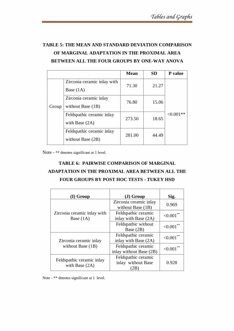
Tables and Graphs
TABLE 5: THE MEAN AND STANDARD DEVIATION COMPARISON
OF MARGINAL ADAPTATION IN THE PROXIMAL AREA
BETWEEN ALL THE FOUR GROUPS BY ONE-WAY ANOVA
Mean SD P value
Group
Zirconia ceramic inlay with
Base (1A) 71.30 21.27
<0.001**
Zirconia ceramic inlay
without Base (1B) 76.80 15.06
Feldspathic ceramic inlay
with Base (2A) 273.50 18.65
Feldspathic ceramic inlay
without Base (2B) 281.00 44.49
Note - ** denotes significant at 1 level.
TABLE 6: PAIRWISE COMPARISON OF MARGINAL
ADAPTATION IN THE PROXIMAL AREA BETWEEN ALL THE
FOUR GROUPS BY POST HOC TESTS - TUKEY HSD
(I) Group (J) Group Sig.
Zirconia ceramic inlay with
Base (1A)
Zirconia ceramic inlay
without Base (1B) 0.969
Feldspathic ceramic
inlay with Base (2A) <0.001
**
Feldspathic without
Base (2B) <0.001
**
Zirconia ceramic inlay
without Base (1B)
Feldspathic ceramic
inlay with Base (2A) <0.001
**
Feldspathic ceramic
inlay without Base (2B) <0.001
**
Feldspathic ceramic inlay
with Base (2A)
Feldspathic ceramic
inlay without Base
(2B)
0.928
Note - ** denotes significant at 1 level.
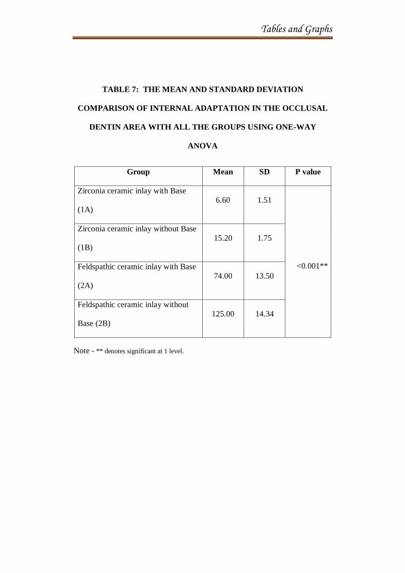
Tables and Graphs
TABLE 7: THE MEAN AND STANDARD DEVIATION
COMPARISON OF INTERNAL ADAPTATION IN THE OCCLUSAL
DENTIN AREA WITH ALL THE GROUPS USING ONE-WAY
ANOVA
Group Mean SD P value
Zirconia ceramic inlay with Base
(1A)
6.60 1.51
<0.001**
Zirconia ceramic inlay without Base
(1B)
15.20 1.75
Feldspathic ceramic inlay with Base
(2A)
74.00 13.50
Feldspathic ceramic inlay without
Base (2B)
125.00 14.34
Note - ** denotes significant at 1 level.

Tables and Graphs
TABLE 8: PAIRWISE COMPARISON OF INTERNAL ADAPTATION
IN THE OCCLUSAL DENTIN AREA WITH ALL THE GROUPS
USING POST HOC TESTS – TUKEY HSD
(I) Group (J) Group P value
Zirconia ceramic inlay with
Base (1A)
Zirconia ceramic inlay without
Base (1B) 0.230
Feldspathic ceramic inlay with
Base (2A) <0.001**
Feldspathic ceramic inlay
without Base (2B) <0.001**
Zirconia ceramic inlay
without Base (1B)
Feldspathic ceramic inlay with
Base (2A) <0.001**
Feldspathic ceramic inlay
without Base (2B)
<0.001**
Feldspathic ceramic inlay
with Base (2A)
Feldspathic ceramic inlay
without Base (2B)
<0.001**
Note - ** denotes significant at 1 level

Tables and Graphs
TABLE 9: THE MEAN AND STANDARD DEVIATION COMPARISON
OF INTERNAL ADAPTATION IN THE AXIAL DENTIN AREA WITH
ALL FOUR GROUPS USING ONE-WAY ANOVA
Mean SD P value
<0.001**
Group
Zirconia ceramic inlay with
Base (1A)
35.40 5.27
Zirconia ceramic inlay without
Base (1B)
52.10 7.55
Feldspathic ceramic inlay with
Base (2A)
19.30 3.68
Feldspathic ceramic inlay
without Base (2B)
56.60 7.18
Note - ** denotes significant at 1 level
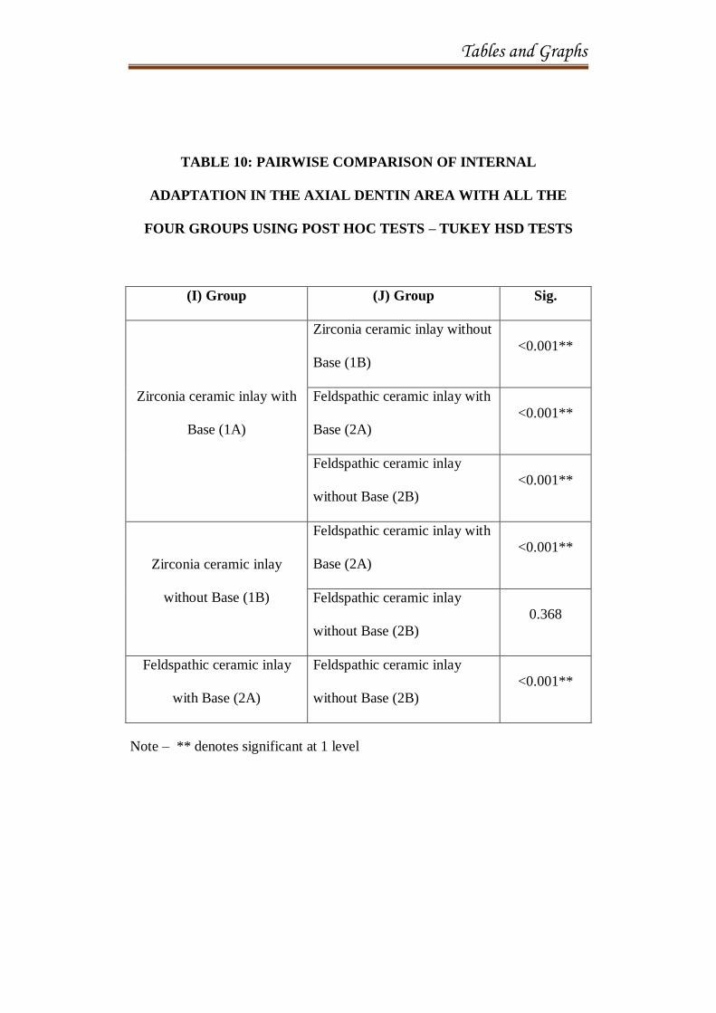
Tables and Graphs
TABLE 10: PAIRWISE COMPARISON OF INTERNAL
ADAPTATION IN THE AXIAL DENTIN AREA WITH ALL THE
FOUR GROUPS USING POST HOC TESTS – TUKEY HSD TESTS
(I) Group (J) Group Sig.
Zirconia ceramic inlay with
Base (1A)
Zirconia ceramic inlay without
Base (1B)
<0.001**
Feldspathic ceramic inlay with
Base (2A)
<0.001**
Feldspathic ceramic inlay
without Base (2B)
<0.001**
Zirconia ceramic inlay
without Base (1B)
Feldspathic ceramic inlay with
Base (2A)
<0.001**
Feldspathic ceramic inlay
without Base (2B)
0.368
Feldspathic ceramic inlay
with Base (2A)
Feldspathic ceramic inlay
without Base (2B)
<0.001**
Note – ** denotes significant at 1 level

Tables and Graphs
TABLE 11: MEAN AND STANDARD DEVIATION COMPARISON OF
INTERNAL ADAPTATION IN THE CERVICAL DENTIN AREA
WITH ALL THE GROUPS USING ONE-WAY ANOVA
Mean SD P value
Group
Zirconia ceramic inlay with
Base (1A)
196.70 10.60
<0.001**
Zirconia ceramic inlay
without Base (1B)
199.90 28.47
Feldspathic ceramic inlay
with Base (2A)
253.00 22.63
Feldspathic ceramic inlay
without Base (2B)
263.00 50.78
Note - ** denotes significant at 1 level.
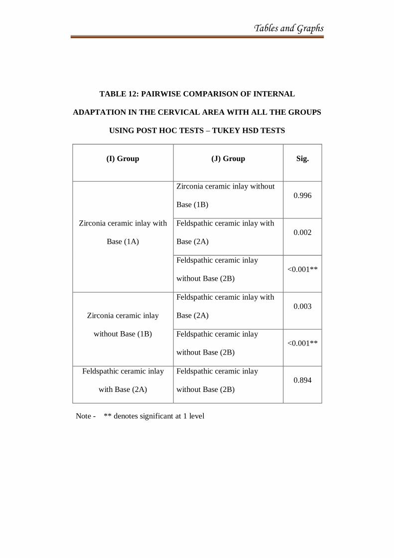
Tables and Graphs
TABLE 12: PAIRWISE COMPARISON OF INTERNAL
ADAPTATION IN THE CERVICAL AREA WITH ALL THE GROUPS
USING POST HOC TESTS – TUKEY HSD TESTS
(I) Group (J) Group Sig.
Zirconia ceramic inlay with
Base (1A)
Zirconia ceramic inlay without
Base (1B)
0.996
Feldspathic ceramic inlay with
Base (2A)
0.002
Feldspathic ceramic inlay
without Base (2B)
<0.001**
Zirconia ceramic inlay
without Base (1B)
Feldspathic ceramic inlay with
Base (2A)
0.003
Feldspathic ceramic inlay
without Base (2B)
<0.001**
Feldspathic ceramic inlay
with Base (2A)
Feldspathic ceramic inlay
without Base (2B)
0.894
Note - ** denotes significant at 1 level

Tables and Graphs
GRAPH 1: SHOWS THE MEAN COMPARISON OF MARGINAL
ADAPTATION IN THE OCCLUSAL AREA WITH ALL THE FOUR
GROUPS USING ONE-WAY ANOVA ANALYSIS
Group 1A – Zirconia inlay with Base
Group 1B – Zirconia inlay without Base
Group 2A – Feldspathic ceramic inlay with Base
Group 2B – Feldspathic ceramic inlay without Base
Group
2B2A1B1A
Occ
lusa
l A
dap
tati
on
µm
200
175
150
125
100
75
50
25
0

Tables and Graphs
GRAPH 2: SHOWS THE MEAN COMPARISON OF THE MARGINAL
ADAPTATION IN THE PROXIMAL AREA WITH ALL THE FOUR
GROUPS USING ONE-WAY ANOVA ANALYSIS
Group 1A – Zirconia inlay with Base
Group 1B – Zirconia inlay without Base
Group 2A – Feldspathic ceramic inlay with Base
Group 2B – Feldspathic ceramic inlay without Base
Group
2B2A1B1A
Pro
xim
al A
dap
tati
on i
n µ
m
300
250
200
150
100
50
0

Tables and Graphs
GRAPH 3: SHOWS THE MEAN COMPARISON OF INTERNAL
ADAPTATION IN THE OCCLUSAL DENTIN AREA WITH ALL THE
FOUR GROUPS USING ONE-WAY ANOVA ANALYSIS
Group 1A – Zirconia inlay with Base
Group 1B – Zirconia inlay without Base
Group 2A – Feldspathic ceramic inlay with Base
Group 2B – Feldspathic ceramic inlay without Base
Group
2B2A1B1A
Occ
lusa
l D
enti
n in µ
m
140
120
100
80
60
40
20
0

Tables and Graphs
GRAPH 4: SHOWS THE MEAN COMPARISON OF THE INTERNAL
ADAPTATION IN THE AXIAL DENTIN AREA WITH ALL THE
FOUR GROUPS USING ONE-WAY ANOVA ANALYSIS
Group 1A – Zirconia inlay with Base
Group 1B – Zirconia inlay without Base
Group 2A – Feldspathic ceramic inlay with Base
Group 2B – Feldspathic ceramic inlay without Base
Group
2B2A1B1A
Axia
l D
enti
n i
n µ
m
60
50
40
30
20
10
0

Tables and Graphs
GRAPH 5: SHOWS THE MEAN COMPARISON OF THE INTERNAL
ADAPTATION IN THE CERVICAL DENTIN AREA WITH ALL THE
FOUR GROUPS USING ONE-WAY ANOVA ANALYSIS
Group 1A – Zirconia inlay with Base
Group 1B – Zirconia inlay without Base
Group 2A – Feldspathic ceramic inlay with Base
Group 2B – Feldspathic ceramic inlay without Base
Group
2B2A1B1A
Cer
vic
al D
enti
n i
n µ
m
280
240
200
160
120
80
40
0

Discussion

Discussion
43
DISCUSSION
Dental restorations are given primarily to rebuild defective tooth
tissues lost by dental caries, tooth wear, fractures or even pre-existing
restorations.49
The direct composites are possible restorative options when
aesthetics, function and durability are primary concerns in addition to
accessibility, visibility and isolation.61
Reproduction of the occlusal
morphology, proximal and intermaxillary contacts and need for a skilled
operator are few predominant limiting factors of direct restorations49
along
with the combined “Negative” effect of composite polymerization shrinkage
and functional stresses.51
On the other hand, indirect restorations such as ceramics are long
lasting as they have better marginal adaptation and are more resistant to
deterioration over time due to wear, fracture and discolouration. But the need
of the hour is to create a strong restoration which is compatible with the
mechanical, biologic and optical properties of the dental tissues which forms
the basis of the Biomimetics approach (MAGNE P, BELSER U 2002) for a
restoration.49
“Biomimetics is a concept of medical research that involves the
investigation of both structures and physical functions of biological
“composites” and the designing of new and improved substitutes.” 39

Discussion
44
The chief goal of this biomimetics approach in restorative dentistry is
to bring back, all the prepared tooth tissues to complete function by creating a
hard tissue bond which ultimately allows functional stresses to pass through
the tooth and bringing back the restoration into the final functional, biologic
and aesthetic result. Meanwhile the adhesive restorative techniques targets for
maximum preservation of sound tooth structure by maintaining the vitality of
the teeth to be restored.39
Partial indirect restorations classified as inlays (without covering the
cusps), onlays (covering at least 1 cusp), and overlays (covering all cusps)
(Felden et al. 1998; Fuzzi and Rappelli 1998; Schulz et al. 2003) enable
conservation of the remaining dental structure, promoting reinforcement of a
tooth compromised by caries or fractures (Fuzzi and Rappelli 1998; Fabianelli
et al. 2006; Guess et al. 2009).44
Earlier, the gold standard for indirect partial restorations were cast
gold inlays whose longevity is an established fact. Their durability, strength,
minimal wear on antagonist teeth and less corrosive properties were
significant for its success.31
Gold inlays remain a possible option for restoring
teeth in the posterior aspects were strength and load bearing is crucial. Then
again, the difficulties faced in manipulation, fabrication and aesthetics which
becomes the foremost criteria, lead to the diminish in use of cast gold.31
Discussion
45
A number of composite resin and ceramic materials are available for
fabricating indirect partial restorations whose mechanical strength is crucial
for the durability of the restoration in the posterior regions.44
Since 1980‟s the hot-pressed ceramics were fabricated using the lost-
wax technique. They were able to press a plasticized ceramic ingot into a
heated investment mould. Ceramics containing high amounts of leucite glass
or optimal pressable ceramics were initially used for this process and later in
2006, lithium disilicate became the second generation of material to use this
method.26
In the mid 1990‟s, the first all-ceramic product with a CAD/ CAM
substructure was introduced. The core consisted of 99.9% alumina on which a
feldspathic ceramic was layered. The use of CAD/CAM technology expanded
machinable ceramic fabrication by allowing scanning, designing, and milling
of either a full-contoured restoration or a single- or multiple-unit framework
by a computer. The available materials for the CAD/CAM processing include
silica-based ceramics, infiltration ceramics, lithium-disilicate ceramics, and
aluminium / zirconium oxide high-performance ceramics.26
Zirconia has emerged as a versatile and promising material among
dental ceramics due to its excellent mechanical properties owing to the
transformation toughening mechanism. Zirconia (ZrO2) is a white oxide of
zirconium which is a polycrystalline ceramic without a glassy phase and exists
in three crystalline forms: monoclinic (m), tetragonal (t) and cubic (c). It has a

Discussion
46
flexural strength of 900–1200MPa and a compression resistance of 2000MPa
with an average load-bearing capacity of 755N. They have fracture loads
ranging between 706N, 2000N and 4100N and all of the studies demonstrated
that in dental restorations zirconia yields higher fracture loads than alumina or
even lithium disilicate. The mechanical properties are similar to that of metals
and its colour to that of tooth. Hence it has been called as „Ceramic Steel‟ by
Garvie.4
With the addition of stabilizing oxides in concentrations less than
those required for complete stabilization obtained at room temperature,
zirconia can also be partially stabilized in a multiphase form, known as
partially stabilized zirconia (PSZ). It consists of cubic zirconia, as the major
phase, and monoclinic and tetragonal zirconia precipitates, as the minor
phase.4
When the whole material is constituted by transformable tetragonal-
zirconia grains it is called Tetragonal zirconia polycrystals (TZP). To date,
Zirconia stabilized with y2O3 (yttrium – oxide) has the best properties for
dental applications. It is basically a fully tetragonal fine-grained zirconia
ceramic material made of 100% small metastable tetragonal grains (y-TZP)
after the addition of approximately 2 to 3 mol% yttrium oxide (y2O3) as a
stabilizing agent. This yttrium-Oxide Partially Stabilised Zirconia
(y-PSZ) [Ceramill Amann Girrbach] was used in this study in Group 1 to
fabricate inlays using CAD/CAM technology.4

Discussion
47
CAD/CAM zirconia restorations are produced according to two
different techniques: “soft machining” of pre-sintered blanks or “hard
machining” of fully sintered blanks.4
Soft machining was the process used in this study to fabricate the
inlays of Group 1 with yttrium-Oxide Partially Stabilised Zirconia (y-PSZ)
[Ceramill Amann Girrbach] which was available as pre-sintered blanks.
The tooth sample used in this study was scanned directly, after which,
a virtual, enlarged framework is designed by sophisticated computer softwares
(CAD). Then, through a CAM milling procedure, a framework with enlarged,
accurately controlled dimension is machined out of the blank. The
sinterization of the framework is completed at high temperature. During
sinterization the framework regains its proper dimensions as it undergoes a
linear volume shrinkage.4
Today, the main classic porcelain ceramics are based on feldspar and
glass or a metal oxide. Glass gives ceramics an aesthetic translucent property,
while oxide ceramics provide high-strength. Feldspathic ceramics are
produced in a powder based form and can be applied to many different
substructure materials. These materials can also be formed into a block to be
used in a milling unit or can be pressed in a mould to form a restoration. The
first generation of heat-pressed dental ceramics contains leucite as reinforcing
crystalline phase. The second generation is lithium disilicate-based.19

Discussion
48
Feldspathic porcelain reinforced with Lithium disilicate glass-ceramics
have been used in this study for fabricating inlays in Group 2 [IPS Emax Press
– IVOCLAR]. Lithium disilicate glass-ceramics for all-ceramic restorations
contain about 65 volume % lithium disilicate as the main crystalline phase,
with about 1% porosity and have mechanical properties far superior to that of
the first generation leucite glass ceramic. They also have relatively low
refractive index making them translucent even with the high crystalline
content in lithium-disilicate crystals.76
The group 2 Feldspathic ceramic inlays were fabricated by hot
pressing the ceramic in layers. It involves pressure moulding and the heated
ceramic ingot is pressed through a heated tube into a mould, where the
ceramic form cools and hardens to the shape of the mould, which is later
recovered after cooling. Hot pressing usually occurs over a 45 mins at a high
temperature to produce the ceramic sub-structure. Then it is stained, glazed or
coated by veneering porcelain, which results in translucent ceramic core,
moderately high flexural strength, excellent fit & excellent aesthetics.76
In indirect restorations, the stress caused by the polymerization
shrinkage is limited to the resin cement layer. The presence of a resin coating
is believed to act as a layer absorbing the tensions, originated by the
polymerization shrinkage of the resin cement and the stress induced by
occlusal load distributed during chewing. Thus, increasing the possibility of
clinical success of the restorations.42
Hence to overcome these issues the resin

Discussion
49
coating technique (RCT) has been proposed by Satoh. M et al in 1994.42
It
consists of hybridization of the exposed dentin followed by the application of
a hydrophobic monomer or a low viscosity resin, protecting the dentine
exposed after the cavity preparation. A considerable reduction has been
observed in the formation of microcracks at the tooth-restoration interface and
an improvement of the bond strength at the interface using the Resin Coating
Protocol.42
Furthermore, in the absence of a liner or base the removal of the
temporary restoration, re-exposure and cleaning of the prepared dentin
surfaces, as well as the positive pressure induced during inlay/onlay insertion,
will frequently induce discomfort in patients. This demands anaesthesia which
is generally not needed with lined cavities. The application of a base or liner
underneath semi-direct and indirect restorations fulfils many requirements,
such as reinforcing undermined cusps, filling undercuts and providing the
necessary geometry for an inlay/onlay restoration. It also represents a
common, non-invasive alternative to surgical crown lengthening in order to
relocate cavity margins from an intra-crevicular to supra-gingival position 51
and facilitates impression technique and cementation procedures.54
Therefore,
the application of a base underneath indirect or chair-side inlays and onlays
has clear technical, biological and clinical advantages over the classical
protocol, which leaves dentin unsealed until the restoration cementation.54

Discussion
50
The elastic modulus (E) of the Flowable composites among, other
physical properties is of great importance to evaluate their potential stress-
absorbing effect and tooth reinforcement effects.54
The optimum “stress
absorbing effect” is thought to be at around 7–7.5 Gpa which is about half of
the elastic modulus of dentin which is 18.6 Gpa (Craig and Peyton). The
Flowable composites were used due to its ease of placement and not requiring
further adjustments, thus eliminating the risk for a mechanical disruption of
the dentinal seal.51
Thus in this study a flowable composite base [TETRIC-N-FLOW -
Ivoclar] was given in Group 1A (y-PSZ) on the pulpal floor and axial wall
alone in the class II inlay cavity and in Group 2 A (IPS Emax Press –
IVOCLAR) on the pulpal floor and axial wall alone in the class II inlay
cavity.
The polymerization shrinkage of composite resins is a drawback for its
use and scientific literature reports that the amount of this volumetric
shrinkage is in the range of 3 to 7% of the starting mass. Polymerization
shrinkage can reduce the bond strength, thus not allowing a close contact
between the restorative material and the tooth structure. Hence they create
gaps at the composite-tooth interface with increased chances of failure of the
restoration.38
This is one of the main causes of marginal failure and
subsequent microleakage.35
The primary concern is the seal at the interface
Discussion
51
between cementum/dentin and the composite resin, because adequate seal is
critical at these margins.35
When applying a base or liner underneath indirect restorations, the
interface quality between the resinous base and luting composite and then
between the luting composite and inlay, resulting from micro-mechanical
retentions or copolymerisation was crucial. Procedures such as soft air
abrasion or airborne particle abrasion are used with the aim to clean the cavity
and to increase micro-mechanical retention between the resinous base and the
luting cement.54
Sandblasting is a pre-treatment procedure done on the inner
surface of indirect restorations which were developed to improve
effectiveness and stability of bond between luting cement and restoration. It
also increases the adhesive area, surface activation and silanization.81
Air abrasion is the pseudo-mechanical, non-rotary method of cutting
and removing dental hard tissue. Studies have shown that the bonding of
enamel and dentin surfaces prepared with air abrasion is much better than that
with conventional carbide burs or acid etching prior to placement of luting
agents.40
With the introduction of flowable and nano-filled composites it is
easier to restore cavities which do not confer with the specifications of GV
Black's concepts.30
Common interface treatment options are grinding, abrasion with
diamond rotary instruments, airborne particle abrasion with aluminum oxide,
sodium bicarbonate, acid etching and combinations of any of these methods.

Discussion
52
Preferred surface treatment methods are acid etching with hydrofluoric acid
solutions (2.5% to 10%) and subsequent application of a silane coupling agent
along with air borne particle abrasion. The lithium-disilicate glass-ceramic
IPS Empress 2 has a high crystalline content and exhibits significantly higher
bond strengths than IPS Empress. The ceramic microstructure has a significant
influence on the fracture resistance of the composite-ceramic adhesion zone.
Hence, a combination of airborne particle abrasion (aluminium oxide), etching
with hydrofluoric acid, and application of a silane coupling agent is
recommended for glass ceramics.12
Retention of zirconia-based ceramic restorations depends on
mechanical roughening of the surface and chemical bonding with adhesive
monomer in special primers or resin cements. An acidic adhesive monomer
such as MDP [10-methacryloyloxydecyl dihydrogen phosphate] bonds to
zirconia-based ceramics. The addition of a MDP-containing bonding/silane
coupling agent to enhance bonding of resin cements has produced positive
results. It was shown that particle air-abrasion, followed by the application of
MDP-containing bonding/silane coupling agent, resulted in increased bond
strength.4
Hence in this study, a soft air abrasion interface treatment was done to
all the 40 teeth cavities with sodium bicarbonate 100 µm at 3 bar pressure for
better micromechanical bonding to the luting cement.41,54
Then the inner
surface of all the 40 inlays were subjected to interface treatment by

Discussion
53
sandblasting with aluminium oxide 27µm at 2 bar pressure to increase the
cohesion between two layers of composite.41,51
The group 1 zirconia ceramic
inlays after sandblasting with aluminium oxide underwent silanization with
Monobond -S silane coupling agent containing MDP. The group 2 feldspathic
ceramic inlays after sandblasting with aluminium oxide underwent etching
with 5% hydrofluoric acid for 60 secs and then silanization with Monobond -S
silane coupling agent.
Simon and Darnell in 2012 stated that the resin cements have the
ability to bond to the tooth structure and the internal surface of the
restoration.77
Luting cements should have good compressive strength to be
able to withstand masticatory forces in the mouth. As resin cements are
bonded to both the tooth structure and the restoration, a high compressive
strength of the cement also increases the fracture resistance of the
restoration.77
Resin cements are approximately 20 times stronger and 130
times tougher in flexure than conventional cements, which make them the
material of choice in the cementation of all-ceramic restorations (Chun and
White 1999).77
As a rule, luting cements should exhibit low film thickness. A low
cement film thickness improves seating of the restoration and decreases
marginal discrepancies which in turn will help reduce plaque accumulation,
periodontal disease, cement dissolution, and eventual secondary caries

Discussion
54
formation. Evidence shows that a lower cement film thickness (less than
50μm) is more advantageous for all-ceramic restorations (Levine 1989 ).77
Dual Cure cements are cured by both light curing and chemical curing,
hence the name “dual.” These types of cements contain both a self-cured
initiator (benzoyl peroxide) and a light-cured initiator (camphoroquinone).
The initial set is usually achieved with light curing to quickly seal the gingival
margins (Vohra et al. 2013). The self- curing component ensures that the
cement will cure underneath restorations that are too thick or too opaque to
allow transmission of light through it (Pegoraro et al 2007). Dual-cured resin
cements although they can set through chemical reaction still require light
curing to achieve a high degree of polymerization.77
The luting cement used in this study was a dual cure resin cement
(Variolink – N – Ivoclar) and was luted in a dental surveyor under 5 kg load
simulating oral masticatory load (mechanical loading ).16
According to
Jorgensen‟s, it has already been demonstrated that marginal adaptation is not
improved with a seating force excesses 5 kg.25
White et al reported that the
film thickness of resin cement was decreased when heavy forces were applied.
In addition, it is suggested that the heavy force should be maintained on the
restoration until the cement setting is completed.18
Marginal adaptation is the foremost important criteria in indirect
restorations, because it influences their longevity and it is known that the
cement layer is challenged by the chewing load. A suitable alternative to

Discussion
55
reduce the stress produced by the masticatory forces and also by the
polymerization shrinkage of the resin cement, is the RCT [resin coating
technique]. This distributes the stress that would concentrate on the cement
layer to the hybridized dentin and the liner material.42
The marginal and internal gap between the inlay and the tooth
substance can influence the longevity of the restoration. The wear,
discolouration, leakage, dissolution, physio-chemical degradation of luting
agent and the restorations ability to withstand loading can be affected by
marginal and internal gap sizes.59
In this present study, the marginal and internal adaptation of class II
Zirconia ceramic inlays fabricated by CAD/CAM were compared with the
Class II Feldspathic ceramic inlays fabricated by hot pressing, with and
without a flowable composite base. After different interface treatments, such
as sandblasting and soft air abrasion were done; the inlays were luted with
dual cure resin cement and evaluated with SEM. The results were recorded
and tabulated. Statistically analysis for inter group evaluation was done using
one-way ANOVA and Post-hoc -Tukey HSD tests.
The results of the present study established that the marginal
adaptation in the occlusal and proximal areas were statistically significant
with zirconia ceramic inlays fabricated by CAD/CAM than the feldspathic
ceramic inlays fabricated by hot pressing. The presence or absence of a base
had no major effect on the marginal adaptation of the ceramic inlays. These

Discussion
56
results were similar to the study done by Simon Addi et al in 200259
where
they evaluated the interface gap sizes of manually and CAD/CAM
manufactured ceramic inlays/onlays. He used the Decim CAD/CAM system
which used yttrium-oxide-partially stabilized zirconia ceramic blocks for
fabricating ceramic inlays (Denzir) and compared this with IPS Empress and
Opc heat pressed ceramic systems. He measured the marginal adaptation in
the occlusal, gingiva-proximal and proximal measuring points of all the
3 systems where the latter two heat pressed systems had wider gap widths
than the Denzir system. Hence the Denzir CAD/CAM system had lower
interface gaps in the margins occlusally and proximally.
On comparing the internal adaptation on the occlusal dentin area, the
present study revealed that the groups which had the flowable composite as a
base showed lesser values when compared to groups which did not receive a
base immaterial of whether they were fabricated with zirconia or feldspathic
ceramic inlays. The zirconia ceramic inlays had a better internal adaptation
with or even without the base than the feldspathic ceramic inlays with base.
Thus, indicating that the presence of a flowable composite base has better
internal adaptation in the occlusal dentin area which is similar to the studies
done by Maria Jose Sandoval and Giovanni Tommaso Rocca et al in 201554
where they measured the marginal and internal adaptation of class II
CAD/CAM ceramic restorations with different resinous bases and interface
treatments.54
Discussion
57
Also, the study on Effect of Ceramic Thickness and Composite Bases
on Stress Distribution of Inlays - A Finite Element Analysis by Letícia
Brandão Durand in 201521
evaluated the effect of cavity depth, ceramic
thickness, and resin bases with different elastic modulus on von Mises stress
patterns of ceramic inlays. It was found that thicker inlays distribute stress
more favourably and bases with low elastic modulus increase stress
concentrations on the internal surface of the ceramic inlay. The increase of
ceramic thickness tends to present more favourable stress distribution,
especially when bonded directly onto the cavity without the use of supporting
materials. When the use of a composite base is required, composite resin with
high elastic modulus and reduced thickness should be preferred21
similar to the
protocols followed in this present study.
The internal adaptation in the axial dentin indicated that the presence
of a flowable composite base proved to have a better internal fit in the axial
dentin region with lesser values observed in the feldspathic ceramic inlays
with base than the zirconia ceramic inlays with base followed by zirconia
ceramic inlay without base and feldspathic ceramic inlays without base.
According to Tuntiprawon and Wilson,17
all ceramic restorations with
smaller gap dimensions at the axial walls and the margins demonstrated the
best compressive strength17
and the results of this study were comparable to
the study done by Petra.C. Guess et al in 201427
where he measured the
marginal and internal fit of heat pressed versus CAD/CAM fabricated all

Discussion
58
ceramic onlays after exposure to thermo-mechanical fatigue and concluded
that heat pressed had better internal fit.27
The internal adaptation in the cervical dentin area showed that there
was not much difference between the zirconia ceramic inlay group and
feldspathic ceramic inlay group either with or without base. Even then the
zirconia ceramic inlays had little lesser values compared to the feldspathic
ceramic inlays.
Chuang et al15
determined the influence of flowable composite linings
on microleakage and internal voids in Class II composite restorations. It was
found that a flowable composite lining in Class II resin filling could
effectively reduce voids in the restoration, but may not necessarily improve
marginal sealing.35
Hence this study incorporated the use of a flowable
composite only on the pulpal floor and axial wall and not on the cervical
dentin area.
Thus, results of the present study were comparable to the one done by
Sedat Guven et al in 2015.28
It suggested that the use of adhesive bonding
materials and resin composites improves the clinical success of the ceramic
inlay restorations and thus significantly increases the fracture resistance.
Zirconium can be used safely in inlay and onlay restorations, due to its high
durability and superior detail ability.

Discussion
59
Also, the study done by Medina et al in 201242
suggested the use of a
RCT (Resin Coating Technique) for better marginal and internal adaptation of
indirect restorations which is comparable to the results of the present study.
The interface treatments such as soft air abrasion and sandblasting
helped in additional micromechanical retention in this study and no
percentages of gaps were observed along the composite and ceramic interface
indicating that the co-polymerisation and soft air abrasion along with
sandblasting was helpful in having a stable interface. This was similar to the
results obtained by Maria Jose Sandoval and Giovanni Tommaso Rocca et al
in 2015 where they measured the marginal and internal adaptation of class II
CAD/CAM ceramic restorations with different resinous bases and interface
treatments.54
Thus, the present study concluded that the marginal adaptation in the
occlusal and proximal areas were comparatively better in the group which
fabricated inlays with zirconia using CAD/CAM than the feldspathic ceramic
group where the inlay was fabricated by hot pressed ceramics. The use of
flowable composites as a base underneath indirect restorations had superior
internal adaptation in the zirconia ceramic inlay and feldspathic ceramic inlay
groups with a flowable composite base.
The placement of base in the cervical dentin area [gingival seat] is still
debatable even though many in vitro studies suggest the use of base in the
cervical dentin. This may be because the increased monomer content in the
Discussion
60
flowable composites which can dissolve the base or the luting cement in oral
conditions with saliva and gingival fluid contamination.15,38
Though, the present study used established protocols to simulate the
oral conditions, the actual in vivo environment cannot be achieved in this type
of experimental study. While, the cavity preparation and application of base
was done according to the principles of Rocca et al 201552
, difficulties such as
isolation, salivary contamination, seepage of gingival fluids are key factors in
the oral environment which cannot be simulated. Moreover, simulation of
pulpal circulation and the thermomechanical loading are other limitations of
the present study. Hence more long term studies are required to evaluate the
marginal and internal adaptation of Class II indirect ceramic restoration in the
in vivo environment.

Summary

Summary
61
SUMMARY
This study was performed to compare and evaluate the marginal and
internal adaptation of Class II Zirconia ceramic inlays to Feldspathic ceramic
inlays with and without a resin base and different interface treatments using
SEM.
40 extracted human mandibular molar teeth were selected and then
they were randomly divided into 4 groups of 10 teeth each per group. All the
teeth underwent Class II [MO] cavity preparation. The groups were
1A – Zirconia ceramic inlay with a flowable composite base which is
fabricated using CAD/CAM
1B – Zirconia ceramic inlay without a base fabricated using
CAD/CAM
2A – Feldspathic ceramic inlay with a flowable composite base
fabricated using hot pressing technique
2B – Feldspathic ceramic inlay without a base fabricated using hot
pressing technique.
Before luting of the inlays all the cavities of 40 teeth underwent soft
air abrasion and the inner surface of all the 40 inlays underwent sandblasting.
Then the inlays were luted with dual cure resin cement under 5 kg mechanical
load on a dental surveyor. Then the samples were cut using diamond disc and
sliced mesio-distally to obtain one side of the undamaged sample.

Summary
62
Then SEM analysis was done for measuring the marginal adaptation in
the occlusal and proximal areas and for measuring the internal adaptation in
the occlusal, axial and cervical dentin areas.
All the SEM values were recorded and tabulated for statistical
analysis. One-way ANOVA and Post hoc Tukey HSD tests were done to
obtain the results.

Conclusion

Conclusion
63
CONCLUSION
Within the limitations of the present study, it was concluded that:
1. The marginal adaptation in the occlusal and proximal area was better
with zirconia ceramic inlays irrespective of whether a base was used or
not.
2. Internal adaptation in the occlusal dentin area was found to be better in
the groups with base than without the base irrespective of whether the
inlays were fabricated with zirconia or feldspathic ceramic.
3. Feldspathic ceramic inlay with base showed the best internal
adaptation in the axial dentin area followed by zirconia inlay with base.
4. When the internal adaptation in the cervical dentin area was checked,
zirconia inlays with and without base showed significantly better
results than the feldspathic ceramic inlay groups with and without
base.
5. Interface treatments done on the cavity with soft air abrasion and
sandblasting done on the inner surface of the inlay helped in producing
a continuous interface between the inlay and the tooth or the inlay and
the composite base.
Hence this study established that the ceramic inlays luted in the
presence of a flowable composite base had better adaptation than the inlays
luted directly to the tooth. Zirconia ceramic inlays (manufactured by

Conclusion
64
CAD/CAM) with a flowable composite resin base had better marginal and
internal adaptation than zirconia ceramic inlays without base and feldspathic
ceramic inlays (manufactured by hot pressing) with and without a base.

Bibliography

Bibliography
BIBLIOGRAPHY
1. Abbate MF, Anthony HL, Willis M. Fox
Comparison of the marginal fit of various ceramic crown systems
Journal of Prosthetic Dentistry 1989;61:5
2. Abraham Sathish, Attur Kailash, Santosh Kumar Singh, Lisa
Chacko N
Aesthetic Inlays
International Journal of Dental Clinics 2011:3(3):62-64.
3. Alonso RC, Sinhoreti MA, Correr Sobrinho L, Consani S, Goes
MF
Effect of resin liners on the microleakage of class v dental composite
restorations
Journal of Applied Oral Science 2004;12(1): 56-61.
4. Ali SA, Karthikeyan S, Deivanai M, Ranjana M
Zirconia: Properties and applications – A review
Pakistan Oral & Dental Journal of Prosthodontics 2014;34:1
5. Anadioti Evanthia, Aquilino SA, Gratton DG, Holloway JA,
Thomas JW, Dendry IL, Qian F
Internal fit of pressed and computer-aided design /computer-aided
manufacturing ceramic crowns made from digital and conventional
impressions.
Journal of Prosthodontics 2015;113:304-309.
65

Bibliography
6. Andreas Aspros
Inlays and Onlays Clinical Experience and Literature Review
Journal of Dental Health, Oral Disorders & Therapy
2015;2(1):00038.
7. Ardekani KT, Ahangari AH, Leila Farahi
Marginal and internal fit of CAD/CAM and split cast made zirconia
copings
Journal of Dental Research, Dental Clinics, Dental Prospects
2012;6:2.
8. Augusti Davide, Augusti Gabriele, Cerutti Francesca and Re Dino
Does Sandblasting Improve Bond Strength between Nano-ceramic
Resin and Two Different Luting Composites?
Bioceramic Dev Appl 2015;5:1.
9. Ausiello P, Rengo S, Davidson CL, Watts CD
Stress distributions in adhesively cemented ceramic and resin
composite class II inlay restorations: a 3D-FEA study
Dental materials Journal 2004;20:862-872.
10. Bergman A Mark
The clinical performance of ceramic inlays: Review
Australian Dental Journal 1999;44:(3):157-168.
11. Blair .K.F, Koeppen R.G, Schwjriz R.S, Davis .R.D.
Microleakage Associated With Resin Composite- Cemented, Cast
Glass Ceramic Restoration.
Bibliography
The International journal of Prosthodontics 1993;6:579-584.
12. Blatz M.B, Sadan .A , Kern.M.
Resin-ceramic bonding: a review of the literature
Journal of Prosthetic Dentistry 2003;89:268-274.
13. Bortolotto T, Onisor I, Krejci I
Proximal direct composite restorations and chairside CAD/CAM
inlays: Marginal adaptation of a two-step self etch adhesive with and
without selective enamel conditioning.
Clinical Oral Investigations 2007;11:35-43.
14. Chabouis HF, Violaine SF, Attal JP
Clinical efficacy of composite versus ceramic inlays and onlays: A
systematic review
Dental Materials Journal 2013;29:1209-1218.
15. Chuang SF, Liu JK, Chao CC, Liao FP, Chen YH
Effects of flowable composite lining and operator experience on
microleakage and internal voids in Class II composite restorations.
Journal of Prosthetic Dentistry 2001;85:177-183.
16. Chu-Jung, Wang, Phillip L. Millsten, Dan Nathanson
Effects of cements, cement space, marginal design, seating aid
materials, and seating force on crown cementation.
The Journal of Prosthetic Dentistry 1992;67:6.
17. Colpani Juliana, Borba Marcia, Della Bona A
Evaluation of marginal and internal fit of ceramic crown copings.

Bibliography
Dental Materials Journal 2013;29:174-180.
18. Darunee Owitayakul, Wipada Lertrid, Chuchai
The comparison of the marginal gaps of zirconia framework luted with
different types of phosphate based-resin cements.
Mahidal Dental Journal 2015.
19. Denry I and Julie A
Ceramics for Dental Applications: A review
Materials 2010;3:351-368.
20. Dietschi D, Olsburgh S, Krejci I, Davidson C.
In vitro evaluation of marginal and internal adaptation after occlusal
stressing of indirect class II composite restorations with different
resinous bases
European Journal of Oral Sciences 2003;111: 73–80.
21. Durand LB, Guimarães JC, Monteiro Junior S, Baratieri LN
Effect of Ceramic Thickness and Composite Bases on Stress
Distribution of Inlays - A Finite Element Analysis.
Brazilian Dental Journal 2015;26(2):146-151.
22. Ender A, Bienz S, Mormann W, Mehl A, Attin T, Stawarczyk B
Marginal adaptation, fracture load and macroscopic failure mode of
adhesive luted PMMA-based CAD/CAM inlays.
Dental Material Journal 2015.

Bibliography
23. Frankenberger R, Lohbauer U, Schaible RB, Nikolanko SA,
Naumann M
Luting of ceramic inlays in vitro: Marginal quality of self etch and etch
and rinse adhesives versus self etch cements.
Dental Materials Journal 2008;24:185-191.
24. Frankenberger R, Hehn J, Hajto J, Kramer N, Koch A,
Roggendorf MJ
Effect of proximal box elevation with resin composite on marginal
quality of ceramic inlays in vitro.
Clinical Oral Investigations 2013;17:177-183.
25. Grajower R, Lewinstein I, Zeltser C.
The effective minimum cement thickness of zinc phosphate cement for
luted non-precious crowns
Journal of Oral Rehabilitation 1985;12:235-45.
26. Gregg A, Helvey
Classification of Dental Ceramics An understanding of dental ceramic
classifications enables the clinician to provide the optimum in strength
and esthetics
Inside Dentistry April 2013.
27. Guess PC, Thaleia Vagkopoulou, Zhang Yu, Wolkewitz, Strub JR
Marginal and internal fit of heat pressed versus CAD/CAM fabricated
all ceramic onlays after exposure to thermo mechanical fatigue
Journal of Dentistry 2014;42:199–209.
Bibliography
28. Guven Sedat, Mehmet Akdoganb, Cihan Ozc, Doganc, Mehmet
Unald, Unale Sahbazf
Three-dimensional finite-element analysis of two ceramic inlay
restorations with different cavity designs.
Biotechnology & Biotechnological Equipment 2015.
29. Hamoudaa IM, Shehataba SH
Fracture resistance of posterior teeth restored with modern restorative
materials.
Journal of Biomedical research 2011;25(6):418-424.
30. Hedge VS and Khatavkar RA
A new dimension to conservative dentistry: Air abrasion
Journal of Conservative Dentistry 2010;13(1):4-8.
31. Hopp CD, Land MF
Considerations for ceramic inlays in posterior teeth: a review
Clinical Cosmetic Investigation Dentistry 2013;5:21-32.
32. Huang Zhuoli, Zhang Lu, Zhu Jingwei, Zhao yiwei, Zhang Xiuyin
Clinical marginal and internal fit of crowns fabricated using different
CAD/CAM technologies.
Journal of Prosthodontics 2014;24:291-295.

Bibliography
33. Ilgenstein I, Zitzmann NU, Buhler J, Wegehaupt FJ, Attin T,
Weiger R, krasti G
Influence of proximal box elevation on the marginal quality and
fracture behaviour of root filled molars restored with CAD/CAM
ceramic or composite onlays.
Clinical Oral Investigations 2015;19(5):1021-8.
34. Karakaya S, Sengun A & Ozer F
Evaluation of internal adaptation in ceramic and composite resin inlays
by silicon replica technique.
Journal of Oral Rehabilitation 2005:32;448–453.
35. Korkmaz Yonca, Ozelb Emre, Nuray Attarc
Effect of Flowable Composite Lining on Microleakage and Internal
Voids in Class II Composite Restorations
Journal of Adhesive Dentistry 2007;9:189-194.
36. Krejci I, Kuster M, Lutz F
Influence of dentinal fluid and stress on marginal adaptation of resin
composites
Journal of Dental Research 1993;71(2):490-494.
37. Liu PR, Essiq ME
Panorama of dental CAD/CAM restorative systems
Compend Contin Education Dentistry 2005;29(8):482-484.

Bibliography
38. Lo Giudice G, Cicciù M, Cervino G, Lizio A, Visco AM
Flowable resin and marginal gap on tooth third medial cavity involving
enamel and radicular cementum: A SEM evaluation of two restoration
techniques.
Indian Journal of Dental Research 2012;23(6).
39. Magne Pascal DMD. PhD
Composite Resins and Bonded Porcelain: The Postamalgam Era?
CDAJ 2006;34(2):1305-47.
40. Mandinić Z, Vulićević ZR, Beloica M, Radović I, Mandić J,
Carević M, Tekić J
The application of air abrasion in dentistry.
Srp Arh Celok Lek.2014 Jan-Feb;142(1-2):99-105.
41. Mattiello Lana DR, Coelho KTM, Coelho Alan AK, Terra GP
A review of surface treatment methods to improve the adhesive
cementation of zirconia based ceramics.
Biomaterials 2013.
42. Medina AD, de Paula AB, de Fucio SB, Puppin-Rontani RM,
Correr-Sobrinho L, Sinhoreti MA
Marginal adaptation of indirect restorations using different resin
coating protocols.
Brazilian Dental Journal 2012;23(6):672-8.

Bibliography
43. Morig. G
Aesthetic all-ceramic restorations: a philosophic and clinical review
Pract Periodontics aesthetic dentistry 1996;8(8):741-9.
44. Morimoto S, Rebello Sampaio FB, Braga MM, Sesma N, Özcan M.
Survival Rate of Resin and Ceramic Inlays, Onlays, and Overlays: A
Systematic Review and Meta-analysis
Journal of Dental Research 2016;95(9):985-94.
45. Mota C.S , Demarco F.F, Camacho G.B, Powers J.M.
Microleakage in ceramic inlays luted with different resin cements.
Journal of Adhesive Dentistry 2003;5(1):63-70.
46. Mou Shu-Hui, Tsongi Chai, Juo-Song Wang and Yuh-Yuan Shiau
Influence of different convergence angles and tooth preparation heights
on the internal adaptation of Cerec crowns.
Journal of Prosthetic Dentistry 2002;87:248:55.
47. Nawafleh NA, Mack F, Evans J, Mackay J, Hatamleh MM
Accuracy and reliability of methods to measure marginal adaptation of
crowns and FDPs: a literature review.
Journal of Prosthodontics 2013;22(5):419-28.
48. Nel Johannes, Driessen CH, Mouton PK
Potential role of CAD/CAM for posterior porcelain restorations.
Journal of Esthetic Dentistry 1994;6(6):282-285.
Bibliography
49. Opdam N, Frankenberger R, Magne P
From ‗Direct Versus Indirect‘ Toward an Integrated Restorative
Concept in the Posterior Dentition.
Operative Dentistry 2016;41:S27-S34.
50. Pol CW DDS, Kalk Warner, DDS, PhD
A Systematic Review of Ceramic Inlays in Posterior Teeth: An
Update.
International Journal of Prosthodontics 2011;25(6):566-75.
51. Rocca GT, Gregor L, Sandoval MJ, Krejci I, Dietschi D
In vitro evaluation of marginal and internal adaptation after occlusal
stressing of indirect class II composite restorations with different
resinous bases and interface treatments. ―Post-fatigue adaptation of
indirect composite restorations‖.
Clinical Oral Investigations 2012;16:1385-1393.
52. Rocca GT, Ricalla N, Krejci I,Dietschi D
Evidence-based concepts and procedures for bonded inlays and onlays.
Part II: Guidelines for cavity preparation and restoration fabrication.
International Journal of Esthetic Dentistry 2015;10(3):392-413.
53. Sadeghi M
The effect of fluid composite as gingival layer on microleakage of
class II composite restorations.
Dental research journal 2007;4:40-47.
Bibliography
54. Sandoval MJ, Rocca GT, Krejci J, Mandikos M, Dietschi D
In viro evaluation of marginal and internal adaptation of class II
CAD/CAM ceramic restorations with different resinous bases and
interface treatments.
Clinical oral Investigations 2015;1449-9.
55. Saltzberg DS, Ceravolo F J, Holstein F, Groom G, Gottsegen R
Scanning electron microscope study of the junction between
restorations and gingival cavosurface margins.
Journal of Prosthetic Dentistry 1976 Nov;36(5):517-22.
56. Seyed Asharaf Ali, Suma Karthigeyan, Mangala Deivanai,
Ranjana Mani
Zirconia: Properties and Application — A Review
Pakistan Oral & Dental Journal of Prosthodontics 2014;34:1.
57. Shah Karishma, Abhilasha Bal
Dental Ceramics- Past, Present and Future – Literature Review
Journal of Dental and Medical Sciences 2016;15:32-39.
58. Silva R.H.B.T, Riberio A.P.D, Catirze A B C E, Pinelli L.A.P, Fais.
L. M. G
Clinical performance of indirect esthetic inlays and onlays for posterior
teeth after 40 months.
Brazilian Journal of Oral Sciences 2009 8:154-158.

Bibliography
59. Simon Addi
Interface gap sizes between manually and CAD/CAM manufactured
ceramic inlays and onlays - in vitro.
Journal of Dentistry 2002.
60. Sjogren Goran
Marginal and internal fit of four different types of ceramic inlays after
luting – An In Vitro study.
Acta Odontologica Scandinavica 1995;53(1):24-28.
61. Smithson Jason, Newsome Philip, Reaney David, Siobhan Owen
Direct or indirect restorations?
International Dentistry– African edition 2011
62. Stappert CFJ, Denner N, Gerds T, Strub JR
Marginal adaptation of different types of all ceramic partial coverage
restorations after exposure to an artificial mouth.
British Dental Journal 2005;199:779-783.
63. Thonemann BM, Federlin M, Schmalz G, Hiller KA.
SEM analysis of marginal expansion and gap formation in Class II
composite restorations.
Dent Materials Journal. 1997 May;13(3):192-7.
64. Van Pelt AW, de Kloet HJ, Van Der Kuy P
Ceramic inlays and onlays
Ned Tijdschr Tandheelkd 1996 Nov;103(11):472-6.
Bibliography
65. Veneziani M
Adhesive restorations in the posterior areas with sub gingival cervical
margins: New classification and differentiated treatment approach.
European Journal of Esthetic Dentistry 2010;5:1.
66. Yeo IS, Yang JH, Lee BJ
In vitro marginal fit of three all ceramic crown systems
Journal of Prosthetic Dentistry 2003;90:5.
67. Yüksel E ,Zaimoğlu A
Influence of marginal fit and cement types on microleakage of all
ceramic crown systems.
Journal of Brazilian Oral Research 2011;25:261-266.
68. Zaruba M, Gohring N, Wegehaupt FJ, Attin T
Influence of proximal margin elevation technique on marginal
adaptaion of ceramic inlays.
Acta Odontologica Scandinavica 2013;71:317-324.
69. Zaruba M, Kasper R, Wegehaupt F, Ender A, Attin T, Mehl A
Marginal adaptation of ceramic and composite inlays in minimally
invasive MOD cavities.
Clinical oral Investigations 2014;18(2):579-587.

Bibliography
TEXTBOOK REFERENCES
70. American Academy of Pediatric Dentistry 2014
Clinical Practice Guidelines Pg no.232-243.
71. American Dental Association 2003
Direct and indirect restorative materials Pg no.463-472.
72. Cohen .G Randell
Expanded use of improved flowable composite.
73. Gerald T. Charbeneau
Principles and practice of operative dentistry
74. Haralad O Heymann, Edward J Swift, Jr, Andre V Ritter
Sturdevant‘s Art & science of Operative dentistry 6th edition
75. Marzouk MA
Operative Dentistry: Modern Theory and Practice
76. Ragavan NR, Arunachalam Lakshmanan
Ceramics in dentistry
Sintering of Ceramics – New Emerging Techniques 2012
77. Sunico Segarra, A. Segarra
Resin Cements: Factors Affecting Clinical Performance.
A Practical Clinical Guide to Resin Cements 2015;9-22

Annexure
Annexure
ANNEXURE


![1.6 Business internal: Activity 01 - IB ECONOMICS€¦ · 12. Complete the following table by calculating the marginal utility values. [4 marks] Table 1: Total utility and marginal](https://static.fdocuments.in/doc/165x107/611da4f0b6807c6a7c3eef71/16-business-internal-activity-01-ib-economics-12-complete-the-following-table.jpg)