Construction of a prophage-free variant of Corynebacterium glutamicum ATCC 13032

Submitted 15 December 2015Accepted 14 April 2016Published 5 May 2016
Corresponding authorsQiang Li, [email protected] He, [email protected]
Academic editorM. Pilar Francino
Additional Information andDeclarations can be found onpage 10
DOI 10.7717/peerj.2012
Copyright2016 Fan et al.
Distributed underCreative Commons CC-BY 4.0
OPEN ACCESS
Comparative analysis of prophage-likeelements in Helicobacter sp. genomesXiangyu Fan, Yumei Li, Rong He, Qiang Li and Wenxing HeSchool of Biological Science and Technology, University of Jinan, Jinan, China
ABSTRACTProphages are regarded as one of the factors underlying bacterial virulence, genomicdiversification, and fitness, and are ubiquitous in bacterial genomes. Informationon Helicobacter sp. prophages remains scarce. In this study, sixteen prophages wereidentified and analyzed in detail. Eight of them are described for the first time. Basedon a comparative genomic analysis, these sixteen prophages can be classified into fourdifferent clusters. Phylogenetic relationships of Cluster A Helicobacter prophages wereinvestigated. Furthermore, genomes of Helicobacter prophages from Clusters B, C,and D were analyzed. Interestingly, some putative antibiotic resistance proteins andvirulence factors were associated with Helicobacter prophages.
Subjects Genomics, Microbiology, VirologyKeywords Helicobacter , Prophage, Phylogeny, Comparative genomics
INTRODUCTIONProphages, a type of phage that integrates into and remains in a bacterial genome, play animportant role in the genomic diversification and fitness cost of bacteria to the infectedhost. As a class of genetic elements, some prophages canmediate horizontal gene transfer inthe evolution of bacterial genomes (Lang, Zhaxybayeva & Beatty, 2012). Because they carryvirulence genes, some prophages make outstanding contributions to bacterial pathogenesis(Penadés et al., 2015) and some have also contributed to the fitness cost of bacteria tothe infected host (Fan et al., 2013). Therefore, it is essential to search for the presence ofprophages in the bacterial genomes and to analyze them. To date, studies have identifiedprophages in a diverse range of hosts, such as Moraxella catarrhalis (Ariff et al., 2015),Lawsonia intracellularis (Vannucci, Kelley & Gebhart, 2013), Bifidobacterium spp. (Lugliet al., 2016; Ventura et al., 2009), Lactococcus spp. (Ventura et al., 2007), Mycobacteriumspp. (Fan, Abd Alla & Xie, 2015; Fan et al., 2014), Streptococcus spp. (Tang et al., 2013), andsome plant-pathogenic bacteria (Varani et al., 2013). However, a systemic investigation ofgenomic information and function of Helicobacter prophages is largely lacking.
Helicobacter is a genus of Gram-negative bacteria, most frequently found in the uppergastrointestinal tract of mammals. One well-known species of the genus is Helicobacterpylori, a carcinogen identified by the World Health Organization (Uemura et al., 2001).H. pylori infection may be associated with gastritis, peptic ulcer, and gastric cancer (Peek &Blaser, 2002). Other non-pylori Helicobacter species such as H. suis, H. felis, H. bizzozeroniiand H. salomonis have been reported and also exhibit carcinogenic potential in animals(O’rourke, Grehan & Lee, 2001). Previous research suggests that Helicobacter phages and
How to cite this article Fan et al. (2016), Comparative analysis of prophage-like elements in Helicobacter sp. genomes. PeerJ 4:e2012;DOI 10.7717/peerj.2012

prophages are unusual (Canchaya, Fournous & Brüssow, 2004). Information onHelicobacterprophages is becoming increasingly available. Two prophage-like elements were detected inHelicobacter acinonychis str. Sheeba (Eppinger et al., 2006). One prophage-like element wasfound within Helicobacter felis ATCC 49179 (Arnold et al., 2011). One prophage, phiHP33,which can be induced by UV irradiation, was found in H. pylori B45 (Lehours et al., 2011).Luo and colleagues (2012) found that the H. pylori str. HP1961 chromosome contains afull-length prophage 1961P. Luo also found thatH. pylori Cuz20,H. pylori India7,H. pyloriB38, H. pylori F16, and H. pylori Gambia94/24 chromosomes all contain a prophage-likeelement (Luo et al., 2012). In addition, two potential prophages were described in H.pylori str. Egypt (Abdel-Haliem & Askora, 2013). These findings suggest that prophagesare common within the Helicobacter genomes. Vale et al. (2015) have demonstrated thatprophages play a role in the diversity of H. pylori. The function of Helicobacter prophagesis nonetheless ill-defined. Some researchers suggest that it is possible to use Helicobacterphages to control some diseases caused by H. pylori (Abdel-Haliem & Askora, 2013).However, if virulence factors and antibiotic resistance genes are found associated withHelicobacter phages or prophages, it is worth reconsidering phage therapy as treatment ofH. pylori infections. As of 1 Oct 2015, eighty-one Helicobacter species genomes have beensequenced and assembled. These comprise an essential dataset for researching the presenceof Helicobacter prophages.
Asmentioned above, it is important that ‘‘hidden’’Helicobacter prophages are identified.In this study, we screened all the available complete Helicobacter sp. genome sequencesdeposited in GenBank for the presence of prophages. We here report the results of ourcomparative genomic analysis, genome content analysis, and prophage-encoded virulenceand antibiotic resistance gene analysis of Helicobacter prophages.
MATERIALS AND METHODSData collection and identification of Helicobacter prophagesEighty-one complete Helicobacter genomes were downloaded from NCBI (the NationalCenter for Biotechnology Information). Helicobacter prophages were identified usinga previously reported method (Fan et al., 2014). In the first place, we used PHAST(http://phast.wishartlab.com/index.html) to analyze bacterial genomes to find candidateprophages. Next, we screened integrase gene from prophage genomes to drop false positivesresults. Finally, based on the presence of significant homology between ORFs (open readingframes) and known phage genes, we obtain Helicobacter prophages.
Genomic and comparative genomic analyses of Helicobacter prophagesProphage flanking sites attL and attR were identified using DNAMAN. Prophagegenes were annotated using Glimmer (Delcher et al., 2007). Dot plot comparisonsof Helicobacter prophage genomes were carried out with Geneious software (Kearseet al., 2012). Global genome comparison was performed using BLASTn, at NCBI(http://blast.ncbi.nlm.nih.gov/Blast.cgi), and results were shown by ACT software. Forall software, default settings were used.
Fan et al. (2016), PeerJ, DOI 10.7717/peerj.2012 2/13

RESULTS AND DISCUSSIONProphages in Helicobacter sp. genomesEighty-one completeHelicobacter sp. genomes (Table S1) were retrieved. Thirteen prohages(Table 1) were detected using a previously reported method (Fan et al., 2014), eight ofthem were novel, and five of them have been described in the literature (Luo et al., 2012).Moreover, seven reported prophages (Table 1) from Helicobacter genomes were notdetected in the screen (Arnold et al., 2011; Eppinger et al., 2006; Lehours et al., 2011; Luo etal., 2012). Two of them, contained in the genomes ofH. acinonychis str. Sheeba andH. felisATCC 49179, have not been designated. We named them phiHac_1 and phiHFELIS_1,respectively. It is worth noting that phiHac_1, phiHFELIS_1 and two other prophagesfrom H. pylori str. Egypt, 8HPE1 and 8HPE2, all lack sequence information. The originalpapers where these prophages were identified did not provide the sequence informationand we cannot retrieve it from the corresponding genomes using our screening method.We therefore discarded them during follow-up analyses. In general, sixteen prophagesare analysed.
The size of all Helicobacter prophage genomes varies between 5.5 kb and 39.3 kb. Basedon the presence of predicted prophage proteins and the length of the prophage genomes,nine sequences were designated as full-length prophages, and seven sequences were labeledprophage-like elements.
Comparative genomics of Helicobacter (pro)phagesWe carried out a comparative genomics analysis of sixteen Helicobacter prophages withknown sequence information using dot plot matrix (Fig. 1). Two Helicobacter phages,KHP30 (Uchiyama et al., 2013) and KHP40 (Uchiyama et al., 2012), were selected as thereference for DotPlots. This revealed that mostHelicobacter (pro)phages can be sorted intoa common group called a ‘cluster’ (designated ‘Cluster A’) based on the similarities of theirgenomes.Helicobacter phages of Cluster A can be further divided into subclusters, accordingto their genomic sequences. These were designated subcluster A1 (containing phiNY40_1,phiK750_1, Sheeba, KHP30, KHP40, 1961P, phiHP33, Cuz20 and India7), subcluster A2(containing Gambia94/24, phiK747_1, phiK749_1 and phiK748_1), subcluster A3 (B38),and subcluster A4 (F16), respectively. OtherHelicobacter phages were grouped into ClusterB (phiHH_1), Cluster C (phiHCD_1), and Cluster D (phiHBZC1_1), as appropriate.
Helicobacter phage Cluster ABased on the similarities of their genomes,Helicobacter Cluster A phages were divided intofour subclusters. Phages belonging to one subcluster are more closely related to each otherthan to phages in the remaining subclusters (Figs. S1 and S2). Some subcluster A1 phages(phiK750_1, Sheeba, KHP30, KHP40, 1961P, phiHP33, Cuz20 and India7) possess 70.57%identity with each other, as determined by multiple genomic sequence alignments inDNAMAN. In addition, a BLASTn comparison of phiNY40_1 and phiK750_1 revealed onemajor sequence segment (8,953 bp) with 81% identity and three segments (3,550 bp, 3,039bp, and 1,997 bp) with identity greater than 76%. Based on the multiple genomic sequencealignments, all subcluster A2 phages displayed 82.79% identity between each other.
Fan et al. (2016), PeerJ, DOI 10.7717/peerj.2012 3/13

Table 1 Genomic features of prophages inHelicobacter genomes.
Prophages Cluster Host Accessionnumbersof bacteria
Coordinates Size Putative attB regions ofprophage-like elements
References
phiK747_1a Cluster A2 Helicobacter pylori UM032 CP005490.3 1500592–1515028 14.4 kb AAACAAATTTTTAAAA this study
phiK749_1a Cluster A2 Helicobacter pylori UM299 CP005491.3 487627–502064 14.4 kb AAACAAATTTTTAAAA this study
phiK750_1a Cluster A1 Helicobacter pylori UM037 CP005492.3 1184664–1213258 28.6 kbd ATTGATAGAAATAAT this study
phiK748_1a Cluster A2 Helicobacter pylori UM298 CP006610.2 167091–181528 14.4 kb AAACAAATTTTTAAAA this study
phiNY40_1a Cluster A1 Helicobacter pylori NY40 AP014523.1 523881–555620 31.7 kbd TTTTTGTGATTGAT this study
phiHH_1a Cluster B Helicobacter hepaticus ATCC 51449 AE017125.1 732167–748393 16.2 kb AATCAAAGTGAGAGA this study
phiHCD_1a Cluster C Helicobacter cetorumMIT 99-5656 CP003481.1 178240–203078 24.8 kbd AAACACTTTTAAA this study
phiHBZC1_1a Cluster D Helicobacter bizzozeronii CIII-1 FR871757.1 1613405–1669733 39.3 kbd CTTTATCAAAATGC this study
Cuz20ab Cluster A1 Helicobacter pylori Cuz20 CP002076.1 186400–215514 29.1 kbd TTATAGCTTATTTCA (Luo et al., 2012)
India7ab Cluster A1 Helicobacter pylori India7 CP002331.1 1217797–1246918 29.1 kbd TTATAGCTTATTTCA (Luo et al., 2012)
B38ab Cluster A3 Helicobacter pylori B38 FM991728.1 1513448–1518986 5.5 kb TTATAG (attL)e (Luo et al., 2012)
Gambia94/24ab Cluster A2 Helicobacter pylori Gambia94/24 CP002332.1 202163–218412 16.3 kb TTATAGCTAATT (attL)TTATAGCTTATTTCA (attR)
(Luo et al., 2012)
phiHac_1bc c Helicobacter acinonychis str. Sheeba AM260522.1 NM 11.6 kb NM (Eppinger et al., 2006)
Sheebaab Cluster A1 Helicobacter acinonychis str. Sheeba AM260522.1 1396699–1425613 28.9 kbd AAGATATCTCTTATT (Eppinger et al., 2006)
F16b Cluster A4 Helicobacter pylori F16 AP011940.1 470905–485827 14.9 kb TTATAGCTTATTTCA (attL)e (Luo et al., 2012)
phiHP33 (B45)b Cluster A1 Helicobacter pylori B45 JF734911.1 NM 24.6 kbd TTATAGCTTATTTCA (attL)TTATAGCTTATTT (attR)
(Lehours et al., 2011)
1961Pb Cluster A1 Helicobacter pylori strain HP1961 Not found NM 26.8 kbd TTATCTTT (Luo et al., 2012)
phiHFELIS_1bc c Helicobacter felis ATCC 49179 FQ670179.2 NM NM NM (Arnold et al., 2011)
8HPE1bc c Helicobacter pylori str. Egypt Not found NM NM NM (Abdel-Haliem & Askora, 2013)
8HPE2bc c Helicobacter pylori str. Egypt Not found NM NM NM (Abdel-Haliem & Askora, 2013)
Notes.NMmeans that these data were not mentioned.
aThose prophages were detected in the screen.bThose prophages had been described in the literature.cThe prophage lack sequence information.dThose prophages are full-length prophage.eAbsent attR from the junction.
Fanetal.(2016),PeerJ,D
OI10.7717/peerj.2012
4/13

Figure 1 Comparative genomic analyses ofHelicobacter prophages. There are 16 Helicobacterprophages and 2 Helicobacter phages. The order of phages was B38, phiNY40_1, phiK750_1, Sheeba,KHP30, KHP40, 1961P, phiHP33, Cuz20, India7, F16, Gambia94/24, phiK747_1, phiK749_1, phiK748_1,phiHH_1, phiHCD_1 and phiHBZC1_1. The clusters of related phages (Clusters A, B, C and D) areshown in the figure. Geneious software was used to carry out dot plot analysis. The word length used is13 bp.
Different subclusters inHelicobacter phageCluster A possess segments ofDNA similarity.Phages of subclusters A2, A3, and A4 all shared sequence similarity with subcluster A1phages (Fig. 2). These are remnant prophage-like elements that have lost sequence segmentsduring evolution. Subcluster A2 prophages retained an upstream region with many virion-associated genes of the subcluster A1 prophages. Subcluster A3 prophage (prophage B38)retained only an incomplete upstream region (5.5 kb) of subclusters A1 and A2 prophages.Subcluster A4 prophage (prophage F16) retained a downstream region containing many
Fan et al. (2016), PeerJ, DOI 10.7717/peerj.2012 5/13
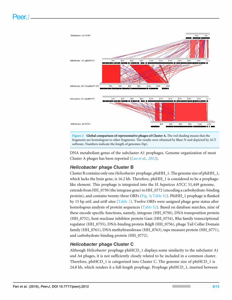
Figure 2 Global comparison of representative phages of Cluster A. The red shading means that thefragments are homologous to other fragments. The results were obtained by Blast-N and depicted by ACTsoftware. Numbers indicate the length of genomes (bp).
DNA metabolism genes of the subcluster A1 prophages. Genome organization of mostCluster A phages has been reported (Luo et al., 2012).
Helicobacter phage Cluster BCluster B contains only oneHelicobacter prophage, phiHH_1. The genome size of phiHH_1,which lacks the lysin gene, is 16.2 kb. Therefore, phiHH_1 is considered to be a prophage-like element. This prophage is integrated into the H. hepaticus ATCC 51,449 genome,extends fromHH_0750 (the integrase gene) toHH_0772 (encoding a carbohydrate-bindingprotein), and contains twenty-three ORFs (Fig. 3; Table S2). PhiHH_1 prophage is flankedby 15 bp attL and attR sites (Table 1). Twelve ORFs were assigned phage gene status afterhomologous analysis of protein sequences (Table S2). Based on database searches, nine ofthese encode specific functions, namely, integrase (HH_0750), DNA transposition protein(HH_0752), host-nuclease inhibitor protein Gam (HH_0754), Rha family transcriptionalregulator (HH_0755), DNA-binding protein RdgB (HH_0756), phage Tail Collar Domainfamily (HH_0761), DNAmethyltransferase (HH_0763), tape measure protein (HH_0771),and carbohydrate-binding protein (HH_0772).
Helicobacter phage Cluster CAlthough Helicobacter prophage phiHCD_1 displays some similarity to the subcluster A1and A4 phages, it is not sufficiently closely related to be included in a common cluster.Therefore, phiHCD_1 is categorized into Cluster C. The genome size of phiHCD_1 is24.8 kb, which renders it a full-length prophage. Prophage phiHCD_1, inserted between
Fan et al. (2016), PeerJ, DOI 10.7717/peerj.2012 6/13
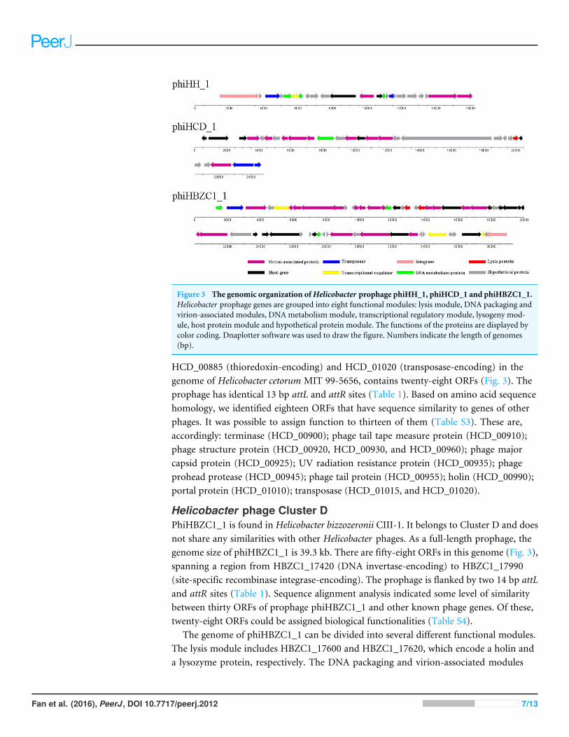
Figure 3 The genomic organization ofHelicobacter prophage phiHH_1, phiHCD_1 and phiHBZC1_1.Helicobacter prophage genes are grouped into eight functional modules: lysis module, DNA packaging andvirion-associated modules, DNA metabolism module, transcriptional regulatory module, lysogeny mod-ule, host protein module and hypothetical protein module. The functions of the proteins are displayed bycolor coding. Dnaplotter software was used to draw the figure. Numbers indicate the length of genomes(bp).
HCD_00885 (thioredoxin-encoding) and HCD_01020 (transposase-encoding) in thegenome of Helicobacter cetorum MIT 99-5656, contains twenty-eight ORFs (Fig. 3). Theprophage has identical 13 bp attL and attR sites (Table 1). Based on amino acid sequencehomology, we identified eighteen ORFs that have sequence similarity to genes of otherphages. It was possible to assign function to thirteen of them (Table S3). These are,accordingly: terminase (HCD_00900); phage tail tape measure protein (HCD_00910);phage structure protein (HCD_00920, HCD_00930, and HCD_00960); phage majorcapsid protein (HCD_00925); UV radiation resistance protein (HCD_00935); phageprohead protease (HCD_00945); phage tail protein (HCD_00955); holin (HCD_00990);portal protein (HCD_01010); transposase (HCD_01015, and HCD_01020).
Helicobacter phage Cluster DPhiHBZC1_1 is found in Helicobacter bizzozeronii CIII-1. It belongs to Cluster D and doesnot share any similarities with other Helicobacter phages. As a full-length prophage, thegenome size of phiHBZC1_1 is 39.3 kb. There are fifty-eight ORFs in this genome (Fig. 3),spanning a region from HBZC1_17420 (DNA invertase-encoding) to HBZC1_17990(site-specific recombinase integrase-encoding). The prophage is flanked by two 14 bp attLand attR sites (Table 1). Sequence alignment analysis indicated some level of similaritybetween thirty ORFs of prophage phiHBZC1_1 and other known phage genes. Of these,twenty-eight ORFs could be assigned biological functionalities (Table S4).
The genome of phiHBZC1_1 can be divided into several different functional modules.The lysis module includes HBZC1_17600 and HBZC1_17620, which encode a holin anda lysozyme protein, respectively. The DNA packaging and virion-associated modules
Fan et al. (2016), PeerJ, DOI 10.7717/peerj.2012 7/13

consist of HBZC1_17440, coding for a phage terminase large subunit; HBZC1_17470,encoding a phage tail protein; phage tail tape measure proteins-encoding HBZC1_17480,HBZC1_17490, and HBZC1_17500; phage tail proteins-encoding HBZC1_17530,HBZC1_17540, HBZC1_17550, HBZC1_17630, HBZC1_17640, and HBZC1_17660;HBZC1_17560, encoding a phage tail sheath-like protein; HBZC1_17670, encoding aphage baseplate protein; capsid proteins-encoding HBZC1_17740 and HBZC1_17750;HBZC1_17860, encoding a portal protein; HBZC1_17880, encoding a phage terminaselarge subunit; and HBZC1_17900, encoding a phage baseplate assembly protein V. TheDNA metabolism module comprises of three genes (HBZC1_17420, HBZC1_17570,and HBZC1_17830), whose predicted protein products are phage DNA invertase, DNAmethyltransferase, and DNA polymerase, respectively. The transcriptional regulatorymodule is composed of HBZC1_17460 (encoding a phage late control D family protein),HBZC1_17930 (coding for the repressor LexA), and HBZC1_17970 (encoding a YcfAfamily protein). The lysogeny module appears to be limited to HBZC1_17990, whosepredicted protein product is a phage integrase.
Putative antibiotic resistance genes and virulence factors associatedwith Helicobacter prophagesExcept for phiHBZC1_1, none of the other characterized Helicobacter prophages containknown antibiotic resistance genes. The protein encoded by HBZC1_17700 shows highsimilarity to multidrug resistance protein D (emrD) of Salmonella enterica subsp. entericaserovar Infantis (Table 2). Multidrug resistance protein D belonging to the major facilitatorsuperfamily facilitates the transport of a variety of antibiotics (Shaheen et al., 2015).
A range of phage-encoded virulence genes was identified within the Helicobacterprophage sequences (Table 2). A DNA methyltransferase-encoding gene was identifiedin most of the analyzed Helicobacter prophages. DNA methyltransferase is thought tocontribute to the specificity of bacterium-host interactions or H. pylori virulence (Vitkuteet al., 2001). Furuta and colleagues (2015) found that DNA methyltransferase genes arerapidly evolving in H. pylori genomes, which facilitates H. pylori adaptation to a newhost. A protein encoded by phiNY40_1 (NY40_0553) displayed 23% identity with aserine/threonine kinase of Thiorhodococcus drewsii. Phosphorylation of proteins usuallyoccurs during interactions between bacterial cells and host cells and plays a role inbacterial pathogenesis (Cozzone, 2005). Serine/threonine kinases are considered to affectcell survival pathways and contribute to H. pylori pathogenesis (King & Obonyo, 2015). Aputative glycosyltransferase is encoded by phiHCD_1. Glycosyltransferases are involvedin biosynthesis of LPS (Luke et al., 2010) that can promote proliferation of gastric cancercells (Tomoda, Kamiya & Suzuki, 2015). An antitoxin component RelB of the addictiontoxin-antitoxin (TA) module system RelBE was identified in phiHBZC1_1. The proteinplays a role in cell survival (Park, Son & Lee, 2013).
CONCLUSIONSIn brief, we present here sixteen Helicobacter prophages. Eight of them were identified forthe first time after mining the sequencedHelicobacter sp. genomes, and the other eight had
Fan et al. (2016), PeerJ, DOI 10.7717/peerj.2012 8/13

Table 2 Putative virulence elements and antibiotic resistance genes inHelicobacter prophages.
Prophage Gene (Accession number) Putative virulence element Querycoverage
E-value Identity
KHP40 ORF24 (BAM34796.1) DNA methyltransferase (Helicobacter pylori) 100% 8e–41 91%KHP30 ORF23 (BAM34765.1) DNA methyltransferase (Helicobacter pylori) 100% 1e–40 92%1961P gp26 (AFC61925.1) DNA methyltransferase (Helicobacter pylori) 100% 6e–44 96%Cuz20 HPCU_00990 (ADO03382.1) DNA methyltransferase (Helicobacter pylori) 100% 2e–42 100%India7 HPIN_06120 (ADU80418.1) DNA methyltransferase (Helicobacter pylori) 100% 2e–47 100%Gambia94/24 HPGAM_01040 (ADU81058.1) DNA methyltransferase (Helicobacter pylori) 100% 1e–45 100%phiK747_1 K747_07685 (AGL67312.1) DNA methyltransferase (Helicobacter pylori) 100% 2e–41 92%phiK749_1 K749_02305 (AGL67850.1) DNA methyltransferase (Helicobacter pylori) 100% 2e–41 92%phiK750_1 K750_05880 (AGL70120.1) DNA methyltransferase (Helicobacter pylori) 95% 4e–37 87%phiK748_1 K748_00765 (AGR63209.1) DNA methyltransferase (Helicobacter pylori) 100% 2e–41 92%phiNY40_1 NY40_0558 (BAO97577.1) Type II methylase (Helicobacter pylori) 100% 0.0 100%phiNY40_1 NY40_0553 (BAO97572.1) Serine/threonine protein kinase (Thiorhodococcus
drewsii)99% 6e–56 23%
phiNY40_1 NY40_0545 (BAO97564.1) DNA methyltransferase (Helicobacter pylori) 100% 2e–43 100%phiHH_1 HH_0763 (AAP77360.1) DNA methyltransferase (Helicobacter sp.MIT 03-1614) 84% 3e–31 97%Sheeba Hac_1629 (CAK00337.1) DNA methyltransferase (Helicobacter pylori) 97% 1e–29 69%phiHCD_1 HCD_00890 (AFI05210.1) Glycosyltransferase (Neisseria meningitidis) 71% 3e–71 17%phiHBZC1_1 HBZC1_17570 (CCB80743.1) DNA methyltransferase (Helicobacter sp.MIT 03-1614) 42% 4e–08 55%phiHBZC1_1 HBZC1_17680 (CCB80754.1) Type VI secretion protein (Herbaspirillum sp. B39) 64% 2.4 28%phiHBZC1_1 HBZC1_17710 (CCB80757.1) DNA methyltransferase (Oceanospirillum beijerinckii) 89% 3e–66 39%phiHBZC1_1 HBZC1_17820 (CCB80754) Addiction module antitoxin RelB (Burkholderia
cenocepacia)91% 2e–24 53%
phiHBZC1_1 HBZC1_17770 (CCB80763.1) DNA adenine methylase (Campylobacter jejuni subsp.jejuni 2008-979)
89% 6e–19 39%
phiHBZC1_1 HBZC1_17780 (CCB80764.1) DNA adenine methylase (Desulfosporosinus acidiphilus) 87% 2e–25 44%phiHBZC1_1 HBZC1_17700 (CCB80756.1) Multidrug resistance protein D (Salmonella enterica
subsp. enterica serovar Infantis)40% 2e–06 31%
been reported in published literature. Based on comparative genomic analyses, the sixteenphages were sorted into four clusters, Clusters A–D, respectively. Cluster A was furtherdivided into four subclusters, subclusters A1–A4. Different subclusters displayed similarityto each other. Subcluster A1 phages are full-length prophages. Subcluster A2, A3 and A4phages are remnant prophage-like elements. The genomes and genetic information of theCluster B, C and D phages were analyzed. Interestingly, several genes encoding antibioticresistance proteins and virulence factors were found within various prophage genomes.These results highlight an important issue, which needs to be resolved before proceedingwith phage therapy for treatment of H. pylori infections. To our knowledge, this is the firstsystematic analysis ofHelicobacter prophages.Withmore forthcomingHelicobacter genomesequences, more Helicobacter prophages will be identified, and the role of prophages inevolution, adaptations and physiology of Helicobacter sp. will be clarified.
Fan et al. (2016), PeerJ, DOI 10.7717/peerj.2012 9/13
ADDITIONAL INFORMATION AND DECLARATIONS
FundingThis work was supported by Shandong Excellent Young Scientist Award Fund(BS2014YY031), Foundation of University of Jinan (XBS1519, XKY1324), NationalNatural Science Foundation of China (31100088, 31300045, 51208290, 31372356),Shandong province science and technology development plan (2013GSF12006), A Projectof Shandong Province Higher Educational Science and Technology Program (YE13), OpenFoundation of Xinjiang Production & Construction Corps Key Laboratory of Protectionand Utilization of Biological Resources in Tarim Basinand (BYBR1405, BRYB1501). Thefunders had no role in study design, data collection and analysis, decision to publish, orpreparation of the manuscript.
Grant DisclosuresThe following grant information was disclosed by the authors:Shandong Excellent Young Scientist Award Fund: BS2014YY031.Foundation of University of Jinan: XBS1519, XKY1324.National Natural Science Foundation of China: 31100088, 31300045, 51208290, 31372356.Shandong province science and technology development plan: 2013GSF12006.Project of Shandong Province Higher Educational Science and Technology Program: YE13.Open Foundation of Xinjiang Production & Construction Corps Key Laboratory ofProtection and Utilization of Biological Resources in Tarim Basinand: BYBR1405,BRYB1501.
Competing InterestsThe authors declare there are no competing interests.
Author Contributions• Xiangyu Fan conceived and designed the experiments, performed the experiments,analyzed the data, wrote the paper, prepared figures and/or tables.
• Yumei Li and Rong He reviewed drafts of the paper.• Qiang Li and Wenxing He contributed reagents/materials/analysis tools, reviewed draftsof the paper.
Data AvailabilityThe following information was supplied regarding data availability:
Raw data were provided as Supplemental Information.
Supplemental InformationSupplemental information for this article can be found online at http://dx.doi.org/10.7717/peerj.2012#supplemental-information.
Fan et al. (2016), PeerJ, DOI 10.7717/peerj.2012 10/13

REFERENCESAbdel-HaliemME, Askora A. 2013. Isolation and characterization of bacteriophages
of Helicobacter pylori isolated from Egypt. Future Virology 8:821–826DOI 10.2217/fvl.13.58.
Ariff A, Wise MJ, Kahler CM, Tay CY, Peters F, Perkins TT, Chang BJ. 2015. NovelMoraxella catarrhalis prophages display hyperconserved non-structural genes despitetheir genomic diversity. BMC Genomics 16:860 DOI 10.1186/s12864-015-2104-1.
Arnold IC, Zigova Z, HoldenM, Lawley TD, Rad R, Dougan G, Falkow S, Bentley SD,Müller A. 2011. Comparative whole genome sequence analysis of the carcinogenicbacterial model pathogen Helicobacter felis. Genome Biology and Evolution 3:302–308DOI 10.1093/gbe/evr022.
Canchaya C, Fournous G, BrüssowH. 2004. The impact of prophages on bacterial chro-mosomes.Molecular Microbiology 53:9–18 DOI 10.1111/j.1365-2958.2004.04113.x.
Cozzone AJ. 2005. Role of protein phosphorylation on serine/threonine and tyrosinein the virulence of bacterial pathogens. Journal of Molecular Microbiology andBiotechnology 9:198–213 DOI 10.1159/000089648.
Delcher AL, Bratke KA, Powers EC, Salzberg SL. 2007. Identifying bacterialgenes and endosymbiont DNA with Glimmer. Bioinformatics 23:673–679DOI 10.1093/bioinformatics/btm009.
Eppinger M, Baar C, Linz B, Raddatz G, Lanz C, Keller H, Morelli G, Gressmann H,AchtmanM, Schuster SC. 2006.Who ate whom? Adaptive Helicobacter genomicchanges that accompanied a host jump from early humans to large felines. PLoSGenetics 2:e120 DOI 10.1371/journal.pgen.0020120.
Fan X, Abd Alla AAE, Xie J. 2015. Distribution and function of prophage phiRv1 andphiRv2 amongMycobacterium tuberculosis complex. Journal of Biomolecular Structureand Dynamics 34:233–238 DOI 10.1080/07391102.2015.1022602.
Fan X, LiW, Zheng F, Xie J. 2013. Bacteriophage inspired antibiotics discovery againstinfection involved biofilm. Critical ReviewsTM in Eukaryotic Gene Expression23:317–326 DOI 10.1615/CritRevEukaryotGeneExpr.2013007717.
Fan X, Xie L, Li W, Xie J. 2014. Prophage-like elements present inMycobacteriumgenomes. BMC Genomics 15:243 DOI 10.1186/1471-2164-15-243.
Furuta Y, KonnoM, Osaki T, Yonezawa H, Ishige T, Imai M, Shiwa Y, Shibata-HattaM, Kanesaki Y, Yoshikawa H, Kamiya S, Kobayashi I. 2015.Microevolutionof virulence-related genes in Helicobacter pylori familial infection. PLoS ONE10:e0127197 DOI 10.1371/journal.pone.0127197.
Kearse M, Moir R,Wilson A, Stones-Havas S, CheungM, Sturrock S, Buxton S, CooperA, Markowitz S, Duran C. 2012. Geneious Basic: an integrated and extendabledesktop software platform for the organization and analysis of sequence data.Bioinformatics 28:1647–1649 DOI 10.1093/bioinformatics/bts199.
King CC, ObonyoM. 2015. Helicobacter pylorimodulates host cell survival regulationthrough the serine-threonine kinase, 3-phosphoinositide dependent kinase 1 (PDK-1). BMCMicrobiology 15:222 DOI 10.1186/s12866-015-0543-0.
Fan et al. (2016), PeerJ, DOI 10.7717/peerj.2012 11/13

Lang AS, Zhaxybayeva O, Beatty JT. 2012. Gene transfer agents: phage-like elements ofgenetic exchange. Nature Reviews Microbiology 10:472–482DOI 10.1038/nrmicro2802.
Lehours P, Vale FF, Bjursell MK, Melefors O, Advani R, Glavas S, Guegueniat J,Gontier E, Lacomme S, Matos AA. 2011. Genome sequencing reveals a phage inHelicobacter pylori.MBio 2:e00239–e00211 DOI 10.1128/mBio.00239-11.
Lugli GA, Milani C, Turroni F, Tremblay D, Ferrario C, Mancabelli L, Duranti S, WardDV, Ossiprandi MC, Moineau S. 2016. Prophages of the genus Bifidobacterium asmodulating agents of the infant gut microbiota. Environmental Microbiology Epubahead of print Dec 2 2015 DOI 10.1111/1462-2920.13154.
Luke NR, Sauberan SL, Russo TA, Beanan JM, Olson R, Loehfelm TW, Cox AD, StMichael F, Vinogradov EV, Campagnari AA. 2010. Identification and characteriza-tion of a glycosyltransferase involved in Acinetobacter baumannii lipopolysaccharidecore biosynthesis. Infection and Immunity 78:2017–2023 DOI 10.1128/IAI.00016-10.
Luo C-H, Chiou P-Y, Yang C-Y, Lin N-T. 2012. Genome, integration, and transductionof a novel temperate phage of Helicobacter pylori. Journal of Virology 86:8781–8792DOI 10.1128/JVI.00446-12.
O’rourke J, GrehanM, Lee A. 2001. Non-pylori Helicobacter species in humans. Gut49:601–606 DOI 10.1136/gut.49.5.601.
Park SJ, SonWS, Lee BJ. 2013. Structural overview of toxin-antitoxin systems in infec-tious bacteria: a target for developing antimicrobial agents. Biochimica et BiophysicaACTA 1834:1155–1167 DOI 10.1016/j.bbapap.2013.02.027.
Peek RM, Blaser MJ. 2002. Helicobacter pylori and gastrointestinal tract adenocarcino-mas. Nature Reviews Cancer 2:28–37 DOI 10.1038/nrc703.
Penadés JR, Chen J, Quiles-Puchalt N, Carpena N, Novick RP. 2015. Bacteriophage-mediated spread of bacterial virulence genes. Current Opinion in Microbiology23:171–178 DOI 10.1016/j.mib.2014.11.019.
Shaheen A, Ismat F, Iqbal M, Haque A, De Zorzi R, Mirza O,Walz T, RahmanM.2015. Characterization of putative multidrug resistance transporters of the majorfacilitator-superfamily expressed in Salmonella Typhi. Journal of Infection andChemotherapy 21:357–362 DOI 10.1016/j.jiac.2015.01.002.
Tang F, Bossers A, Harders F, Lu C, Smith H. 2013. Comparative genomic analysis oftwelve Streptococcus suis (pro) phages. Genomics 101:336–344DOI 10.1016/j.ygeno.2013.04.005.
Tomoda A, Kamiya S, Suzuki H. 2015. Helicobacter pylori and Pathogenesis. BioMedResearch International 2015:304768.
Uchiyama J, Takeuchi H, Kato S-I, Gamoh K, Takemura-Uchiyama I, UjiharaT, Daibata M, Matsuzaki S. 2013. Characterization of Helicobacter pylori bac-teriophage KHP30. Applied and Environmental Microbiology 79:3176–3184DOI 10.1128/AEM.03530-12.
Uchiyama J, Takeuchi H, Kato S-I, Takemura-Uchiyama I, Ujihara T, DaibataM, Matsuzaki S. 2012. Complete genome sequences of two Helicobacter pylori
Fan et al. (2016), PeerJ, DOI 10.7717/peerj.2012 12/13

bacteriophages isolated from Japanese patients. Journal of Virology 86:11400–11401DOI 10.1128/JVI.01767-12.
Uemura N, Okamoto S, Yamamoto S, Matsumura N, Yamaguchi S, YamakidoM,Taniyama K, Sasaki N, Schlemper RJ. 2001. Helicobacter pylori infection and thedevelopment of gastric cancer. New England Journal of Medicine 345:784–789DOI 10.1056/NEJMoa001999.
Vale F, Vadivelu J, OleastroM, Breurec S, Engstrand L, Perets T, Mégraud F, LehoursP. 2015. Dormant phages of Helicobacter pylori reveal distinct populations in Europe.Scientific Reports 5:14333 DOI 10.1038/srep14333.
Vannucci FA, Kelley MR, Gebhart CJ. 2013. Comparative genome sequencing identifiesa prophage-associated genomic island linked to host adaptation of Lawsoniaintracellularis infections. Veterinary Research 44:49 DOI 10.1186/1297-9716-44-49.
Varani AM,Monteiro-Vitorello CB, Nakaya HI, Van Sluys M-A. 2013. The role ofprophage in plant-pathogenic bacteria. Annual Review of Phytopathology 51:429–451DOI 10.1146/annurev-phyto-081211-173010.
VenturaM, Turroni F, Lima-Mendez G, Foroni E, Zomer A, Duranti S, Giubellini V,Bottacini F, Horvath P, Barrangou R. 2009. Comparative analyses of prophage-likeelements present in bifidobacterial genomes. Applied and Environmental Microbiology75:6929–6936 DOI 10.1128/AEM.01112-09.
VenturaM, Zomer A, Canchaya C, O’Connell-MotherwayM, Kuipers O, TurroniF, Ribbera A, Foroni E, Buist G,Wegmann U. 2007. Comparative analyses ofprophage-like elements present in two Lactococcus lactis strains. Applied andEnvironmental Microbiology 73:7771–7780 DOI 10.1128/AEM.01273-07.
Vitkute J, Stankevicius K, Tamulaitiene G, Maneliene Z, Timinskas A, Berg DE,Janulaitis A. 2001. Specificities of eleven different DNA methyltransferasesof Helicobacter pylori strain 26695. Journal of Bacteriology 183:443–450DOI 10.1128/JB.183.2.443-450.2001.
Fan et al. (2016), PeerJ, DOI 10.7717/peerj.2012 13/13