LECTURE 12 th EARLY AMBULATION AND GAIT TRAINING FOR PATIENT WITH LOWER EXTREMITY BURN.
Upload
janice-thorntonCategory
view
215download
1
Common Pediatric Disorders of the Lower
Extremity Affecting Gait
Gregory A. Schmale, MDChildren’s Hospital and Regional Medical Center
5/01/06

Objectives:Objectives:
1. DescribeDescribe the commonly seen pediatric disorders involving gait, children's feet, and children's
legs, including problems in normal normal developmentdevelopment (and the ages at which these problems are commonly seen).

Objectives:Objectives:
2. Discuss the evaluationevaluation of common pediatric foot, gait, and leg disorders.
3. Describe their optimal management.optimal management.

Approach • Learn the range of normalLearn the range of normal
– It’s huge– “Normal” changes with growth and
development– Before saying something is “normal”,
rule out the pathologies
• Know the common pathologiescommon pathologies
““The eye sees what the mind knows”The eye sees what the mind knows”

Common and often benign orthopaedic concerns
• In-toeing• Out-toeing• Bowed legs• Knock-knees• Flat feet

Pathologies
• Cerebral Palsy• Hip dysplasia• Legg-Calve-Perthes’s disease• Slipped Capital Femoral Epiphysis• Clubfoot

Systematic approach - Where’s the source?
• Hip joint• Thigh (femur)• Knee joint• Leg (tibia)• Ankle joint• Foot (tarsals and metatarsals)X
X

Group pathologies by age
• Newborns and infants (< 1 yr)• Toddlers (1-3 yr)• Older children (4-10 yr)• Pre-teens and teens (> 10 yrs)

Is in-toeing a problem?
• Not painful in and of itself• Not associated with early arthritis• Can be associated with knee pain
and patellofemoral problems• May be a cosmetic problem
Why does thisthis patient in-toe?

History
• What is the specific concern?• Who is concerned?• When does it manifest?• Duration? • Improving or worsening?
Evaluation
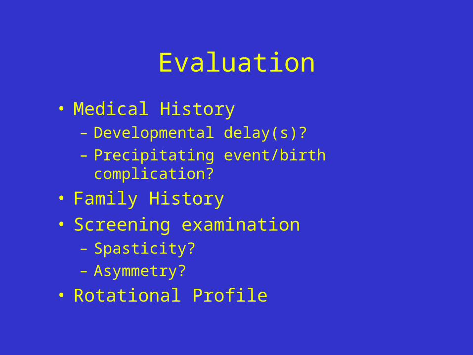
• Medical History– Developmental delay(s)?– Precipitating event/birth complication?
• Family History• Screening examination
– Spasticity?– Asymmetry?
• Rotational Profile
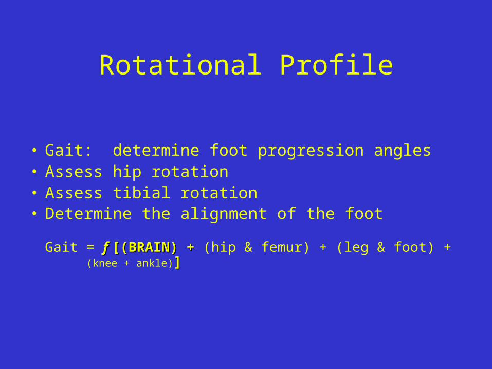
Rotational Profile
• Gait: determine foot progression angles• Assess hip rotation• Assess tibial rotation• Determine the alignment of the foot
Gait = f f [(BRAIN) + [(BRAIN) + (hip & femur) + (leg & foot) + (knee + ankle)]]

Rotational Profile• Gait: foot progression angles

Rotational Profile• Range of normal: foot progression angles

Structural toeing and bowingStructural toeing and bowing
• Terminology:Terminology:
– ““Normal” - within two standard deviations of Normal” - within two standard deviations of the meanthe mean
– Version: the normal twist to a boneVersion: the normal twist to a bone– Torsion: abnormal twist to a boneTorsion: abnormal twist to a bone– Medial = internalMedial = internal– Lateral = externalLateral = external

Rotational Profile• Gait: foot progression angles

Rotational Profile• Gait: foot progression angles

Rotational Profile
• Gait: determine foot progression angles• Assess hip rotationAssess hip rotation• Assess tibial rotation• Determine the alignment of the foot
Where is the source???Where is the source???

Assessing hip rotation
MedialRotationHip
LateralRotationHip

Assessing hip rotation
QuickTime™ and aMotion JPEG OpenDML decompressor
are needed to see this picture.

Normals: medial femoral rotation

Normals: lateral femoral rotation

Is the hip rotation normal?Is the hip rotation normal?
• Within two standard deviations of the mean?Within two standard deviations of the mean?• Symmetric?Symmetric?• Painless?Painless?• Without spasticity?Without spasticity?
What What is the cause of the increased is the cause of the increased medial (or lateral) rotation?medial (or lateral) rotation?

Causes of excess rotationCauses of excess rotation• Soft tissues vs. bony anatomySoft tissues vs. bony anatomy
• Hip joint - soft tissue contracturesHip joint - soft tissue contractures– Newborns have an posterior capsular Newborns have an posterior capsular
contracture, producing excessive lateral contracture, producing excessive lateral rotation of the hipsrotation of the hips
• Femoral antetorsion - bony anatomyFemoral antetorsion - bony anatomy– produces excessive medial rotation at the produces excessive medial rotation at the
hiphip

What is femoral anteversion?
Leftfoot
LeftfootLeft
foot
Anteversion Excessive anteversionequalsantetorsion
Femoral antetorsion produces intoeingFemoral antetorsion produces intoeing

Femoral antetorsion
• Usually 3-5 yo girls• Sits in the “W”• “Kissing patellae”• “Egg-beater” run• Severe if > 90°• Resolves with growth -
no association with osteoarthritis

Femoral antetorsion

Rotational Profile
• Gait: determine foot progression angles• Assess hip rotation• Assess tibial rotationAssess tibial rotation• Determine the alignment of the foot
Where is the source???

Tibia• TorsionTorsion
– Tibial torsion can lead to intoeing:Tibial torsion can lead to intoeing:
Internal or medial tibial torsion is Internal or medial tibial torsion is a twist to the leg, pointing the toe a twist to the leg, pointing the toe inwardsinwards

Assessing tibial torsion:
• Thigh-foot angle• Transmalleolar axis
•Determine axes
•Measure angles

Assessing tibial rotation
L TFA R TFA

Assessing tibial rotation
QuickTime™ and aMotion JPEG OpenDML decompressor
are needed to see this picture.

Assessing tibial rotation
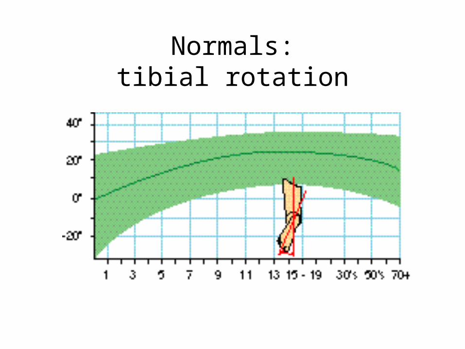
Normals:tibial rotation

Medial tibial torsion

FootFoot
• Metatarsus adductus curves the foot Metatarsus adductus curves the foot inwardsinwards
• Searching great toe pulls the foot Searching great toe pulls the foot inwardsinwards
• Flatfoot may produce out-toeing from Flatfoot may produce out-toeing from ““wringing-outwringing-out” of the foot:” of the foot:
Supinated forefoot with valgus Supinated forefoot with valgus heelheel

Assessing alignment of the foot
• Shape of the foot• Heel-bisector angle

Metatarsus Adductus• Majority are flexible• Adductus resolves by 3-4 yrs• 10% stiff and may benefit from casting

Assessing foot alignment
PrettyMuchNormal

Toeing and bowing:Toeing and bowing:Determining the source
• Excessive medial rotation of hips?
Does he have it? NONO on antetorsion, but YES on excessive medial rotationYES on excessive medial rotation
• Internally rotated thigh-foot angle = internal tibial torsion? NoNo
• Curved foot = metatarsus adductus? NoNo

In SummaryIn Summary
• Femoral antetorsion produces excessive medial Femoral antetorsion produces excessive medial rotation at the hip which leads to in-toeingrotation at the hip which leads to in-toeing
• Medial tibial torsion is a twist to the leg, Medial tibial torsion is a twist to the leg, pointing the foot inwardspointing the foot inwards
• Metatarsus adductus curves the foot inwardsMetatarsus adductus curves the foot inwards• A searching or abducted great toe produces in-A searching or abducted great toe produces in-
toeingtoeing

A five year old girl presents with A five year old girl presents with knock-knees and intoeing. knock-knees and intoeing.
You should obtain a rotational profile You should obtain a rotational profile and…and…
1.1. refer to orthopaedics for bracing or surgeryrefer to orthopaedics for bracing or surgery
2.2. have the child put her shoes on the opposite feet and have the child put her shoes on the opposite feet and recheck her in a yearrecheck her in a year
3.3. just recheck her in a yearjust recheck her in a year
4.4. obtain an AP pelvis radiograph and full length lower obtain an AP pelvis radiograph and full length lower extremity films to look for hip dysplasiaextremity films to look for hip dysplasia

How to treat intoeing?
• Shoe wedges? No.
• Twister cables? No.
• Observation? YesYes.

Pathologies to considerPathologies to consider“Why is there an abnormal range of “Why is there an abnormal range of
motion of the motion of the hiphip?”?”
• Infants and toddlersInfants and toddlers– Hip dysplasiaHip dysplasia
– Neuromuscular diseaseNeuromuscular disease -Cerebral palsy• ToddlersToddlers
– Legg-Calve-Perthes disease• Pre-teensPre-teens
– Legg-Calve-Perthes disease– Slipped Capital femoral epiphysis

QuickTime™ and aCinepak decompressor
are needed to see this picture.

The most likely diagnosis is…The most likely diagnosis is…
1.1. cerebral palsycerebral palsy
2.2. arthrogryposisarthrogryposis
3.3. Perthe’s diseasePerthe’s disease
4.4. septic arthritis of the hipseptic arthritis of the hip
5.5. hip dysplasiahip dysplasia
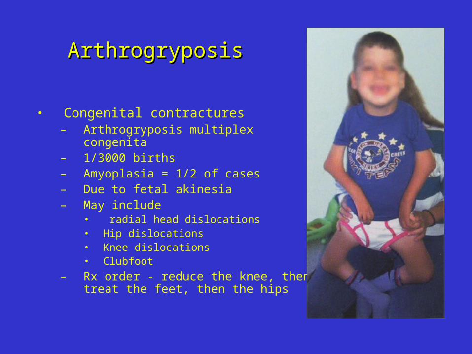
ArthrogryposisArthrogryposis
• Congenital contractures– Arthrogryposis multiplex congenita– 1/3000 births– Amyoplasia = 1/2 of cases– Due to fetal akinesia– May include
• radial head dislocations• Hip dislocations• Knee dislocations• Clubfoot
– Rx order - reduce the knee, then treat the feet, then the hips

ArthrogryposisArthrogryposis
Amyoplasia– Classic arthrogryposis– Muscle replaced by fibrous
tissue– Multiple congenital contractures– 60% with all limbs affected,
» Lower only in 25%» Upper only in 15%
– Normal IQ– Surgery changes the range of
the arc of motion, not the total arc itself

The most likely diagnosis is…The most likely diagnosis is…
1.1. cerebral palsycerebral palsy
2.2. arthrogryposisarthrogryposis
3.3. Perthe’s diseasePerthe’s disease
4.4. septic arthritis of the hipseptic arthritis of the hip
5.5. hip dysplasiahip dysplasia


The most likely diagnosis is…The most likely diagnosis is…
1.1. cerebral palsycerebral palsy
2.2. arthrogryposisarthrogryposis
3.3. Perthe’s diseasePerthe’s disease
4.4. septic arthritis of the hipseptic arthritis of the hip
5.5. hip dysplasiahip dysplasia

Bilateral hip dislocations

Developmental dysplasia of the hip (DDH)
• Incidence – dislocation 1:1000– neonatal hip instability 1:100
• Increased risks for first-born, girls, breech positioning, family history
• L>R

DDH detection• Newborn nursery exam
– Galiazzi test– Ortolani test– Barlow test– Good up to 2-3 mos of age
• Loss of abduction, pistoning
• Pavlik harness for instability or dislocated hip

DDH detection
• Ultrasound (dedicated center)– Better at > 2 wks of age– Dynamic exam
• Radiography– Gold standard– Best after 6-8 weeks of age

Rx for dysplasia -REFER• PavlikPavlik for both dysplastic and
dislocated hips– Never exceed about four weeks of Pavlik
treatment for a persistently dislocated hip
• Unstable hips deserve a referral to Unstable hips deserve a referral to orthopaedicsorthopaedics
• Abduction orthoses may help correct hip dysplasia in the older child

Hip dysplasia
• Early treatment enables quick resolution
• Delayed treatment risks a poor result/multiple surgeries
• Over-treatment is generally benign for the located hip

QuickTime™ and aMotion JPEG OpenDML decompressor
are needed to see this picture.

R hip after OR, fem short, pelvic osteotomy

Cerebral palsyCerebral palsy
• Mild developmental delays?Mild developmental delays?• Mild spasticity or increased tone?Mild spasticity or increased tone?• Asymmetry of motion, tone, reflexes?Asymmetry of motion, tone, reflexes?
You may be the first to make the You may be the first to make the diagnosisdiagnosis

Perthes ds• Peak age of onset 3-8yr• Spontaneous osteonecrosis of the femoral
head• Follow with serial radiographs• Prognosis depends on age of onset / severity
– < 6 yrs at onset, less than whole-head involvement do better
• Rx- decrease synovitis and weight bearing

Perthes ds

Perthes ds

Slipped capital femoral epiphysis
• Peak incidence in pre-teens, 50% obese (50% not!)
• Anterior thigh or knee pain• Bilateral in cases of endocrinopathy or renal ds• Dx - AP and frog pelvis * radiograph• If present, immediate wheel chair and referral

Slipped capital femoral epiphysis

Slipped capital femoral epiphysis

Knee angular deformities
• Genu varum - bowing
• Genu valgum - knock-knees
What’s normal?What’s normal?

•Maximum varus at birth•Maximum valgus > 10°, ages 3 - 4 yrs•At maturity, mean is ~ 6° anatomic valgus
Physiologic genu valgum

Bowing or genu varum
• Physiologic bowing• Pathologic bowing
– Rickets– Tibia vara– Skeletal dysplasia

Apparent bowing

Vit-D deficient/resistant rickets

Bowing of tibia vara

Knock- knees or genu valgum
• Physiologic
• Pathologic
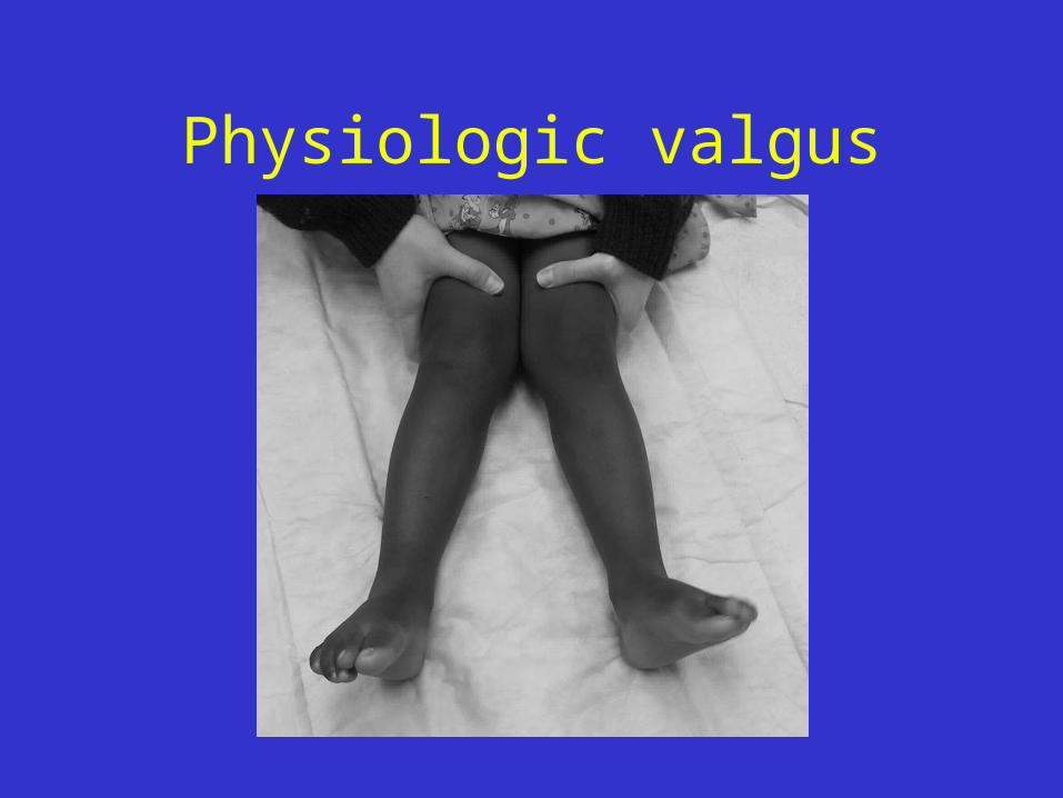
Physiologic valgus

•Maximum varus at birth•Maximum valgus > 10°, ages 3 - 4 yrs•At maturity, mean is ~ 6° anatomic valgus
Physiologic genu valgum

Knock- knees• Pathologic genu valgum
– Rickets - later onset such as with renal osteodystrophy, because the disease is active when knock knees are the norm
– Skeletal dysplasias• Diastrophic dysplasia• Morquio’s syndrome• Ellis-van Creveld or chondroectodermal dysplasia• Spondyloepiphyseal and multiple epiphyseal dysplasias

Pathologies to consider - legleg
•AngulationAngulation or bowing of the tibia–Very unusual!Very unusual!
•Antero-lateral Antero-lateral ?neurofibromatosis??neurofibromatosis?
•Postero-medial Postero-medial ?leg length difference??leg length difference?
•Antero-medial Antero-medial ?fibular deficiency??fibular deficiency?

Pathologies to consider: footfoot
Flatfoot
• All infants have it• Most children have it• More than 15% of adults have it

Flexible flatfoot
• Often resolves with growth• Not affected by specific shoes, heel
cups, or UCBL inserts• Not correlated with disability in
military populations• May be protective against stress
fractures

More foot pathologies to consider
• Stiff or rigid metatarsus adductus • Clubfoot• Calcaneovalgus• Cavovarus foot

Clubfoot
• Incidence 1:1000• Talipes equinovarus• True congenital vs positional• Cavus, adductus, varus, equinus• If present, examine hips carefully!

Clubfoot treatment• Serial manipulations and casting• Begin first week of life, if possible• Perform weekly• 90% of routine clubfoot respond

Calcaneovalgus foot
• Most common foot deformity at birth
• Forefoot abducted, ankle dorsiflexed - foot lies on anterior leg
• Resolves spontaneously
• Associated with hip dysplasia

Cavovarus foot
• High arch = cavus• Heel in varus• Often rigid• Look to spinal cord or peripheral
nervous system

Out-toeing (Less commonly seen)
Causes:• External rotation contracture at the hip?• Lateral tibial torsion?• Flatfoot?• Little hope of improvement over time,
unlessunless it’s a result of flatfoot

Summary: Normal Development
• Femoral anteversion: 30° at birth, only 10° at maturity (= lateral rotation)– Femoral antetorsion improves over time
• Tibial version: 0° at birth, 15° externally rotated at maturity (= laterally rotation)– Medial tibial torsion improves over time
• Growth: lateral rotation of both femur and tibia– In-toeing decreases with growthIn-toeing decreases with growth

Summary
• Most toe-ing and bow-ing deformities are benign– Resolution may take many yearsmany years
• Use history and exam to rule-out the pathologic causes
• ReassureReassure for what appear to be non-pathologic but extreme cases– Check back for re-exam, 6-12 months
• Beware unilateral deformities and those associated with pain– Radiographs indicated

Who needs a referral for toeing and bowing?
• Over three years of age with documented progression of deformity
• Stiff metatarsus adductus• Bowing
– below the 5th percentile for height– marked asymmetry or lateral thrust with
ambulation
• Marked knock-knees or in-toeing in patients over 8 years of age

Who needs a referral?
1) A newborn with a hip click?2) A newborn with a hip clunk?3) A ten year old girl with marked out-
toeing on the side of groin pain?4) A newborn with flat feet?

References:• Herring, JA: Tachdjian’s Pediatric Orthpaedics,
WB Saunders, Philadelphia, 2002.• Staheli, LT: Fundamentals of Pediatric
Orthopedics, Raven Press, New York, 1992.• Staheli, LT: Practice of Pediatric Orthopedics,
Lippincott, 2002.• Tolo, VT: “In-toeing and Out-toeing,” Lovell
and Winter’s Pediatric Orthopaedics, 4th ed., Morrissey and Weinstein, eds., Lippincott-Raven, Philadelphia, 1996.
• Wenger, DA and M Rang: The Art and Practice of Pediatric Orthopaedics, Raven, New York, 1993.