Common Garlic ( Allium sativum ) has Potent Anti- Activity ... · Rajinder Kaur1#, Atul Tiwari1#,...
27
Common Garlic (Allium sativum) has Potent Anti-Bacillus anthracis Activity among Various Commonly Used Spices and Herbs Rajinder Kaur 1# , Atul Tiwari 1# , Manish Manish 2 , Indresh K Maurya 1 , Rakesh Bhatnagar 2 and Samer Singh 1 * Affiliations: 1. Department of Microbial Biotechnology, Panjab University, Chandigarh-160014, India 2. School of Biotechnology, Jawaharlal Nehru University, New Delhi-110067, India # Authors contributed equally to this work * Corresponding Author: Samer Singh, Department of Microbial Biotechnology, Panjab University, Chandigarh-160014, India; Phone: +91-172-2534092; E-mail: [email protected] Key Words: Anthrax, Garlic, Traditional medicine, Edible plants, Bacillus anthracis, Allium sativum peer-reviewed) is the author/funder. All rights reserved. No reuse allowed without permission. The copyright holder for this preprint (which was not . http://dx.doi.org/10.1101/162214 doi: bioRxiv preprint first posted online Jul. 11, 2017;
Transcript of Common Garlic ( Allium sativum ) has Potent Anti- Activity ... · Rajinder Kaur1#, Atul Tiwari1#,...
Common Garlic (Allium sativum) has Potent Anti-Bacillus anthracis Activity among Various Commonly Used Spices and Herbs
Rajinder Kaur1#, Atul Tiwari1#, Manish Manish2, Indresh K Maurya1, Rakesh Bhatnagar2 and
Samer Singh1*
Affiliations:
1. Department of Microbial Biotechnology, Panjab University, Chandigarh-160014, India
2. School of Biotechnology, Jawaharlal Nehru University, New Delhi-110067, India
# Authors contributed equally to this work
*Corresponding Author:
Samer Singh, Department of Microbial Biotechnology, Panjab University, Chandigarh-160014,
India; Phone: +91-172-2534092; E-mail: [email protected]
Key Words: Anthrax, Garlic, Traditional medicine, Edible plants, Bacillus anthracis, Allium
sativum
peer-reviewed) is the author/funder. All rights reserved. No reuse allowed without permission. The copyright holder for this preprint (which was not. http://dx.doi.org/10.1101/162214doi: bioRxiv preprint first posted online Jul. 11, 2017;
Abstract: Anthrax remains an important but relatively neglected disease of animals and humans. Its control in animals can be easily achieved by employing available vaccines. It is highly preventable in animals as well as humans. However, poverty, lack of information and appropriate medical support in the poor endemic areas of the world are some of the major obstacles in its control. We hypothesize that some of the commonly available plants in poor endemic areas which are used as food and also employed in traditional medicine may have potent anti-Bacillus anthracis activity in a form amenable to use in the endemic areas. Evaluation of the aqueous extracts made from various commonly available plants for anti-Bacillus anthracis activity indicated Garlic (Allium sativum) as the most promising candidate. It consistently inhibited the growth of Bacillus anthracis in agar-well diffusion assay and decreased the viable cell counts in liquid-broth cultures by 6-logs within 6-12 h. Characterization of the bioactive fractions of Aqueous Garlic Extract by GC-MS analysis indicated the presence of previously unreported constituents such as phthalic acid derivatives, acid esters, phenyl group containing compounds, steroids etc. The anti-Bacillus anthracis activity in the Aqueous Garlic Extract displayed acceptable thermostability (>80% anti-Bacillus anthracis activity retained on incubation at 50oC for 12 h) and did not antagonize the activity of FDA-approved antibiotics used for anthrax control. Further work should be undertaken to explore the possible application/use of Aqueous Garlic Extract in preventing anthrax incidences in endemic areas.
peer-reviewed) is the author/funder. All rights reserved. No reuse allowed without permission. The copyright holder for this preprint (which was not. http://dx.doi.org/10.1101/162214doi: bioRxiv preprint first posted online Jul. 11, 2017;
Introduction: Anthrax remains one of the major enzootic diseases in many poorer regions of the world, particularly sub-Saharan Africa, Asia, Central and South America [1,2]. The causative agent Bacillus anthracis is endemic in this part of the world due to several reasons. Major factors include favorable environmental conditions, availability of a wide range of hosts that includes all hoofed animals (e.g., cattle, horses, sheep, etc.), inadequate surveillance and poor healthcare facilities. There are four types of anthrax based on the route of infection, i.e., cutaneous, gastrointestinal, inhalational and injection-related. The cutaneous or skin anthrax is the most commonly observed form followed by gastrointestinal anthrax in natural settings [2,3]. Inhalational or pulmonary anthrax is the rarest form and mostly associated with the exposure to large dosage of Bacillus anthracis spores present in air mainly due to bioterror activity [4]. Animals get infected with the pathogen by grazing on contaminated pastures or eating infected carcasses. They frequently develop gastrointestinal and cutaneous forms of anthrax [1,2]. Humans also acquire these forms of the anthrax while handling the infected animals, their skins, carcasses or eating the contaminated meat. As expected, the gastrointestinal and cutaneous form of anthrax is most commonly reported in the pastoral population in endemic regions due to their close association with cattle and wild animals. As per a recent estimate about 2000-20,000 human cases may be occurring worldwide per year [1]. However, these estimates could be gross underestimate of the underlying anthrax problem due to the similarity of the symptoms of anthrax infection with common diseases. Gastrointestinal anthrax initially shows the general symptoms of diarrhea, and the inhalational anthrax initially shows the symptoms of flu. Proper management of anthrax is hindered due to the lack of education, absence of surveillance, unavailability of the diagnostic tools and the lack of medical infrastructure that is needed to treat anthrax in fulminant stage [1,2,3]. Bacillus anthracis is one of the most preferred biological warfare agents. Human anthrax has always drawn a lot of attention as it could be resulting from biological warfare or bioterror activity. In modern times, anthrax has gained a lot of notoriety due to ‘The Sverdlovsk anthrax outbreak of 1979’ that possibly resulted from the leakage of spores from a military facility and the well highlighted anthrax spore attacks in USA through postal service in the year 2001 after September 11 terrorist attacks [3]. A lot has been done to prevent, diagnose and treat anthrax in a timely manner [3,4,5]. Vaccines are available for human as well as veterinary use [1,2,3]. Diagnostic kits are available to detect the possible exposure to Bacillus anthracis. Various antibiotics are available to treat the possible Bacillus anthracis infection as it is sensitive to number of antibiotics such as penicillin, ciprofloxacin etc. [4,6]. As per the recent guidelines published by ‘Centers for Disease Control’ and ‘Anthrax prevention expert panel’, extended combination antibiotic treatment with three or more antibiotics is recommended for the treatment of suspected exposure to anthrax spores due to the inability of antibiotics to kill spores and the observed delay in germination of the spores in experimental host animals [4,7]. Prophylactic antibiotic therapy for 60 days is recommended for the treatment of the inhalational anthrax in conjunction with available anthrax vaccines [4,6,8]. However, to treat the fulminant stage anthrax, when toxemia and bacteremia have appeared, the ability to treat patients is less than
peer-reviewed) is the author/funder. All rights reserved. No reuse allowed without permission. The copyright holder for this preprint (which was not. http://dx.doi.org/10.1101/162214doi: bioRxiv preprint first posted online Jul. 11, 2017;
satisfactory in most parts of the world. It is primarily due to the way the pathogenesis of anthrax by Bacillus anthracis works. Once the toxins produced by Bacillus anthracis, i.e., Lethal toxin (LeTx) and Edema Toxin (EdTx) have reached a critical threshold level in the body, use of antibiotics to treat the disease is ineffective as antibiotics cannot not inhibit the action of toxins. It may lead to sudden death of infected animal or human being [1,2]. In the absence of sufficient medical support in anthrax endemic regions of Asia and Africa, people still use and rely on traditional medicine. Traditional medicine is based on use of various plants or animal products to treat different ailments both in humans as well as animals. This system of medicine is based on empirical evidence and the traditional knowledge of symptomatic treatment gathered over thousands of years to treat such diseases. The symptoms of anthrax are so similar to other less serious diseases that in the absence of appropriate diagnostic tests there is always a high probability of getting wrong diagnosis or treatment. The identification of anti-Bacillus anthracis activity in commonly available edible plants or plant parts could be a big help in fighting the endemic anthrax in poorer parts of the world. The conscious evidence based inclusion of such plant(s) or the plant part(s) in the regular diet could effectively decrease the anthrax incidences. They may also be employed in decontaminating the supposed contaminated sites. With this aim in mind, the current study had been undertaken to evaluate and characterize various commonly available edible plants which are used as food items as well as traditional medicine for various indications, for their ability to kill Bacillus anthracis. We evaluated a number of edible plants commonly available in Indian subcontinent which are indicated for different ailments and also purported to have antimicrobial or healing properties, i.e., Aegle marmelos (Bael), Allium sativum (Garlic), Allium cepa (Onion), Berberis asiatica (Daruharidra), Cynodon dactylon (common grass), Azadiracta indica (Neem), Coriandrum sativum (Coriander), Curcuma longa (Turmeric), Mangifera indica (Mango), Morus indica (Shahtoot), Ocimum sanctum (Tulsi), Ocimum gratissimum (Ram Tulsi), Psidium guajava (Guava), Zingiber officinale (Ginger) [9,10]. Although various herbal extracts or decoctions have been used in traditional medicine for treating anthrax like symptoms in humans and animals their characterization and validation had been lacking [9,10]. During the course of our study a study by Elisha et al. 2016 had also appeared that evaluated the acetone extracts of 9 medicinal plants from South Africa against Bacillus anthracis Sterne strain [11]. The acetone extracts from plants Maesa lanceolata, Bolusanthus speciosus, Hypericum roeperianum, Morus mesozygia and Pittosporum viridiflorum displayed good anti-Bacillus anthracis activity but variable cytotoxicity to vertebrate cells. In our endeavor to identify commonly used edible plants that may be used as anti-Bacillus anthracis agent in the poorer and remote regions, we tried to identify plants with sufficiently high concentration of water soluble/extractable anti-Bacillus anthracis constituents so that it would not require any complex organic phase extraction for its effective application in the field. In our study, Bacillus anthracis Sterne strain – a pXO1 virulence plasmid harboring but pXO2 virulence plasmid lacking attenuated stain that is also used in the veterinary anthrax vaccine, was used as a surrogate for assessing the effect of various plant extracts on the wild type pathogenic Bacillus anthracis strains. A number of plants such as Allium sativum (Garlic), Azadiracta indica (Neem), Mangifera indica (Mango), Berberis asiatica (Daruharidra), Psidium guajava (Guava) and Allium cepa (Onion) displayed varying level of anti-Bacillus anthracis activity in our study. Azadiracta indica or Neem had been already indicated to have
peer-reviewed) is the author/funder. All rights reserved. No reuse allowed without permission. The copyright holder for this preprint (which was not. http://dx.doi.org/10.1101/162214doi: bioRxiv preprint first posted online Jul. 11, 2017;
anti-Bacillus anthracis activity in Dr. Duke's Phytochemical and Ethnobotanical Databases [10]. The Allium sativum or Garlic, a common spice and herb used in food, appeared to contain the highest concentration of antimicrobial activity among the tested plants in our comparative analysis of the aqueous extracts. The Aqueous Garlic Extract (AGE) exposure changed the morphology of Bacillus anthracis cells within 3 h and decreased the number of viable cells in the liquid growth medium by 6 logs within 6-12 h. Preliminary characterization of the bioactive principles present in AGE which may be responsible for its anti-Bacillus anthracis activity, indicated it to be different from the ones already ascribed in the literature for the general antibacterial activity of Garlic [9,10,12,13]. Furthermore, the AGE did not seem to antagonize the activity of antibiotics approved by Food and Drug Administration, USA (FDA) for the anthrax control suggesting its safer interaction with the antibiotics. Material and Methods Bacterial strains and Chemicals: The commonly used laboratory strain Escherichia coli DH5α and Bacillus anthracis Sterne 34F2 - an avirulent (pXO1+, pXO2-) vaccine strain were used in the current study. All bacterial growth media such as Muller-Hinton Broth (MHB), Muller-Hinton Agar (MHA), Luria Broth (LB), Luria Agar (LA), Nutrient Agar (NA), Nutrient Broth (NB) and other media components were purchased from HiMedia Laboratories Ltd (India). The antibiotics and other routine chemicals were from Sigma Aldrich Inc (St. Louis, MO, USA). The organic solvents used were from Merck Millipore (Merck Life Science Private Limited, India). Collection of plant samples: The dried cloves/bulbs of Garlic (Allium sativum) and Onion (Allium cepa), and the dried uncrushed rhizomes of turmeric (Curcuma longa) were purchased from the local market of Chandigarh, India. In case of other plants, the fresh leaves from the identified plants growing in the Panjab University, Chandigarh, India premises were collected in sterile polythene bags on the day of experimentation and immediately transported back to the laboratory for use. The voucher specimens of Garlic (Allium sativum) were deposited in the herbarium of the Panjab University, Chandigarh (PAN) with Acc. No. 21146, 21147 and 21148. The Garlic batch with Acc. No. 21146 was used for all the experiments described in the current study including where it was compared (referred as sample No. 1) with other batches of Garlic (Acc. No. 21147 and 21148 referred as sample No. 2 and 3, respectively) for the presence of water extractable anti-Bacillus anthracis activity. Preparation of the aqueous plant extract: Two grams of freshly plucked leaves, bulbs or rhizomes were surface sterilized by 70% ethanol (except turmeric, which was used as such) followed by drying for 15 minutes at room temperature under aseptic conditions. To get the aqueous extract, the samples were put individually in sterile mortar, 5 ml of sterile ultrapure water was added, and the sample was crushed using a sterile pestle followed by separating out the insoluble portion by passing the
peer-reviewed) is the author/funder. All rights reserved. No reuse allowed without permission. The copyright holder for this preprint (which was not. http://dx.doi.org/10.1101/162214doi: bioRxiv preprint first posted online Jul. 11, 2017;
suspension through a sterile Whatman No. 1 filter paper. The soluble filtered aqueous extracts of plants were kept at 40 C until use (usually within 2-4 h) unless noted otherwise. Anti-Bacillus anthracis activity assessment of aqueous plant extracts: Agar-well diffusion assay: The overnight grown Bacillus anthracis Sterne strain culture was used to re-inoculate the fresh Muller-Hinton Broth (MHB) medium and the culture was allowed to reach the optical density at 600nm (OD600) of 0.3 -0.4. This culture was harvested, diluted in fresh medium to adjust its OD600 to 0.1 then further diluted so as to get evenly distributed lawn of the cells when 50-100 μl of diluted culture is spread on the Muller-Hinton Agar (MHA) plate or a mat when mixed with molten 1% MHA and incubated at 370C for 12-16 h. The test wells in the agar plate were made by punching holes using the wide mouth of a 200 μl pipette tips.
For estimating and comparing the growth inhibitory potential of different plant extracts fixed volume of the plant extracts were loaded in the different wells of the same MHA plate. The antibiotic Rifampicin (2-8 μg) was used as a positive control for growth inhibition (+ Ctrl) while ultrapure water that was employed as a solvent/diluent for the extracts was used as a negative control for growth inhibition (- Ctrl) in the assay. The plates were incubated for 12-72 h at 37oC and the zones of inhibition (ZOI) were estimated at different time intervals. The loss of anti-Bacillus anthracis activity of Aqueous Garlic Extract (AGE) on incubation at different temperatures, both above and below the room temperature, was evaluated by incubating the 200μl aliquots of AGE at 4oC to 100oC for 0 h to 15 days, followed by residual activity assessment by agar-well diffusion assay as described above. The observed zone of inhibition (ZOI) was used as a measure of remaining/residual anti-Bacillus anthracis activity. The relative zone of inhibition (%) was calculated as equal to ‘(Observed ZOI for sample incubated at test temperature (0C) / Observed ZOI for given sample incubated at 4oC temperature)*100. Growth assay: The overnight grown culture of Bacillus anthracis Sterne strain was re-inoculated in MHB medium, incubated for 1.5 - 2 h at 370C with agitation (150 rpm) to allow it to reach OD600 of about 0.4, then diluted with MHB medium to adjust its OD600 to 0.100. Different volumes (0-500 μl) of aqueous extract of Garlic (Allium sativum) or AGE (40% w/v) were added to the 5 ml of the diluted broth cultures, followed by incubation at 370 C. The change in the optical density, i.e., OD600 at regular time intervals (0 - 24 h) was monitored to assess the effect of the extract on the growth of Bacillus anthracis. Sample storage and verification/identification (DNA barcoding): DNA from the Garlic (Allium sativum) sample (voucher deposited in the herbarium of the Panjab University, Chandigarh (PAN) with Acc. No. 21146) was extracted using Plant DNA isolation kit (Hi-Media). The conserved region of the rbcL gene was PCR amplified using the forward primer 5' TGTAAAACGACGGCCAGTATGTCACCACAAACAGAGACTAAAGC 3' and the reverse primer 5' CAGGAAACAGCTATGACGTAAAATCAAGTCCACCACG 3'. The program used was 1 min of initial denaturation at 94°C; 30 cycles of 45 seconds of denaturation at 94°C, 30 sec of annealing at 60°C, 1 min 30 sec of extension at 72°C; followed by a final extension of 10 min at
peer-reviewed) is the author/funder. All rights reserved. No reuse allowed without permission. The copyright holder for this preprint (which was not. http://dx.doi.org/10.1101/162214doi: bioRxiv preprint first posted online Jul. 11, 2017;
72°C. The amplified product was purified and sequenced on commercially available Sanger sequencing platform. The sequence of the rbcL gene fragment was aligned with reference sequences using nucleotide blast available at https://blast.ncbi.nlm.nih.gov/Blast.cgi?PROGRAM=blastn&PAGE_TYPE=BlastSearch&BLAST_ SPEC=&LINK_LOC=blasttab&LAST_PAGE=blastn.
Morphological characterization of Aqueous Garlic Extract (AGE) exposed Bacillus anthracis cells by Scanning Electron Microscopy: The exponentially growing Bacillus anthracis cells were exposed to AGE for 0, 3 and 8 h followed by processing for observation by scanning electron microscope. Briefly, the Bacillus anthracis cells were fixed with 2% glutaraldehyde for 1hr at room temperature, washed with phosphate buffer (100mM, pH 7.2), incubated with Osmium tetroxide for 1 h, dehydrated by serial passage in 10, 30, 50, 75, 90, and 100% ethanol and finally sputter coated with gold. The images were taken at different magnification using JSM 6100 (JEOL) Scanning Electron Microscope (SAIF, CIL, DST-supported facility at Panjab University, Chandigarh). Evaluations of the ability of Aqueous Garlic Extract (AGE) to kill vegetative cells: The exponentially growing Bacillus anthracis culture (OD600 ∼0.4) was centrifuged at 10,000 g for 5 minutes to pellet down the cells. The pelleted cells were washed with saline or MHB medium, followed by resuspension in the normal saline or MHB medium so as to have approximately 106 colony forming units (CFU) /ml. It was supplemented with AGE at a final concentration of 0 to 3.6% w/v and incubated at 37oC for 0 - 24 h with shaking (150rpm), followed by plating of the AGE exposed Bacillus anthracis culture on MHA (2% agar) medium plates and incubation of the plates at 37oC for 15-18 h. The colonies obtained were counted and the remaining CFU/ml in the AGE supplemented MHB medium or saline suspension was calculated for different AGE exposure durations. Plasmid loss assay: To assess the possibility of AGE induced curing or loss of pXO1 plasmid, the Bacillus anthracis cells were grown in the presence of different concentration of AGE (0-3.6% w/v) that retarded/delayed or inhibited the growth of Bacillus anthracis cells in the broth culture. The cultures exposed to different concentrations of AGE were plated on MHA plates and the colonies that developed on incubation at 37oC for 12-16 h were examined for the loss of pXO1 by assessing the presence of pXO1 encoded toxin gene pagA (encodes for Protective Antigen or PA) by colony PCR. The primer set used for pXO1 borne gene pagA amplification included a forward primer PA-1588F 5’GCA TTT GGA TTT AAC GAA CCG A 3’ and a reverse primer PA-2016R 5’TCC ATC TTG CCG TAA ACT AGA A 3’, and that for the chromosomal gene phoP amplification included a forward primer PhoP-F 5’GCG CCC ATG GGC ATG AAC AAT CG 3’ and a reverse primer PhoP-R 5’GCG CCT CGA GTT CAT CCC CTT TTG GC 3’. The PCR condition used for the colony PCR was 30 cycles of ’45 sec of denaturation at 94°C, 30 sec of annealing at 58°C for pagA/ 60°C for phoP, 1 min 30 sec of extension at 72°C’, followed by 10 min of final extension at 72°C.
peer-reviewed) is the author/funder. All rights reserved. No reuse allowed without permission. The copyright holder for this preprint (which was not. http://dx.doi.org/10.1101/162214doi: bioRxiv preprint first posted online Jul. 11, 2017;
Thin Layer Chromatography (TLC), TLC - Bioautography and Mass Spectrometry: A 5-10 µl aliquot of the filtered Aqueous Garlic Extract (AGE) was applied 1 cm away from the base of silica gel 60 TLC plate (Merck Millipore). The applied sample was air dried for 10 min at 37oC, followed by developing the chromatogram with Toluene-Acetone (7:3) mixture as the developing solvent system. The chromatograms were run in duplicate. The developed chromatograms were air dried by keeping them at 37oC for 20 min so that the solvents get completely evaporated from the chromatograms. The chromatograms were observed under short- and long- UV radiation. One set of chromatogram was used for bioautography to test the ability of fractionated components to inhibit the growth of Bacillus anthracis and the other set was used as the reference chromatogram - that was later used for mass spectrometry to identify the bioactive principles with anti-Bacillus anthracis activity. The positions of UV-fluorescent bands on chromatogram were marked. For bioautography, the developed chromatograms were placed in petri-plates then the molten 1% MHA medium mixed with exponentially growing Bacillus anthracis cells was poured over them, followed by incubation at 37oC for 15-18 h. The presence/location of the zone of growth inhibition on the chromatograms was noted. For mass spectrometry the areas corresponding to the zone of inhibition and encompassing the UV-fluorescent bands were scratched from the duplicate chromatogram with the help of sterile blades, placed in separate 1.5 ml tubes containing 100μl of absolute ethanol and then processed for Gas chromatography – Mass spectrometry (GC-MS) analysis. As a control for the process the butanolic Garlic extract was also prepared and processed for GC-MS analysis. The samples were analyzed using Thermo Scientific Gas Chromatograph - Mass Spectrometer or GC-MS (Thermo Trace 1300 GC and MS Thermo TSQ 8000) fitted with column TG 5MS (30m X 0.25mm, 0.25µm) that employed NIST 2.0 Library to identify potential compound hits. Interaction of Aqueous Garlic Extract (AGE) with antibiotics used for Anthrax treatment: The AGE was evaluated for its relative potency and possible antagonistic interaction with the commonly employed antibiotics used to treat anthrax, i.e., Amoxicillin (Am), Cefixime (Ce), Ciprofloxacin (C), Doxycycline (Dox), Levofloxacin (L), Penicillin (P), Rifampicin(R), Sulfamethoxazole (S), Tetracycline(T) using agar well diffusion assay as described above. The antibiotics Ciprofloxacin, Doxycycline, Levofloxacin and Penicillin are approved by FDA for anthrax treatment [6] while Sulfamethoxazole, a Folic acid biosynthesis inhibitor, known to be ineffective against Bacillus anthracis [8] was used as a negative control. The ZOI remaining after different durations of incubation at 37oC were noted. The shape of ZOI was also ascertained to assess the interaction between AGE and different antibiotics. Results: A number of common plants have anti-Bacillus anthracis activity: The aqueous extracts from different plants were analyzed for their ability to prevent the growth of Bacillus anthracis cells using agar well diffusion assay (Figure 1A-B). The growth inhibition potential assessment of the aqueous extracts from edible plants from one representative evaluation experiment is presented in Table 1. Among the tested plants, Garlic (Allium sativum) that generally forms part of a regular human diet, was found to be the most potent in terms of its anti-Bacillus anthracis activity content when compared on wet weight basis (Figure 1A-B, Table 1). The aqueous extract of Garlic produced a zone of growth inhibition (ZOI) measuring about
peer-reviewed) is the author/funder. All rights reserved. No reuse allowed without permission. The copyright holder for this preprint (which was not. http://dx.doi.org/10.1101/162214doi: bioRxiv preprint first posted online Jul. 11, 2017;
18 mm upto 72 hr of incubation similar to the positive control Rifampicin (8µg) while those produced by others such as Neem (Azadirachta indica) and Mango (Mangifera indica) were comparatively smaller measuring just about 10-11 mm after 12-24 hours of incubation that too disappeared by 72 h of incubation. The extracts from Daruharidra (Berberis asiatica), Onion (Allium cepa) and Guava (Psidium guajava) did not produce a ZOI in the shown agar-well diffusion assay when 50 μl of the 40 % w/v aqueous extract was evaluated (Figure 1A-B, Table 1). However, based upon multiple activity evaluation experiments, it may be concluded that the activity in extracts of Neem, Mango, Daruharidra, Onion, and Guava are variable (batch to batch, plant to plant, and time to time) and on an average the anti-Bacillus anthracis activity content in the aqueous extracts made from different plants would be in the order Garlic (Allium sativum)>> Neem (Azadirachta indica), Mango (Mangifera indica)> Daruharidra (Berberis asiatica), Onion (Allium cepa), Guava (Psidium guajava). As the Garlic extract showed minimum variability from experiment to experiment and batch to batch, it was chosen for further characterization.
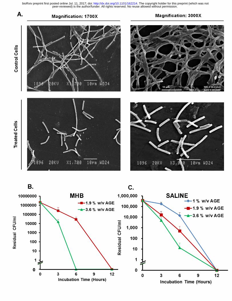
Aqueous Garlic (Allium sativum) Extract (AGE) inhibits growth of Bacillus anthracis in a dose dependent manner: We evaluated the ability of AGE in inhibiting the growth of exponentially growing Bacillus anthracis in MHB medium. The supplementation of MHB medium with different quantities of AGE (0-3.6% w/v) inhibited the growth of exponentially growing Bacillus anthracis culture in a dose dependent manner (Figure 1C). The supplementation of MHB at concentrations ≥1.9% w/v of AGE completely inhibited growth of Bacillus anthracis while lower concentrations retarded/delayed the growth in a dose dependent manner. AGE induces morphological changes in Bacillus anthracis: To assess the possible mechanism of anti-Bacillus anthracis activity the morphology of Bacillus anthracis cells growing in cultures supplemented with 0% w/v AGE (control) and 1.9% w/v AGE (treated) for different durations were examined using light microscopy and scanning electron microscopy. The control Bacillus anthracis culture which was not exposed to AGE displayed presence of cells in long chains as expected (Fig. 2 A top part, control cells, and see also Fig. 1D) while the culture exposed to 1.9% w/v AGE, a growth inhibitory concentration, showed appearance of cells in relatively shorter chains and as single cells (Fig. 2A bottom part, treated cells) within 3 hours of incubation, indicating potential disruption/dissolution or weakening of the cell walls by AGE. Incubation for 8 h resulted in the appearance of debris (data not shown). AGE is bactericidal to vegetative Bacillus anthracis cells: The bactericidal activity of AGE was assessed by exposing Bacillus anthracis cells in the log phase of growth with different concentration of AGE in the presence of growth medium or saline followed by estimating the number of surviving cells/ml (i.e., CFU/ml) after different time intervals (0-12 h). The AGE at concentration 1.9% w/v and above was found to cause decrease in the viability (CFU) of vegetative Bacillus anthracis cells by as much as 6 logs (Fig. 2B and C) within 6-12 h. However, 1 % w/v AGE which could only retard the growth in the presence of MHB medium (See Fig 1C; data not shown in Fig 2B) was found to kill Bacillus anthracis cells in
peer-reviewed) is the author/funder. All rights reserved. No reuse allowed without permission. The copyright holder for this preprint (which was not. http://dx.doi.org/10.1101/162214doi: bioRxiv preprint first posted online Jul. 11, 2017;
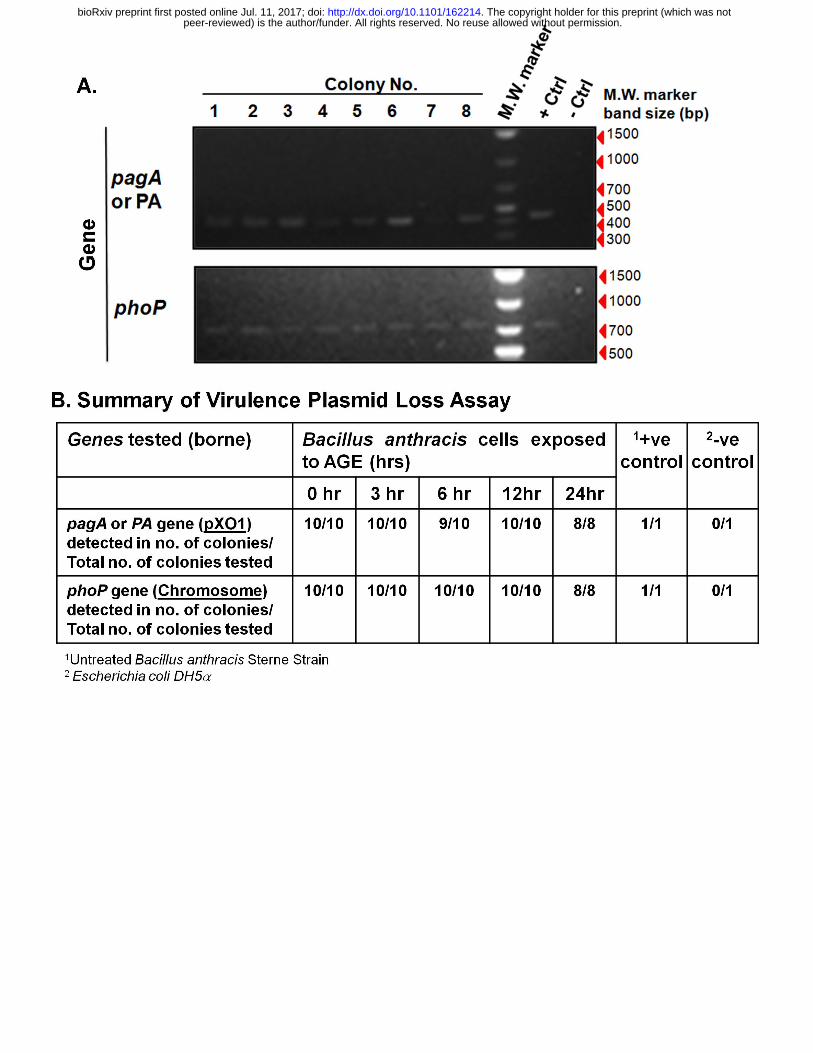
the absence of MHB (Fig. 2C) indicating increased efficacy of AGE in killing Bacillus anthracis cells in the absence of growth medium components. AGE does not induce virulence plasmid pXO1 loss from Bacillus anthracis Sterne strain: The colonies that developed on MHA plate on inoculation of Bacillus anthracis culture exposed to sub-inhibitory concentration of AGE (1% w/v) for different durations (0 - 24 h), were examined for the presence of pagA gene - a virulence plasmid pXO1 borne gene, and phoP gene – a chromosome borne gene using polymerase chain reaction (Figure 3). The exposure to sub-inhibitory concentration of AGE for 24 h did not cause the loss of pXO1 plasmid as apparent from the specific amplification of the plasmid borne pagA gene in 100% of the random colonies (8 out of 8) screened (Fig. 3 A). The sampled colonies formed by the culture of Bacillus anthracis cells exposed to 1% w/v AGE for different durations, i.e., 0 - 24 h also consistently tested positive for the presence of pagA gene indicating retention of pXO1 plasmid and the inability of AGE to induce virulence plasmid pXO1 loss or curing (Fig. 3 B). Similar results were obtained with colonies formed from residual surviving cells present in the Bacillus anthracis culture exposed to growth inhibitory concentration of AGE, i.e., >1.9% w/v for shorter durations (1 – 3 h; data not shown). Autobiography of AGE and Mass spectrometry of bioactive component: To characterize the bioactive constituents in AGE, the conditions for its separation into bioactive entities were performed by coupling the analytical TLC separation of AGE (Supplementary Figure 1A) with bioautography (Sup. Figure 1B) followed by elution of the potential bioactive components from the corresponding place on the duplicate TLC chromatogram of AGE that was developed in parallel and subjecting the eluted fractions to GC-MS for detection of the ions that may indicate the identity of bioactive compound (Figure Sup. Figure 2). Among the tested solvent systems, the solvent system comprising of Toluene-Acetone (7:3) that performed best in separating two UV – fluorescent spots/components in AGE on silica TLC (Sup. Figure 1A, compare chromatogram No. 3 of bottom panel with others) was used to fractionate AGE on silica TLC followed by bioautography of the same (Sup. Figure 1B). The ability to inhibit Bacillus anthracis growth was found to correspond with positions of UV-fluorescent spots (Sup. Figure 1B; encircled in white in right panel: AGE TLC plate). The silica around and including UV-fluorescent spots observed in the AGE fractions that corresponded to anti-Bacillus anthracis activity (growth inhibition) on bioautography were labeled G1 and G2 (Sup. Figure 2A), isolated and processed for GC-MS analysis. The GC-MS profile of G1 and G2 are shown as Sup. Figure 2B and the partial list of potential compound hits as ascertained using the NIST 2.0 library are provided in Table 2 while the complete list can be found in the supplementary information (Sup. Figure 3 and Sup. Figure 4). GC-MS analysis was done to ascertain the presence of compounds which are already reported to be responsible for the antimicrobial activity of Garlic against various pathogenic bacteria [9,10,12,13,14]. Surprisingly, the ions generated did not seem to indicate the presence of the well-known bioactive organosulfur compounds in Garlic, e.g., alliin, S-allyl-L-cysteine, γ-glutamyl-S-allyl-L-cysteine, and allicin etc. though other class of compounds with potential antimicrobial activity, e.g., phthalic acid derivatives, acid esters, phenyl group containing compounds, steroids were identified (Table 2, Sup. Figure 3 and Sup. Figure 4). We speculated the absence of the well-known antibacterial compounds in our
peer-reviewed) is the author/funder. All rights reserved. No reuse allowed without permission. The copyright holder for this preprint (which was not. http://dx.doi.org/10.1101/162214doi: bioRxiv preprint first posted online Jul. 11, 2017;
bioactive fraction to be the result of previously unidentified constituents or our use of water as extractant and the methodology adopted that would have led to the reduced extraction, decomposition and/or loss of the previously reported bioactive compounds. GC-MS analysis of the butanolic extract of Garlic was performed to assess the validity of this speculation. The butanolic extract indeed contained well known allyl derivatives as well as multiple S-containing compounds (Sup. Figure 5) supporting the speculation. The identity of the Garlic sample was also verified by sequencing of the rbcL gene fragment amplified from the Garlic sample DNA. The sequence of the amplified rbcL gene fragment displayed more than 99% identity to the gene bank deposited rbcL gene sequences of Allium sativum (data not shown), confirming the authenticity of the Garlic sample. Anti-Bacillus anthracis activity of AGE is stable near room temperature: The ability of the anti-Bacillus anthracis activity of AGE to tolerate different temperatures was evaluated to assess its potential field use. Based upon the ZOI produced by AGE samples incubated at different temperatures (0-100oC) for different durations (0-15 days), it can be inferred that the anti-Bacillus anthracis activity of AGE is quite stable around room temperature (20-30oC), and also moderately stable at temperatures encountered near tropics during the summer season (see Figure 4A and 4B). It retained more than 80% of anti-Bacillus anthracis activity on incubation at 40oC and 50oC for >1 day and 12 h, respectively. At 4oC incubation temperature, AGE did not show any loss in activity upto 15 days. However, on incubation at 100oC temperature the anti-Bacillus anthracis activity of AGE was lost in about 10 minutes (data not shown); suggesting at least S-allylcysteine [15] which is reportedly quite stable at 100oC would not be the bioactive constituent responsible for the anti-Bacillus anthracis activity shown by AGE. Different batches of Garlic show similar anti-Bacillus anthracis activity: The bioactive content of plant products are known to vary with origin, age and time, so for using any plant product for any particular purpose the plant or plant products from different origin or of age must consistently display desired characteristics within acceptable limits. To this end we evaluated garlic samples collected/bought from local market over a period of time (April 2016 to December 2016) for anti- Bacillus anthracis activity using agar-well diffusion assay. All the three samples evaluated displayed robust and similar anti-Bacillus anthracis activity as apparent from the production of 21-23 mm size ZOIs (Figure 4C and 4D). AGE does not antagonize the activity of antibiotics recommended for the treatment of possible Bacillus anthracis exposure: To evaluate if AGE may affect the action of antibiotics prescribed for anthrax treatment, Amoxicillin (Am), Cefixime (Ce), Ciprofloxacin (C), Doxycycline (Dox), Levofloxacin (L), Penicillin (P), Rifampicin (R), Tetracycline (T), [4,6] were assayed for possible interaction using agar well diffusion assay, as done before. All the antibiotics which are currently in use for anthrax treatment inhibited the growth of Bacillus anthracis as expected (Figure. 4E and 4F and Sup. Fig 6). The AGE did not appear to antagonize the activity of antibiotics used in the anthrax control [6] as summarized in Figure 4F, rather it appeared that the AGE may be potentially
peer-reviewed) is the author/funder. All rights reserved. No reuse allowed without permission. The copyright holder for this preprint (which was not. http://dx.doi.org/10.1101/162214doi: bioRxiv preprint first posted online Jul. 11, 2017;
showing synergy with antibiotics. These observations suggest that Garlic in the form of AGE could be one of the readily available natural anti-Bacillus anthracis agents, though largely unexplored, that can be potentially employed as preventive as well as therapeutic agent after further characterization. Discussion: Anthrax remains a disease of greater concern in poorer parts of the world that also have larger share of livestock but relatively poor veterinary healthcare infrastructure such as in Indian subcontinent and Africa [1,2]. Its reemergence in the areas which were earlier declared anthrax free is also on rise as recently observed in parts of Russia and Africa [1]. A better improved management and prevention strategy needs to be put in place before it is too late. In Indian context, as per the epidemiological data from 1991 to 2010 collated by National Animal Disease Referral Expert System (NADRES), India, it remains one of the top ten killer diseases of the livestock [16]. The condition in other regions of Indian subcontinent could be similar. Mostly the livestock in poorer remote regions bear the brunt of anthrax infection [1,2]. Though antibiotics and vaccines would remain the mainstay of controlling anthrax in immediate future, their availability and reach in poorer regions may remain limited. The issue of antibiotic resistance may also arise sooner or later in Bacillus anthracis as being observed in other more common pathogens. In this direction, the discovery of new antimicrobials [17] or agents that may affect the pathogenicity of organisms by modulating the quorum sensing [18,19] or cause loss of virulence plasmids would be useful in our fight against the pathogens [20,21,22]. Plants are an important source of antimicrobial compounds and have played an important role in controlling diseases [19,20,23,24,25]. They still remain the primary source of medicine for both human and livestock in poorer and remote parts of the world. Identification of edible plants that may have anti-Bacillus anthracis activity would have direct impact on the prevention of anthrax and decreasing its occurrence in the poorer endemic regions. The extracts from a number of plants from diverse geographical origins, mostly employing organic solvent extraction, have been indicated in literature to have anti-Bacillus anthracis activity [11,26,27,28,29]. In the current study, a number of edible plants commonly found in Indian subcontinent [9,10] were evaluated for anti-Bacillus anthracis activity. Although extraction of bioactive compounds from plants is more commonly performed using organic solvents such as ethanol, methanol, acetone, chloroform etc., we had consciously chosen water as the extractant in the current study because of its wider availability and ease of use in the field as compared to any other extractant. In the current study, a number of plants displayed water extractable anti-Bacillus anthracis activity including Garlic that had been indicated previously [30]. The Aqueous Garlic Extract (AGE) exhibited a robust growth inhibition activity against Bacillus anthracis producing large ZOI which was also sustained for 72 h of incubation. A number of plants such as Turmeric, Onion and Tulsi, which are variously attributed to have high antimicrobial/bactericidal activity against a number of pathogens were found to be relatively inactive in the form tested, while Neem and Mango seemed to be moderately active [9,10]. It is expected, as the plant extracts are supposed to contain a number of bioactive chemicals whose synergistic interactions are responsible for their overall activity against specific pathogens and hence the medicinal prescription [10]. Among the tested aqueous extracts of various plants, the
peer-reviewed) is the author/funder. All rights reserved. No reuse allowed without permission. The copyright holder for this preprint (which was not. http://dx.doi.org/10.1101/162214doi: bioRxiv preprint first posted online Jul. 11, 2017;
extract from Garlic was found to be the most potent that reproducibly inhibited Bacillus anthracis growth. However, the extract employed in the current study seemed to be relatively less active as compared to that used by Sasaki and Kita, 2003 [30]. One of the reasons could be the difference in the bioactive content present in different preparations used, i.e., fresh garlic vs. dried powder and the methodology employed. The vegetative cells of the Bacillus anthracis are known to be responsible for the pathogenesis primarily through production of the anthrax toxins. Any agent that may kill vegetative cells of Bacillus anthracis [31,32] or that do not allow germination of spores are potent anti-Bacillus anthracis agents [33]. When we tested the AGE for such activity, the AGE at concentrations ≥1.9% w/v displayed robust killing of vegetative cells of Bacillus anthracis and decreased the cell counts by more than 6 logs within 6-12 h of exposure indicating it to have a potent anti-Bacillus anthracis activity that may be further explored for controlling anthrax infection. Virulence plasmids, pXO1 and pXO2 are responsible for the pathogenicity of Bacillus anthracis. If somehow Bacillus anthracis cells can be cured of these plasmids they would become avirulent [3,34]. Some small molecules such as acridine dyes are known to cause plasmid loss at sub-inhibitory concentration making virulent strains non-pathogenic [21,27,39]. The ability of any safer anti-Bacillus anthracis agent to cause loss of the virulence plasmid(s) at sub-inhibitory concentration would further increase its utility to control anthrax. When we evaluated the AGE for such a plasmid curing ability at both growth sub-inhibitory and inhibitory dosages the results negated any such potential use of Garlic as no plasmid loss was observed at any tested concentration. Bioactive content identification or characterization of any plant origin product is required to credibly correlate the constituent and their effect to recommend its preventive or curative use [18,23,35,36]. The attempt to characterize the bioactive constituents present in AGE with anti-Bacillus anthracis bioactivity revealed the presence of chemical entities that belonged to different class of potential bioactive compounds such as phthalic acid derivatives, acid esters, phenyl group containing compounds, steroids etc. but conspicuously the well-known bioactive compounds such as alliin, ajoene, S-allyl-L-cysteine, γ-glutamyl-S-allyl-L-cysteine, allicin etc. [12] were absent suggesting compounds other than the well-knowns to be responsible for the observed anti-Bacillus anthracis activity. Further characterization of the bioactive fraction(s) is underway. Although garlic is used widely in food in the cooked form its intake in the uncooked raw form is relatively limited. The characterization of thermal stability of the anti-Bacillus anthracis activity present in AGE indicated it to be relatively stable at room temperature but quite unstable at 100oC - the temperature generally achieved during cooking. These observations combined with the previous observations that indicate raw Garlic or Garlic extract to be safe for consumption or injection [37] suggests that the usage of Garlic, in the raw form, may be helpful in controlling the incidence of anthrax in endemic areas. Antimicrobial agents when present in combination(s) may synergize or antagonize each other’s activity [8,38,39]. This kind of information is needed for successful usage of any new potential antimicrobial agent. When we tested AGE for its interaction with FDA approved antibiotics for
peer-reviewed) is the author/funder. All rights reserved. No reuse allowed without permission. The copyright holder for this preprint (which was not. http://dx.doi.org/10.1101/162214doi: bioRxiv preprint first posted online Jul. 11, 2017;
anthrax prevention it was found to not antagonize the activity of antibiotics used for anthrax control, suggesting absence of any negative interaction. Immediate future work may be focused on testing the efficacy of AGE alone or in combination with other common food ingredients in decreasing the Bacillus anthracis load in spiked animal tissue samples (meat) and the characterization of the bioactive fraction(s). Although AGE did not seem to promote virulence plasmid loss in our assay, ascertaining its effect on the expression of virulence factors, e.g., Protective Antigen (PA), Lethal Factor (LF), Edema Factor (EF), and on quorum sensing would remain a priority to increase its potential utility in the anthrax control. Evaluation of the ability of Garlic to prevent the occurrence of gastrointestinal anthrax or evaluation of AGE as injectable for therapy purpose as it is well tolerated in model animals [37] may be some other areas of exploration. Further work needs to be undertaken to explore the possibility of employing Garlic or AGE to decrease the incidence of anthrax in endemic areas. Acknowledgement: The work was supported by a research grant ‘Ramalingaswami Fellowship (BT/RLF/Re-Entry/50/2011)’ from DBT, India to SS. Partial funding support to the laboratory of SS from DST-PURSE through Panjab University, Chandigarh is also duly acknowledged. The research funding agencies had no role in study design, data collection and analysis, decision to publish, or preparation of the manuscript. Author Contributions: Conceived and designed the experiments: SS. Performed the experiments: RK, AT. Analyzed the data: RK, AT, SS. Contributed reagents/materials/analysis tools/discussion: RK, AT, MM, RB and SS. Wrote the paper: SS. References: 1. Shadomy S, Idrissi AE, Raizman E, Bruni M, Palamara E, et al. (2016) Anthrax outbreaks: a
warning for improved prevention, control and heightened awareness. 2. Turnbull P, editor (2008) Anthrax in humans and animals. 4th ed. Geneva, Switzerland: WHO
Press, World Health Organization. 3. Kaur M, Singh S, Bhatnagar R (2013) Anthrax vaccines: present status and future prospects.
Expert Rev Vaccines 12: 955-970. 4. CDC (2016) Anthrax: Recommendations. 5. Turnbull PCB, Böhm R, Cosivi O, Doganay M, Hugh-Jones ME, et al. Guidelines for the
Surveillance and Control of Anthrax in Humans and Animals. 3rd ed: World Health Organization, Emerging and other Communicable Diseases, Surveillance and Control.
6. FDA (2016) Drugs: Drug Information Related to Anthrax 7. Hendricks KA, Wright ME, Shadomy SV, Bradley JS, Morrow MG, et al. (2014) Centers for
disease control and prevention expert panel meetings on prevention and treatment of anthrax in adults. Emerg Infect Dis 20.
8. Weiss S, Altboum Z, Glinert I, Schlomovitz J, Sittner A, et al. (2015) Efficacy of Single and Combined Antibiotic Treatments of Anthrax in Rabbits. Antimicrob Agents Chemother 59: 7497-7503.
9. Duke JA, Bogenschutz-Godwin MJ, duCellier J, Duke P-AK (2002) Handbook of medicinal herbs. Boca Raton: CRC Press LLC.
peer-reviewed) is the author/funder. All rights reserved. No reuse allowed without permission. The copyright holder for this preprint (which was not. http://dx.doi.org/10.1101/162214doi: bioRxiv preprint first posted online Jul. 11, 2017;
10. U.S. Department of Agriculture ARS (1992-2016) Dr. Duke's Phytochemical and Ethnobotanical Databases. U.S. Department of Agriculture, Agricultural Research Service
11. Elisha IL, Dzoyem JP, Botha FS, Eloff JN (2016) The efficacy and safety of nine South African medicinal plants in controlling Bacillus anthracis Sterne vaccine strain. BMC Complement Altern Med 16: 5.
12. Goncagul G, Ayaz E (2010) Antimicrobial effect of garlic (Allium sativum). Recent Pat Antiinfect Drug Discov 5: 91-93.
13. Sharifi-Rad J, Mnayer D, Tabanelli G, Stojanovic-Radic ZZ, Sharifi-Rad M, et al. (2016) Plants of the genus Allium as antibacterial agents: From tradition to pharmacy. Cell Mol Biol (Noisy-le-grand) 62: 57-68.
14. Rana SV, Pal R, Vaiphei K, Sharma SK, Ola RP (2011) Garlic in health and disease. Nutr Res Rev 24: 60-71.
15. Kodera Y, Suzuki A, Imada O, Kasuga S, Sumioka I, et al. (2002) Physical, chemical, and biological properties of s-allylcysteine, an amino acid derived from garlic. J Agric Food Chem 50: 622-632.
16. Rahman H (2012) Epidemiology of anthrax in India. Indian Council of Agricultural Research, New Delhi.
17. Blaskovich MA, Butler MS, Cooper MA (2017) Polishing the tarnished silver bullet: the quest for new antibiotics. Essays Biochem 61: 103-114.
18. Bhardwaj AK, Vinothkumar K, Rajpara N (2013) Bacterial quorum sensing inhibitors: attractive alternatives for control of infectious pathogens showing multiple drug resistance. Recent Pat Antiinfect Drug Discov 8: 68-83.
19. Ta CA, Arnason JT (2015) Mini Review of Phytochemicals and Plant Taxa with Activity as Microbial Biofilm and Quorum Sensing Inhibitors. Molecules 21: E29.
20. Dastidar SG, Kristiansen JE, Molnar J, Amaral L (2013) Role of Phenothiazines and Structurally Similar Compounds of Plant Origin in the Fight against Infections by Drug Resistant Bacteria. Antibiotics (Basel) 2: 58-72.
21. Molnar J, Foldeak S, Nakamura MJ, Rausch H, Domonkos K, et al. (1992) Antiplasmid activity: loss of bacterial resistance to antibiotics. APMIS Suppl 30: 24-31.
22. Spengler G, Molnar A, Schelz Z, Amaral L, Sharples D, et al. (2006) The mechanism of plasmid curing in bacteria. Curr Drug Targets 7: 823-841.
23. Cowan MM (1999) Plant products as antimicrobial agents. Clin Microbiol Rev 12: 564-582. 24. Mahady GB (2005) Medicinal plants for the prevention and treatment of bacterial infections.
Curr Pharm Des 11: 2405-2427. 25. Imam MU, Ismail M, Ooi DJ, Azmi NH, Sarega N, et al. (2016) Are bioactive-rich fractions
functionally richer? Crit Rev Biotechnol 36: 585-593. 26. Akinpelu DA, Adegboye MF, Adeloye OA, Okoh AI (2008) Biocidal activity of partially
purified fractions from methanolic extract of Garcinia kola (Heckel) seeds on bacterial isolates. Biol Res 41: 277-287.
27. Mbwambo ZH, Erasto P, Nondo RO, Innocent E, Kidukuli AW (2011) Antibacterial and cytotoxic activities of Terminalia stenostachya and Terminalia spinosa. Tanzan J Health Res 13: 119-124.
28. Moshi MJ, Mbwambo ZH (2005) Some pharmacological properties of extracts of Terminalia sericea roots. J Ethnopharmacol 97: 43-47.
29. Taher M, Susanti D, Rezali MF, Zohri FS, Ichwan SJ, et al. (2012) Apoptosis, antimicrobial and antioxidant activities of phytochemicals from Garcinia malaccensis Hk.f. Asian Pac J Trop Med 5: 136-141.
30. Sasaki J, Kita J (2003) Bacteriocidal activity of garlic powder against Bacillus anthracis. J Nutr Sci Vitaminol (Tokyo) 49: 297-299.
peer-reviewed) is the author/funder. All rights reserved. No reuse allowed without permission. The copyright holder for this preprint (which was not. http://dx.doi.org/10.1101/162214doi: bioRxiv preprint first posted online Jul. 11, 2017;
31. Omotade TO, Bernhards RC, Klimko CP, Matthews ME, Hill AJ, et al. (2014) The impact of inducing germination of Bacillus anthracis and Bacillus thuringiensis spores on potential secondary decontamination strategies. J Appl Microbiol 117: 1614-1633.
32. Committee on Prepositioned Medical Countermeasures for the Public IoM, Clare Stroud, Kristin Viswanathan, Tia Powell, and Robert R. Bass, editor (2012) Prepositioning Antibiotics for Anthrax. Washington (DC): National Academies Press (US).
33. Akoachere M, Squires RC, Nour AM, Angelov L, Brojatsch J, et al. (2007) Identification of an in vivo inhibitor of Bacillus anthracis spore germination. J Biol Chem 282: 12112-12118.
34. Levy H, Weiss S, Altboum Z, Schlomovitz J, Glinert I, et al. (2012) Differential contribution of Bacillus anthracis toxins to pathogenicity in two animal models. Infect Immun 80: 2623-2631.
35. Friedman M (2015) Antibiotic-resistant bacteria: prevalence in food and inactivation by food-compatible compounds and plant extracts. J Agric Food Chem 63: 3805-3822.
36. Guil-Guerrero JL, Ramos L, Moreno C, Zúñiga-Paredesa JC, Carlosama-Yepez M, et al. (2016) Antimicrobial activity of plant-food by-products: A review focusing on the tropics. Livestock Science 189: 32-49.
37. (HMPC) CoHMP (2016) Assessment report on Allium sativum L., bulbus. London. EMA/HMPC/7686/2013 EMA/HMPC/7686/2013.
38. Acar JF (2000) Antibiotic synergy and antagonism. Med Clin North Am 84: 1391-1406. 39. Wolfart K, Spengler G, Kawase M, Motohashi N, Molnar J, et al. (2006) Synergistic
interaction between proton pump inhibitors and resistance modifiers: promoting effects of antibiotics and plasmid curing. In Vivo 20: 367-372.
peer-reviewed) is the author/funder. All rights reserved. No reuse allowed without permission. The copyright holder for this preprint (which was not. http://dx.doi.org/10.1101/162214doi: bioRxiv preprint first posted online Jul. 11, 2017;
Figure Legends:
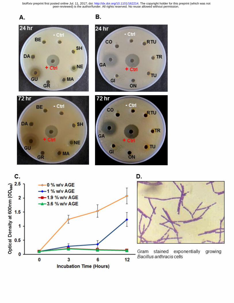
Figure 1. Common edible plants have anti-Bacillus anthracis activity. The aqueous plant
extracts prepared from different plants were placed in the wells made in the solidified MHA
medium followed by spreading of exponentially growing Bacillus anthracis culture on the plate
and incubation at 37oC for upto 72 h (see Materials and Methods for details). The growth pattern
of the test organism on the MHA plates varied as apparent from different diameters of the zone
of growth inhibition (ZOI) - (A) Bael (Aegle marmelos) leaf: BE; Daruharidra (Berberis asiatica)
leaf: DA.; Guava (Psidium guajava) leaf: GU; common grass (Cynodon dactylon): GR; Mango
(Mangifera indica) leaf: MA; Neem (Azadiracta indica) leaf: NE; Shahtoot (Morus indica) leaf:
SH; (B) Coriander (Coriandrum sativum) leaf: CO; Garlic (Allium sativum) bulb: GA; Ginger
(Zingiber officinale) rhizome: GI; Onion (Allium cepa) bulb: ON; Tulsi (Ocimum sanctum) leaf:
TU; Turmeric (Curcuma longa) rhizome: TR; Ram Tulsi (Ocimum gratissimum) leaf: RTU. The
Aqueous Garlic Extract (AGE) appeared to be the most potent based upon the ZOI produced by
the aqueous extracts made from equal wet weight of plant parts tested (See Table 1). The ZOI
initially produced by Neem leaf and Mango leaf extract were short lived and disappeared by 48
h of incubation (See Table 1). Each plate included antibiotic Rifampicin as a positive control (+
Ctrl), and ultrapure water as solvent control (- Ctrl). Note: The aqueous extract from Guava leaf
did not produce any ZOI in the displayed plate. (C) Aqueous Garlic (Allium sativum) Extract
(AGE) inhibits growth of Bacillus anthracis. The exponentially growing Bacillus anthracis culture
was diluted in fresh MHB growth medium so as to have optical density at 600nm (OD600) of 0.1,
supplemented with 0-3.6% w/v of AGE and the growth was monitored by measuring OD600 at
different time intervals. Note: The aqueous Garlic (Allium sativum) extract supplementation of ≥
1.9% w/v inhibited the growth of exponentially growing Bacillus anthracis culture. The values
shown are average of three independent experiments (n=3) and the error bars represent
standard error of mean. (D) Photograph of Gram stained exponentially growing Bacillus
anthracis cells (0% w/v AGE).
Figure 2. Aqueous Garlic (Allium sativum) Extract (AGE) kills Bacillus anthracis cells. (A)
The exposure to AGE induces morphological changes in Bacillus anthracis cells. Scanning
Electron Microscopy (SEM) image of the exponentially growing Bacillus anthracis cells (top
panel; control cells) and those exposed to 1.9% w/v AGE for 3 h (bottom panel; treated cells)
indicate the disintegration of longer chains and appearance of smaller chains and single cells in
the AGE exposed Bacillus anthracis culture (compare control cells in top panel with AGE treated
peer-reviewed) is the author/funder. All rights reserved. No reuse allowed without permission. The copyright holder for this preprint (which was not. http://dx.doi.org/10.1101/162214doi: bioRxiv preprint first posted online Jul. 11, 2017;
cells in the bottom panel). The exposure of exponentially growing Bacillus anthracis cells to
AGE (0 - 3.6% w/v at 37oC) in MHB medium (B) as well as normal saline (C) results in
progressive loss of the cells viability as indicated by the progressive decrease in the remaining
CFU/mL with increase in exposure time. Note the survival of exponentially growing Bacillus
anthracis cells appears to be differentially affected by the presence of growth medium
components on AGE exposure. The CFU/ml values are average of three independent
experiments (n=3) and the error bars represent standard deviation.
Figure 3. Aqueous Garlic Extract (AGE) at sub-inhibitory concentration does not promote
virulence plasmid ‘pXO1’ loss from Bacillus anthracis Sterne strain. The Bacillus anthracis
cells were grown in MHB medium in the presence of sub-inhibitory concentration of AGE (1%
w/v) and the loss of virulence plasmid pXO1 from cells was assessed at different time intervals
(0 - 24 h). The presence of pXO1 borne pagA gene as well as genome borne phoP gene was
assessed by PCR amplification of the said genes using gene specific primers (see materials
and methods). (A) The analysis of eight random colonies picked from 24 h old AGE exposed
culture for the presence of pagA and phoP genes are shown. All colonies examined retained the
pXO1 plasmid based upon the PCR amplification of pagA gene. Genomic DNA from Bacillus
anthracis Sterne strain was used as positive control and that of Escherichia coli DH5α strain as
negative control for the PCR based analysis. (B) The overall summary of the virulence plasmid
pXO1 loss assay is presented in tabular form. Bacillus anthracis cells seem to not lose pXO1 on
AGE exposure at all the time points tested (0 - 24 h).
Figure 4. Characterizations of Aqueous Garlic (Allium sativum) Extract (AGE). (A-B)
Exposure of AGE to higher temperature decreases its potency to inhibit growth of Bacillus
anthracis. The result of agar-well diffusion assay to assess the anti-Bacillus anthracis activity
remaining in AGE incubated for 6 h at various temperatures (4-50oC) is shown in (A). The 50µl
aliquots from the AGE extracts incubated at different temperatures, as labeled in figure, were
loaded in the wells made in MHA plate along with 8µg of antibiotic Rifampicin as positive control
in the well that is marked as positive (+) followed by overnight incubation of the plate at 37oC.
(B) The relative decrease in potency of AGE incubated upto 15 days at temperatures 4-50oC to
inhibit Bacillus anthracis growth was assessed in terms of zone of growth inhibition (ZOI)
observed in agar-well diffusion assays as shown in (A). The data indicated incremental loss in
the anti-Bacillus anthracis activity of AGE with increase in temperature and the incubation
peer-reviewed) is the author/funder. All rights reserved. No reuse allowed without permission. The copyright holder for this preprint (which was not. http://dx.doi.org/10.1101/162214doi: bioRxiv preprint first posted online Jul. 11, 2017;
duration. The data is presented as ZOI with respect to (WRT) that shown by AGE incubated at
4oC, which did not seem to lose activity and consistently gave a ZOI of 17-19 mm upto 15 days
of incubation. The freshly prepared AGE does not seem to lose anti-Bacillus anthracis activity
rapidly near room temperature (see data for upto 40oC incubation). The AGE incubated at 40
oC
retained >80% activity even after incubation for 1 day. (C-D) Garlic samples/batches display
similar anti-Bacillus anthracis activity. The Garlic samples purchased at different times from
local market of Chandigarh, India were evaluated for the presence of bioactive principles to
ascertain the relative consistency of the batches in terms of the anti-Bacillus anthracis activity
profile. (C) The aqueous extracts made from different Garlic sample batches were compared for
the relative potency against Bacillus anthracis using agar-well diffusion assay as described
before. All three batches displayed similar potency as apparent from their ZOI. (D) Summary of
the assay shown in (C). Note: The identity of sample no. 1 was further verified as Allium sativum
through sequencing of the rbcL gene (see material methods). (E-F) Aqueous Garlic Extract
(AGE) does not antagonize the anti-Bacillus anthracis activity of commonly employed antibiotics
for anthrax control. (E) The antibiotics commonly employed for anthrax control, i.e., Amoxicillin
(Am), Cefixime (Ce), Ciprofloxacin (C), Doxycycline (Dox) Levofloxacin (L) Penicillin (P)
Rifampicin (R) Tetracycline(T) along with Sulfamethoxazole (S), an antibiotic known to be
ineffective against Bacillus anthracis were assayed for possible interaction with AGE using agar
well diffusion assay. The top and bottom panels in (E) are from experiment performed with
different concentration of the indicated test substances. Except Sulfamethoxazole, all antibiotics
and AGE inhibited the growth of Bacillus anthracis as expected. (F) Summary of the interaction
of AGE with antibiotics in inhibiting Bacillus anthracis growth is shown in (E). The AGE did not
interact negatively with antibiotics currently employed for anthrax control rather displayed some
degree of synergy (see the shape of ZOI produced around wells loaded with different antibiotics
in (E)). Summary of the relative ability of different tested substances in inhibiting Bacillus
anthracis growth in agar-well diffusion assay is provided as Sup. Figure. 6.
peer-reviewed) is the author/funder. All rights reserved. No reuse allowed without permission. The copyright holder for this preprint (which was not. http://dx.doi.org/10.1101/162214doi: bioRxiv preprint first posted online Jul. 11, 2017;
Table 1. Inhibition of Bacillus anthracis growth by aqueous (sterile ultrapure water) extract of indicated plant part
S. No.
Name of the plant (common
name) - plant part used (L: Leaf; B: bulb; Fruit:
F; Rhizome: R)
Site of collection Zone of Inhibition (in mm) after incubation for
Volume of crude aqueous extract 12 h 24 h 48 h 72 h
1. Aegle marmelos (Bael) – L
Panjab University campus, Chandigarh (Chd.)
0 0 0 0 50µl
2. Allium cepa (Onion) – B
Local market, Chd. 0 0 0 0 50µl
3. Allium sativum (Garlic) – B
Local market, Chd. 19 19 18 18 50µl
4. Azadirachta indica (Neem) – L
Panjab University campus, Chd.
11 10 0 0 50µl
5. Berberis asiatica (Daruharidra) - L
Panjab University campus, Chd.
0 0 0 0 50µl
6. Coriandrum sativum (Coriander) – L
Local market, Chd. 0 0 0 0 50µl
7. Curcuma longa (Turmeric) – R
Local market, Chd. 0 0 0 0 50µl
8. Cynodon dactylon (Grass) - L
Panjab University campus, Chd.
0 0 0 0 50µl
9. Mangifera indica (Mango)-L
Panjab University campus, Chd.
10 10 0 0 50µl
10. Ocimum sanctum/ Ocimum tenuiflorum (Tulsi) - L
Panjab University campus, Chd.
0 0 0 0 50µl
11. Ocimum gratissimum (Ram Tulsi) -L
Panjab University campus, Chd.
0 0 0 0 50µl
12. Morus indica (Sahtoot)
-L Panjab University campus,
Chd. 0 0 0 0 50µl
13. Psidium guajava (Guvava) - L
Panjab University campus, Chd.
0 0 0 0 50µl
14. Zingiber officinale (Ginger) – R
Local market, Chd. 0 0 0 0 50µl
- Ctrl Solvent control (Ultrapure Water) 0 0 0 0 50µl
+ Ctrl Rifampicin (8µg) 18 18 18 17 +
peer-reviewed) is the author/funder. All rights reserved. No reuse allowed without permission. The copyright holder for this preprint (which was not. http://dx.doi.org/10.1101/162214doi: bioRxiv preprint first posted online Jul. 11, 2017;
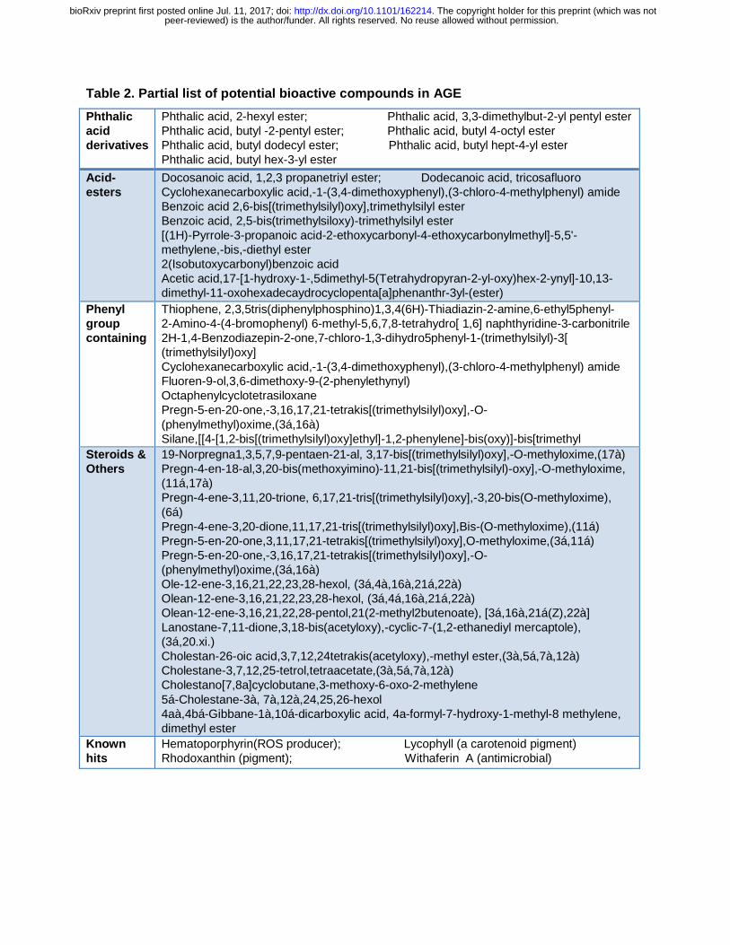
Table 2. Partial list of potential bioactive compounds in AGE
Phthalic acid derivatives
Phthalic acid, 2-hexyl ester; Phthalic acid, 3,3-dimethylbut-2-yl pentyl ester Phthalic acid, butyl -2-pentyl ester; Phthalic acid, butyl 4-octyl ester Phthalic acid, butyl dodecyl ester; Phthalic acid, butyl hept-4-yl ester Phthalic acid, butyl hex-3-yl ester
Acid-esters
Docosanoic acid, 1,2,3 propanetriyl ester; Dodecanoic acid, tricosafluoro Cyclohexanecarboxylic acid,-1-(3,4-dimethoxyphenyl),(3-chloro-4-methylphenyl) amide Benzoic acid 2,6-bis[(trimethylsilyl)oxy],trimethylsilyl ester Benzoic acid, 2,5-bis(trimethylsiloxy)-trimethylsilyl ester [(1H)-Pyrrole-3-propanoic acid-2-ethoxycarbonyl-4-ethoxycarbonylmethyl]-5,5'-methylene,-bis,-diethyl ester 2(Isobutoxycarbonyl)benzoic acid Acetic acid,17-[1-hydroxy-1-,5dimethyl-5(Tetrahydropyran-2-yl-oxy)hex-2-ynyl]-10,13-dimethyl-11-oxohexadecaydrocyclopenta[a]phenanthr-3yl-(ester)
Phenyl group containing
Thiophene, 2,3,5tris(diphenylphosphino)1,3,4(6H)-Thiadiazin-2-amine,6-ethyl5phenyl- 2-Amino-4-(4-bromophenyl) 6-methyl-5,6,7,8-tetrahydro[ 1,6] naphthyridine-3-carbonitrile 2H-1,4-Benzodiazepin-2-one,7-chloro-1,3-dihydro5phenyl-1-(trimethylsilyl)-3[ (trimethylsilyl)oxy] Cyclohexanecarboxylic acid,-1-(3,4-dimethoxyphenyl),(3-chloro-4-methylphenyl) amide Fluoren-9-ol,3,6-dimethoxy-9-(2-phenylethynyl) Octaphenylcyclotetrasiloxane Pregn-5-en-20-one,-3,16,17,21-tetrakis[(trimethylsilyl)oxy],-O-(phenylmethyl)oxime,(3á,16à) Silane,[[4-[1,2-bis[(trimethylsilyl)oxy]ethyl]-1,2-phenylene]-bis(oxy)]-bis[trimethyl
Steroids & Others
19-Norpregna1,3,5,7,9-pentaen-21-al, 3,17-bis[(trimethylsilyl)oxy],-O-methyloxime,(17à) Pregn-4-en-18-al,3,20-bis(methoxyimino)-11,21-bis[(trimethylsilyl)-oxy],-O-methyloxime, (11á,17à) Pregn-4-ene-3,11,20-trione, 6,17,21-tris[(trimethylsilyl)oxy],-3,20-bis(O-methyloxime), (6á) Pregn-4-ene-3,20-dione,11,17,21-tris[(trimethylsilyl)oxy],Bis-(O-methyloxime),(11á) Pregn-5-en-20-one,3,11,17,21-tetrakis[(trimethylsilyl)oxy],O-methyloxime,(3á,11á) Pregn-5-en-20-one,-3,16,17,21-tetrakis[(trimethylsilyl)oxy],-O-(phenylmethyl)oxime,(3á,16à) Ole-12-ene-3,16,21,22,23,28-hexol, (3á,4à,16à,21á,22à) Olean-12-ene-3,16,21,22,23,28-hexol, (3á,4á,16à,21á,22à) Olean-12-ene-3,16,21,22,28-pentol,21(2-methyl2butenoate), [3á,16à,21á(Z),22à] Lanostane-7,11-dione,3,18-bis(acetyloxy),-cyclic-7-(1,2-ethanediyl mercaptole), (3á,20.xi.) Cholestan-26-oic acid,3,7,12,24tetrakis(acetyloxy),-methyl ester,(3à,5á,7à,12à) Cholestane-3,7,12,25-tetrol,tetraacetate,(3à,5á,7à,12à) Cholestano[7,8a]cyclobutane,3-methoxy-6-oxo-2-methylene 5á-Cholestane-3à, 7à,12à,24,25,26-hexol 4aà,4bá-Gibbane-1à,10á-dicarboxylic acid, 4a-formyl-7-hydroxy-1-methyl-8 methylene, dimethyl ester
Known hits
Hematoporphyrin(ROS producer); Lycophyll (a carotenoid pigment) Rhodoxanthin (pigment); Withaferin A (antimicrobial)
peer-reviewed) is the author/funder. All rights reserved. No reuse allowed without permission. The copyright holder for this preprint (which was not. http://dx.doi.org/10.1101/162214doi: bioRxiv preprint first posted online Jul. 11, 2017;
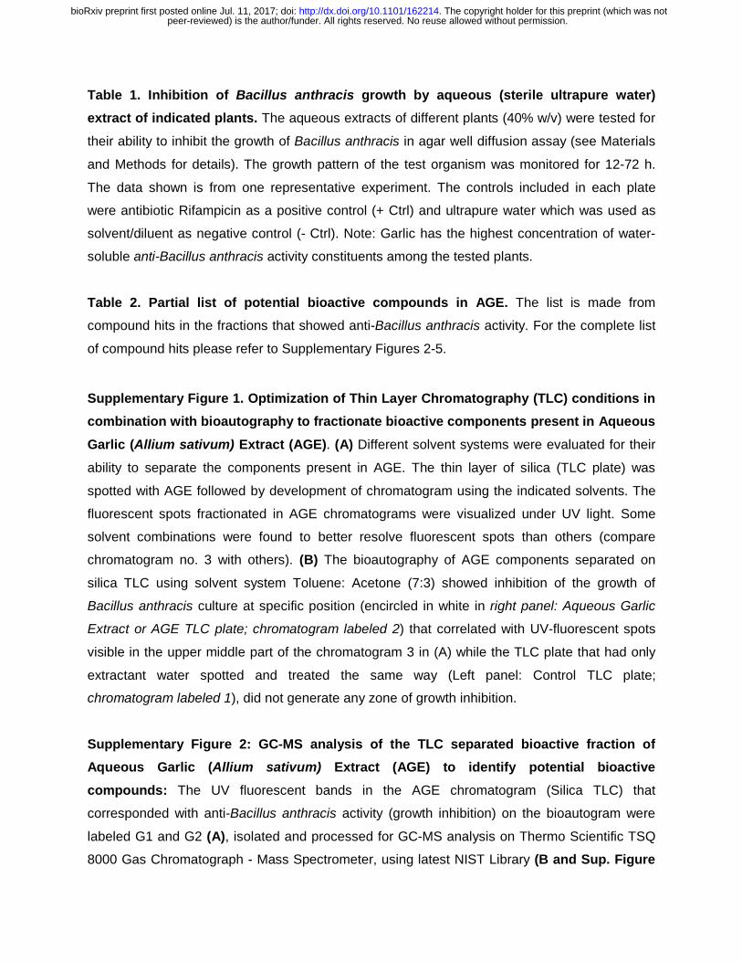
Table 1. Inhibition of Bacillus anthracis growth by aqueous (sterile ultrapure water)
extract of indicated plants. The aqueous extracts of different plants (40% w/v) were tested for
their ability to inhibit the growth of Bacillus anthracis in agar well diffusion assay (see Materials
and Methods for details). The growth pattern of the test organism was monitored for 12-72 h.
The data shown is from one representative experiment. The controls included in each plate
were antibiotic Rifampicin as a positive control (+ Ctrl) and ultrapure water which was used as
solvent/diluent as negative control (- Ctrl). Note: Garlic has the highest concentration of water-
soluble anti-Bacillus anthracis activity constituents among the tested plants.
Table 2. Partial list of potential bioactive compounds in AGE. The list is made from
compound hits in the fractions that showed anti-Bacillus anthracis activity. For the complete list
of compound hits please refer to Supplementary Figures 2-5.
Supplementary Figure 1. Optimization of Thin Layer Chromatography (TLC) conditions in
combination with bioautography to fractionate bioactive components present in Aqueous
Garlic (Allium sativum) Extract (AGE). (A) Different solvent systems were evaluated for their
ability to separate the components present in AGE. The thin layer of silica (TLC plate) was
spotted with AGE followed by development of chromatogram using the indicated solvents. The
fluorescent spots fractionated in AGE chromatograms were visualized under UV light. Some
solvent combinations were found to better resolve fluorescent spots than others (compare
chromatogram no. 3 with others). (B) The bioautography of AGE components separated on
silica TLC using solvent system Toluene: Acetone (7:3) showed inhibition of the growth of
Bacillus anthracis culture at specific position (encircled in white in right panel: Aqueous Garlic
Extract or AGE TLC plate; chromatogram labeled 2) that correlated with UV-fluorescent spots
visible in the upper middle part of the chromatogram 3 in (A) while the TLC plate that had only
extractant water spotted and treated the same way (Left panel: Control TLC plate;
chromatogram labeled 1), did not generate any zone of growth inhibition.
Supplementary Figure 2: GC-MS analysis of the TLC separated bioactive fraction of
Aqueous Garlic (Allium sativum) Extract (AGE) to identify potential bioactive
compounds: The UV fluorescent bands in the AGE chromatogram (Silica TLC) that
corresponded with anti-Bacillus anthracis activity (growth inhibition) on the bioautogram were
labeled G1 and G2 (A), isolated and processed for GC-MS analysis on Thermo Scientific TSQ
8000 Gas Chromatograph - Mass Spectrometer, using latest NIST Library (B and Sup. Figure
peer-reviewed) is the author/funder. All rights reserved. No reuse allowed without permission. The copyright holder for this preprint (which was not. http://dx.doi.org/10.1101/162214doi: bioRxiv preprint first posted online Jul. 11, 2017;
3 and 4). The well-known antimicrobials reported in Garlic, i.e., allicin, ajoene, allyl sulphides,
could not be detected but other class of compounds with potential antimicrobial activity, e.g.,
phthalic acid derivatives, acid esters, phenyl group containing compounds, steroids, were
detected (Sup. Figure 3 and 4). However, the GC-MS analysis of butanolic extract of garlic (C)
showed the presence of various allyl derivatives as well as multiple S-containing compounds
(Sup. Figure 5) suggesting the possible loss of allyl compounds in our processing of Garlic or
their absence in the AGE as bioactive constituents.
Supplementary Figure 3. GC-MS profile of bioactive G1 spot from AGE and the list of compound hits Supplementary Figure 4. GC-MS profile of bioactive G2 spot from AGE and the list of compound hits
Supplementary Figure 5. GC-MS profile of butanolic extract of Garlic and the list of compound hits
Supplementary Figure 6. Zone of inhibition of antibiotics used in anthrax control and AGE
peer-reviewed) is the author/funder. All rights reserved. No reuse allowed without permission. The copyright holder for this preprint (which was not. http://dx.doi.org/10.1101/162214doi: bioRxiv preprint first posted online Jul. 11, 2017;
peer-reviewed) is the author/funder. All rights reserved. No reuse allowed without permission. The copyright holder for this preprint (which was not. http://dx.doi.org/10.1101/162214doi: bioRxiv preprint first posted online Jul. 11, 2017;
peer-reviewed) is the author/funder. All rights reserved. No reuse allowed without permission. The copyright holder for this preprint (which was not. http://dx.doi.org/10.1101/162214doi: bioRxiv preprint first posted online Jul. 11, 2017;
peer-reviewed) is the author/funder. All rights reserved. No reuse allowed without permission. The copyright holder for this preprint (which was not. http://dx.doi.org/10.1101/162214doi: bioRxiv preprint first posted online Jul. 11, 2017;
peer-reviewed) is the author/funder. All rights reserved. No reuse allowed without permission. The copyright holder for this preprint (which was not. http://dx.doi.org/10.1101/162214doi: bioRxiv preprint first posted online Jul. 11, 2017;