COMMON ENT INSTRUMENTS PRITHWIRAJ MAITI R.G.KAR … · COMMON ENT INSTRUMENTS PRITHWIRAJ MAITI...
30
COMMON ENT INSTRUMENTS PRITHWIRAJ MAITI R.G.KAR MEDICAL COLLEGE, KOLKATA, INDIA. INDEX Ear instruments. Nose instruments. Throat instruments. EAR 1. Head Mirror It is a concave mirror used to reflect light from the Bull's eye lamp onto the part being examined. It has a focal length of approximately 25 cm. The examiner sees through the hole in the centre of the mirror. Diameter of the mirror is 89 mm (3½″) and that of the central hole is 19 mm (3/4″).
Transcript of COMMON ENT INSTRUMENTS PRITHWIRAJ MAITI R.G.KAR … · COMMON ENT INSTRUMENTS PRITHWIRAJ MAITI...

COMMON ENT INSTRUMENTS
PRITHWIRAJ MAITI
R.G.KAR MEDICAL COLLEGE, KOLKATA, INDIA.
INDEX
Ear instruments.
Nose instruments.
Throat instruments.
EAR
1. Head Mirror
It is a concave mirror used to reflect light from the Bull's eye lamp onto the part being
examined.
It has a focal length of approximately 25 cm.
The examiner sees through the hole in the centre of the mirror.
Diameter of the mirror is 89 mm (3½″) and that of the central hole is 19 mm (3/4″).
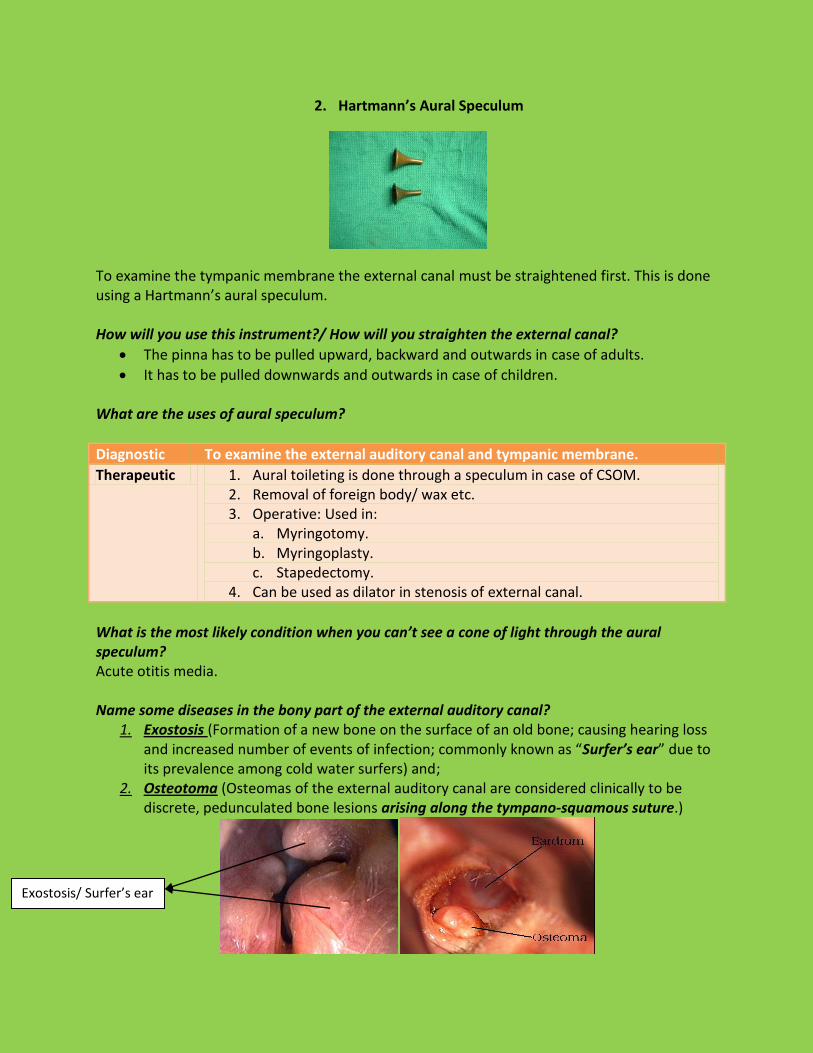
2. Hartmann’s Aural Speculum
To examine the tympanic membrane the external canal must be straightened first. This is done using a Hartmann’s aural speculum. How will you use this instrument?/ How will you straighten the external canal?
The pinna has to be pulled upward, backward and outwards in case of adults.
It has to be pulled downwards and outwards in case of children. What are the uses of aural speculum?
Diagnostic To examine the external auditory canal and tympanic membrane.
Therapeutic 1. Aural toileting is done through a speculum in case of CSOM. 2. Removal of foreign body/ wax etc. 3. Operative: Used in:
a. Myringotomy. b. Myringoplasty. c. Stapedectomy.
4. Can be used as dilator in stenosis of external canal.
What is the most likely condition when you can’t see a cone of light through the aural speculum? Acute otitis media. Name some diseases in the bony part of the external auditory canal?
1. Exostosis (Formation of a new bone on the surface of an old bone; causing hearing loss and increased number of events of infection; commonly known as “Surfer’s ear” due to its prevalence among cold water surfers) and;
2. Osteotoma (Osteomas of the external auditory canal are considered clinically to be discrete, pedunculated bone lesions arising along the tympano-squamous suture.)
Exostosis/ Surfer’s ear
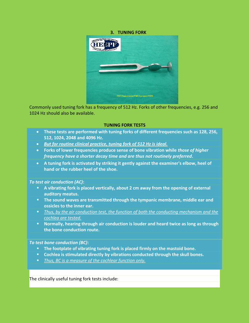
3. TUNING FORK
Commonly used tuning fork has a frequency of 512 Hz. Forks of other frequencies, e.g. 256 and 1024 Hz should also be available.
TUNING FORK TESTS
These tests are performed with tuning forks of different frequencies such as 128, 256, 512, 1024, 2048 and 4096 Hz.
But for routine clinical practice, tuning fork of 512 Hz is ideal.
Forks of lower frequencies produce sense of bone vibration while those of higher frequency have a shorter decay time and are thus not routinely preferred.
A tuning fork is activated by striking it gently against the examiner's elbow, heel of hand or the rubber heel of the shoe.
To test air conduction (AC): A vibrating fork is placed vertically, about 2 cm away from the opening of external
auditory meatus. The sound waves are transmitted through the tympanic membrane, middle ear and
ossicles to the inner ear. Thus, by the air conduction test, the function of both the conducting mechanism and the
cochlea are tested. Normally, hearing through air conduction is louder and heard twice as long as through
the bone conduction route.
To test bone conduction (BC): The footplate of vibrating tuning fork is placed firmly on the mastoid bone. Cochlea is stimulated directly by vibrations conducted through the skull bones. Thus, BC is a measure of the cochlear function only.
The clinically useful tuning fork tests include:
(a) Rinne test
In this test, air conduction of the ear is compared with its bone conduction. A vibrating tuning fork is placed on the patient's mastoid and when he stops hearing,
it is brought beside the meatus. If he still hears, AC is more than BC. Alternatively, the patient is asked to compare the loudness of sound heard through air
and bone conduction. Rinne test is called positive when AC is longer or louder than BC. It is seen in normal persons or those having sensorineural deafness. A negative Rinne (BC > AC) is seen in conductive deafness. A negative Rinne indicates a minimum air-bone gap of 15-20 dB.
A prediction of air-bone gap can be made if tuning forks of 256, 512 and 1024 Hz are used:
256 Hz 512 Hz 1024 Hz Air bone gap
=/- + 20-30 dB
- - + 30-45 dB
- - - 45-60 dB
False negative Rinne: It is seen in severe unilateral sensorineural hearing loss. Patient does not perceive any sound of tuning fork by air conduction but responds to bone conduction testing. This response to bone conduction is, in reality, from the opposite ear because of transcranial transmission of sound. In such cases, correct diagnosis can be made by masking the non-test ear with Barany's noise box while testing for bone conduction. Weber test will further help as it gets lateralised to the better ear.
(b) Weber test
In this test, a vibrating tuning fork is placed in the middle of the forehead or the vertex and the patient is asked in which ear the sound is heard.
Normally, it is heard equally in both ears. It is lateralised to the worse ear in conductive deafness and to the better ear in
sensorineural deafness. In weber test, sound travels directly to the cochlea via bone. Lateralisation of sound in weber test with a tuning fork of 512 Hz implies a conductive
loss of 15-25 dB in ipsilateral ear or a sensorineural loss in the contralateral ear.

(c) Absolute bone conduction (ABC) test
Bone conduction is a measure of cochlear function. In ABC test, patient's bone conduction is compared with that of the examiner
(presuming that the examiner has normal hearing). External auditory meatus of both the patient and examiner should be occluded (by
pressing the tragus inwards), to prevent ambient noise entering through AC route. In conductive deafness, the patient and the examiner hear the fork for the same duration
of time. In sensorineural deafness, the patient hears the fork for a shorter duration.
Test Normal Conductive deafness SN deafness
Rinne AC > BC (Rinne positive) BC > AC (Rinne negative) AC > BC
Weber Not lateralised Lateralised to poorer ear Lateralised to better ear
ABC Same as examiner's Same as examiner's Reduced
Tuning fork tests: (A) Testing for air conduction. (B) Testing for bone conduction. (C) Weber test.

What are the parts of a tuning fork? 1. Base plate/ footplate. 2. Shaft. 3. Prongs that vibrate and produce sound.
Where the prongs should be hit? Against a resilient surface. Why the prongs of a tuning fork shouldn’t be hit against a hard surface? Because then it will produce overtones.
4. Sympson’s Aural Syringe
Parts of the instrument 1. Nozzle, 2. Cylinder, 3. Plunger.
Uses of the instrument 1. Removal of foreign body and wax. 2. Removal of otomycotic plug. 3. Aural toilet.
Technique of syringing the ear
Patient is seated with ear to be syringed towards the examiner.
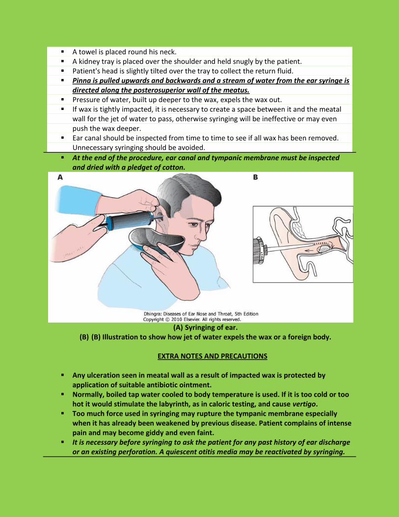
A towel is placed round his neck. A kidney tray is placed over the shoulder and held snugly by the patient. Patient's head is slightly tilted over the tray to collect the return fluid. Pinna is pulled upwards and backwards and a stream of water from the ear syringe is
directed along the posterosuperior wall of the meatus. Pressure of water, built up deeper to the wax, expels the wax out. If wax is tightly impacted, it is necessary to create a space between it and the meatal
wall for the jet of water to pass, otherwise syringing will be ineffective or may even push the wax deeper.
Ear canal should be inspected from time to time to see if all wax has been removed. Unnecessary syringing should be avoided.
At the end of the procedure, ear canal and tympanic membrane must be inspected and dried with a pledget of cotton.
(A) Syringing of ear.
(B) (B) Illustration to show how jet of water expels the wax or a foreign body.
EXTRA NOTES AND PRECAUTIONS
Any ulceration seen in meatal wall as a result of impacted wax is protected by application of suitable antibiotic ointment.
Normally, boiled tap water cooled to body temperature is used. If it is too cold or too hot it would stimulate the labyrinth, as in caloric testing, and cause vertigo.
Too much force used in syringing may rupture the tympanic membrane especially when it has already been weakened by previous disease. Patient complains of intense pain and may become giddy and even faint.
It is necessary before syringing to ask the patient for any past history of ear discharge or an existing perforation. A quiescent otitis media may be reactivated by syringing.
What are the contraindications of syringing? Ear canal irrigation is contraindicated if there is a known or suspected:
a. Tympanic membrane perforation (including a patent myringotomy tube), b. Monomeric or dimeric tympanic membrane (a thin, weak area of the membrane where
one or two layers have healed after perforation), c. The presence of vegetable matter such as a bean or a pea, d. The presence of a watch or hearing aid battery, e. Evidence of purulent exudate filling the canal, or a f. History of ear surgery.
How will you remove a vegetable foreign body? By a Jobson Horne’s Probe with Ring Curette. What are the complications of syringing?
1. Vasovagal attack. 2. Tympanic membrane rupture and perforation. 3. Injury of external auditory canal and otitis externa. 4. Laryrinthine stimulation and vertigo.
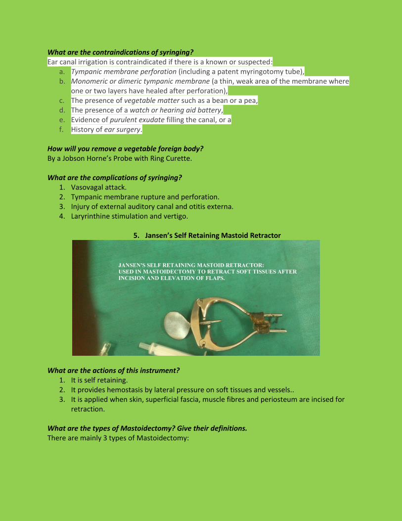
5. Jansen’s Self Retaining Mastoid Retractor
What are the actions of this instrument? 1. It is self retaining. 2. It provides hemostasis by lateral pressure on soft tissues and vessels.. 3. It is applied when skin, superficial fascia, muscle fibres and periosteum are incised for
retraction.
What are the types of Mastoidectomy? Give their definitions. There are mainly 3 types of Mastoidectomy:
Cortical/ Simple Mastoidectomy
It is an exenteration of all accessible mastoid air cells preserving the posterior meatal wall.
Modified Radical Mastoidectomy
It is an operation to eradicate disease of the attic and mastoid, both of which are exteriorised into the external auditory canal by removal of the posterior meatal and lateral attic walls. Tympanic membrane remnant, functioning ossicles and the reversible mucosa and function of the eustachian tube are preserved. These structures are necessary to reconstruct hearing mechanism at the time of surgery or in a 2nd stage operation.
Radical Mastoidectomy
It is an operation to eradicate disease of the middle ear and mastoid in which mastoid, middle ear, attic and the antrum are exteriorised into the external ear by removal of posterior meatal wall. All remnants of tympanic membrane, malleus, incus (not the stapes) chorda tympani and the mucoperiosteal lining are removed, and the opening of eustachian tube closed by packing a piece of muscle or cartilage into the eustachian tube. What is the preferred local anaesthetic in ENT surgeries?
Preferred anaesthetic: Xylocaine.
Strength: 2%.
Dosage: 4-7 ml/kg body weight.
Ingradient: 1 ml of xylocaine includes 21.3 mg Lingocaine hydrochloride + 0.003 mg adrenaline.
Preferred route: Nasal intubation.
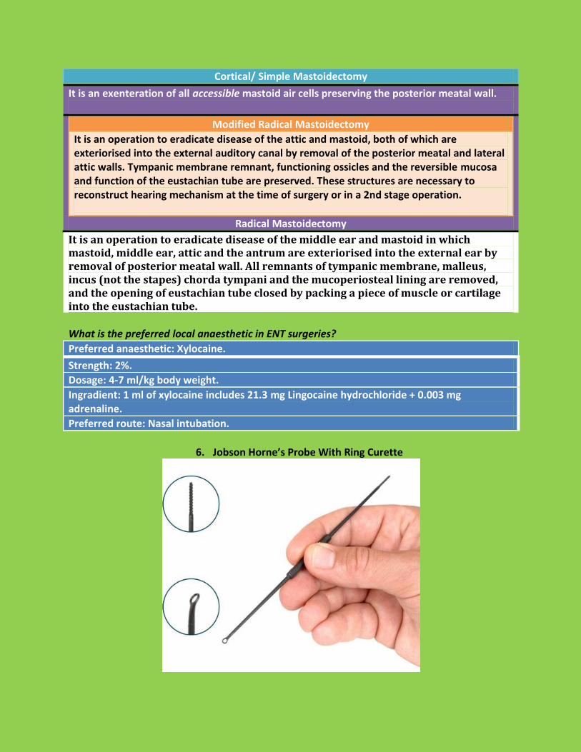
6. Jobson Horne’s Probe With Ring Curette
If you look closely, you will find that one end of the probe is shaped like a ring. This end may be used to hook out wax or foreign bodies from the ear canal.
` The other end of the instrument is sharp and serrated. An ear wick can be fashioned out of this end by rolling cotton on to it and used to mop ear discharge.
What are the uses of this instrument? Serrated probe 1. It is used as cotton wool swab carrier to clean aural discharge in
otitis externa and otitis media. 2. It can be used to trace a sinus tract. 3. It can be used to apply local anaesthesia of the meatus before
maxillary antral lavage.
Curette It is used to remove wax/ foreign body/ granulation tissue.
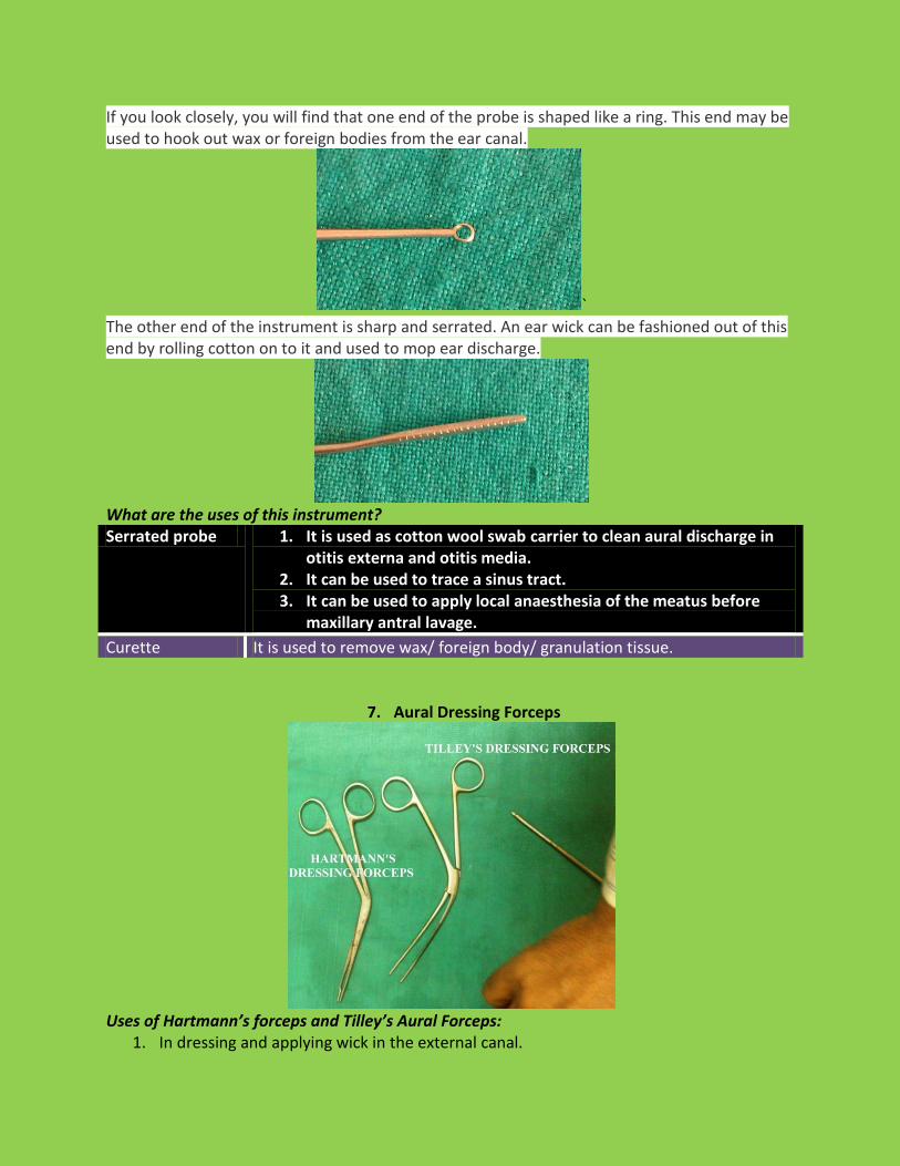
7. Aural Dressing Forceps
Uses of Hartmann’s forceps and Tilley’s Aural Forceps:
1. In dressing and applying wick in the external canal.
2. Removal of foreign body and crusts from the external auditory canal. 3. In ear surgery.
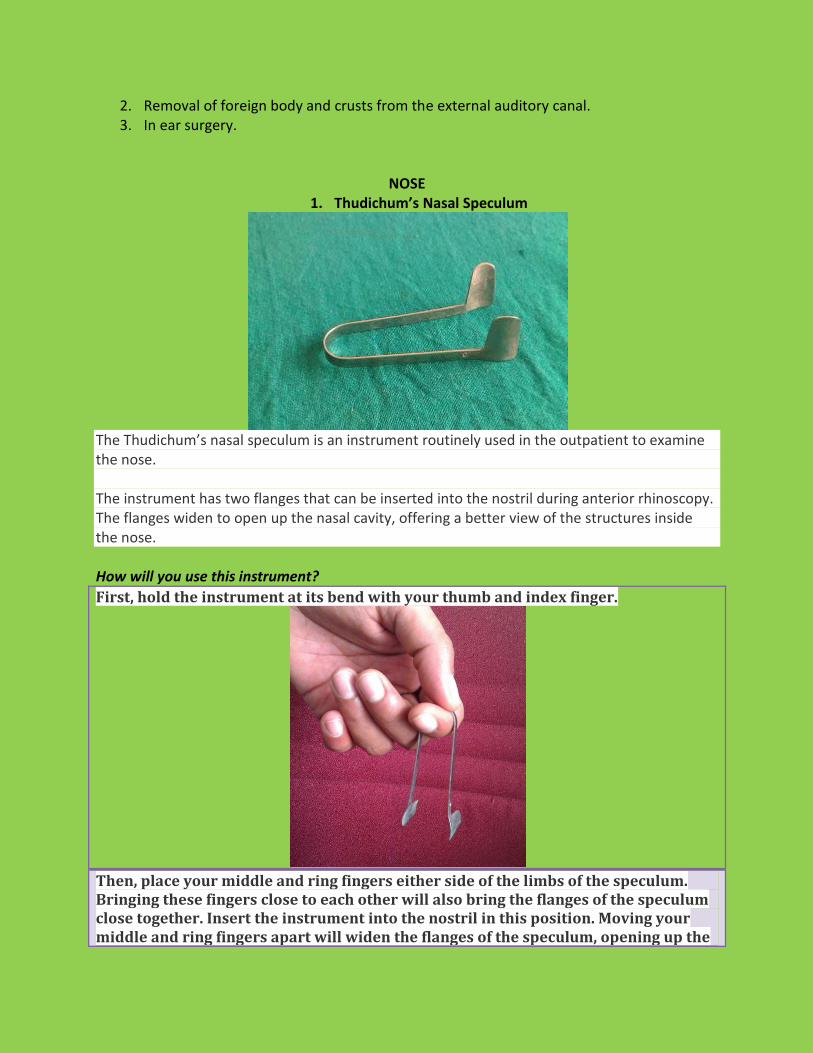
NOSE 1. Thudichum’s Nasal Speculum
The Thudichum’s nasal speculum is an instrument routinely used in the outpatient to examine the nose. The instrument has two flanges that can be inserted into the nostril during anterior rhinoscopy. The flanges widen to open up the nasal cavity, offering a better view of the structures inside the nose. How will you use this instrument?
First, hold the instrument at its bend with your thumb and index finger.
Then, place your middle and ring fingers either side of the limbs of the speculum. Bringing these fingers close to each other will also bring the flanges of the speculum close together. Insert the instrument into the nostril in this position. Moving your middle and ring fingers apart will widen the flanges of the speculum, opening up the

nasal cavity in the process.
What are the uses of this instrument? Diagnostic For anterior rhinoscopy.
Therapeutic Used in various nasal procedures and operations like: a. Foreign body removal. b. Antral wash out. c. Nasal packing. d. Polypectomy. e. SMR and septoplasty.
What are the structures seen in anterior rhinoscopy?
1. Nasal vestibule. 2. Nasal passage. 3. Nasal septum. 4. Floor of the nose. 5. Lateral wall of the nose: [Ant. Part of inferior and middle turbinates and Inferior and
middle meatus.]
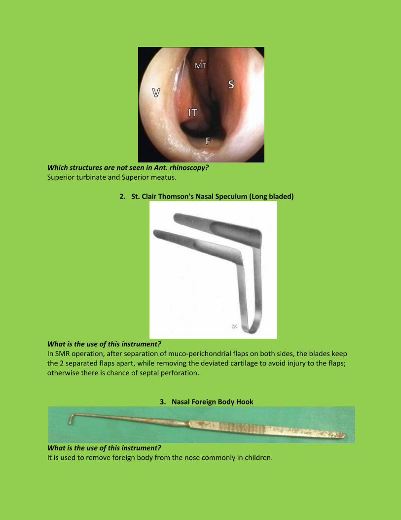
Which structures are not seen in Ant. rhinoscopy? Superior turbinate and Superior meatus.
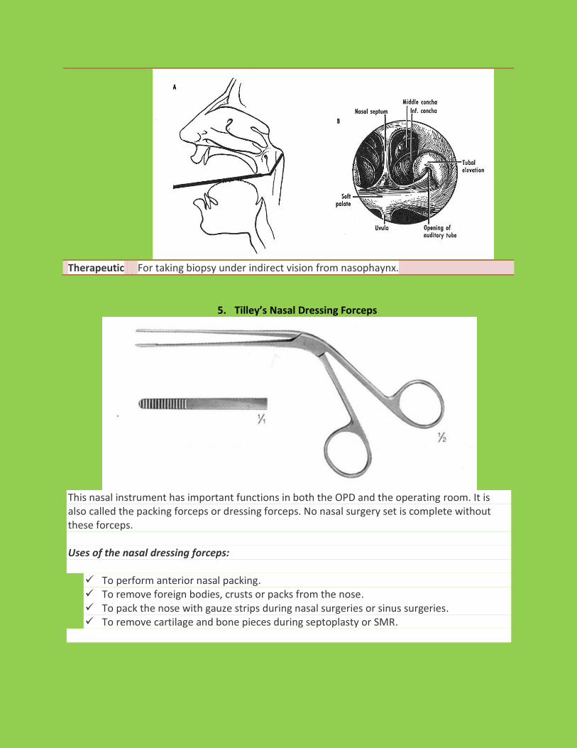
2. St. Clair Thomson’s Nasal Speculum (Long bladed)
What is the use of this instrument? In SMR operation, after separation of muco-perichondrial flaps on both sides, the blades keep the 2 separated flaps apart, while removing the deviated cartilage to avoid injury to the flaps; otherwise there is chance of septal perforation.
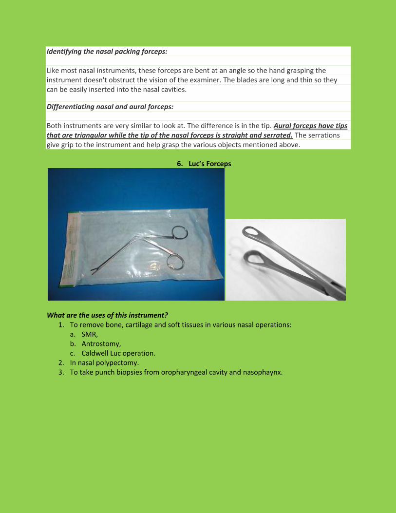
3. Nasal Foreign Body Hook
What is the use of this instrument? It is used to remove foreign body from the nose commonly in children.

What are the histories a child coming with in case of a nasal foreign body? 1. History of a recent foreign body insertion in the nose. 2. History of unilateral foul smelling nasal discharge and nasal obstruction: This is very
much diagnostic of an old nasal foreign body in children. How will you manage the case?
Nasal FB in children should be removed early by a doctor trained in ENT, as faulty technique can cause damage to the nasal mucosa/ the foreign body may be displaced backwards and aspirated into the bronchial tree.
Proper holding of the child by an assistant is important. The FB is usually removed by a nasal foreign body hook in case of round objects (like peas/ beans etc.).
The FB can be removed by a forceps in case of buttons/ papers/ sponges etc.
Tip of the hook is introduced along the roof of the nose and placed behind the FB.
The tip is rotated and FB is lodged in the angle of the hook and brought out.
In a non-cooperative child/ in case of impacted FB; general anaesthesia is required.
4. Posterior Nasal Space (PNS) Mirror
Uses:
Diagnostic Examination of the post nasal space by a procedure called posterior rhinoscopy, an out-patient procedure. The mirror is warmed into normal body temperature and introduced into the oral cavity while the tongue is depressed with a tongue depressor. The mirror is turned upwards in order to examine the post nasal space.
Therapeutic For taking biopsy under indirect vision from nasophaynx.
5. Tilley’s Nasal Dressing Forceps
This nasal instrument has important functions in both the OPD and the operating room. It is also called the packing forceps or dressing forceps. No nasal surgery set is complete without these forceps. Uses of the nasal dressing forceps: To perform anterior nasal packing. To remove foreign bodies, crusts or packs from the nose. To pack the nose with gauze strips during nasal surgeries or sinus surgeries. To remove cartilage and bone pieces during septoplasty or SMR.
Identifying the nasal packing forceps: Like most nasal instruments, these forceps are bent at an angle so the hand grasping the instrument doesn't obstruct the vision of the examiner. The blades are long and thin so they can be easily inserted into the nasal cavities.
Differentiating nasal and aural forceps: Both instruments are very similar to look at. The difference is in the tip. Aural forceps have tips that are triangular while the tip of the nasal forceps is straight and serrated. The serrations give grip to the instrument and help grasp the various objects mentioned above.
6. Luc’s Forceps
What are the uses of this instrument?
1. To remove bone, cartilage and soft tissues in various nasal operations: a. SMR, b. Antrostomy, c. Caldwell Luc operation.
2. In nasal polypectomy. 3. To take punch biopsies from oropharyngeal cavity and nasophaynx.

7. Lichtwitz Antrum Puncture Trocar And Cannula
Use: Antrum is punctured through inferior meatus, usually under local anaesthesia, as it is the most dependent and most accessible area for drainage. Diagnostic 1. To confirm presence of pus in the maxillary sinus and diagnosis of
chronic sinusitis (Proof puncture method). 2. Cytological examination of antral wash out fluid.
Therapeutic 1. For lavaging the maxillary sinus in chronic sinusitis and oroantral fistula.
2. Introduction of indwelling polythene tube in chronic sinusitis in children.
3. Introduction of medication in the antrum.
8. Tilley’s Harpoon And Antral Burr
Uses: Tilley’s harpoon It is used to puncture the medial wall of the antrum at the inferior meatus in
antrostomy operation in case of chronic sinusitis/ along with CL operation.
Tilley’s antral burr It is used to dilate and smoothen the antrostomy opening following puncture by the harpoon.

PART 3: THROAT
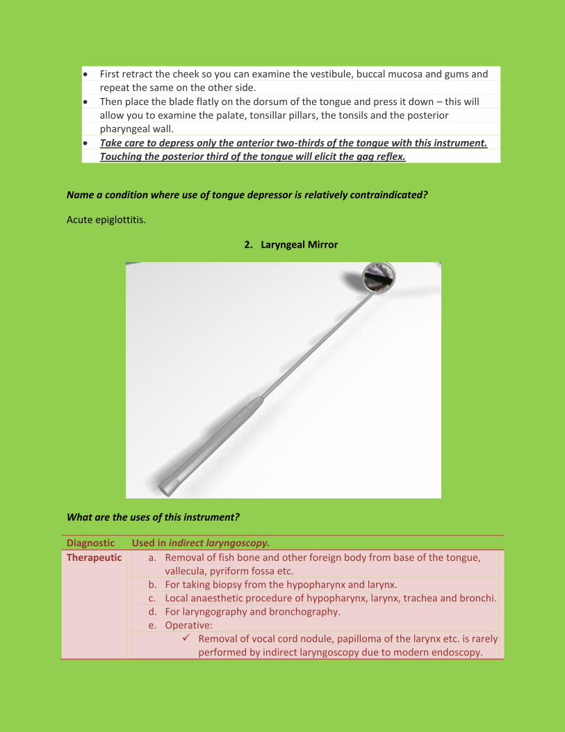
1. Lack’s Tongue Depressor
This is a very commonly used OPD instrument. It has two blades at right angles to each other. One of them is slightly wider than the other and is completely flat. This is the part of the tongue depressor that is inserted into the oral cavity. The other blade is narrower and has a slight curve at its free end, like a handle. This is the part of the instrument that is held in your hand. Uses of the tongue depressor:
Examination of the oral cavity – vestibule, buccal mucosa, gums, floor of the mouth.
Examination of the oropharynx and posterior pharyngeal wall.
Used in posterior rhinoscopy, along with the postnasal mirror.
For the ‘cold spatula test’: to assess the nasal airway/ patency in the OPD.
To perform minor procedures in the oral cavity.
To take a throat swab or a swab from the tonsil. How to use the tongue depressor:
Hold the instrument by the narrower blade that acts as a handle.
Insert the other blade into the oral cavity.
First retract the cheek so you can examine the vestibule, buccal mucosa and gums and repeat the same on the other side.
Then place the blade flatly on the dorsum of the tongue and press it down – this will allow you to examine the palate, tonsillar pillars, the tonsils and the posterior pharyngeal wall.
Take care to depress only the anterior two-thirds of the tongue with this instrument. Touching the posterior third of the tongue will elicit the gag reflex.
Name a condition where use of tongue depressor is relatively contraindicated?
Acute epiglottitis.
2. Laryngeal Mirror
What are the uses of this instrument?
Diagnostic Used in indirect laryngoscopy.
Therapeutic a. Removal of fish bone and other foreign body from base of the tongue, vallecula, pyriform fossa etc.
b. For taking biopsy from the hypopharynx and larynx. c. Local anaesthetic procedure of hypopharynx, larynx, trachea and bronchi. d. For laryngography and bronchography. e. Operative:
Removal of vocal cord nodule, papilloma of the larynx etc. is rarely performed by indirect laryngoscopy due to modern endoscopy.

How indirect laryngoscopy is performed?
Patient is seated opposite the examiner. He should sit erect with the head and
chest leaning slightly towards the examiner.
He is asked to protrude his tongue which is wrapped in gauze and held by the
examiner between the thumb and middle finger.
Index finger is used to keep the upper lip or moustache out of the way.
Gauze piece is used to get a firm grip of the tongue and to protect it against
injury by the lower incisors.
Laryngeal mirror (size 4 to 6) which has been warmed and tested on the back of
hand is introduced into the mouth and held firmly against the uvula and soft
palate.
Light is focussed on the laryngeal mirror and patient is asked to breathe quietly.
To see movements of the cords, patient is asked to take deep inspiration
(abduction of cords), say "Aa" (adduction of cords) and "Eee" (for adduction and
tension). Movements of both the cords are compared.
What are the structures are seen by indirect laryngoscopy?
Larynx. Epiglottis, aryepiglottic folds, arytenoids, cuneiform and corniculate cartilages, ventricular bands, ventricles, true cords, anterior commissure, posterior commissure, subglottis and rings of trachea.
Laryngopharynx: Both pyriform fossae, postcricoid region, posterior wall of laryngopharynx.
Oropharynx: Base of tongue, lingual tonsils, valleculae, medial and lateral glossoepiglottic folds.

Name some types of laryngoscopy.
1. Direct laryngoscopy.
2. Indirect laryngoscopy.
3. Fibre-optic flexible laryngoscopy.
4. Prism-optically enhanced laryngoscopy.
5. Micro- laryngoscopy.
6. Video- laryngoscopy.
7. Stroboscopy.
What are the differences between direct and indirect laryngoscopy?
Points Direct laryngoscopy Indirect laryngoscopy
Anaesthesia It can only be done under general anaesthesia.
No/ little need of anaesthesia; if needed, only local anaesthesia is sufficient.
View It provides a much better and direct view of larynx.
It provides an indirect view of the larynx; so it can’t provide a visualization of the hidden areas of hypopharynx and larynx.
Risk Risk of injury to lips, tongue, teeth and laryngeal edema is there.
Minimal risk.
Position of the patient
Patient lies supine. Head is elevated by 10-15 cm by placing a pillow under the occiput or by raising head flap of the operation table. Neck is flexed on thorax and the head extended on atlanto-
Patient is seated opposite the examiner. He should sit erect with the head and chest leaning slightly towards the examiner. He is asked to protrude his tongue which is wrapped in gauze and
occipital joint (Barking-dog position). held by the examiner between the thumb and middle finger. Index finger is used to keep the upper lip or moustache out of the way.
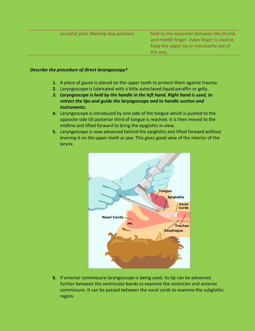
Describe the procedure of direct laryngoscopy?
1. A piece of gauze is placed on the upper teeth to protect them against trauma. 2. Laryngoscope is lubricated with a little autoclaved liquid paraffin or gelly. 3. Laryngoscope is held by the handle in the left hand. Right hand is used, to
retract the lips and guide the laryngoscope and to handle suction and instruments.
4. Laryngoscope is introduced by one side of the tongue which is pushed to the opposite side till posterior third of tongue is reached. It is then moved to the midline and lifted forward to bring the epiglottis in view.
5. Laryngoscope is now advanced behind the epiglottis and lifted forward without levering it on the upper teeth or jaw. This gives good view of the interior of the larynx.
6. If anterior commissure laryngoscope is being used, its tip can be advanced further between the ventricular bands to examine the ventricles and anterior commissure. It can be passed between the vocal cords to examine the subglottic region.
7. Following structures are examined serially: Base of tongue, Right and left valleculae, Epiglottis (its tip, lingual and laryngeal surfaces), Right and left pyriform sinuses, Aryepiglottic folds, Arytenoids, Post-cricoid region, Both false cords, anterior and posterior commissure, right and left
ventricles, Right and left vocal cords and subglottic area. Mobility of vocal cords should also be observed.
TONSILLECTOMY SET
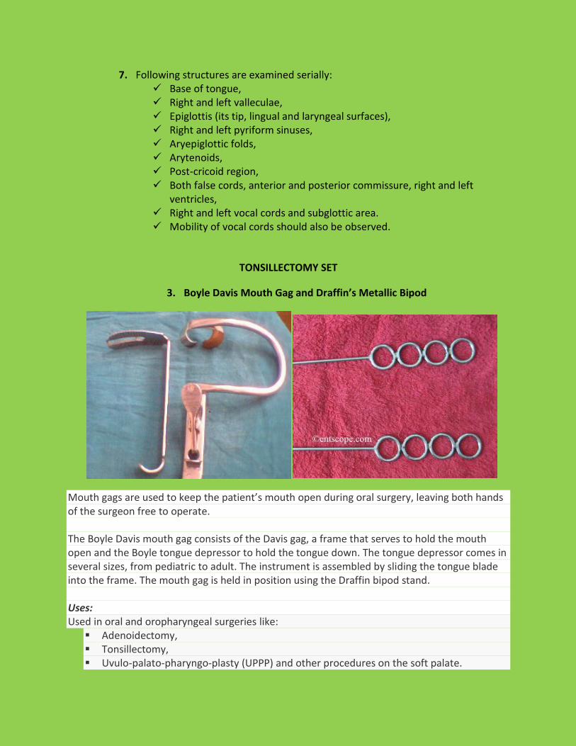
3. Boyle Davis Mouth Gag and Draffin’s Metallic Bipod
Mouth gags are used to keep the patient’s mouth open during oral surgery, leaving both hands of the surgeon free to operate. The Boyle Davis mouth gag consists of the Davis gag, a frame that serves to hold the mouth open and the Boyle tongue depressor to hold the tongue down. The tongue depressor comes in several sizes, from pediatric to adult. The instrument is assembled by sliding the tongue blade into the frame. The mouth gag is held in position using the Draffin bipod stand. Uses: Used in oral and oropharyngeal surgeries like: Adenoidectomy, Tonsillectomy, Uvulo-palato-pharyngo-plasty (UPPP) and other procedures on the soft palate.
Procedures on the hard palate like cyst or tumor excision. Precautions:
It cannot be used to perform procedures on the tongue as it is completely held down by the tongue blade.
This instrument can cause injury to the lips and teeth. Care must be taken while applying the mouth gag to avoid getting the lips caught in it.
Opening the mouth excessively with the gag can cause dislocation of the temporomandibular joint.
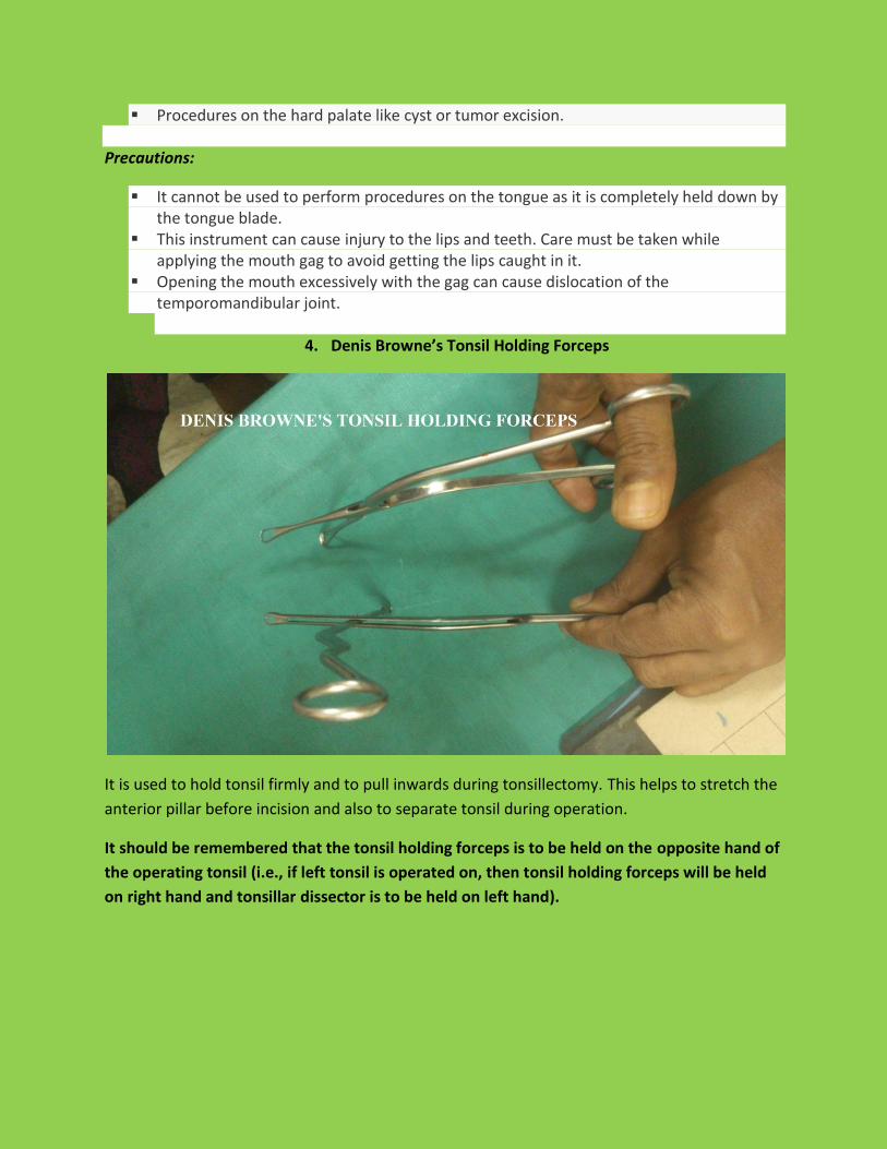
4. Denis Browne’s Tonsil Holding Forceps
It is used to hold tonsil firmly and to pull inwards during tonsillectomy. This helps to stretch the
anterior pillar before incision and also to separate tonsil during operation.
It should be remembered that the tonsil holding forceps is to be held on the opposite hand of
the operating tonsil (i.e., if left tonsil is operated on, then tonsil holding forceps will be held
on right hand and tonsillar dissector is to be held on left hand).
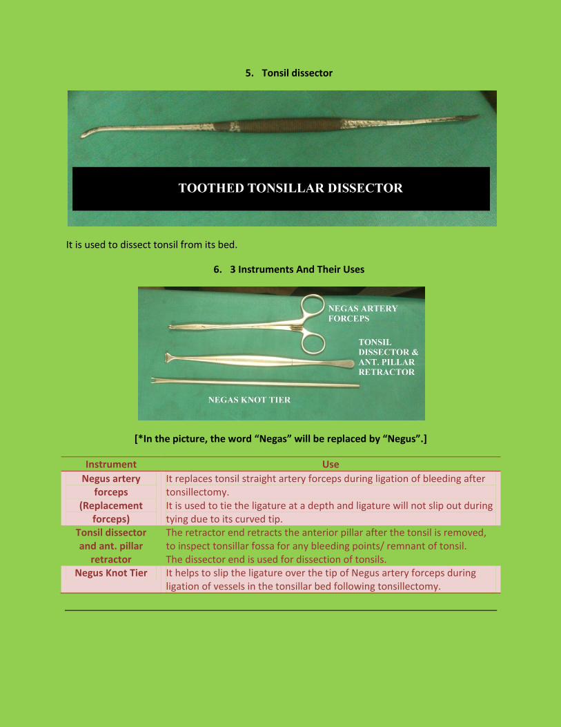
5. Tonsil dissector
It is used to dissect tonsil from its bed.
6. 3 Instruments And Their Uses
[*In the picture, the word “Negas” will be replaced by “Negus”.]
Instrument Use
Negus artery forceps
(Replacement forceps)
It replaces tonsil straight artery forceps during ligation of bleeding after tonsillectomy. It is used to tie the ligature at a depth and ligature will not slip out during tying due to its curved tip.
Tonsil dissector and ant. pillar
retractor
The retractor end retracts the anterior pillar after the tonsil is removed, to inspect tonsillar fossa for any bleeding points/ remnant of tonsil. The dissector end is used for dissection of tonsils.
Negus Knot Tier It helps to slip the ligature over the tip of Negus artery forceps during ligation of vessels in the tonsillar bed following tonsillectomy.
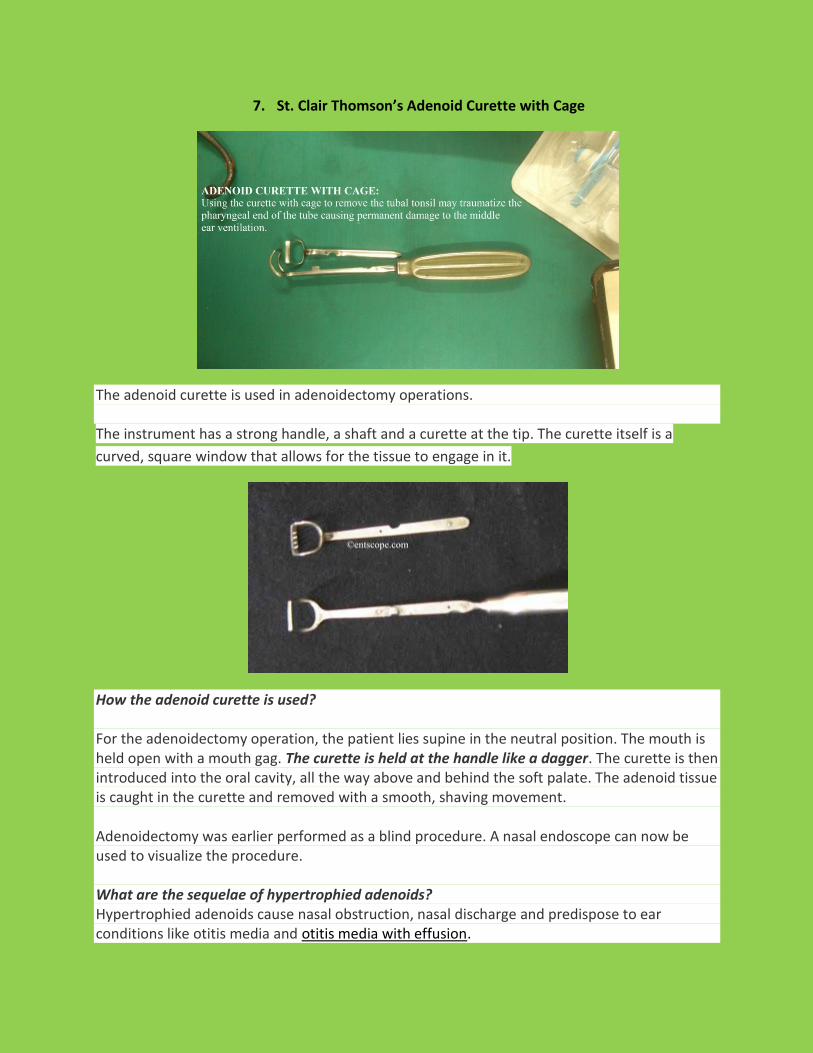
7. St. Clair Thomson’s Adenoid Curette with Cage
The adenoid curette is used in adenoidectomy operations. The instrument has a strong handle, a shaft and a curette at the tip. The curette itself is a
curved, square window that allows for the tissue to engage in it.
How the adenoid curette is used? For the adenoidectomy operation, the patient lies supine in the neutral position. The mouth is held open with a mouth gag. The curette is held at the handle like a dagger. The curette is then introduced into the oral cavity, all the way above and behind the soft palate. The adenoid tissue is caught in the curette and removed with a smooth, shaving movement. Adenoidectomy was earlier performed as a blind procedure. A nasal endoscope can now be used to visualize the procedure. What are the sequelae of hypertrophied adenoids? Hypertrophied adenoids cause nasal obstruction, nasal discharge and predispose to ear conditions like otitis media and otitis media with effusion.

More information: Endoscopic adenoidectomy achieves better results and lesser complications as the
procedure is performed under visualization. Endoscopic adenoidectomy can be performed using endoscopic instruments and/or
microdebrider.
TRACHEOSTOMY SET
8. Fuller’s Bivalve Metallic Tracheostomy Tube
It consists of outer tube and inner tube/ cannula.
What is the advantage of a bivalve tube?
Bivalve tube acts itself as a tracheal dilator. So it helps in introduction into tracheostome
without assistance of tracheal dilator.
Shield
Fenestra
Why the inner tube is longer than outer tube?
So that when the tube gets blocked, on removal of the inner tube, the blockage is cleared.
Why there is an opening on the postero-superior wall of the inner tube?
It helps in decannulation, i.e., determines whether normal air passage is established on blocking
the tracheostomy stroma.
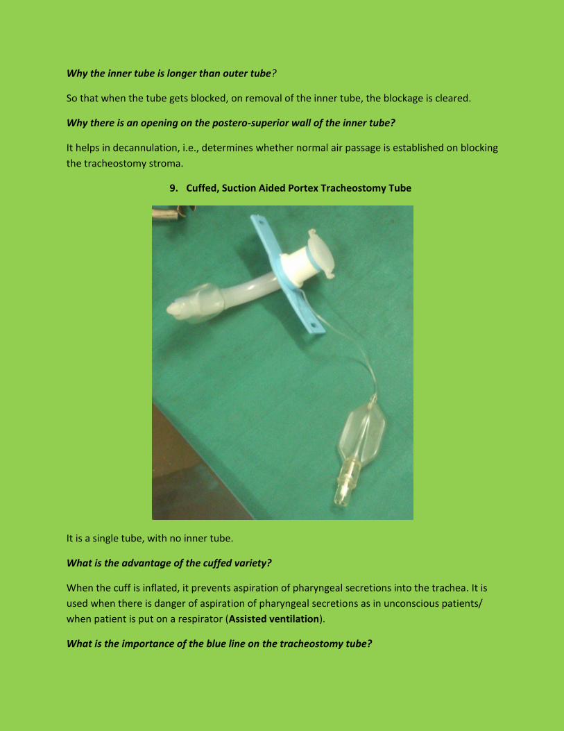
9. Cuffed, Suction Aided Portex Tracheostomy Tube
It is a single tube, with no inner tube.
What is the advantage of the cuffed variety?
When the cuff is inflated, it prevents aspiration of pharyngeal secretions into the trachea. It is
used when there is danger of aspiration of pharyngeal secretions as in unconscious patients/
when patient is put on a respirator (Assisted ventilation).
What is the importance of the blue line on the tracheostomy tube?
The blue line is radio-opaque in the X-Ray. So it may be used to detect the position of the tube
if it falls.
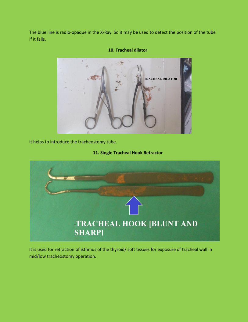
10. Tracheal dilator
It helps to introduce the tracheostomy tube.
11. Single Tracheal Hook Retractor
It is used for retraction of isthmus of the thyroid/ soft tissues for exposure of tracheal wall in
mid/low tracheostomy operation.
Thank you….
If you find any mistake in this document, please
mail it to [email protected].
Resources Used
Dhingra’s Diseases of Ear, Nose and Throat.
S.K.Dey’s ENT Textbook.
Wikipedia.
Picture courtesy:
Some pictures are taken from ENT dept. of
R.G.Kar Medical College [Thanks to Rishi Ranjan
Shrivastava].
Rests are taken from various sources of
internet.
20-Nov-13
XPrithwiraj Maiti
Student, R.G.KAR MEDICAL COLLEGE