
Common Elbow, Wrist, and Hand injuries in Youth Sports Elbow, Wrist, and Hand injuries in Youth...
92
Common Elbow, Wrist, and Hand injuries in Youth Sports Jamie Monica, MD February 21, 2015
Transcript of Common Elbow, Wrist, and Hand injuries in Youth Sports Elbow, Wrist, and Hand injuries in Youth...
Common Elbow, Wrist, and Hand injuries
in Youth Sports
Jamie Monica, MD
February 21, 2015

I have no disclosures
Elbow injuries
Epidemiology
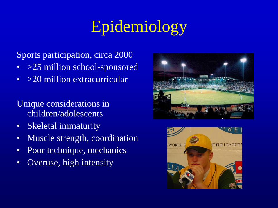
Sports participation, circa 2000
• >25 million school-sponsored
• >20 million extracurricular
Unique considerations in children/adolescents
• Skeletal immaturity
• Muscle strength, coordination
• Poor technique, mechanics
• Overuse, high intensity
Sports-Specialized Intensive Training
and the Risk of Injury in Young Athletes:
A Clinical Case-Control Study.
• 1200 youth athletes
• Early specialization in a single sport is one
of the strongest predictors of injury
• Athletes in the study who specialized
were 70% to 93% more likely to be
injured than children who played multiple
sports
Am J Sports Med. 2015 Feb 2 Jayanthi NA1, LaBella CR2, Fischer D3, Pasulka J4, Dugas LR3
http://www.ncbi.nlm.nih.gov/pubmed/?term=Jayanthi%20NA%5BAuthor%5D&cauthor=true&cauthor_uid=25646361
http://www.ncbi.nlm.nih.gov/pubmed/?term=Jayanthi%20NA%5BAuthor%5D&cauthor=true&cauthor_uid=25646361
Recommendations
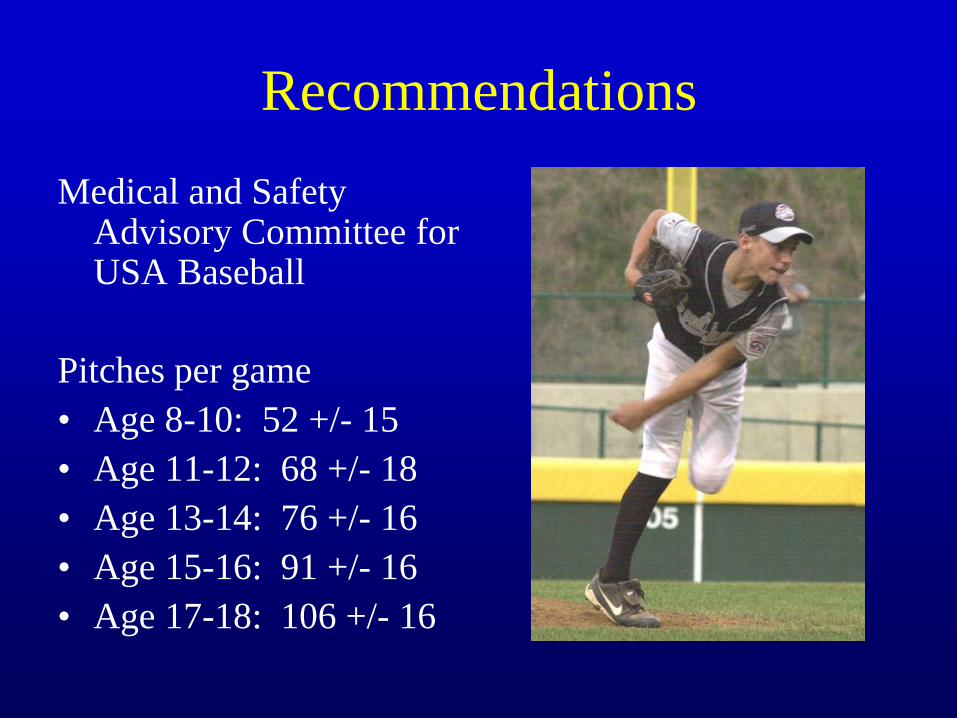
Medical and Safety Advisory Committee for USA Baseball
Pitches per game
• Age 8-10: 52 +/- 15
• Age 11-12: 68 +/- 18
• Age 13-14: 76 +/- 16
• Age 15-16: 91 +/- 16
• Age 17-18: 106 +/- 16
Anatomy
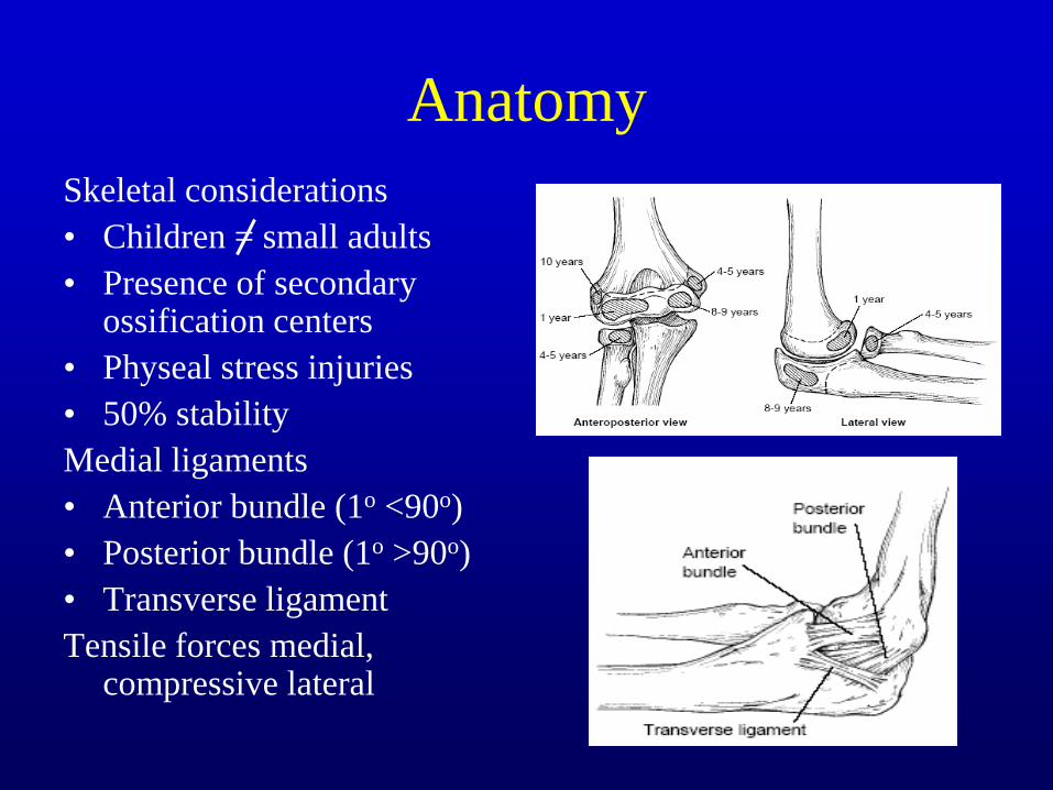
Skeletal considerations
• Children = small adults
• Presence of secondary ossification centers
• Physeal stress injuries
• 50% stability
Medial ligaments
• Anterior bundle (1o <90o)
• Posterior bundle (1o >90o)
• Transverse ligament
Tensile forces medial, compressive lateral
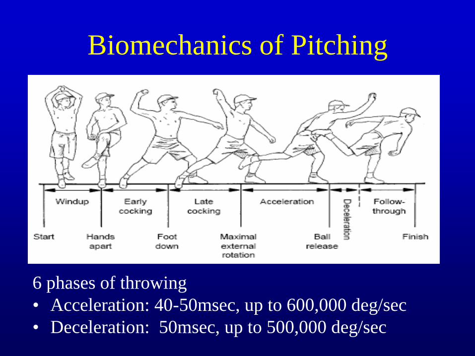
Biomechanics of Pitching
6 phases of throwing
• Acceleration: 40-50msec, up to 600,000 deg/sec
• Deceleration: 50msec, up to 500,000 deg/sec

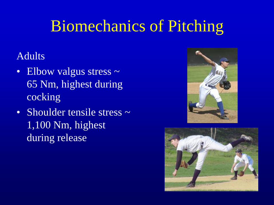
Biomechanics of Pitching
Adults
• Elbow valgus stress ~
65 Nm, highest during
cocking
• Shoulder tensile stress ~
1,100 Nm, highest
during release
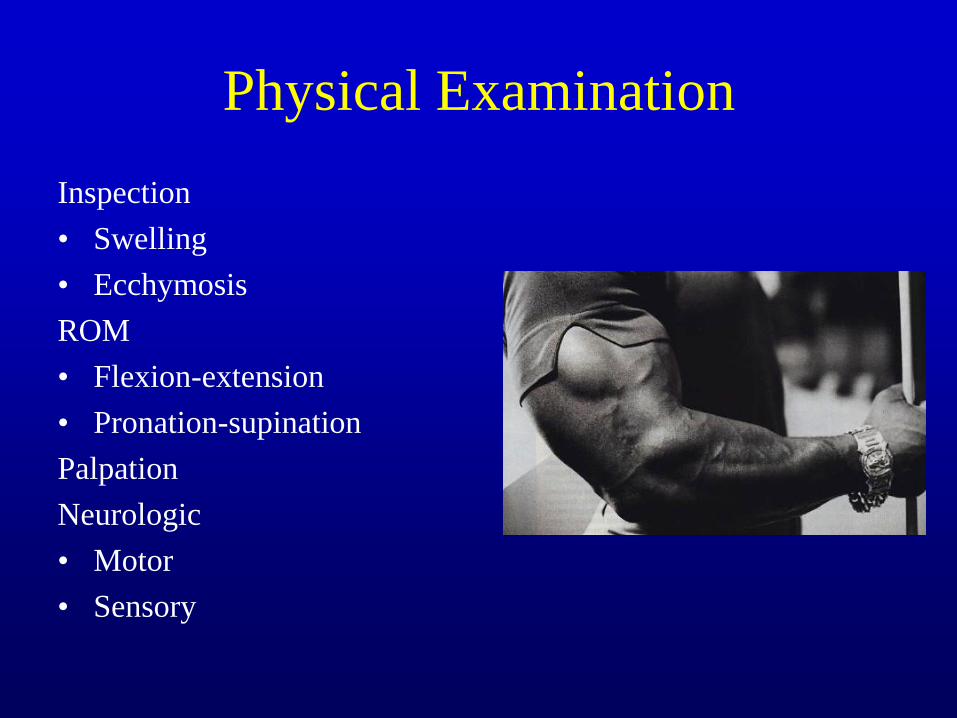
Physical Examination
Inspection
• Swelling
• Ecchymosis
ROM
• Flexion-extension
• Pronation-supination
Palpation
Neurologic
• Motor
• Sensory

Physical Examination
Valgus instability
• Anterior bundle (A)
– Elbow flexed 20-30o
– Valgus stress
• Posterior bundle (B)
– Elbow flexed 90o,
forearm supinated
– Pull outward on thumb
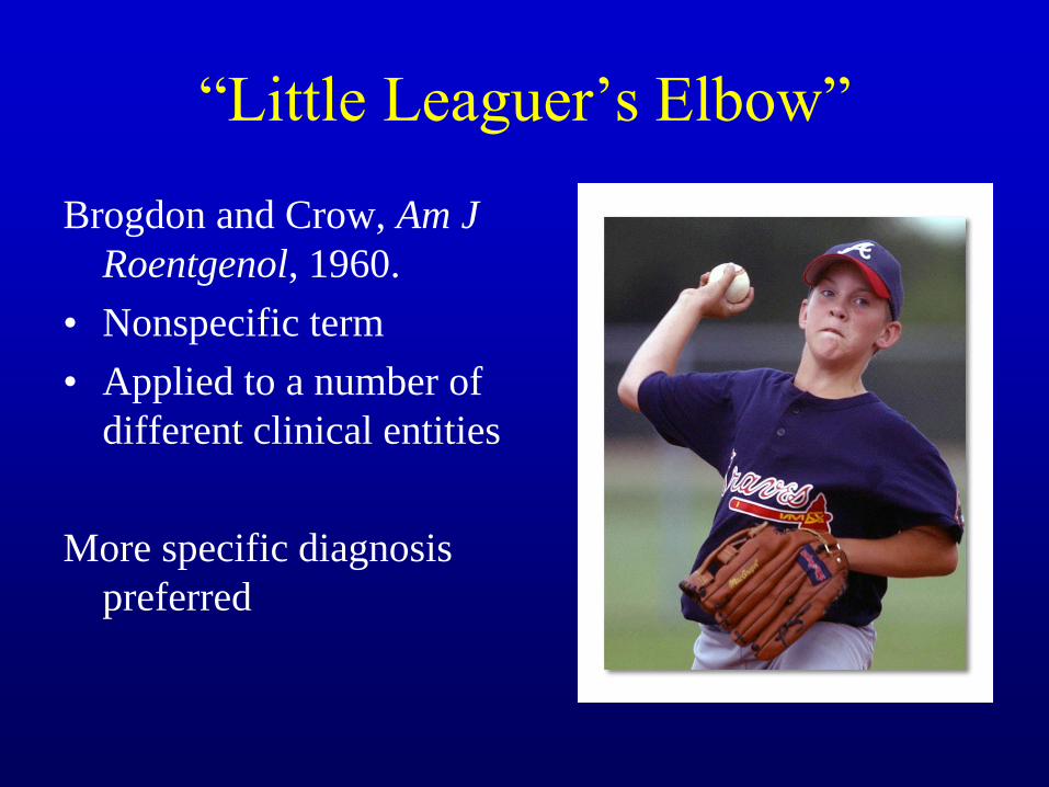
“Little Leaguer’s Elbow”
Brogdon and Crow, Am J
Roentgenol, 1960.
• Nonspecific term
• Applied to a number of
different clinical entities
More specific diagnosis
preferred
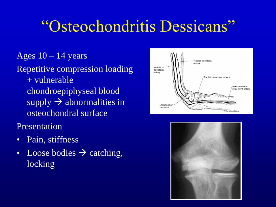
“Osteochondritis Dessicans”
Ages 10 – 14 years
Repetitive compression loading
+ vulnerable
chondroepiphyseal blood
supply abnormalities in
osteochondral surface
Presentation
• Pain, stiffness
• Loose bodies catching,
locking

“Osteochondritis Dessicans”
Diagnosis
• Radiographs
• MRI
Classification
• Multiple
• Radiographic, arthroscopic
• Defilice et al., 2001
• Keys: cartilage integrity, fragment stability

“Osteochondritis Dessicans”
Treatment
• Stable lesion
– Rest, NSAIDs, PT
– Drilling in situ
• Unstable lesion
– Arthroscopy vs. arthrotomy
– Debridement
– Internal fixation
– Loose body excision
– Marrow stimulation

Panner’s Disease
Panner, Acta Radiol, 1927.
<10 years of age
Fissuring irregulatiry
fragmentation
reossification
remodeling, resolution of
symptoms
Entire capitellar involvement
Self-limiting
“Perthes of the elbow”
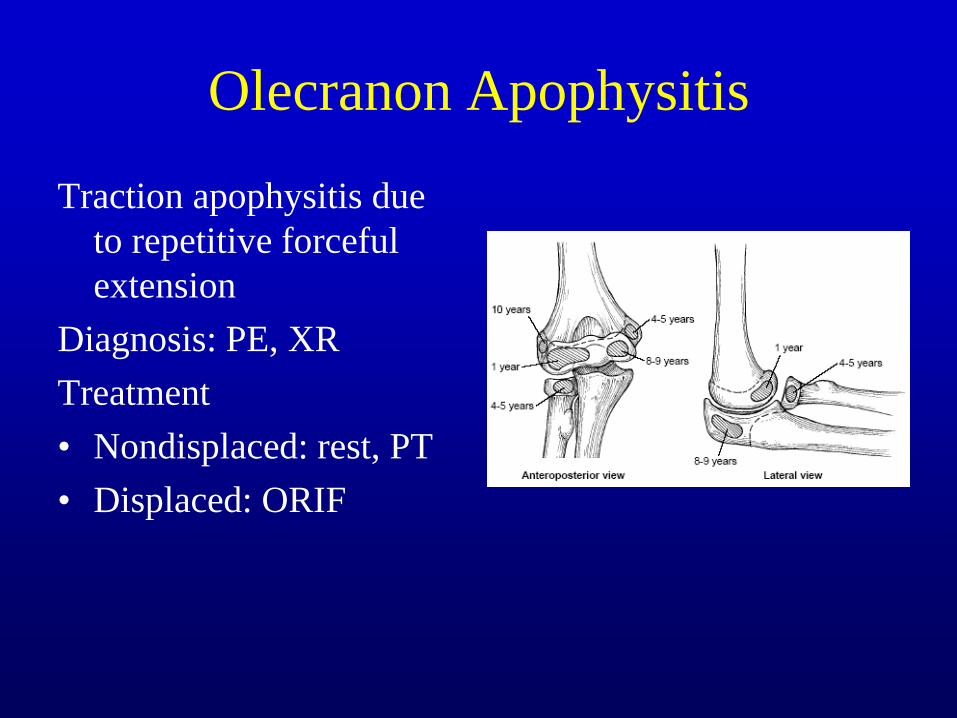
Olecranon Apophysitis
Traction apophysitis due
to repetitive forceful
extension
Diagnosis: PE, XR
Treatment
• Nondisplaced: rest, PT
• Displaced: ORIF

Medial Epicondylitis
“Golfer’s elbow”
Pathophysiology
• Repetitive valgus stress inflammation of flexor-pronator origin
Diagnosis
• PE, MRI
Treatment
• Rest, NSAIDs, bracing
• Injections?
• Debridement

Medial Epicondyle Fracture
In skeletally immature
athletes, may have
physeal injury/avulsion
Diagnosis: XR, MRI
• Physeal widening
Treatment
• Nondisplaced: rest, PT
• Displaced: ORIF?
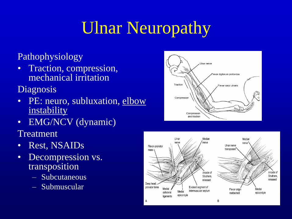
Ulnar Neuropathy
Pathophysiology
• Traction, compression, mechanical irritation
Diagnosis
• PE: neuro, subluxation, elbow instability
• EMG/NCV (dynamic)
Treatment
• Rest, NSAIDs
• Decompression vs. transposition – Subcutaneous
– Submuscular
• What is the main
difference between
Panner’s disease
and OCD lesion of
the elbow?
• A. Panner’s disease is
associated with other
comorbidities.
• B. OCD has a favorable
prognosis.
• C. Panner’s disease
occurs in patient’s under
10.
• D. Patient’s with Panner’s
disease often do not have
pain.

Wrist Injuries
Wrist injuries
Variable presentations
Sport-specific
Special considerations in competitive athletes – Return to play
– Performance expectations
– Protective/assistive devices
– Psychosocial
– Financial

Epidemiology Little information regarding incidence and prevalence
Torjussen et al, AJSM, 2005
• 12% wrist/hand injuries in elite snowboarders
Logan, BJSM, 2004
• 28% wrist/hand injuries in rock climbers
Jacobson et al, JOSPT, 2004
• 16% wrist/hand injuries in cheerleaders
Similar numbers in other sports
• Basketball
• Skating
• Soccer
• Gymnastics
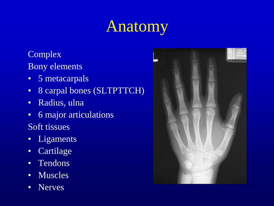
Anatomy
Complex
Bony elements
• 5 metacarpals
• 8 carpal bones (SLTPTTCH)
• Radius, ulna
• 6 major articulations
Soft tissues
• Ligaments
• Cartilage
• Tendons
• Muscles
• Nerves
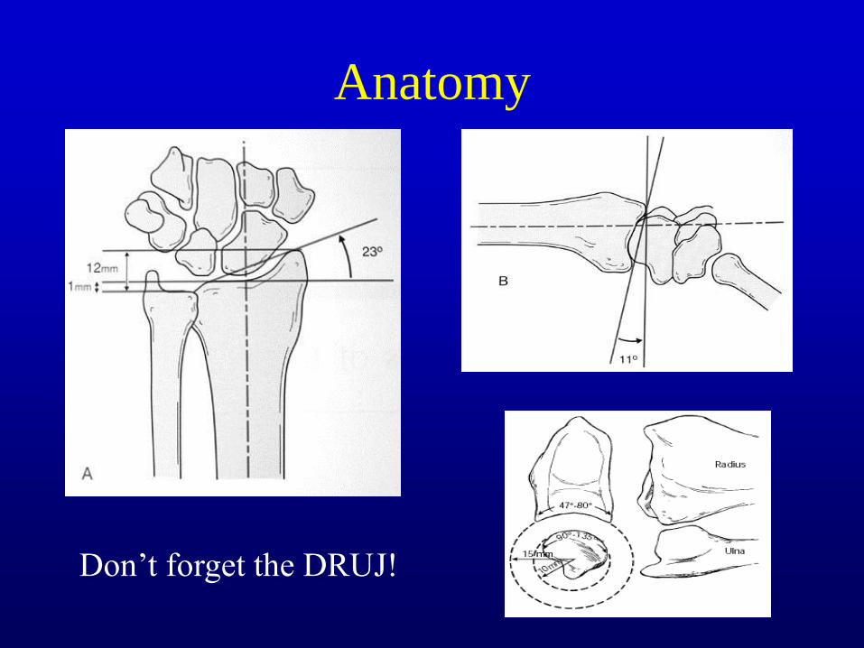
Anatomy
Don’t forget the DRUJ!
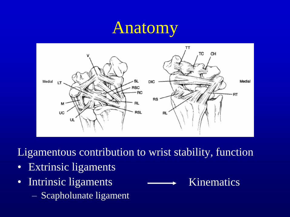
Anatomy
Ligamentous contribution to wrist stability, function
• Extrinsic ligaments
• Intrinsic ligaments
– Scapholunate ligament
Kinematics

Physical examination: observation
Deformity
Swelling
Ecchymosis
Wounds
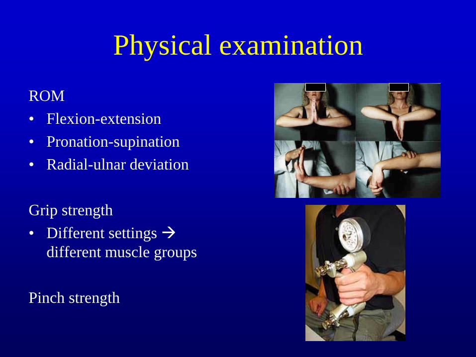
Physical examination
ROM
• Flexion-extension
• Pronation-supination
• Radial-ulnar deviation
Grip strength
• Different settings
different muscle groups
Pinch strength
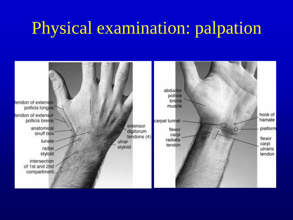
Physical examination: palpation

Radiographic evaluation
Expedited diagnosis critical
Earlier utilization of radiographic imaging to guide treatment
Common modalities
• Plain radiographs
• Computed tomography (CT)
• Magnetic resonance imaging (MRI)
• Fluorosocpy
• ?anesthetic arthrogram
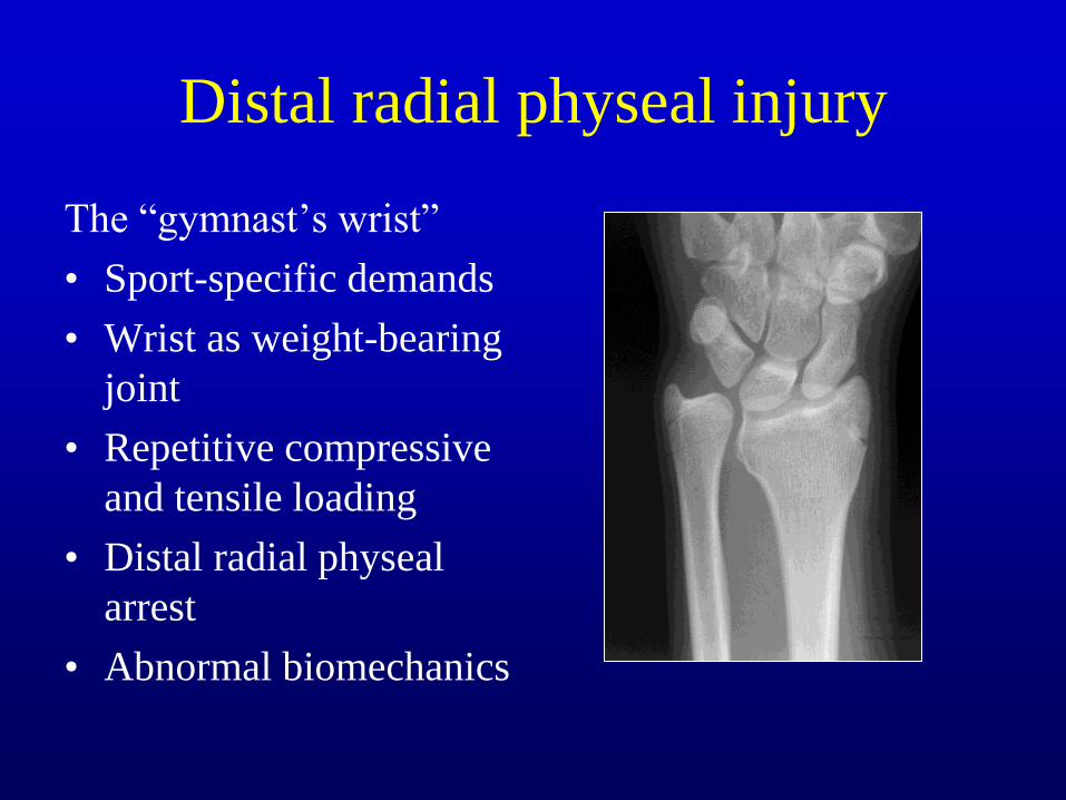
Distal radial physeal injury
The “gymnast’s wrist”
• Sport-specific demands
• Wrist as weight-bearing
joint
• Repetitive compressive
and tensile loading
• Distal radial physeal
arrest
• Abnormal biomechanics
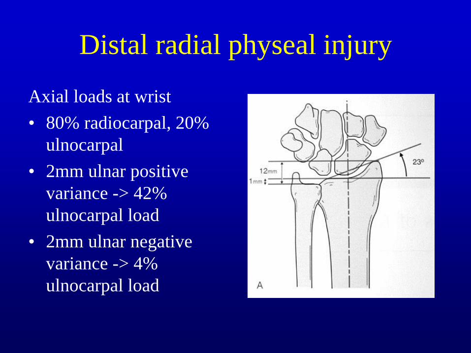
Distal radial physeal injury
Axial loads at wrist
• 80% radiocarpal, 20%
ulnocarpal
• 2mm ulnar positive
variance -> 42%
ulnocarpal load
• 2mm ulnar negative
variance -> 4%
ulnocarpal load
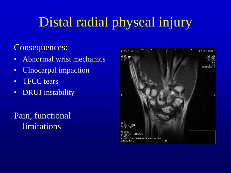
Distal radial physeal injury
Consequences:
• Abnormal wrist mechanics
• Ulnocarpal impaction
• TFCC tears
• DRUJ instability
Pain, functional
limitations
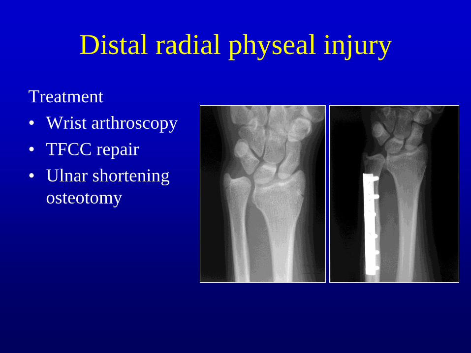
Distal radial physeal injury
Treatment
• Wrist arthroscopy
• TFCC repair
• Ulnar shortening
osteotomy

Scaphoid fractures
Most commonly fractured
carpal bone
• Fall onto outstretched
hand, wrist extended
• Pain, swelling
• Tenderness at anatomic
snuffbox or scaphoid
tubercle

Scaphoid fractures
Difficulties in radiographic
diagnosis
• Complex shape of the
scaphoid
• Proximal row kinematics
with wrist motion
• “scaphoid view” with ulnar
deviation of wrist
• “radiographically occult”
fractures

Scaphoid fractures
Challenges:
• Difficult to diagnose
• Require long time for
healing (2-3months)
• Significant nonunion
rate
• Risk of osteonecrosis
• Risk of arthrosis
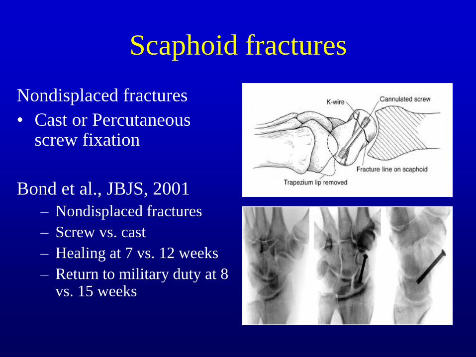
Scaphoid fractures
Nondisplaced fractures
• Cast or Percutaneous screw fixation
Bond et al., JBJS, 2001
– Nondisplaced fractures
– Screw vs. cast
– Healing at 7 vs. 12 weeks
– Return to military duty at 8 vs. 15 weeks

Scaphoid fractures
Displaced fractures
• Risk of nonunion
• Risk of arthrosis
• Treatment: ORIF!
Nonunion, osteonecrosis
• ORIF with bone graft
• Vascularized bone graft
• Salvage procedures
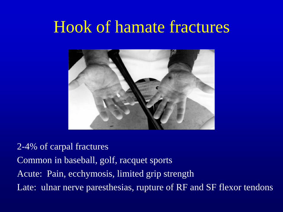
Hook of hamate fractures
2-4% of carpal fractures
Common in baseball, golf, racquet sports
Acute: Pain, ecchymosis, limited grip strength
Late: ulnar nerve paresthesias, rupture of RF and SF flexor tendons
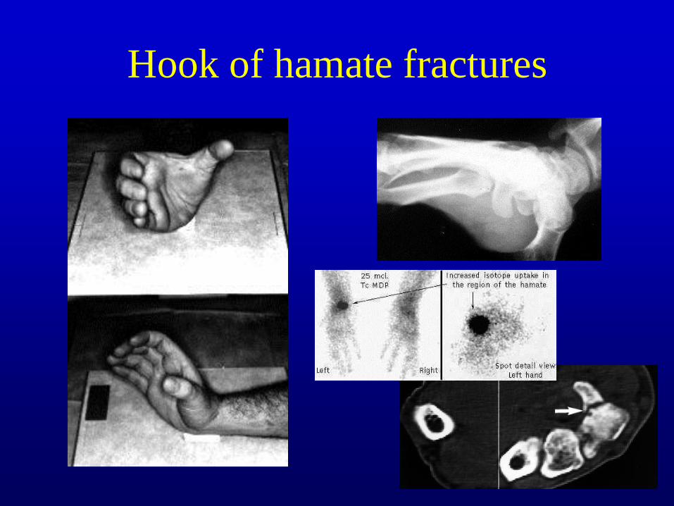
Hook of hamate fractures

Hook of hamate fractures
Treatment:
• Acute
– Cast/splint immobilization
– excision
• Chronic
– ORIF
– Excision
• Postop recovery
– ROM at 1 week
– Sports at 6-8 weeks
– Painful scar!

TFCC tears
What is the TFCC?
• Triangular fibrocartilage complex
• Multiple components
– ECU subsheath
– Ulnar collateral ligament
– Meniscal homologue
– Articular disk/ TFC
– DRUL/ PRUL
– UL/ UT ligaments
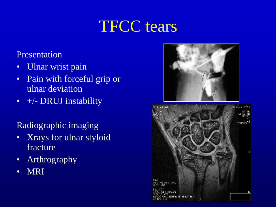
TFCC tears
Presentation
• Ulnar wrist pain
• Pain with forceful grip or ulnar deviation
• +/- DRUJ instability
Radiographic imaging
• Xrays for ulnar styloid fracture
• Arthrography
• MRI
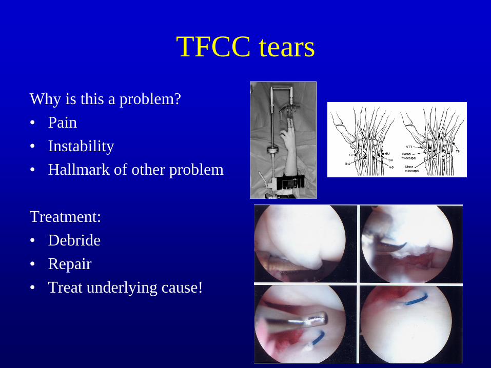
TFCC tears
Why is this a problem?
• Pain
• Instability
• Hallmark of other problem
Treatment:
• Debride
• Repair
• Treat underlying cause!

Ulnar styloid fractures
Often associated with
distal radius fractures
Present with painful
nonunion
Treatment considerations
• Size of fragment
• TFCC tear
• DRUJ stability

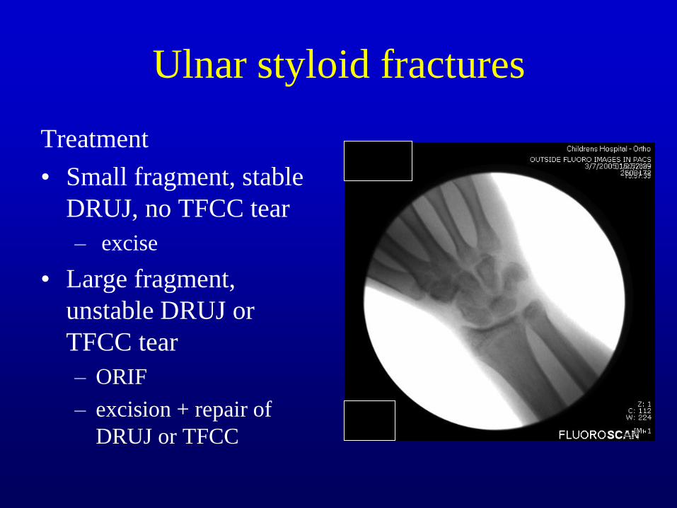
Ulnar styloid fractures
Treatment
• Small fragment, stable
DRUJ, no TFCC tear
– excise
• Large fragment,
unstable DRUJ or
TFCC tear
– ORIF
– excision + repair of
DRUJ or TFCC

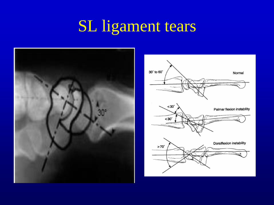
SL ligament tears
Scapholunate ligament
• Critical in wrist kinematics,
linking scaphoid to lunate
• Fall onto outstretched hand
• Tear pain, instability
• Failure leads to predictable
progression to early wrist
arthritis

SL ligament tears
Radiographic evaluation
• Widening of SL interval (>3mm)
• Cortical ring sign
• Trapezoid shape of lunate
• Increased scapholunate angle (>60 degrees)
SL ligament tears

DRUJ instability
“The forgotten joint”
Complex; difficult to diagnose
Instability usually associated
with distal radius or ulnar
styloid fracture, TFCC
injury
Pronation ulna goes dorsally
Radiographic evaluation
• Plain radiographs
• CT (in multiple positions)
• +/- MRI
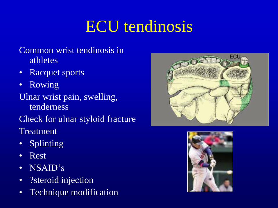
ECU tendinosis
Common wrist tendinosis in athletes
• Racquet sports
• Rowing
Ulnar wrist pain, swelling, tenderness
Check for ulnar styloid fracture
Treatment
• Splinting
• Rest
• NSAID’s
• ?steroid injection
• Technique modification

ECU subluxation
Acute injury > overuse
Examination
• Ulnar deviation, supination,
flexion
• Injection test
Acute treatment
• Casting in pronation and
wrist extension
Surgical treatment
• Repair vs. reconstruction

• What is the
treatment for
negative scaphoid
xrays in a patient
with snuffbox
tenderness?
• A. ORIF
• B. Thumb spica
cast or splint and
referral to a hand
specialist
• C. Immediate MRI
• D. Repeat xrays in
one day after
immobilization

Hand Injuries

Epidemiology
Fetter-Zarzeka & Joseph, Pediatr Emerg Care, 2002
• Review of 382 hand injuries over 8 months, urban pediatric ER
• Lacerations (30%), fractures (16%), infections (4%)
• <2 years: fingertip injuries
• 12-16 years: sports-related hand injuries
Epidemiology
Hastings & Simmons, Clin Orthop, 1984
• 354 pediatric hand fractures, 2 year follow-up
• Small percentage of injuries -> large percentage of complications and poor outcomes
• Malunion risks
– Failure to obtain adequate x-rays
– False assumptions about remodelling
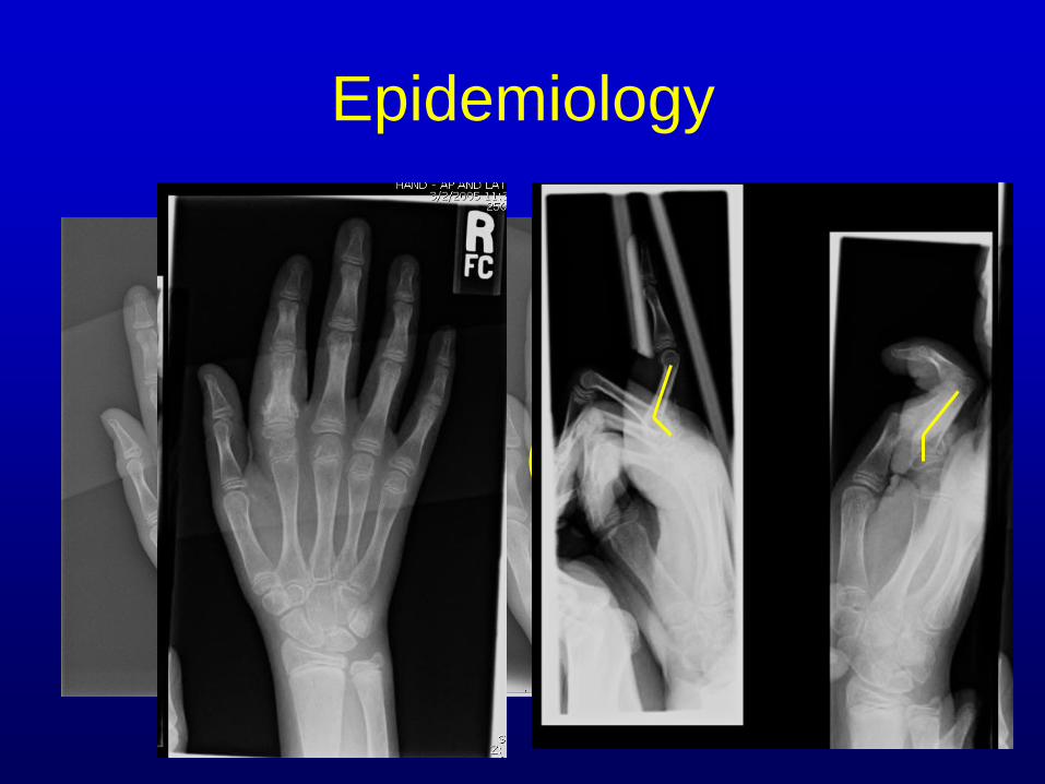
Epidemiology
Epidemiology
Hastings & Simmons, Clin Orthop, 1984
• Problem fractures:
– Open
– Displaced articular
– SH I of distal phalanx
– Phalangeal neck
• Key: recognition of problematic injuries

Unique considerations
Challenges in examination
Presence of physis
Small size of musculo-skeletal structures
Need for more restrictive immobilization
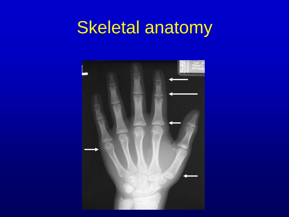
Skeletal anatomy
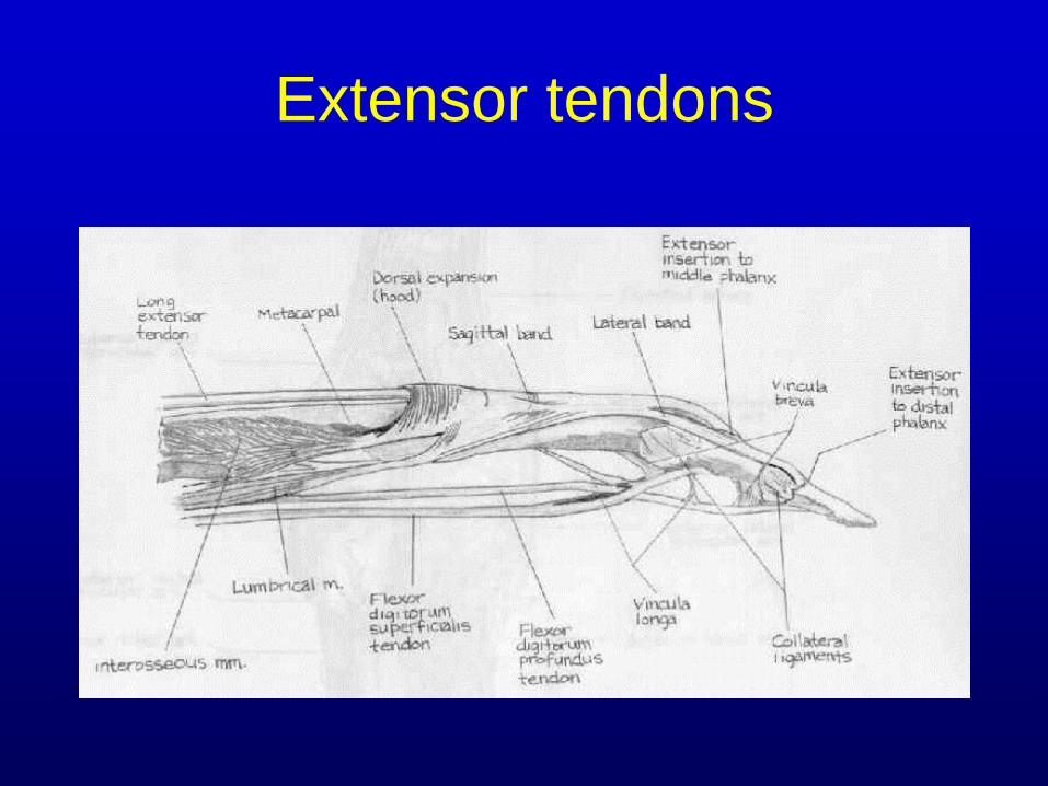
Extensor tendons

Flexor tendons
FDP and FDS tendons
Camper’s chiasm
Vascular supply
• vinculae

Examination: observation
Deformity
Swelling
Ecchymosis
Wounds
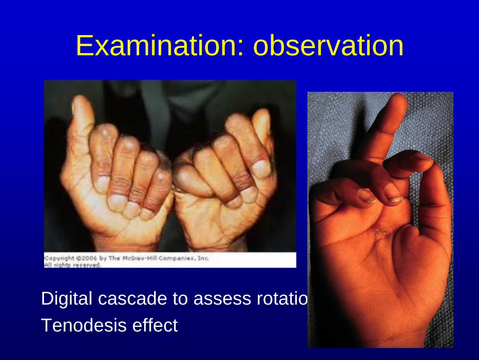
Examination: observation
Digital cascade to assess rotation
Tenodesis effect
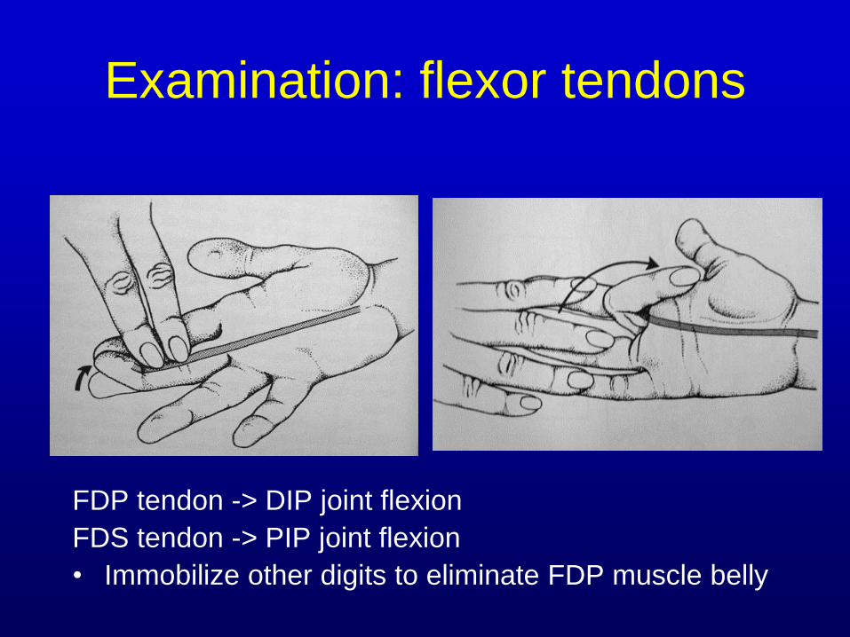
Examination: flexor tendons
FDP tendon -> DIP joint flexion
FDS tendon -> PIP joint flexion
• Immobilize other digits to eliminate FDP muscle belly

Examination: extensor tendons
Terminal tendon -> DIP
Central slip -> PIP
Sagittal bands -> MCP
Intrinsic muscles ->
• MCP flexion
• IP extension

Examination: nerves
Digital nerves
(Radial and ulnar)
• 2-point discrimination
• Threshold testing
• Warm immersion
testing
Median
• Opposition, finger
flexion
Ulnar
• Pinch, crossing
fingers
Radial
• Finger extension

Fractures
Non-physeal (64%)
Non-displaced (55%)
Closed (95%)
Salter-Harris classification of physeal fractures
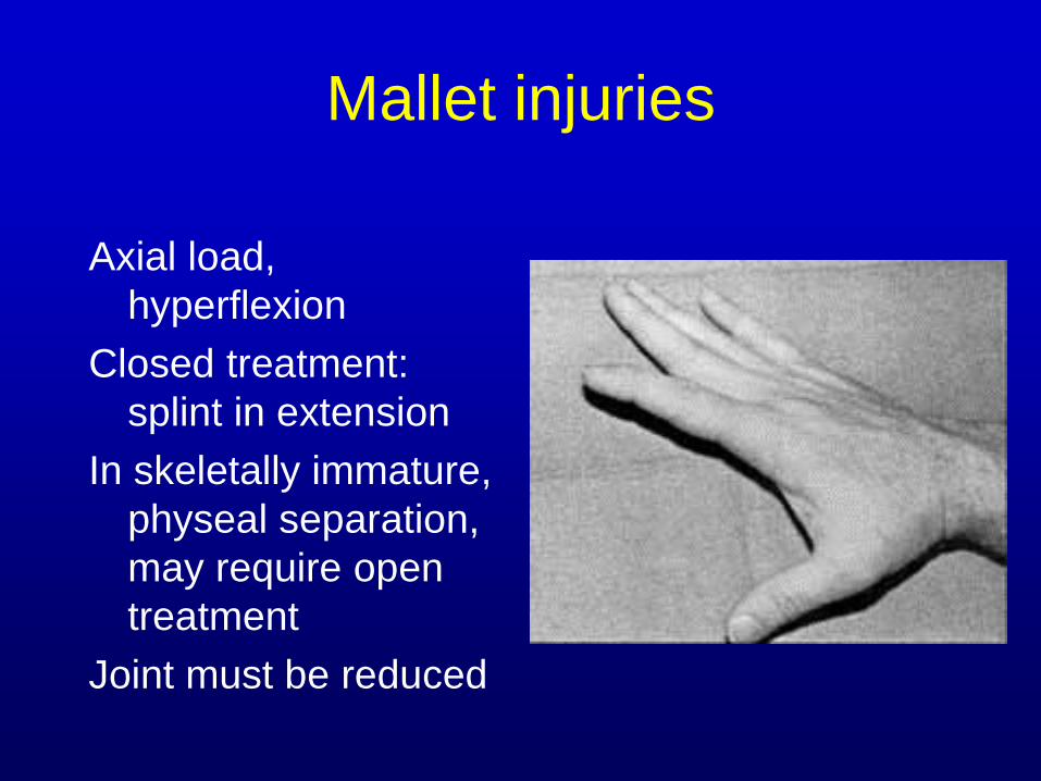
Mallet injuries
Axial load,
hyperflexion
Closed treatment:
splint in extension
In skeletally immature,
physeal separation,
may require open
treatment
Joint must be reduced

Mallet injuries

Seymour’s fracture*
Physeal fracture of distal phalanx with nailbed
laceration
• Incarceration of germinal matrix
High index of suspicion
Requires nail removal, I&D, nailbed repair, fracture
reduction
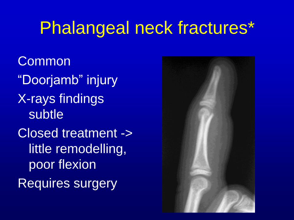
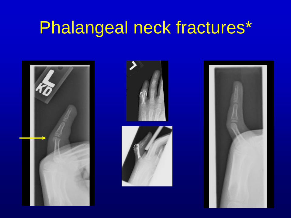
Phalangeal neck fractures*
Common
“Doorjamb” injury
X-rays findings
subtle
Closed treatment ->
little remodelling,
poor flexion
Requires surgery
Phalangeal neck fractures*

Intercondylar phalangeal
fracture Any displacement in
any joint of the hand
is indication for hand
surgery consultation
“Flake” of bone in joint
is osteochondral
fragment until proven
otherwise

Phalangeal shaft fracture
Assess for rotational malalignment!
Most amenable to closed reduction, immobilization
Unstable, irreducible injuries require surgical stabilization

Phalangeal physeal fractures
Salter-Harris II of small
finger most common
• “extra-octave” fracture
• Usually amenable to
closed reduction
• Check x-rays after
buddy taping
• Cast immobilization
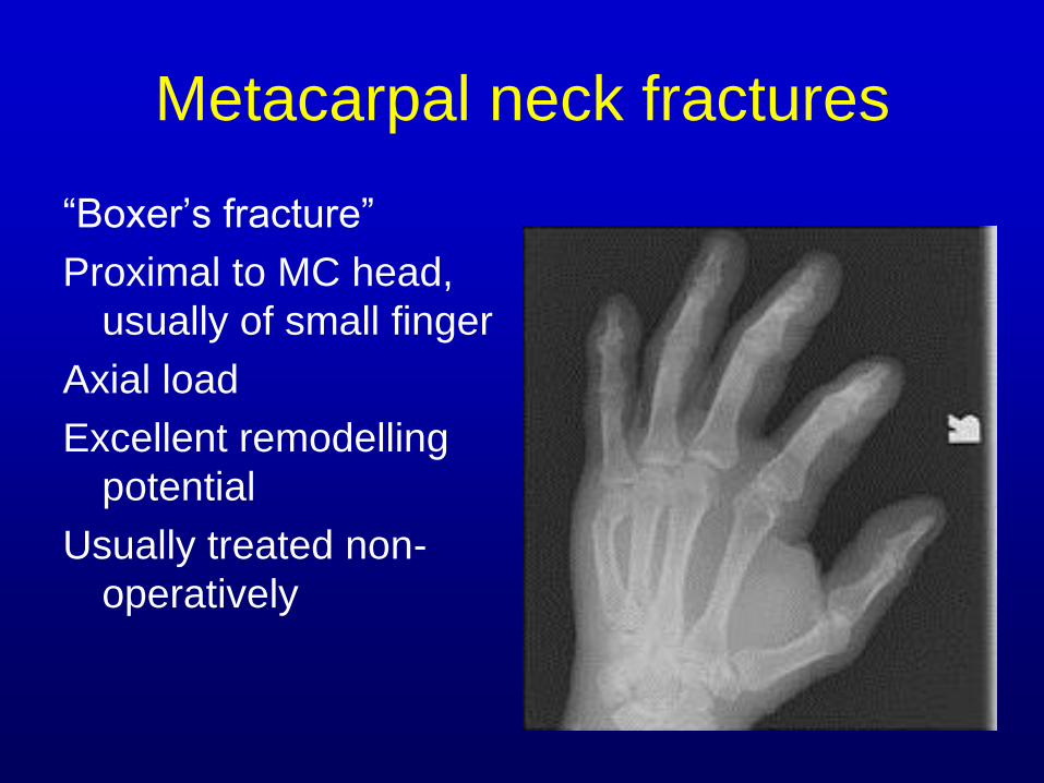
Metacarpal neck fractures
“Boxer’s fracture”
Proximal to MC head,
usually of small finger
Axial load
Excellent remodelling
potential
Usually treated non-
operatively
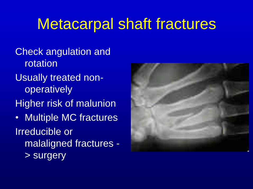
Metacarpal shaft fractures
Check angulation and
rotation
Usually treated non-
operatively
Higher risk of malunion
• Multiple MC fractures
Irreducible or
malaligned fractures -
> surgery
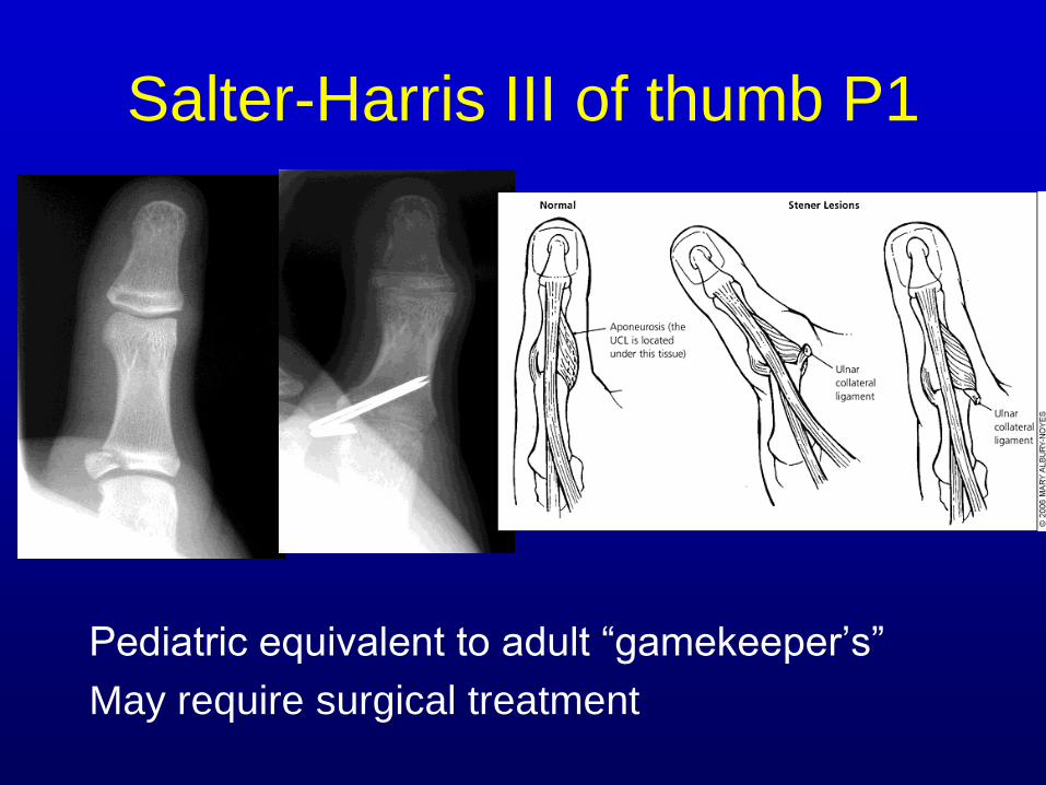
Salter-Harris III of thumb P1
Pediatric equivalent to adult “gamekeeper’s”
May require surgical treatment
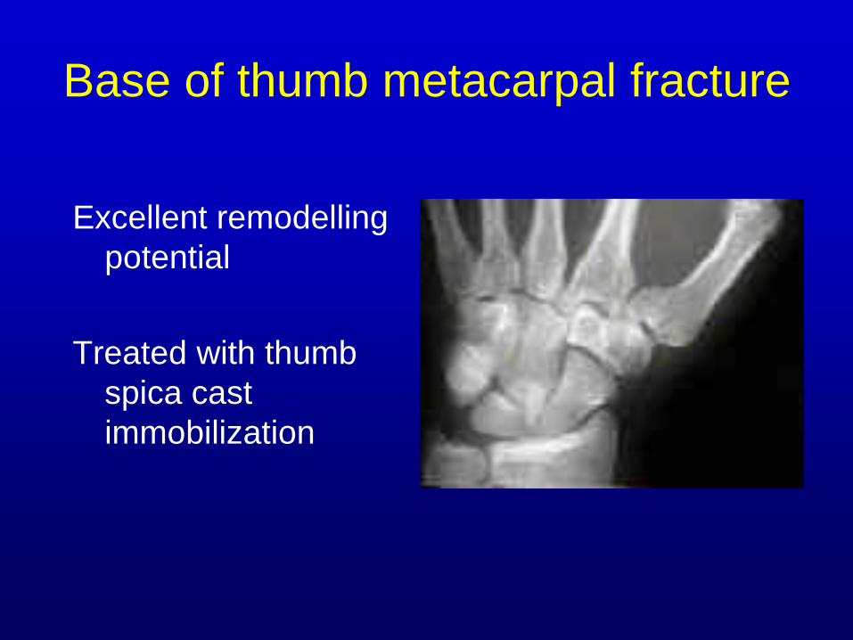
Base of thumb metacarpal fracture
Excellent remodelling
potential
Treated with thumb
spica cast
immobilization
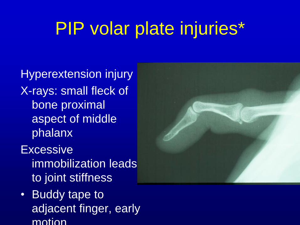
PIP volar plate injuries*
Hyperextension injury
X-rays: small fleck of
bone proximal
aspect of middle
phalanx
Excessive
immobilization leads
to joint stiffness
• Buddy tape to
adjacent finger, early
motion
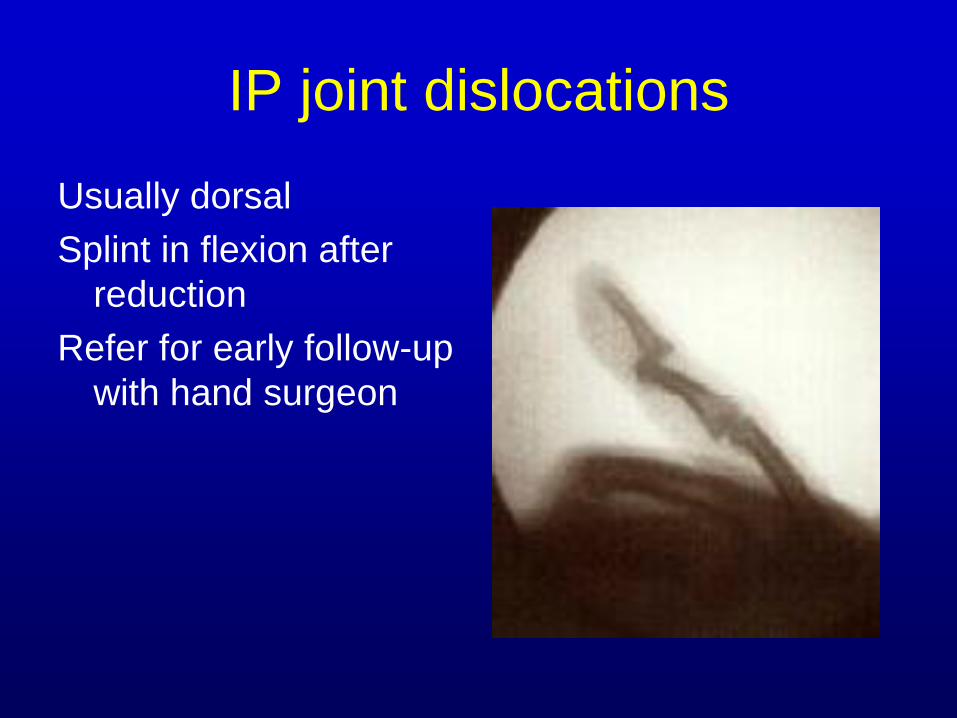
IP joint dislocations
Usually dorsal
Splint in flexion after
reduction
Refer for early follow-up
with hand surgeon
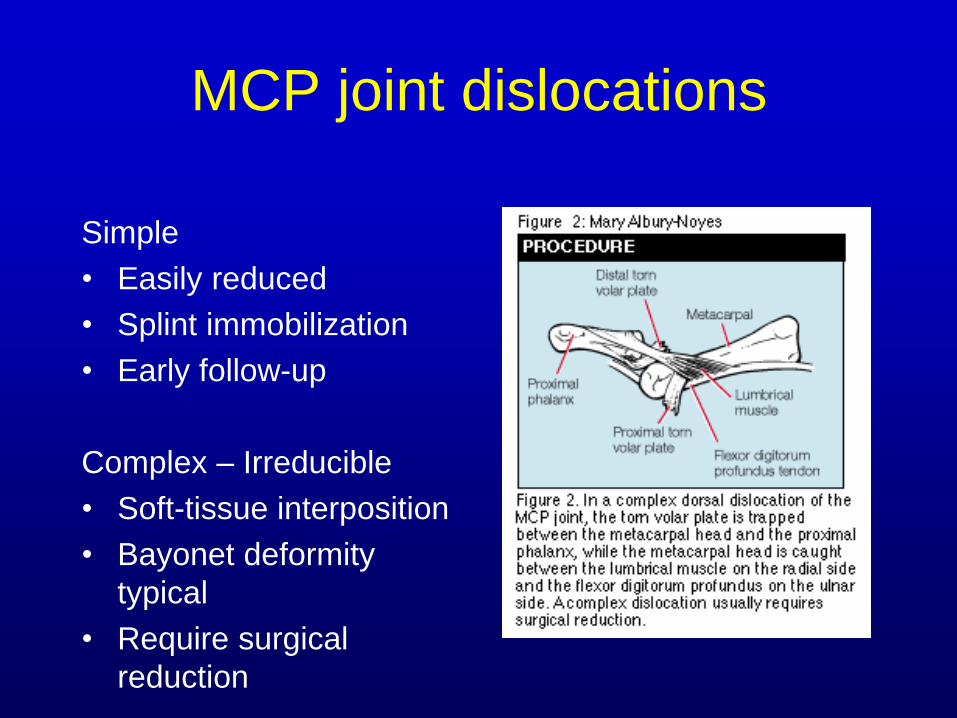
MCP joint dislocations
Simple
• Easily reduced
• Splint immobilization
• Early follow-up
Complex – Irreducible
• Soft-tissue interposition
• Bayonet deformity
typical
• Require surgical
reduction
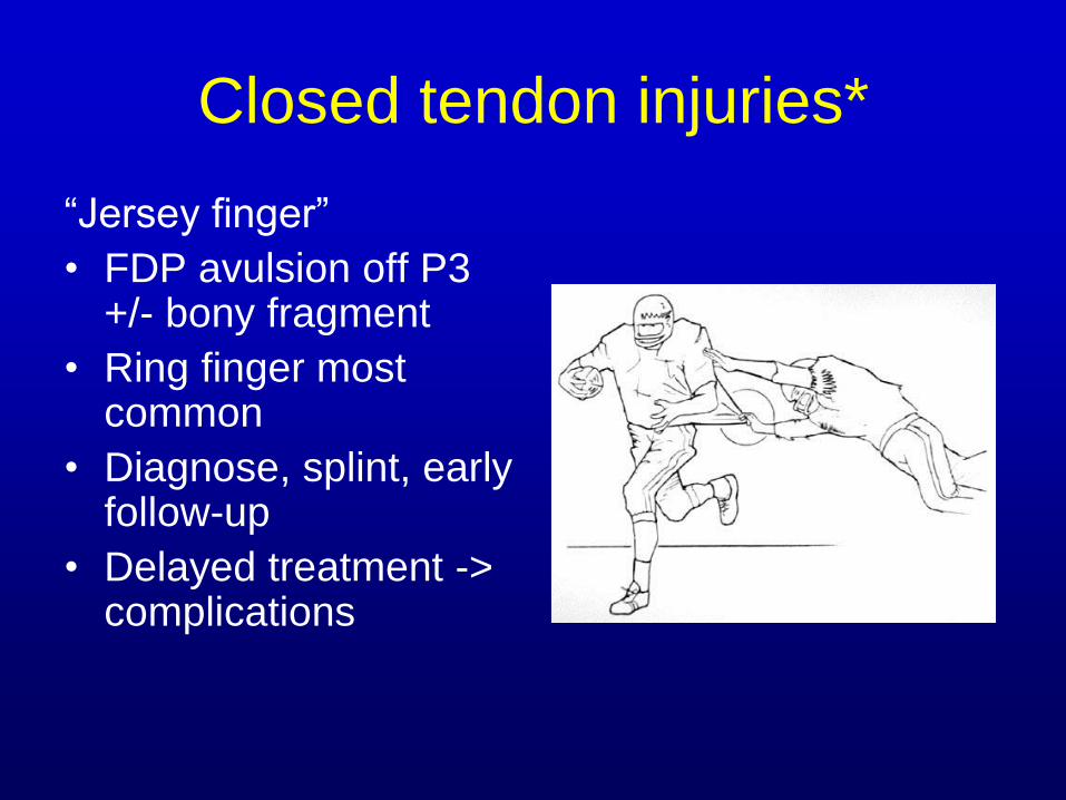
Closed tendon injuries*
“Jersey finger”
• FDP avulsion off P3 +/- bony fragment
• Ring finger most common
• Diagnose, splint, early follow-up
• Delayed treatment -> complications

Summary
Thorough examination critical
• Tenodesis/digital cascade
• Neurovascular status
• Appropriate x-rays
Joint displacement unacceptable
Avoid overtreatment of volar plate injuries
Close all wounds loosely (absorbable
sutures)
How soon should a “Jersey” finger be
referred to a hand specialist?
A. Never-I can take care of that, I practice in
Jersey.
B. I should refer them to the ED.
C. Within one week
D. Within 2 weeks

Thank You