Commensal-derived metabolites govern Vibrio cholerae ... · RESEARCH Open Access Commensal-derived...
18
RESEARCH Open Access Commensal-derived metabolites govern Vibrio cholerae pathogenesis in host intestine Jin Sun You 1,2† , Ji Hyun Yong 1,2† , Gwang Hee Kim 1,2 , Sungmin Moon 2,3 , Ki Taek Nam 2,3 , Ji Hwan Ryu 2,3 , Mi Young Yoon 1,2,4* and Sang Sun Yoon 1,2,4* Abstract Background: Recent evidence suggests that the commensal microbes act as a barrier against invading pathogens and enteric infections are the consequences of multi-layered interactions among commensals, pathogens, and the host intestinal tissue. However, it remains unclear how perturbations of the gut microbiota compromise host infection resistance, especially through changes at species and metabolite levels. Results: Here, we illustrate how Bacteroides vulgatus, a dominant species of the Bacteroidetes phylum in mouse intestine, suppresses infection by Vibrio cholerae, an important human pathogen. Clindamycin (CL) is an antibiotic that selectively kills anaerobic bacteria, and accordingly Bacteroidetes are completely eradicated from CL-treated mouse intestines. The Bacteroidetes-depleted adult mice developed severe cholera-like symptoms, when infected with V. cholerae. Germ-free mice mono-associated with B. vulgatus became resistant to V. cholerae infection. Levels of V. cholerae growth-inhibitory metabolites including short-chain fatty acids plummeted upon CL treatment, while levels of compounds that enhance V. cholerae proliferation were elevated. Furthermore, the intestinal colonization process of V. cholerae was well-simulated in CL-treated adult mice. Conclusions: Overall, we provide insights into how a symbiotic microbe and a pathogenic intruder interact inside host intestine. We identified B. vulgatus as an indigenous microbial species that can suppress intestinal infection. Our results also demonstrate that commensal-derived metabolites are a critical determinant for host resistance against V. cholerae infection, and that CL pretreatment of adult mice generates a simple yet useful model of cholera infection. Keywords: Vibrio cholerae, Gut microbiota, Colonization resistance, Short-chain fatty acids, Metabolomics, Bacteroides vulgatus, Clindamycin, Amino sugars Background Vibrio cholerae is the causative agent of pandemic diarrheal disease, cholera. While cholera toxin (CT) and toxin- coregulated pilus (TCP) are known to be the major virulence determinants, its pathogenic mechanisms are starting to be understood as consequences of interaction with indigenous microbes, collectively termed gut microbiota [1–4]. A key feature of the gut microbiota is its protective capacity against enteropathogenic infections, termed “colonization resistance” [5–7]. This property can be ascribed to the microbial ecosystem that is formed within the host intestine. This microbial ecosystem is an ever-changing community of various microbial species regulated by a complex network of microbiota-intrinsic and microbiota-extrinsic factors [8]. Microbiota-intrinsic factors such as interactions between the gut-residing species serve as the primary determinant of community composition. Such interac- tions include interbacterial niche competition [9] and secretion of antimicrobial substances [10]. For example, Bacteroides species that reside in the human gut, such as Bacteroides fragilis and Bacteroides uniformis, utilise © The Author(s). 2019 Open Access This article is distributed under the terms of the Creative Commons Attribution 4.0 International License (http://creativecommons.org/licenses/by/4.0/), which permits unrestricted use, distribution, and reproduction in any medium, provided you give appropriate credit to the original author(s) and the source, provide a link to the Creative Commons license, and indicate if changes were made. The Creative Commons Public Domain Dedication waiver (http://creativecommons.org/publicdomain/zero/1.0/) applies to the data made available in this article, unless otherwise stated. * Correspondence: [email protected]; [email protected] † Jin Sun You and Ji Hyun Yong contributed equally to this work. 1 Department of Microbiology and Immunology, Yonsei University College of Medicine, 50-1 Yonsei-ro, Seodaemun-gu Seoul, Seoul 03722, Korea Full list of author information is available at the end of the article You et al. Microbiome (2019) 7:132 https://doi.org/10.1186/s40168-019-0746-y
Transcript of Commensal-derived metabolites govern Vibrio cholerae ... · RESEARCH Open Access Commensal-derived...

RESEARCH Open Access
Commensal-derived metabolites governVibrio cholerae pathogenesis in hostintestineJin Sun You1,2†, Ji Hyun Yong1,2†, Gwang Hee Kim1,2, Sungmin Moon2,3, Ki Taek Nam2,3, Ji Hwan Ryu2,3,Mi Young Yoon1,2,4* and Sang Sun Yoon1,2,4*
Abstract
Background: Recent evidence suggests that the commensal microbes act as a barrier against invading pathogensand enteric infections are the consequences of multi-layered interactions among commensals, pathogens, and thehost intestinal tissue. However, it remains unclear how perturbations of the gut microbiota compromise hostinfection resistance, especially through changes at species and metabolite levels.
Results: Here, we illustrate how Bacteroides vulgatus, a dominant species of the Bacteroidetes phylum in mouseintestine, suppresses infection by Vibrio cholerae, an important human pathogen. Clindamycin (CL) is an antibiotic thatselectively kills anaerobic bacteria, and accordingly Bacteroidetes are completely eradicated from CL-treated mouseintestines. The Bacteroidetes-depleted adult mice developed severe cholera-like symptoms, when infected withV. cholerae. Germ-free mice mono-associated with B. vulgatus became resistant to V. cholerae infection. Levels of V.cholerae growth-inhibitory metabolites including short-chain fatty acids plummeted upon CL treatment, whilelevels of compounds that enhance V. cholerae proliferation were elevated. Furthermore, the intestinalcolonization process of V. cholerae was well-simulated in CL-treated adult mice.
Conclusions: Overall, we provide insights into how a symbiotic microbe and a pathogenic intruder interactinside host intestine. We identified B. vulgatus as an indigenous microbial species that can suppress intestinalinfection. Our results also demonstrate that commensal-derived metabolites are a critical determinant for hostresistance against V. cholerae infection, and that CL pretreatment of adult mice generates a simple yet usefulmodel of cholera infection.
Keywords: Vibrio cholerae, Gut microbiota, Colonization resistance, Short-chain fatty acids, Metabolomics,Bacteroides vulgatus, Clindamycin, Amino sugars
BackgroundVibrio cholerae is the causative agent of pandemic diarrhealdisease, cholera. While cholera toxin (CT) and toxin-coregulated pilus (TCP) are known to be the majorvirulence determinants, its pathogenic mechanisms arestarting to be understood as consequences of interactionwith indigenous microbes, collectively termed gutmicrobiota [1–4]. A key feature of the gut microbiotais its protective capacity against enteropathogenic
infections, termed “colonization resistance” [5–7]. Thisproperty can be ascribed to the microbial ecosystemthat is formed within the host intestine. This microbialecosystem is an ever-changing community of variousmicrobial species regulated by a complex network ofmicrobiota-intrinsic and microbiota-extrinsic factors [8].Microbiota-intrinsic factors such as interactions
between the gut-residing species serve as the primarydeterminant of community composition. Such interac-tions include interbacterial niche competition [9] andsecretion of antimicrobial substances [10]. For example,Bacteroides species that reside in the human gut, such asBacteroides fragilis and Bacteroides uniformis, utilise
© The Author(s). 2019 Open Access This article is distributed under the terms of the Creative Commons Attribution 4.0International License (http://creativecommons.org/licenses/by/4.0/), which permits unrestricted use, distribution, andreproduction in any medium, provided you give appropriate credit to the original author(s) and the source, provide a link tothe Creative Commons license, and indicate if changes were made. The Creative Commons Public Domain Dedication waiver(http://creativecommons.org/publicdomain/zero/1.0/) applies to the data made available in this article, unless otherwise stated.
* Correspondence: [email protected]; [email protected]†Jin Sun You and Ji Hyun Yong contributed equally to this work.1Department of Microbiology and Immunology, Yonsei University College ofMedicine, 50-1 Yonsei-ro, Seodaemun-gu Seoul, Seoul 03722, KoreaFull list of author information is available at the end of the article
You et al. Microbiome (2019) 7:132 https://doi.org/10.1186/s40168-019-0746-y

membrane attack complex/perforin toxins BSAP-1 andBSAP-2 for intraspecies antagonism [11]. Microbiota-ex-trinsic factors include inflammation, diet, and antibiotictreatment. However, the aforementioned microbiota-in-trinsic and extrinsic factors do not operate in exclusion ofanother. For example, compositional shift induced by anexternal factor may, in turn, modulate the host immuneresponse via production of specific metabolites [8].Recent studies have demonstrated that antibiotic
treatments alter the gut microbiota in humans andother mammals [12–15], and increase the susceptibilityof the host to infections by various enteric pathogenssuch as Shigella flexneri [16], Salmonella enterica [17],Clostridium difficile [18, 19], and vancomycin-resistantEnterococcus [20]. The abolishing effect of antibiotictreatment on host resistance to infection is most likelyimplemented through a multitude of factors, includingsuppression of specific microbial species, alteration ofthe metabolomic landscape as a result of the changedmicrobiome composition, and/or host responses [21–23].These findings prompted us to investigate the effectsof different classes of antibiotics on the gut microbiotaand how they relate to host resistance against V. cho-lerae infection.In this study, we uncover a unique microbiota-ex-
trinsic treatment that induces severe cholera-likesymptoms in adult mice, that are otherwise completelyresistant. Furthermore, we show that a dramatic shiftin metabolome production profile accounts for thecompromised infection resistance in the host. This re-port highlights the importance of commensal-derivedmetabolites as a crucial determinant of host suscepti-bility to enteric infection.
ResultsClindamycin-treated adult mice exhibited dramatic gutmicrobiota compositional changes and becamesusceptible to V. cholerae infectionColonization of enteric pathogens occurs dependingon the composition of indigenous microbes inside thehost intestine [5, 24, 25]. In order to observe the ef-fects of gut microbiota compositional changes on hostinfection resistance under diverse experimental condi-tions, we treated adult C57BL/6 specific pathogen-free(SPF) mice (8 weeks of age) with three different antibi-otics that have a distinct mode of bacterial killing;streptomycin (SM), vancomycin (VAN), and clindamy-cin (CL). SM is a broad-spectrum antibiotic that tar-gets both Gram-positive and Gram-negative bacteria.VAN is an effective antibiotic against Gram-positives[26], while CL is known to selectively kill anaerobes[27, 28]. After daily treatment for 5 days, mouse feceswere collected to analyze microbiota composition by16S rRNA gene sequencing. Five bacterial phyla were
found to have characteristic distributions amonggroups (Fig. 1a–d). In the untreated control group,Bacteroidetes was the most abundant phylum and themembers of that single phylum occupied approximately80% of the entire microbiota population (Fig. 1a). UponSM treatment, the Verrucomicrobia phylum emerged,while the Bacteroidetes phylum maintained its pre-dominant occupancy (Fig. 1b). VAN treatment resultedin multiplication of bacterial cells belonging to theVerrucomicrobia and Proteobacteria phyla, both ofwhich are Gram-negatives (Fig. 1c). As a result of CLtreatment, the Bacteroidetes phylum was eradicated,while the Firmicutes phylum remained largely un-changed in number (Fig. 1d). This observation is con-sistent with previous findings that CL is effective intreating infections of Bacteroides fragilis, a major spe-cies of the Bacteroidetes phylum [29, 30]. On the otherhand, the Proteobacteria phylum underwent explosiveexpansion during CL treatment, further demonstratingthat CL is not effective in inhibiting the growth offacultative anaerobes (Fig. 1d).Subsequently, the mice of each antibiotic group were
infected with 5 × 10⁸ CFU of the V. cholerae 7thpandemic strain, N16961, to monitor susceptibility toinfection and infection-induced microbiota compos-ition changes. In the control and SM-treated groups,microbiota composition did not change much follow-ing V. cholerae infection (Fig. 1e, f). In response toN16961 infection, the relative abundance of theBacteroidetes phylum increased in the VAN-pretreatedgroup (Fig. 1g vs. c). In CL-treated mice, the relativeabundance of the Firmicutes phylum increased, whilethat of the Proteobacteria phylum decreased in re-sponse to V. cholerae infection (Fig. 1h). Importantly,among the Proteobacteria phylum detected in Fig. 1h,~ 8.81% was found to be derived from V. cholerae-spe-cific 16S rRNA gene sequence (Fig. 3). This result isworthy of particular attention because V. cholerae-spe-cific sequence was not detected in the Proteobacteriapopulations in other groups (Fig. 1f, g).Our results indicate that V. cholerae may actively
colonize the adult mouse intestine under conditionscreated by CL treatment. To gain more insight into theoutcomes of V. cholerae infection, we investigatedwhether any anatomical change was induced in thegastrointestinal tract ranging from the small intestineto the rectum. In the first three experimental groups,mouse tissues appeared similar to one another and nomajor changes were observed following V. cholerae in-fection (Fig. 1i–k). In contrast, infection-induced phe-notypes were clearly demonstrated in the CL-treatedgroup for the following indications (Fig. 1l). First, fluidaccumulation was clearly observed in the small intes-tine (red arrowheads). Second, the size of the cecum
You et al. Microbiome (2019) 7:132 Page 2 of 18
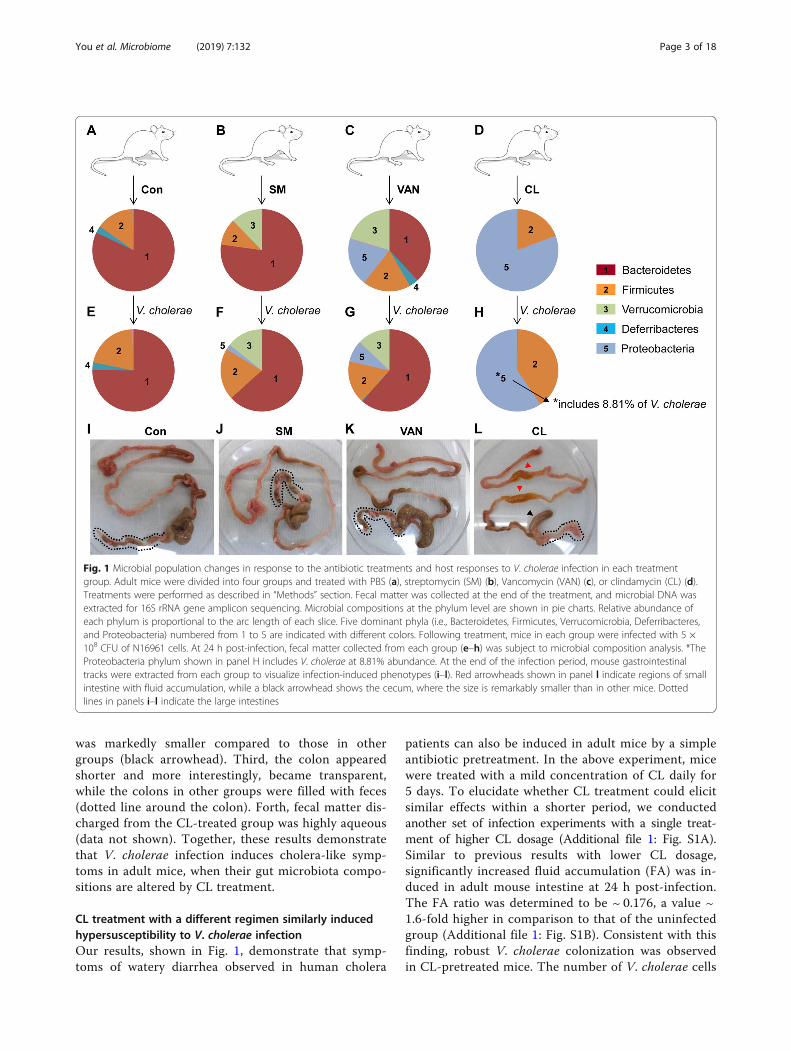
was markedly smaller compared to those in othergroups (black arrowhead). Third, the colon appearedshorter and more interestingly, became transparent,while the colons in other groups were filled with feces(dotted line around the colon). Forth, fecal matter dis-charged from the CL-treated group was highly aqueous(data not shown). Together, these results demonstratethat V. cholerae infection induces cholera-like symp-toms in adult mice, when their gut microbiota compo-sitions are altered by CL treatment.
CL treatment with a different regimen similarly inducedhypersusceptibility to V. cholerae infectionOur results, shown in Fig. 1, demonstrate that symp-toms of watery diarrhea observed in human cholera
patients can also be induced in adult mice by a simpleantibiotic pretreatment. In the above experiment, micewere treated with a mild concentration of CL daily for5 days. To elucidate whether CL treatment could elicitsimilar effects within a shorter period, we conductedanother set of infection experiments with a single treat-ment of higher CL dosage (Additional file 1: Fig. S1A).Similar to previous results with lower CL dosage,significantly increased fluid accumulation (FA) was in-duced in adult mouse intestine at 24 h post-infection.The FA ratio was determined to be ~ 0.176, a value ~1.6-fold higher in comparison to that of the uninfectedgroup (Additional file 1: Fig. S1B). Consistent with thisfinding, robust V. cholerae colonization was observedin CL-pretreated mice. The number of V. cholerae cells
Fig. 1 Microbial population changes in response to the antibiotic treatments and host responses to V. cholerae infection in each treatmentgroup. Adult mice were divided into four groups and treated with PBS (a), streptomycin (SM) (b), Vancomycin (VAN) (c), or clindamycin (CL) (d).Treatments were performed as described in “Methods” section. Fecal matter was collected at the end of the treatment, and microbial DNA wasextracted for 16S rRNA gene amplicon sequencing. Microbial compositions at the phylum level are shown in pie charts. Relative abundance ofeach phylum is proportional to the arc length of each slice. Five dominant phyla (i.e., Bacteroidetes, Firmicutes, Verrucomicrobia, Deferribacteres,and Proteobacteria) numbered from 1 to 5 are indicated with different colors. Following treatment, mice in each group were infected with 5 ×10⁸ CFU of N16961 cells. At 24 h post-infection, fecal matter collected from each group (e–h) was subject to microbial composition analysis. *TheProteobacteria phylum shown in panel H includes V. cholerae at 8.81% abundance. At the end of the infection period, mouse gastrointestinaltracks were extracted from each group to visualize infection-induced phenotypes (i–l). Red arrowheads shown in panel l indicate regions of smallintestine with fluid accumulation, while a black arrowhead shows the cecum, where the size is remarkably smaller than in other mice. Dottedlines in panels i–l indicate the large intestines
You et al. Microbiome (2019) 7:132 Page 3 of 18

recovered from the small intestine (SI) or cecum wasremarkably larger in CL-treated vs. control group. V.cholerae cells of > 109 CFU were grown per gram sam-ple in SI and cecum, respectively. Likewise, ~ 109 CFUwas also detected in fecal pellets shed from CL-treatedmice (Additional file 1: Fig. S1C). In contrast, V. cho-lerae cells were not detected at all in feces dischargedfrom the control group. Given that an infection dosageof 5 × 10⁸ CFU was used, these results demonstratethat (i) V. cholerae cells were almost completely elimi-nated inside the intestine of regular adult mice while(ii) the same number of inoculated cells was morehighly proliferated in the CL-pretreated adult mouseintestine.
Germ-free mice transplanted with feces of CL-treatedmice were susceptible to V. cholerae infectionPrevious studies have revealed that CL treatment canmodulate the production of pro-inflammatory cytokines,especially in response to lipopolysaccharide [31–33].Furthermore, CL at sub-lethal concentrations stimulateneutrophil phagocytosis in vitro against Staphylococcus
aureus [34] and Bacteroides spp. [35]. While our resultsstrongly suggest that CL-induced changes in gutmicrobiota composition are associated with elevatedsusceptibility to V. cholerae infection, these previousreports also indicate that enhanced V. cholerae infect-ivity in adult mice might be attributed to CL-inducedmodulation of the host immune response. To addressthis issue, we conducted fecal microbiota transplant-ation (FMT) to germ-free (GF) mice and testedwhether the GF mice with reconstituted intestinalmicrobiota develop differential resistance to V. cho-lerae infection. Fecal pellets freshly collected fromPBS- or CL-treated mice were resuspended in PBS andtransferred to GF mice via oral gavage. Following a 3-day transplant stabilization period, 5 × 10⁸ N16961cells were infected (Fig. 2a). GF mice transplanted withPBS-treated control fecal pellets developed strongresistance to the infection. Viable V. cholerae was re-covered neither from the SI of all six mice nor fromthe cecum isolated from five out of six mice (Fig. 2b).While varying degrees of N16961 cells were detectedin feces discharged from the GF mice transplanted
Fig. 2 Germ-free mice transplanted with feces of CL-treated mice are susceptible to V. cholerae infection. a Schematic diagram of theexperimental procedure. Germ-free mice (C57BL/6, 8~9 weeks old, n = 6 per group) received fecal suspensions (50 μL) derived from PBS- or CL-treated SPF mice. Following 3 days of stabilization, transplanted mice were challenged with V. cholerae infection (5 × 10⁸ CFU) for 24 h. b The SIand cecum were removed from each mouse for tissue homogenization. Fresh fecal matter was collected to prepare fecal suspensions. Aliquots oflysates or suspensions were serially diluted to count CFU of V. cholerae on LB agar supplemented with 200 μg/mL SM. Values are displayed on alog scale as mean ± SEM for each group. *P < 0.05 versus bacterial CFU detected in the control group. **P < 0.001 versus bacterial CFUs detectedin the control group. c In a separate set of experiments (n = 3 per group), small intestines were extracted for tissue homogenization, and aliquotsof homogenates were assessed for detection of CT by ELISA. *P < 0.01 versus CT level in the control group
You et al. Microbiome (2019) 7:132 Page 4 of 18

with control feces, four out of six mice produced fecesthat contained ten or fewer N16961 cells per gram(Fig. 2b). In contrast, N16961 colonization occurredmore actively in the GF mice transplanted with CL-treated and therefore Bacteroidetes-depleted feces. Incecum and feces, > 104-fold and > 103.4-fold higherbacterial colonization were observed, respectively,compared with the control group (Fig. 2b).We next asked whether production of CT, a critical
virulence determinant, was accordingly increasedduring active V. cholerae colonization. As shown inFig. 2c, in response to V. cholerae infection, CTproduction was significantly increased in mice trans-planted with fecal suspensions of CL-treated mice.Since CL would not be much left in the feces dis-charged from the CL-treated mice, our results demon-strate that altered gut microbiota composition inducedby CL treatment is directly responsible for renderingthe murine host more susceptible to V. choleraeinfection.
Bacteroides vulgatus is the predominant species in theBacteroidetes phylum and it inhibited V. cholerae growthin vivoBased on our results presented in Fig. 1, elevated V.cholerae infection in CL-pretreated mice could be as-cribed either to the depletion of the Bacteroidetes orto the expansion of Proteobacteria phylum. Meanwhile,we noticed the followings. First, the Proteobacteriapopulation similarly sufficiently propagated in VAN-treated mice, a group that did not develop cholera-likesymptoms (Fig. 1c). Second, the relative abundance ofthe Bacteroidetes phylum increased from ~ 38% (be-fore infection) to ~ 61% (after infection) in VAN-treated mice (Fig. 1c, g). Based on these results, wepostulated that the lack of cholera-like symptoms inthe VAN-treated group was due not only to the pres-ence of the Bacteroidetes phylum but also to the infec-tion-induced expansion of its population. Wehypothesized that the Bacteroidetes phylum, whichpredominantly occupies the adult mouse intestine,plays a critical role in protecting its host from V. cho-lerae infection.To provide an insight into which bacterial species
are core members of the Bacteroidetes phylum, weperformed a species-level population analysis using16S rRNA gene sequencing. When necessary, we con-ducted PCR reactions with species-specific primer setsand verified the sequences of amplification products(data not shown). Three distinct bacterial species weredetermined to belong to the Bacteroidetes phylum;Bacteroides vulgatus, Parabacteroides goldsteinii, andBacteroides caccae. In control mice, B. vulgatus wasfound to be present in the largest quantity, with a
relative abundance greater than 50% of the entirepopulation (Fig. 3). In response to VAN treatment, P.goldsteinii propagated, whereas B. vulgatus was dimin-ished in population size. Of note, however, the relativeabundance of the B. vulgatus population was recovered toits original level following V. cholerae infection. P. goldstei-nii has been reclassified from Bacteroides goldsteinii [36]and a strain isolated from human intestine was resistantto VAN treatment [37]. As shown in Fig. 1d, l, no 16SrRNA gene sequences matching any of these three spe-cies were detected in feces discharged from CL-treatedmice either before or after V. cholerae infection (Fig. 3).Because B. vulgatus was the most dominant species
in the adult mouse intestine, we next assessed, under amore defined condition, the effect of B. vulgatus onaltering host susceptibility to V. cholerae infection. Tothis end, we mono-associated GF mice with a suspen-sion of B. vulgatus cells, grown anaerobically in vitro(Fig. 4a). B. vulgatus colonies cultivated straight frommouse fecal suspensions were subject to RAPD assayand an identical amplification pattern was observedbetween isolated colonies, thereby leading us to con-clude that most colonies were clonal (Additional file 2:Figure S2). As shown in Fig. 4a, GF mice were alsomono-associated with heat-killed B. vulgatus beforebeing infected with V. cholerae cells as an additionalnegative control. At 24 h post-infection, significantlyreduced numbers of V. cholerae cells were recoveredin animals that were mono-associated with live B.vulgatus. In SI and colon, ~ 830-fold and ~ 40-fold lessV. cholerae colonization were observed, respectively, ascompared with the PBS control group. Likewise, V.cholerae cell numbers, recovered from feces of the GFmice transplanted with live B. vulgatus, was ~ 75-foldlower than the PBS control (Fig. 4b). Importantly, V.cholerae colonization was never decreased in theanimals mono-associated with heat-inactivated B.vulgatus cells (Fig. 4b). These results suggest thatactive B. vulgatus cells, when present in adult mouseintestine, can suppress V. cholerae colonization.We next explored whether B. vulgatus also effectively
inhibits V. cholerae colonization in the infant mousemodel that has been used to study V. cholerae infec-tion [38, 39]. Five-day-old infant mice were dividedinto two groups, with the first group being trans-planted with B. vulgatus and the other group servingas a negative control (Fig. 4c). B. vulgatus, when trans-planted in the intestine of the infant mouse, can alsoeffectively suppress V. cholerae colonization and fluidaccumulation (Fig. 4d). V. cholerae colonization wasdecreased ~ 4.47-fold in the presence of transplantedB. vulgatus. Although the difference between these twogroups was less pronounced than what was observed inexperiments with adult mice, V. cholerae colonization and
You et al. Microbiome (2019) 7:132 Page 5 of 18

infection-induced fluid accumulation were both re-duced significantly in B. vulgatus-transplanted infantmice (Fig. 4d).
Metabolome profiles revealed the characteristicmetabolites that directly modulate intestinal V. choleraegrowthWe next conducted a metabolome analysis to examinewhether metabolomic profiles are altered in responseto CL treatment and/or V. cholerae infection. We pos-tulated that alterations of metabolite levels influencedhost susceptibility to V. cholerae infection. Cecal con-tents were subject to sample preparation for CE-TOF/MS, as described in the “Methods” section. Among themetabolites detected in our analysis, a total of 273 me-tabolites were successfully quantified and their relativequantities between samples are presented in a heat
map (Fig. 5). A table that lists all 273 metabolites andtheir relative amounts in four samples is shown assupplementary information (a supplementary excelfile). Of the many differences between the metabolo-mic profiles of each group, the most noticeable one isthat red and green line clusters representing com-pounds in large and small quantities, respectively, aregenerally reversed in control SPF vs. CL-treated group(column 1 vs. 3, Fig. 5). Such striking alterations inmetabolome profiles can be interpreted as a conse-quence of the dramatic microbiota compositionalchanges, as shown in Fig. 1. Interestingly, a smallerdegree of profile change was observed in response toV. cholerae infection in both experimental groups (col-umn 1 vs. 2 and column 3 vs. 4, Fig. 5). These resultsdemonstrate that (i) CL treatment induces more sub-stantial changes in metabolome profiles than does V.
Fig. 3 Species-level microbiota populations in response to antibiotic treatments and after V. cholerae infection. Distribution of commensalmicrobial species at the phylum (far-left column) and species (second column) levels are presented by colors in 8 different experimentalconditions. Con, SM, VAN, and CL in the first low indicate microbial samples of corresponding antibiotic treatments and Con-Vc, SM-Vc, VAN-Vc,and CL-Vc indicate samples of the same groups following V. cholerae infection. Three species were determined to belong to Bacteroidetes(brown) and Proteobacteria (gray) phyla and 17 species belonging to the Firmicutes (orange) phylum were identified. Only bacterial species witha relative abundance of > % of the total microbiota population were selected and displayed. Relative abundance of each species is indicated bycolor-coded boxes. For example, brown boxes mean that the abundance of a given species under a specific condition is > 50%. A completeelimination of Bacteroides vulgatus is indicated with **, while a pink box denoted by * indicates 8.81% V. cholerae occupancy
You et al. Microbiome (2019) 7:132 Page 6 of 18

cholerae infection, and (ii) CL-induced changes on me-tabolite level might have created an environment thatstrongly facilitates V. cholerae growth and pathogenesis.Table 1 shows a list of metabolites that were de-
tected in the greatest quantities in control SPF (com-pounds 1~13) or CL-treated (compounds 14~27) mice.Out of the 13 molecules detected in the SPF mouse
ceca, all except for cholic acid were not detected inCL-treated mice. It is of particular interest that (iso)butyric acid, propionic acid, and (iso) valeric acid,collectively termed short-chain fatty acid (SCFA) areamong the most abundantly detected metabolites incontrol group (Table 1). Fourteen compounds (14~27),whose relative quantities are greater than 1.0E-03,
Fig. 4 B. vulgatus inhibits V. cholerae growth in vivo. a Schematic diagram of the experimental procedure. Germ-free mice (C57BL/6, 8~9 weeksold, n = 3 or 4 per group) ingested 50 μL PBS (n = 3), heat-inactivated (autoclaved) B. vulgatus cell suspension (n = 4), or live B. vulgatus cellsuspension (n = 4). The B. vulgatus cell suspensions contained 2 × 10⁹ CFU/mL. At 24 h post-association, mice were challenged with V. choleraeinfection (5 × 10⁸ CFU) for 24 h. b Viable V. cholerae cells in SI, colon, or fecal pellets were enumerated and presented in log-scale. **P < 0.001versus bacterial CFU in the control and heat-inactivated Bv groups. c Schematic diagram of the experimental procedure. Five-day-old infant mice(n = 4 or 6 per group) ingested 50 μL PBS (n = 6) or B. vulgatus cell suspension (n = 4). The B. vulgatus cell suspensions contained 1 × 10⁹ CFU/mL. At 12 h post-association, mice were challenged with V. cholerae infection (1 × 10⁸ CFU) for 12 h. d Viable V. cholerae cells in intestinal extractwere enumerated and presented in log-scale. **P < 0.05 versus bacterial CFU in the control group. At the end of the infection period, fluidaccumulation ratio was calculated by the equation of (intestine weight)/[(total body weight)-(intestine weight)]. *P < 0.05 versus thecontrol group
You et al. Microbiome (2019) 7:132 Page 7 of 18

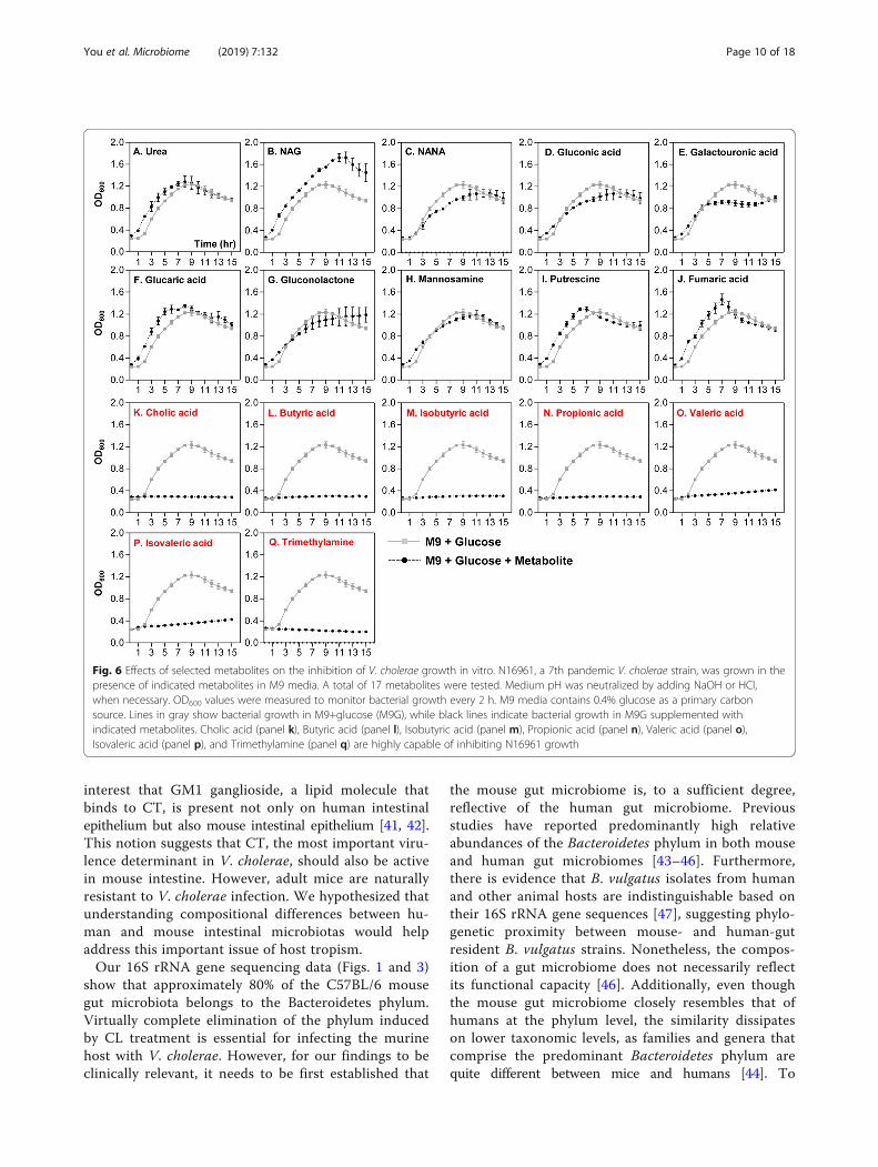
were selected and presented as dominant metabolitesin the CL-treated group (Table 1, bottom portion).Relative quantities of those molecules were either non-detectable (compounds 14, 21, 23, and 27) or consider-ably smaller in the control SPF group. These resultsfurther support the notion that distinct profile changeoccurred in response to CL treatment. We next exam-ined how V. cholerae growth is controlled by each ofthese major compounds. Of the 27 listed compounds,17 were selected based on the availability and wemonitored V. cholerae in vitro growth in M9 mediasupplemented with each metabolite. Growth curveexperiments shown in Fig. 6 were conducted to revealcompounds that can inhibit V. cholerae growth. M9media used in this particular set of growth experimentscontain 0.4% glucose as primary carbon source, therebyenabling us to monitor whether the extraneously addedmetabolite promotes or inhibits V. cholerae growth. Seven
metabolites shown in panels K~Q clearly inhibited V.cholerae growth. Importantly, these compounds werepresent in the largest quantities in regular SPF mouseintestines, but either absent or barely detectable in CL-treated mouse intestines (Table 1). Prominent growthpromotion was observed when the culture medium in-cluded NAG (Fig. 6b), a metabolite detected at sufficientlyhigh level in CL-treated mice. Supplementation with urea(Fig. 6a), glucaric acid (Fig. 6f), putrescine (Fig. 6i), orfumaric acid (Fig. 6j) resulted in a slight growth stimula-tion, especially during the early stage of growth. Again,these metabolites were detected more abundantly in CL-treated mice, than in the SPF control mice (Table 1).When N16961 growth was assessed in M9 media with anindicated metabolite as the sole carbon source, eachmetabolite’s ability to stimulate V. cholerae growth wasclearly demonstrated (Fig. 7). Consistent with resultshown in Fig. 6b, NAG was most potent in stimulating V.
Fig. 5 Heat map constructed from hierarchical cluster analysis (HCA) of cecal metabolites. Metabolites (n = 273) that exhibit similar detectionpatterns in four samples are clustered, and the distance between clusters is shown as a tree diagram. The degree of red or green color indicatesa larger or smaller amount of a given metabolite. Columns 1 and 2 display metabolome profiles of regular SPF mice, while 3 and 4 shows thoseof CL-treated groups. Columns 2 and 4 are metabolome profiles after V. cholerae infection in each group
You et al. Microbiome (2019) 7:132 Page 8 of 18

cholerae growth, with OD600 values reaching ~ 1.05(Fig. 7b). Next to NAG, gluconic acid (Fig. 7d) andgluconolactone (Fig. 7g) were effective at promoting V.cholerae growth. As expected, seven inhibitory metabo-lites (cholic acid, butyric acid, isobutyric acid, propionicacid, valeric acid, isovaleric acid, and trimethylamine) stillprevented growth of V. cholerae (Fig. 7k–q). Together,these results illustrate that CL-induced changes in thehost intestine are clearly reflected on metabolite level andsuch major alterations of metabolomic profiles signifi-cantly affect V. cholerae intestinal growth.
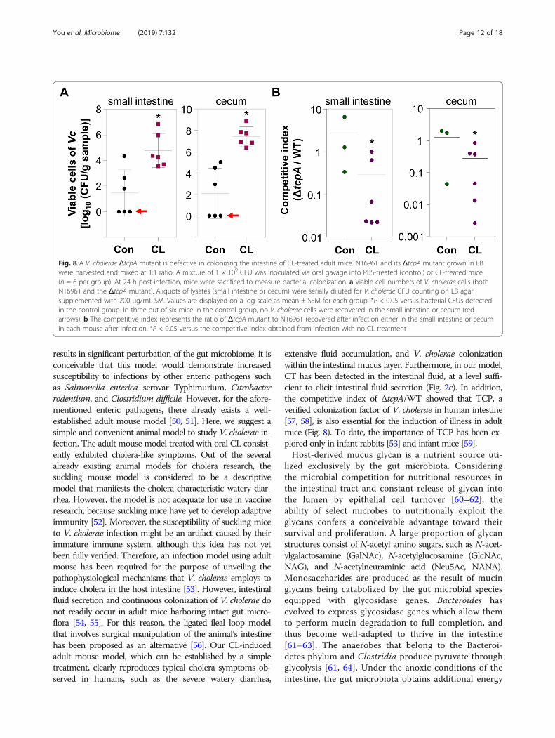
CL-treated adult mice can serve as a model forinvestigating V. cholerae pathogenesisOur results demonstrate that infection-induced pheno-types, such as intestinal colonization and fluid accu-mulation, are clearly observed in CL-pretreated adultmice. Therefore, we finally examined whether or notCL-treated mice can be utilized as a model to delineateV. cholerae infection. To address this issue, two groupsof mice were infected with 1:1 mixture of N16961 andits ΔtcpA mutant, a well-characterized V. cholerae mu-tant defective in colonizing the intestine [40]. We thencompared colonization capabilities of these two strains
by calculating the competitive index. In this particularset of experiments, fluid accumulation and bacterialcolonization were again substantially increased in CL-treated mice, but not in PBS-treated control group(Additional file 3: Figure S3). V. cholerae cells werecompletely eliminated in three out of six mice incontrol group (red arrow lines, Fig. 8a). Importantly,ΔtcpA mutant exhibited significantly reduced colonizationcapabilities, in comparison to WT N16961 (Fig. 8b). Theaverage values of the competitive indices (ΔtcpA/WT)were ~ 0.29 and ~ 0.28 in the small intestine and cecum ofCL-treated mice, respectively. In contrast, meaningfuldifferences in colonization capabilities between twostrains were not detected, when untreated adult micewere used (Fig. 8b). These results suggest that CLpretreatment not only induces active bacterialcolonization but also creates a valuable environmentwe can exploit to better understand the pathogenesisof V. cholerae.
DiscussionA long-standing unresolved question in cholera re-search is how V. cholerae has evolved as a human-spe-cific pathogen in nature. It has been of particular
Table 1 Metabolites found to be dominant in regular SPF and CL-treated mouse intestines and their impacts on V. cholerae growth
Twenty-seven metabolites that were present exclusively in either group were selected among 273 compounds. Metabolites detected most abundantly in thecontrol group (No.1~13) or in the CL-treated group (No.14~27) are listed in the order of quantity. The growth-inhibiting or growth-promoting capability of a givenmetabolite is displayed semi-qualitatively with a minus (−) or plus (+) sign, respectively. For example, “+” indicates a metabolite with mild growth-promotingcapability, whereas “---” indicates that the metabolite induced strong growth suppression. ND not detected, NT not tested, m/z mass-to-charge ratio, MT migrationtime, RT retention time, Vc Vibrio cholerae. Relative quantity of a metabolite is calculated by the equation: (peak area of the metabolite)/(peak areas of internalstandards × sample amount). Peaks are assigned based on the signal-to-noise ratio (S/N) of 3 or above.
You et al. Microbiome (2019) 7:132 Page 9 of 18
interest that GM1 ganglioside, a lipid molecule thatbinds to CT, is present not only on human intestinalepithelium but also mouse intestinal epithelium [41, 42].This notion suggests that CT, the most important viru-lence determinant in V. cholerae, should also be activein mouse intestine. However, adult mice are naturallyresistant to V. cholerae infection. We hypothesized thatunderstanding compositional differences between hu-man and mouse intestinal microbiotas would helpaddress this important issue of host tropism.Our 16S rRNA gene sequencing data (Figs. 1 and 3)
show that approximately 80% of the C57BL/6 mousegut microbiota belongs to the Bacteroidetes phylum.Virtually complete elimination of the phylum inducedby CL treatment is essential for infecting the murinehost with V. cholerae. However, for our findings to beclinically relevant, it needs to be first established that
the mouse gut microbiome is, to a sufficient degree,reflective of the human gut microbiome. Previousstudies have reported predominantly high relativeabundances of the Bacteroidetes phylum in both mouseand human gut microbiomes [43–46]. Furthermore,there is evidence that B. vulgatus isolates from humanand other animal hosts are indistinguishable based ontheir 16S rRNA gene sequences [47], suggesting phylo-genetic proximity between mouse- and human-gutresident B. vulgatus strains. Nonetheless, the compos-ition of a gut microbiome does not necessarily reflectits functional capacity [46]. Additionally, even thoughthe mouse gut microbiome closely resembles that ofhumans at the phylum level, the similarity dissipateson lower taxonomic levels, as families and genera thatcomprise the predominant Bacteroidetes phylum arequite different between mice and humans [44]. To
Fig. 6 Effects of selected metabolites on the inhibition of V. cholerae growth in vitro. N16961, a 7th pandemic V. cholerae strain, was grown in thepresence of indicated metabolites in M9 media. A total of 17 metabolites were tested. Medium pH was neutralized by adding NaOH or HCl,when necessary. OD600 values were measured to monitor bacterial growth every 2 h. M9 media contains 0.4% glucose as a primary carbonsource. Lines in gray show bacterial growth in M9+glucose (M9G), while black lines indicate bacterial growth in M9G supplemented withindicated metabolites. Cholic acid (panel k), Butyric acid (panel l), Isobutyric acid (panel m), Propionic acid (panel n), Valeric acid (panel o),Isovaleric acid (panel p), and Trimethylamine (panel q) are highly capable of inhibiting N16961 growth
You et al. Microbiome (2019) 7:132 Page 10 of 18

compensate for such limitations, we have demonstratedthat our key findings are reproduced when GF mice aremono-associated with B. vulgatus and infected with V.cholerae (Fig. 4a, b). Given such considerations, it is likelythat the relative abundance of B. vulgatus is an importantdetermining factor for V. cholerae infectivity not only inmurine hosts but in humans as well. An important ques-tion would be how abundantly B. vulgatus cells that sharesimilar functions with mouse isolates are present inhuman intestine.Based on our species-level population changes,
Escherichia coli explosively proliferated during CLtreatment and became the most dominant component(Fig. 3). This finding suggests that elevated susceptibilityto V. cholerae infection in CL-pretreated mouse couldalso be due to the presence of E. coli cells in largequantity. In line with this notion, an E. coli strain with
robust catalase activity was found to stimulateenhanced V. cholerae intestinal colonization in neo-natal mice [1]. In the same work, increased V. choleraecolonization occurred in VAN-treated adult mice [1].In the current study, however, cholera-like symptomsincluding fluid accumulation in the small intestine andproduction of watery fecal matter were not observed inthe VAN-treated group (Fig. 1k). We thereforeconclude that the loss of B. vulgatus is a much morecritical determinant over increased E. coli for host sus-ceptibility to V. cholerae infection.In the present study, we found that CL treatment increases
V. cholerae infectivity in the normally resistant adult mice.Previous studies have reported that antibiotic administrationtriggers dysbiosis in the gut microbiota [48, 49], and suchperturbations lead to suppression of host resistance againstvarious enteropathogenic infections. Since CL treatment
Fig. 7 Effects of selected metabolites on the stimulation of V. cholerae growth in vitro. Bacterial growth was conducted as described in Fig. 6. Toexamine growth-stimulating capabilities of metabolites, M9 media lacking in glucose was used in experiments. Lines in gray show bacterialgrowth in M9 with no carbon source, while black lines indicate bacterial growth in M9 supplemented with indicated metabolites. Compoundsshown in purple font promoted bacterial growth, albeit to varying degrees. The degree of growth-promoting capability of a given metabolite isdisplayed semi-qualitatively with “+++”, “++”, or “+” in Table 1. “+++” indicates the metabolite with the strongest growth-promoting capability,while the “+” means that the metabolite induced a mild growth promotion
You et al. Microbiome (2019) 7:132 Page 11 of 18
results in significant perturbation of the gut microbiome, it isconceivable that this model would demonstrate increasedsusceptibility to infections by other enteric pathogens suchas Salmonella enterica serovar Typhimurium, Citrobacterrodentium, and Clostridium difficile. However, for the afore-mentioned enteric pathogens, there already exists a well-established adult mouse model [50, 51]. Here, we suggest asimple and convenient animal model to study V. cholerae in-fection. The adult mouse model treated with oral CL consist-ently exhibited cholera-like symptoms. Out of the severalalready existing animal models for cholera research, thesuckling mouse model is considered to be a descriptivemodel that manifests the cholera-characteristic watery diar-rhea. However, the model is not adequate for use in vaccineresearch, because suckling mice have yet to develop adaptiveimmunity [52]. Moreover, the susceptibility of suckling miceto V. cholerae infection might be an artifact caused by theirimmature immune system, although this idea has not yetbeen fully verified. Therefore, an infection model using adultmouse has been required for the purpose of unveiling thepathophysiological mechanisms that V. cholerae employs toinduce cholera in the host intestine [53]. However, intestinalfluid secretion and continuous colonization of V. cholerae donot readily occur in adult mice harboring intact gut micro-flora [54, 55]. For this reason, the ligated ileal loop modelthat involves surgical manipulation of the animal’s intestinehas been proposed as an alternative [56]. Our CL-inducedadult mouse model, which can be established by a simpletreatment, clearly reproduces typical cholera symptoms ob-served in humans, such as the severe watery diarrhea,
extensive fluid accumulation, and V. cholerae colonizationwithin the intestinal mucus layer. Furthermore, in our model,CT has been detected in the intestinal fluid, at a level suffi-cient to elicit intestinal fluid secretion (Fig. 2c). In addition,the competitive index of ΔtcpA/WT showed that TCP, averified colonization factor of V. cholerae in human intestine[57, 58], is also essential for the induction of illness in adultmice (Fig. 8). To date, the importance of TCP has been ex-plored only in infant rabbits [53] and infant mice [59].Host-derived mucus glycan is a nutrient source uti-
lized exclusively by the gut microbiota. Consideringthe microbial competition for nutritional resources inthe intestinal tract and constant release of glycan intothe lumen by epithelial cell turnover [60–62], theability of select microbes to nutritionally exploit theglycans confers a conceivable advantage toward theirsurvival and proliferation. A large proportion of glycanstructures consist of N-acetyl amino sugars, such as N-acet-ylgalactosamine (GalNAc), N-acetylglucosamine (GlcNAc,NAG), and N-acetylneuraminic acid (Neu5Ac, NANA).Monosaccharides are produced as the result of mucinglycans being catabolized by the gut microbial speciesequipped with glycosidase genes. Bacteroides hasevolved to express glycosidase genes which allow themto perform mucin degradation to full completion, andthus become well-adapted to thrive in the intestine[61–63]. The anaerobes that belong to the Bacteroi-detes phylum and Clostridia produce pyruvate throughglycolysis [61, 64]. Under the anoxic conditions of theintestine, the gut microbiota obtains additional energy
Fig. 8 A V. cholerae ΔtcpA mutant is defective in colonizing the intestine of CL-treated adult mice. N16961 and its ΔtcpA mutant grown in LBwere harvested and mixed at 1:1 ratio. A mixture of 1 × 10⁹ CFU was inoculated via oral gavage into PBS-treated (control) or CL-treated mice(n = 6 per group). At 24 h post-infection, mice were sacrificed to measure bacterial colonization. a Viable cell numbers of V. cholerae cells (bothN16961 and the ΔtcpA mutant). Aliquots of lysates (small intestine or cecum) were serially diluted for V. cholerae CFU counting on LB agarsupplemented with 200 μg/mL SM. Values are displayed on a log scale as mean ± SEM for each group. *P < 0.05 versus bacterial CFUs detectedin the control group. In three out of six mice in the control group, no V. cholerae cells were recovered in the small intestine or cecum (redarrows). b The competitive index represents the ratio of ΔtcpA mutant to N16961 recovered after infection either in the small intestine or cecumin each mouse after infection. *P < 0.05 versus the competitive index obtained from infection with no CL treatment
You et al. Microbiome (2019) 7:132 Page 12 of 18

by exploiting the pyruvate through anaerobic catabol-ism. Predominant end products of the anaerobicfermentation are short-chain fatty acids (SCFAs), suchas propionic acid, butyric acid, and valeric acid [61].In this study, the dramatic changes in the cecal
metabolomes of CL-treated mice strongly correlatewith the compositional shift of the microbiota. Deple-tion of SCFAs and relatively high occurrence of aminosugars in the ceca of CL-treated mice indicate thatcatabolism of mucin glycan, a proliferation strategy formicrobes belonging to the Bacteroidetes phylum, be-came inactive upon CL treatment.Utilization of the host glycan is also important for
the invasion of V. cholerae into the host intestine.When the pathogen enters the intestinal tract, the nextstrategy for its proliferation is to colonize the epithelialcells covered with a viscous layer of mucus. V. choleraeeffectively responds to the mucus glycan and catabolizethe polysaccharides to acquire competitive advantagein host intestine [65–68]. Glucosamine-6-phosphate(GlcNP-6), a common intermediate molecule sharedbetween the catabolic pathways of GlcNAc andNeu5Ac, was reported to be indispensable for V.cholerae motility [68]. Chemotaxis mediated by motil-ity toward the nutrient sources is important forcolonization and proliferation of V. cholerae [68].
Consistent with this notion, depletion of sialic acid(Neu5Ac) transporters (encoded in VC1777-1779)attenuates colonization [69]. During this process, V.cholerae needs to overcome a number of challengessuch as acid stress, antimicrobial peptides, host im-mune response, and SCFAs [64, 66, 68]. The reductionof intestinal SCFAs in patients with diarrhea andrecovery of normal SCFA levels after conventionaltreatment support that the SCFAs could be a contrib-uting factor to eubiosis of the intestinal microbiota[70]. In general, SCFAs are considered to be an effect-ive barrier against pathogenic invasion, although someenteric pathogens exploit SCFAs to sense their sur-roundings and to decide whether or not to expresstheir arsenal of virulence factors [71].Our results suggest that catabolism of N-acetyl
amino sugars and subsequent accumulation of SCFAsin the distal intestine are largely managed by specificgut microbiota (Fig. 3). Elimination of the anaerobicBacteroides and Clostridium genera caused by CLtreatment results in drastic changes of intestinal me-tabolites as summarized in Fig. 9. Especially, propionicand butyric acid mainly produced by Bacteroides andClostridium, respectively, are dramatically reducedafter CL treatment, thus leaving a variety of metabo-lites (NAG, NANA, gluconate, gluconolactone, etc.)
Fig. 9 Summary of CL-induced microbiota composition changes and its impact on host susceptibility to V. cholerae infection. A remarkable shiftin commensal microbiota population occurs in response to treatment with CL. Under normal condition, B. vulgatus, and to a lesser degree, alsoClostridium spp. metabolize mucin glycan and accumulation of metabolites including SCFAs that suppress V. cholerae intestinal growth ensues.Metabolites that increased in quantities upon CL treatment, such as amino sugars, potentiate V. cholerae intestinal growth
You et al. Microbiome (2019) 7:132 Page 13 of 18

available for utilization by V. cholerae (Fig. 9). Ofparticular note, gluconate and its intermediate metabo-lites were found to enhance the colonization andvirulence of V. cholerae within the intestine [72]. Inthe perturbed intestinal environment, V. cholerae read-ily acquires nutrient sources as there is no competitionwith the host glycan-degrading and thus SCFA-producinggut microbiota (mainly Bacteroides and Clostridium)genera.
ConclusionsIn conclusion, we identified a commensal bacterialspecies, B. vulgatus, that plays a significant role ininhibiting V. cholerae colonization in the adult mouseintestine. Moreover, we propose a simple antibiotic-treated mouse model to delineate V. cholerae infection,which might prove a useful tool for cholera vaccine re-search. We hope that the results presented in thepresent study will stimulate future investigations tobetter understand the in vivo probiotic function of B.vulgatus against V. cholerae infection and to devisebetter strategies for tackling this virulent and clinicallyimportant human pathogen.
MethodsBacterial strains and growth conditionVibrio cholerae O1 serotype N16961 was used as amodel pathogen in all experiments [73]. N16961 wasroutinely grown in Luria-Bertani (LB) broth (10 gNaCl, 10 g tryptone, and 5 g yeast extract per L) or onLB agar plates (15 g agar per L) at 37 °C under aerobiccondition. In order to selectively isolate N16961 fromfeces, SI, cecum, and colon, we used streptomycin(Duchefa) at a 200 μg/mL concentration. Another se-lective medium used to isolate V. cholerae from intes-tinal contents was thiosulfate-citrate-bile salt-sucrose(TCBS) medium. For enumeration of intestinal V. cho-lerae loads, mouse intestines were manually extractedfrom infected animals. The SIs, ceca, and colons werethen homogenized in PBS (Sigma-Aldrich) and centri-fuged at 1000 rpm for 1 min. The supernatants wereserially diluted in PBS, and spotted on an LB agar platecontaining streptomycin. B. vulgatus strains wereisolated from C57BL/6 mouse intestines using Bacter-oides Bile Esculin (KisanBio, Korea) agar plates. Subse-quently, B. vulgatus strains were cultivated on GifuAnaerobic Media (GAM) broth (KisanBio, Korea) in ananaerobic chamber maintained at 90% N2, 5% CO2,and 5% H2. All anaerobic culture media weredeoxygenated for at least 24 h prior to usage. RAPDanalysis was performed using six different B. vulgatuscolonies recovered independently from two different
CL-treated mice, following procedures described else-where [1].
Mouse husbandry and antibiotic treatmentExperiments were performed using both SPF and GFmice in a C57BL/6 genetic background. SPF mice wereobtained from Orient Bio (Sungnam, Korea), and GFmice were generated by the Yonsei University College ofMedicine GF mouse facility. Mice were provided sterilewater and autoclaved food. GF status was confirmed bynegative microbial growth in mouse feces. GF mice werebred and housed in flexible-film isolators (Class Bio-logically Clean Ltd., Madison, WI). When necessary, GFmice were transferred to isocages (Tecniplast Inc., Italy)for experimental grouping. All mouse experiments wereconducted according to the guidelines provided by theDepartment of Animal Resources of Yonsei BiomedicalResearch Institute. The Committee on the Ethics ofAnimal Experiments at Yonsei University College ofMedicine approved this study (Permit numbers, 2017-0210 and 2018-0250).For the antibiotic treatment presented in Fig. 1, 8- to
9-week-old C57BL/6 female mice were administeredeither SM (1 mg), VAN (250 μg), and CL (1 mg), orPBS by oral gavage once a day for 5 days. The dosageof the antibiotics was chosen to induce dysbiosis butnot complete elimination of commensal species of thegut microbiota. Each oral gavage treatment did not ex-ceed 100 μl. For the antibiotic treatment shown inAdditional file 2: Figure S2, mice were treated with asingle dose of CL (10 mg).
V. cholerae infection and GF mouse manipulationV. cholerae cells grown aerobically overnight at 37 °Cwere diluted 100-fold to inoculate fresh LB brothmedium. The sub-cultured broth was incubated in ashaking incubator at 37 °C for 4 h. Finally, V. choleraecells were centrifuged at 13,000 rpm for 10 min, andthe resulting pellet was suspended in sterile PBS to theconcentration of 1010 CFU per mL. For infection, 8- to9-week-old mice were orally infected with 50 μl of V.cholerae cell suspension containing approximately 5 ×10⁸ CFU of V. cholerae cells. The fluid accumulationratio was calculated by using the equation (intestineweight) / (total body weight-intestine weight). CT levelwas quantified by CT ELISA [38]. To enumerate V.cholerae cells in mouse feces, fecal pellets were physic-ally disrupted to homogeneity, and fecal suspensionswere serially diluted for CFU counting. Shortly aftereuthanasia, SI, cecum, and colon samples were manu-ally extracted from each mouse and homogenized in 5mL of PBS. The sample homogenates were centrifuged
You et al. Microbiome (2019) 7:132 Page 14 of 18

at low speed for remnant tissue removal, and theresulting supernatant was used for CFU counting.FMT was performed to transplant the antibiotic-
treated gut microbiota into GF mice. SPF mice weretreated either with CL or PBS as per the proceduredescribed above, and fresh feces were collected 24 hpost-antibiotic treatment. In each antibiotic-treatedand control group, there were three mice, and twofecal pellets were collected from each mouse. The fecalpellets collected were pooled per group and suspendedin 600 μl of PBS. The suspension was quick-centri-fuged to remove the debris, and the resultingsupernatants were orally administered. Each GF mousereceived 50 μl of FMT solution by oral gavage. For B.vulgatus mono-association, B. vulgatus was first seededwith thawed stock and passaged at least twice and lessthan 4 times for maximum viability. B. vulgatus cellswere grown in GAM broth under anaerobic conditions.The cultured B. vulgatus cells were resuspended inPBS, and 108 CFU cells were delivered orogastrically toGF mice. Infant mouse infection was performedfollowing procedures described previously [39].
Competitive index assayTo proceed with the competitive index assay, lacZ-negative wild-type strain N16961 and lacZ-positiveΔtcpA were cultured as per the aforementioned V. cho-lerae culture protocol. Sub-cultured wild-type andΔtcpA cells were centrifuged at 13,000 rpm for 2 minand then resuspended in PBS. Each strain was mixedat a 1:1 ratio, and 100 μl of the prepared mixture (total109 CFU) was administered to 8- to 9-week-old femaleC57BL/6 mice (either untreated or CL-treated) by oralgavage. Each mouse was infected with 5 × 10⁸ CFU ofeach strain.
Amplicon sequencing for microbiome profilingThe extraction method for bacterial DNA was per-formed using a PowerMax Soil DNA Isolation Kit (MOBIO). Each sequenced sample was prepared accordingto the Illumina 16S Metagenomic Sequencing Libraryprotocols to amplify the V3 and V4 regions (519F-806R). The DNA quality was measured by PicoGreenand Nanodrop. Input gDNA (10 ng) was PCR ampli-fied. The barcoded fusion primer sequences used foramplifications were as follows: 341F: 5′-CCTACGGGNGGCWGCAG, and 806R: 5′-GACTACHVGGG-TATCTAATCC. The final purified product was thenquantified using qPCR according to the qPCR Quanti-fication Protocol Guide (KAPA Library Quantificationkits for Illumina Sequencing platforms) and qualifiedusing a LabChip GX HT DNA High Sensitivity Kit(PerkinElmer, MA, USA). Paired-end (2 × 300 bp)
sequencing was performed by Macrogen using theMiSeq™ platform (Illumina, San Diego, CA, USA). Forspecies identification, sequenced reads were alignedusing blast and NCBI 16S microbial database (versiondownloaded 2018.01.14). Taxonomy information wasassigned only to OTUs that met both query coverage> 85% and identity percent > 85%. OTUs that did notmeet these two criteria were left unassigned. Ingeneral, microbial species was identified with ~ 99%identity with a reference sequence.
Metabolite extractionFor extracting ionic metabolites, approximately 50 mgof cecal contents was dissolved in MilliQ watercontaining internal standards (H3304-1002, HumanMetabolome Technologies (HMT), Tsuruoka, Yama-gata, Japan) at a ratio of 1:9 (w/v). After centrifugation,the supernatant was centrifugally filtered through aMillipore 5000-Da cutoff filter (UltrafreeMC-PLHCC,HMT) to remove macromolecules (9100×g, 4 °C, 60 min)for subsequent analysis with capillary electrophoresistime-of-flight mass spectrometry (CE-TOFMS).
Metabolome analysisMetabolome analysis was conducted at HMT using theBasic Scan package with CE-TOFMS for ionic metabo-lites, based on the methods described previously [74, 75].Briefly, CE-TOFMS analysis was carried out using anAgilent CE capillary electrophoresis system equippedwith an Agilent 6210 time-of-flight mass spectrometer,Agilent 1100 isocratic HPLC pump, Agilent G1603A CE-MS adapter kit, and Agilent G1607A CE-ESI-MS sprayerkit (Agilent Technologies, Waldbronn, Germany). Thesystems were controlled by Agilent G2201AA Chem-Station software version B.03.01 for CE (Agilent Tech-nologies) and connected by a fused silica capillary (50μm i.d.×80 cm total length) with commercial electro-phoresis buffer (H3301-1001 and H3302-1021 forcation and anion analyses, respectively, HMT) as theelectrolyte. The spectrometer was scanned from m/z50 to 1000 [74]. Peaks were extracted using theautomatic integration software MasterHands (KeioUniversity, Tsuruoka, Yamagata, Japan) to obtain peakinformation including m/z, peak area, and migrationtime (MT) for CE-TOFMS analysis [76]. Signal peakscorresponding to isotopomers, adduct ions, and otherproduct ions of known metabolites were excluded, andremaining peaks were annotated according to theHMT metabolite database based on m/z values withthe MTs determined by TOFMS. Areas of the anno-tated peaks were then normalized based on internalstandard levels and sample amounts to obtain relativelevels of each metabolite. Hierarchical cluster analysis
You et al. Microbiome (2019) 7:132 Page 15 of 18

(HCA) was performed using the proprietary softwaresupplied by HMT, PeakStat.
V. cholerae in vitro growth curve experimentsN16961 was grown in M9 media supplemented with 17metabolites at 20 mM each, with the exception of N-acetylglucosamine (GluNAc), N-acetylneuraminic acid(NANA), glucaric acid, gluconolactone, mannosamine,and cholic acid. These metabolites were added at con-centrations of 4, 1, 4, 10, 1, and 0.2 mM, respectively.N16961 growth was monitored by measuring OD600
values spectrophotometrically. To examine growth-inhi-biting capabilities of the metabolites, N16961 was grownin M9 plus 0.4% glucose. However, M9 salt without glu-cose was used to assess the capabilities of the metabo-lites to promote the growth of V. cholerae.
Statistical analysisData are expressed as mean ± standard error of themean (SEM). Unpaired Student’s t test was used todetermine whether differences between groups weresignificant. A p value < 0.05 was considered to indicatestatistical significance. All experiments were repeatedfor reproducibility.
Supplementary informationSupplementary information accompanies this paper at https://doi.org/10.1186/s40168-019-0746-y.
Additional file 1: Figure S1. Effects of a single-dose treatment of CLon host resistance to V. cholerae infection.
Additional file 2: Figure S2. Random Amplified Polymorphic DNA(RAPD) analysis of B. vulgatus clones.
Additional file 3: Figure S3. A V. cholerae ΔtcpA mutant is defective incolonizing the intestine of CL-treated adult mouse.
AbbreviationsCL: Clindamycin; CT: Cholera toxin; FA: Fluid accumulation; FMT: Fecalmicrobiota transplantation; GalNAc: N-acetylgalactosamine; GAM: Gifuanaerobic media; GF: Germ-free; HCA: Hierarchical cluster analysis; LB: Luria-Bertani; NAG, GlcNAc: N-acetyl glucosamine; NANA: N-acetylneuraminic acid;SCFA: Short-chain fatty acids; SM: Streptomycin; SPF: Specific pathogen free;TCBS: Thiosulfate-citrate-bile salt-sucrose; TCP: Toxin-coregulated pilus;VAN: Vancomycin
AcknowledgementsNot applicable.
Authors’ contributionsMYY and SSY conceptualized and designed the experiments. JSY, JHY, andGHK performed experiments and analyzed experimental results. JSY, JHY,GHK, MYY, and SSY drafted the manuscript. SM, KTN, and JHR providedgerm-free mice and valuable comments. All authors read and approved thefinal manuscript.
FundingThis work was supported by grants from the National Research Foundation(NRF) of Korea, which is funded by the Korean Government,2017R1A2A2A05019987 and 2017M3A9F3041233, 2019R1A6A1A03032869,and 2017R1A1A1A05001200. This work was also supported by HealthFellowship Foundation, as well as Korea Institute of Planning and Evaluation
for Technology in Food, Agriculture, Forestry and Fisheries (IPET) throughAgricultural Microbiome R&D Program, funded by Ministry of Agriculture,Food and Rural Affairs (MAFRA) (918003041SB010).
Availability of data and materialsUnprocessed amplicon sequencing results (shown in Fig. 1 and Additional file 1:Figure S1) are provided in the supplementary spreadsheet. In addition, acomplete list of metabolites and their relative quantities are displayed in thesupplementary spreadsheet. Additional data will become available upon request.
Ethics approval and consent to participateAll animal studies were performed in compliance with the guidelinesprovided by the Department of Animal Resources of Yonsei BiomedicalResearch Institute. The Committee on the Ethics of Animal Experiments atthe Yonsei University College of Medicine approved this study (permitnumber 2017-0210).
Consent to publicationNot applicable.
Competing interestsThe authors declare that they have no competing interests.
Author details1Department of Microbiology and Immunology, Yonsei University College ofMedicine, 50-1 Yonsei-ro, Seodaemun-gu Seoul, Seoul 03722, Korea. 2BrainKorea 21 PLUS Project for Medical Sciences, Yonsei University College ofMedicine, Seoul 03722, Korea. 3Severance Biomedical Science Institute,Yonsei University College of Medicine, Seoul 03722, Korea. 4Institute forImmunology and Immunological Diseases, Yonsei University College ofMedicine, Seoul 03722, Korea.
Received: 25 June 2019 Accepted: 3 September 2019
References1. Yoon MY, Min KB, Lee KM, Yoon Y, Kim Y, Oh YT, et al. A single gene of a
commensal microbe affects host susceptibility to enteric infection. NatCommun. 2016;7:11606.
2. Hsiao A, Ahmed AM, Subramanian S, Griffin NW, Drewry LL, Petri WA Jr, etal. Members of the human gut microbiota involved in recovery from Vibriocholerae infection. Nature. 2014;515(7527):423–6.
3. Midani FS, Weil AA, Chowdhury F, Begum YA, Khan AI, Debela MD, et al.Human gut microbiota predicts susceptibility to Vibrio cholerae infection. JInfect Dis. 2018;218(4):645–53.
4. Zhao W, Caro F, Robins W, Mekalanos JJ. Antagonism toward the intestinalmicrobiota and its effect on Vibrio cholerae virulence. Science. 2018;359(6372):210–3.
5. Yurist-Doutsch S, Arrieta MC, Vogt SL, Finlay BB. Gastrointestinal microbiota-mediated control of enteric pathogens. Annu Rev Genet. 2014;48:361–82.
6. McKenney PT, Pamer EG. From Hype to Hope: The gut microbiota in entericinfectious disease. Cell. 2015;163(6):1326–32.
7. Kamada N, Seo SU, Chen GY, Nunez G. Role of the gut microbiota inimmunity and inflammatory disease. Nat Rev Immunol. 2013;13(5):321–35.
8. Schmidt TSB, Raes J, Bork P. The human gut microbiome: from associationto modulation. Cell. 2018;172(6):1198–215.
9. Anderson MC, Vonaesch P, Saffarian A, Marteyn BS, Sansonetti PJ. Shigellasonnei encodes a functional T6SS used for interbacterial competition andniche occupancy. Cell Host Microbe. 2017;21(6):769–76 e3.
10. Kommineni S, Bretl DJ, Lam V, Chakraborty R, Hayward M, Simpson P, et al.Bacteriocin production augments niche competition by enterococci in themammalian gastrointestinal tract. Nature. 2015;526(7575):719–22.
11. Garcia-Bayona L, Comstock LE. Bacterial antagonism in host-associatedmicrobial communities. Science. 2018;361(6408).
12. Dethlefsen L, Huse S, Sogin ML, Relman DA. The pervasive effects of anantibiotic on the human gut microbiota, as revealed by deep 16S rRNAsequencing. PLoS Biol. 2008;6(11):e280.
13. Russell SL, Gold MJ, Reynolds LA, Willing BP, Dimitriu P, Thorson L, et al.Perinatal antibiotic-induced shifts in gut microbiota have differential effectson inflammatory lung diseases. J Allergy Clin Immunol. 2015;135(1):100–9.
You et al. Microbiome (2019) 7:132 Page 16 of 18

14. Gasparrini AJ, Crofts TS, Gibson MK, Tarr PI, Warner BB, Dantas G. Antibioticperturbation of the preterm infant gut microbiome and resistome. GutMicrobes. 2016;7(5):443–9.
15. Dethlefsen L, Relman DA. Incomplete recovery and individualized responsesof the human distal gut microbiota to repeated antibiotic perturbation.Proc Natl Acad Sci U S A. 2011;108(Suppl 1):4554–61.
16. Sprinz H, Kundel DW, Dammin GJ, Horowitz RE, Schneider H, Formal SB. Theresponse of the germfree guinea pig to oral bacterial challenge withEscherichia coli and Shigella flexneri. Am J Pathol. 1961;39:681–95.
17. Sekirov I, Tam NM, Jogova M, Robertson ML, Li Y, Lupp C, et al. Antibiotic-induced perturbations of the intestinal microbiota alter host susceptibilityto enteric infection. Infect Immun. 2008;76(10):4726–36.
18. Buffie CG, Jarchum I, Equinda M, Lipuma L, Gobourne A, Viale A, et al.Profound alterations of intestinal microbiota following a single dose ofclindamycin results in sustained susceptibility to Clostridium difficile-induced colitis. Infect Immun. 2012;80(1):62–73.
19. Theriot CM, Koenigsknecht MJ, Carlson PE Jr, Hatton GE, Nelson AM, Li B, et al.Antibiotic-induced shifts in the mouse gut microbiome and metabolomeincrease susceptibility to Clostridium difficile infection. Nat Commun. 2014;5:3114.
20. Ubeda C, Taur Y, Jenq RR, Equinda MJ, Son T, Samstein M, et al.Vancomycin-resistant Enterococcus domination of intestinal microbiota isenabled by antibiotic treatment in mice and precedes bloodstream invasionin humans. J Clin Invest. 2010;120(12):4332–41.
21. Vogt SL, Pena-Diaz J, Finlay BB. Chemical communication in the gut: Effectsof microbiota-generated metabolites on gastrointestinal bacterialpathogens. Anaerobe. 2015;34:106–15.
22. Ng KM, Ferreyra JA, Higginbottom SK, Lynch JB, Kashyap PC, Gopinath S, etal. Microbiota-liberated host sugars facilitate post-antibiotic expansion ofenteric pathogens. Nature. 2013;502(7469):96–9.
23. Jump RL, Polinkovsky A, Hurless K, Sitzlar B, Eckart K, Tomas M, et al. Metabolomicsanalysis identifies intestinal microbiota-derived biomarkers of colonization resistancein clindamycin-treated mice. PLoS One. 2014;9(7):e101267.
24. Caballero S, Kim S, Carter RA, Leiner IM, Susac B, Miller L, et al. Cooperatingcommensals restore colonization resistance to vancomycin-resistantEnterococcus faecium. Cell Host Microbe. 2017;21(5):592–602 e4.
25. Sekirov I, Finlay BB. The role of the intestinal microbiota in enteric infection.J Physiol. 2009;587(Pt 17):4159–67.
26. Chua K, Howden BP. Treating Gram-positive infections: vancomycin updateand the whys, wherefores and evidence base for continuous infusion ofanti-Gram-positive antibiotics. Curr Opin Infect Dis. 2009;22(6):525–34.
27. Hecht DW. Prevalence of antibiotic resistance in anaerobic bacteria:worrisome developments. Clin Infect Dis. 2004;39(1):92–7.
28. Lusk RH, Fekety FR Jr, Silva J Jr, Bodendorfer T, Devine BJ, Kawanishi H, et al.Gastrointestinal side effects of clindamycin and ampicillin therapy. J InfectDis. 1977;135(Suppl):S111–9.
29. Douglas RL, Kislak JW. Treatment of Bacteroides fragilis bacteremia withclindamycin. J Infect Dis. 1973;128(4):569–71.
30. Tomioka S, Kobayashi Y. Bacteriological studies on Bacteroides fragilisinfections and treatment with clindamycin for intravenous injection(author's transl). Jpn J Antibiot. 1977;30(1):30–5.
31. Hirata N, Hiramatsu K, Kishi K, Yamasaki T, Ichimiya T, Nasu M. Pretreatmentof mice with clindamycin improves survival of endotoxic shock bymodulating the release of inflammatory cytokines. Antimicrob AgentsChemother. 2001;45(9):2638–42.
32. Kishi K, Hirai K, Hiramatsu K, Yamasaki T, Nasu M. Clindamycin suppressesendotoxin released by ceftazidime-treated Escherichia coli O55:B5 andsubsequent production of tumor necrosis factor alpha and interleukin-1beta. Antimicrob Agents Chemother. 1999;43(3):616–22.
33. Nakano T, Hiramatsu K, Kishi K, Hirata N, Kadota J, Nasu M. Clindamycinmodulates inflammatory-cytokine induction in lipopolysaccharide-stimulated mouse peritoneal macrophages. Antimicrob Agents Chemother.2003;47(1):363–7.
34. Veringa EM, Verhoef J. Clindamycin at subinhibitory concentrationsenhances antibody- and complement-dependent phagocytosis by humanpolymorphonuclear leukocytes of Staphylococcus aureus. Chemotherapy.1987;33(4):243–9.
35. Veringa EM, Lambe DW Jr, Ferguson DA Jr, Verhoef J. Enhancement ofopsonophagocytosis of Bacteroides spp. by clindamycin in subinhibitoryconcentrations. J Antimicrob Chemother. 1989;23(4):577–87.
36. Sakamoto M, Benno Y. Reclassification of Bacteroides distasonis, Bacteroidesgoldsteinii and Bacteroides merdae as Parabacteroides distasonis gen. nov.,
comb. nov., Parabacteroides goldsteinii comb. nov. and Parabacteroidesmerdae comb. nov. Int J Syst Evol Microbiol. 2006;56(Pt 7):1599–605.
37. Song Y, Liu C, Lee J, Bolanos M, Vaisanen ML, Finegold SM. "Bacteroidesgoldsteinii sp. nov." isolated from clinical specimens of human intestinalorigin. J Clin Microbiol. 2005;43(9):4522–7.
38. Oh YT, Park Y, Yoon MY, Bari W, Go J, Min KB, et al. Cholera toxinproduction during anaerobic trimethylamine N-oxide respiration ismediated by stringent response in Vibrio cholerae. J Biol Chem. 2014;289(19):13232–42.
39. Yoon SS, Mekalanos JJ. 2,3-butanediol synthesis and the emergence of theVibrio cholerae El Tor biotype. Infect Immun. 2006;74(12):6547–56.
40. Rhine JA, Taylor RK. TcpA pilin sequences and colonization requirements forO1 and O139 vibrio cholerae. Mol Microbiol. 1994;13(6):1013–20.
41. Umesaki Y, Setoyama H. Immune responses of mice to orally administeredasialo GM1-specific rabbit IgG in the presence or absence of cholera toxin.Immunology. 1992;75(2):386–8.
42. Sato E, Uezato T, Fujita M, Nishimura K. Developmental profiles ofglycolipids in mouse small intestine. J Biochem. 1982;91(6):2013–9.
43. Arumugam M, Raes J, Pelletier E, Le Paslier D, Yamada T, Mende DR, et al.Enterotypes of the human gut microbiome. Nature. 2011;473:174.
44. Nagpal R, Wang S, Solberg Woods LC, Seshie O, Chung ST, Shively CA, et al.Comparative microbiome signatures and short-chain fatty acids in mouse,rat, non-human primate, and human feces. Front Microbiol. 2018;9:2897.
45. Nguyen TL, Vieira-Silva S, Liston A, Raes J. How informative is the mouse forhuman gut microbiota research? Dis Model Mech. 2015;8(1):1–16.
46. The Human Microbiome Project C, Huttenhower C, Gevers D, Knight R,Abubucker S, Badger JH, et al. Structure, function and diversity of thehealthy human microbiome. Nature. 2012;486:207.
47. Dick LK, Bernhard AE, Brodeur TJ, Santo Domingo JW, Simpson JM, WaltersSP, et al. Host distributions of uncultivated fecal Bacteroidales bacteriareveal genetic markers for fecal source identification. Appl EnvironMicrobiol. 2005;71(6):3184–91.
48. Lange K, Buerger M, Stallmach A, Bruns T. Effects of antibiotics on gutmicrobiota. Dig Dis. 2016;34(3):260–8.
49. Modi SR, Collins JJ, Relman DA. Antibiotics and the gut microbiota. J ClinInvest. 2014;124(10):4212–8.
50. Bouladoux N, Harrison OJ, Belkaid Y. The mouse model of infection withCitrobacter rodentium. Curr Protoc Immunol. 2017;119:19.5.1-.5.25.
51. Deng H, Yang S, Zhang Y, Qian K, Zhang Z, Liu Y, et al. Bacteroides fragilisprevents Clostridium difficile infection in a mouse model by restoring gutbarrier and microbiome regulation. Front Microbiol. 2018;9:2976.
52. Klose KE. The suckling mouse model of cholera. Trends Microbiol. 2000;8(4):189–91.53. Ritchie JM, Rui H, Bronson RT. Waldor MK. Back to the future: studying
cholera pathogenesis using infant rabbits. mBio. 2010;1(1):e00047–10.54. Angelichio MJ, Spector J, Waldor MK, Camilli A. Intestinal population
dynamics in the suckling mouse model of infection. Infection andImmunity. 1999;67(8):3733.
55. Nygren E, Li BL, Holmgren J, Attridge SR. Establishment of an adult mousemodel for direct evaluation of the efficacy of vaccines against Vibriocholerae. Infect Immun. 2009;77(8):3475–84.
56. Sawasvirojwong S, Srimanote P, Chatsudthipong V, Muanprasat C. An adultmouse model of Vibrio cholerae-induced diarrhea for studyingpathogenesis and potential therapy of cholera. PLoS Negl Trop Dis. 2013;7(6):e2293.
57. Herrington DA, Hall RH, Losonsky G, Mekalanos JJ, Taylor RK, Levine MM.Toxin, toxin-coregulated pili, and the toxR regulon are essential for Vibriocholerae pathogenesis in humans. J Exp Med. 1988;168(4):1487–92.
58. Attridge SR, Wallerstrom G, Qadri F, Svennerholm AM. Detection ofantibodies to toxin-coregulated pili in sera from cholera patients. InfectImmun. 2004;72(3):1824–7.
59. Krebs SJ, Taylor RK. Protection and attachment of Vibrio cholerae mediatedby the toxin-coregulated pilus in the infant mouse model. J Bacteriol. 2011;193(19):5260–70.
60. Fletcher CM, Coyne MJ, Villa OF, Chatzidaki-Livanis M, Comstock LE. AGeneral O-Glycosylation System Important to the Physiology of a MajorHuman Intestinal Symbiont. Cell. 2009;137(2):321–31.
61. Hooper LV, Midtvedt T, Gordon JI. How host-microbial interactionsshape the nutrient environment of the mammalian intestine. Annu RevNutr. 2002;22:283–307.
62. Backhed F, Ley RE, Sonnenburg JL, Peterson DA, Gordon JI. Host-bacterialmutualism in the human intestine. Science. 2005;307(5717):1915–20.
You et al. Microbiome (2019) 7:132 Page 17 of 18

63. Rakoff-Nahoum S, Foster KR, Comstock LE. The evolution of cooperationwithin the gut microbiota. Nature. 2016;533:255.
64. Sun Y, O'Riordan MXD. Regulation of bacterial pathogenesis by intestinalshort-chain Fatty acids. Advances in applied microbiology. 2013;85:93–118.
65. Fu Y, Ho BT, Mekalanos JJ. Tracking Vibrio cholerae cell-cell interactionsduring infection reveals bacterial population dynamics within intestinalmicroenvironments. Cell Host and Microbe. 2018;23(2):274-81.e2.
66. Almagro-Moreno S, Boyd EF. Sialic acid catabolism confers a competitiveadvantage to pathogenic vibrio cholerae in the mouse intestine. InfectImmun. 2009;77(9):3807–16.
67. Maria Spagnuolo A, DiRita V, Kirschner D. A model for Vibrio choleraecolonization of the human intestine. Journal of Theoretical Biology.2011;289:247–58.
68. Reddi G, Pruss K, Cottingham KL, Taylor RK, Almagro-Moreno S. Catabolismof mucus components influences motility of Vibrio cholerae in the presenceof environmental reservoirs. PLOS ONE. 2018;13(7):e0201383.
69. McDonald ND, Lubin J-B, Chowdhury N, Boyd EF. Host-derived sialic acidsare an important nutrient source required for optimal bacterial fitness invivo. mBio. 2016;7(2):e02237–15.
70. Ramakrishna BS, Mathan VI. Colonic dysfunction in acute diarrhoea: the roleof luminal short chain fatty acids. Gut. 1993;34(9):1215.
71. Baumler AJ, Sperandio V. Interactions between the microbiota andpathogenic bacteria in the gut. Nature. 2016;535(7610):85–93.
72. Patra T, Koley H, Ramamurthy T, Ghose AC, Nandy RK. The Entner-Doudoroffpathway is obligatory for gluconate utilization and contributes to thepathogenicity of Vibrio cholerae. J Bacteriol. 2012;194(13):3377–85.
73. Lee KM, Park Y, Bari W, Yoon MY, Go J, Kim SC, et al. Activation of choleratoxin production by anaerobic respiration of trimethylamine N-oxide inVibrio cholerae. J Biol Chem. 2012;287(47):39742–52.
74. Ohashi Y, Hirayama A, Ishikawa T, Nakamura S, Shimizu K, Ueno Y, et al.Depiction of metabolome changes in histidine-starved Escherichia coli byCE-TOFMS. Mol Biosyst. 2008;4(2):135–47.
75. Ooga T, Sato H, Nagashima A, Sasaki K, Tomita M, Soga T, et al.Metabolomic anatomy of an animal model revealing homeostaticimbalances in dyslipidaemia. Mol Biosyst. 2011;7(4):1217–23.
76. Sugimoto M, Wong DT, Hirayama A, Soga T, Tomita M. Capillaryelectrophoresis mass spectrometry-based saliva metabolomics identifiedoral, breast and pancreatic cancer-specific profiles. Metabolomics. 2010;6(1):78–95.
Publisher’s NoteSpringer Nature remains neutral with regard to jurisdictional claims inpublished maps and institutional affiliations.
You et al. Microbiome (2019) 7:132 Page 18 of 18