Combined pulmonary fibrosis and emphysema syndrome: a ...
15
Monaldi Arch Chest Dis 2011; 75: 4, 220-234 REVIEW Combined pulmonary fibrosis and emphysema syndrome: a radiologic perspective I.B. Oliva 1 , F. Cortopassi 2 , C.L. Rochester 3 , A.N. Rubinowitz 1 Introduction Chronic obstructive pulmonary disease (COPD) and pulmonary fibrosis (PF) are chronic progressive lung diseases both of which account for significant morbidity and mortality. COPD is currently predicted to become the third leading cause of death worldwide in 2020 [1, 2]. The im- pact is so great because it affects such a large pop- ulation during the prime of their life. Pulmonary fi- brosis is a progressive disease with average an sur- vival of 3 to 5 years following diagnosis [3]. Both of these diseases have different etiologies with dis- tinct pathogenic, clinical, functional, radiological, and pathological characteristics. The syndrome of combined pulmonary fibrosis and emphysema (CPFE) is a newly recognized syndrome which has a much worse prognosis than emphysema or fibro- sis when occurring in isolation. Combined pulmonary fibrosis and emphysema syndrome was first described in a series of eight patients by Wiggins et al. in 1990 [4]. Some have questioned whether CPFE is really a new and dis- tinct disease, or simply represents the coexistence of the two distinct pathological alterations. How- ever, more recently, CPFE has been characterized as an individual entity that is separate from both pulmonary fibrosis and emphysema alone because when occurring together they are associated with distinct symptoms and clinical manifestations [5]. Computed tomography (CT) of the chest is the most important modality used in the diagnosis of CPFE. It shows emphysema of the upper lung zones and fibrosis predominating in the lower lung zones. Fibrotic changes are seen on the chest CT as honeycombing, architectural distortion, reticular markings, traction bronchiectasis, and occasional- ly ground glass opacities (representing fine fibro- sis), all with a peripheral distribution. Clinically, the patients demonstrate severe hypoxemia with normal to slightly reduced lung volumes and se- verely reduced diffusing lung capacity for carbon monoxide (DLCO). CPFE syndrome is often ac- companied by pulmonary arterial hypertension, which worsens with disease progression and nega- tively impacts patient prognosis. Given that radiology plays a key role in early di- agnosis of CPFE syndrome, our intention is to not only elucidate the clinical manifestations of this dis- ease but also its the radiologic features, reviewing fundamental points and findings of this syndrome. Pulmonary Fibrosis: definition and background The American Thoracic Society/European Respiratory Society consensus statement lists sev- Keywords: Idiopathic pulmonary fibrosis, Chronic obstructive pulmonary disease, Combined pulmonary fibrosis and emphysema, Computed Tomography. 1 Department of Diagnostic Radiology, Yale New Haven Hospital; Yale University School of Medicine, New Haven, CT, 2 Yale New Haven Hospital; Yale University School of Medicine, New Haven, CT, 3 Section of Pulmonary and Critical Care, Yale University School of Medicine, New Haven, CT; VA Connecticut Healthcare System, West Haven, CT, U.S.A. Correspondence: Isabel B. Oliva, Assistant Professor of Department of Diagnostic Radiology, Yale New Haven Hospital; Yale University School of Medicine, 333 Cedar St, NP 2 2003, New Haven, CT U.S.A. 06520-8042; e-mail: [email protected] ABSTRACT: Combined pulmonary fibrosis and emphysema syndrome: a radiologic perspective. I.B. Oliva, F. Cortopassi, C.L. Rochester, A.N. Rubinowitz. Chronic obstructive pulmonary disease (that in- cludes emphysema) results in significant morbidity and mortality worldwide. Idiopathic pulmonary fibrosis (IPF) is also a chronic and progressive parenchymal lung dis- ease with an average survival of less than 5 years after di- agnosis. Combined pulmonary fibrosis and emphysema (CPFE) is an important but still underdiagnosed syn- drome. Its diagnosis is based on the radiological findings at computed tomography which consists of emphysema of the upper lung zones and fibrosis of the lower lung zones. Since this syndrome has a very bad prognosis, even worse than isolated finding of emphysema or fibrosis alone, ear- ly recognition and rapid treatment are important. In this article we will review and elucidate the radiologic ap- pearance of this syndrome and highlight its clinical im- portance. Monaldi Arch Chest Dis 2011; 75: 4, 220-234.
Transcript of Combined pulmonary fibrosis and emphysema syndrome: a ...

Monaldi Arch Chest Dis2011; 75: 4, 220-234 REVIEW
Combined pulmonary fibrosis and emphysema syndrome:
a radiologic perspectiveI.B. Oliva1, F. Cortopassi2, C.L. Rochester3, A.N. Rubinowitz1
Introduction
Chronic obstructive pulmonary disease(COPD) and pulmonary fibrosis (PF) are chronicprogressive lung diseases both of which accountfor significant morbidity and mortality. COPD iscurrently predicted to become the third leadingcause of death worldwide in 2020 [1, 2]. The im-pact is so great because it affects such a large pop-ulation during the prime of their life. Pulmonary fi-brosis is a progressive disease with average an sur-vival of 3 to 5 years following diagnosis [3]. Bothof these diseases have different etiologies with dis-tinct pathogenic, clinical, functional, radiological,and pathological characteristics. The syndrome ofcombined pulmonary fibrosis and emphysema(CPFE) is a newly recognized syndrome which hasa much worse prognosis than emphysema or fibro-sis when occurring in isolation.
Combined pulmonary fibrosis and emphysemasyndrome was first described in a series of eightpatients by Wiggins et al. in 1990 [4]. Some havequestioned whether CPFE is really a new and dis-tinct disease, or simply represents the coexistenceof the two distinct pathological alterations. How-ever, more recently, CPFE has been characterizedas an individual entity that is separate from bothpulmonary fibrosis and emphysema alone because
when occurring together they are associated withdistinct symptoms and clinical manifestations [5].Computed tomography (CT) of the chest is themost important modality used in the diagnosis ofCPFE. It shows emphysema of the upper lungzones and fibrosis predominating in the lower lungzones. Fibrotic changes are seen on the chest CT ashoneycombing, architectural distortion, reticularmarkings, traction bronchiectasis, and occasional-ly ground glass opacities (representing fine fibro-sis), all with a peripheral distribution. Clinically,the patients demonstrate severe hypoxemia withnormal to slightly reduced lung volumes and se-verely reduced diffusing lung capacity for carbonmonoxide (DLCO). CPFE syndrome is often ac-companied by pulmonary arterial hypertension,which worsens with disease progression and nega-tively impacts patient prognosis.
Given that radiology plays a key role in early di-agnosis of CPFE syndrome, our intention is to notonly elucidate the clinical manifestations of this dis-ease but also its the radiologic features, reviewingfundamental points and findings of this syndrome.
Pulmonary Fibrosis: definition and background
The American Thoracic Society/EuropeanRespiratory Society consensus statement lists sev-
Keywords: Idiopathic pulmonary fibrosis, Chronic obstructive pulmonary disease, Combined pulmonary fibrosis and emphysema,Computed Tomography.
1 Department of Diagnostic Radiology, Yale New Haven Hospital; Yale University School of Medicine, New Haven, CT,2 Yale New Haven Hospital; Yale University School of Medicine, New Haven, CT,3 Section of Pulmonary and Critical Care, Yale University School of Medicine, New Haven, CT; VA Connecticut Healthcare
System, West Haven, CT, U.S.A.
Correspondence: Isabel B. Oliva, Assistant Professor of Department of Diagnostic Radiology, Yale New Haven Hospital; YaleUniversity School of Medicine, 333 Cedar St, NP 2 2003, New Haven, CT U.S.A. 06520-8042; e-mail: [email protected]
ABSTRACT: Combined pulmonary fibrosis and emphysemasyndrome: a radiologic perspective. I.B. Oliva, F. Cortopassi,C.L. Rochester, A.N. Rubinowitz.
Chronic obstructive pulmonary disease (that in-cludes emphysema) results in significant morbidity andmortality worldwide. Idiopathic pulmonary fibrosis (IPF)is also a chronic and progressive parenchymal lung dis-ease with an average survival of less than 5 years after di-agnosis. Combined pulmonary fibrosis and emphysema(CPFE) is an important but still underdiagnosed syn-
drome. Its diagnosis is based on the radiological findingsat computed tomography which consists of emphysema ofthe upper lung zones and fibrosis of the lower lung zones.Since this syndrome has a very bad prognosis, even worsethan isolated finding of emphysema or fibrosis alone, ear-ly recognition and rapid treatment are important. In thisarticle we will review and elucidate the radiologic ap-pearance of this syndrome and highlight its clinical im-portance.Monaldi Arch Chest Dis 2011; 75: 4, 220-234.
221
COMBINED PULMONARY FIBROSIS AND EMPHYSEMA SYNDROME
en idiopathic interstitial pneumonias (IIP’s) basedon histopathological patterns. The seven clinico-pathological entities include: nonspecific intersti-tial pneumonia (NSIP), organizing pneumonia(OP), acute interstitial pneumonia (AIP), respirato-ry bronchiolitis-interstitial lung disease (RB-ILD),desquamative interstitial pneumonia (DIP), lym-phocytic interstitial pneumonia (LIP), and usualinterstitial pneumonia (UIP) [5]. UIP is the mostcommon of the IIP’s accounting for 50-60% ofcases [6]. The histological pattern of UIP may de-velop secondary to dust exposure (e.g., asbestosis),drug toxicity, and collagen-vascular diseases (e.g.,rheumatoid arthritis), or it can be seen in the set-ting of chronic hypersensitivity pneumonitis [5].In many cases, after detailed clinical evaluation,no etiology is found so the morphologic pattern ofUIP is considered synonymous with the clinicalsyndrome of idiopathic pulmonary fibrosis (IPF).
Histopathologic characteristics of UIP includeheterogeneous areas of pulmonary fibrosis with fi-broblastic foci in association with areas of normallung, interstitial inflammation, and honeycombchanges with the temporal heterogeneity being themost characteristic feature [5, 7]. There may alsobe mild inflammatory cell infiltration to a lesserextent [8]. The factors that initiate and maintainthe inflammatory and fibrotic responses observedin IPF remain unknown but the current hypothesisis that IPF pathogenesis involves different mecha-nisms that include repetitive lung injury, deposi-tion of collagen and extracellular matrix, inflam-mation, proliferation of fibroblasts, in the settingof an inappropriate healing response [9].
Idiopathic pulmonary fibrosis is a chronic andprogressive fibrosing interstitial lung disease witha highly variable clinical course [10-12] and an av-erage survival of less than 5 years after diagnosis.IPF has a worse prognosis when compared withother causes of pulmonary fibrosis. It affects ap-proximately 14-20 per 100,000 people in the gen-eral population [13, 14] and currently has no cura-tive treatment. Lung transplant is an option for se-lect patients and can help increase short term sur-vival but is not without its own complications.
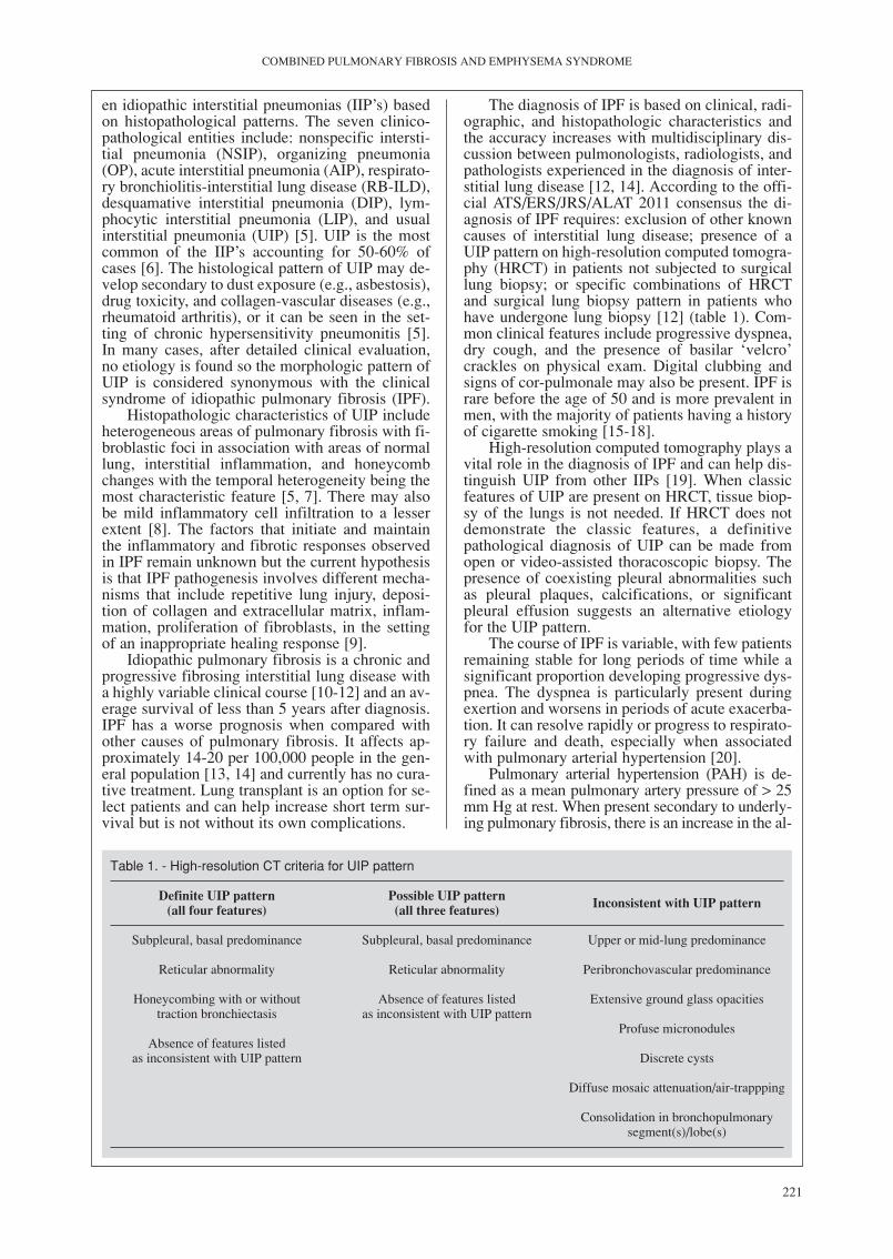
The diagnosis of IPF is based on clinical, radi-ographic, and histopathologic characteristics andthe accuracy increases with multidisciplinary dis-cussion between pulmonologists, radiologists, andpathologists experienced in the diagnosis of inter-stitial lung disease [12, 14]. According to the offi-cial ATS/ERS/JRS/ALAT 2011 consensus the di-agnosis of IPF requires: exclusion of other knowncauses of interstitial lung disease; presence of aUIP pattern on high-resolution computed tomogra-phy (HRCT) in patients not subjected to surgicallung biopsy; or specific combinations of HRCTand surgical lung biopsy pattern in patients whohave undergone lung biopsy [12] (table 1). Com-mon clinical features include progressive dyspnea,dry cough, and the presence of basilar ‘velcro’crackles on physical exam. Digital clubbing andsigns of cor-pulmonale may also be present. IPF israre before the age of 50 and is more prevalent inmen, with the majority of patients having a historyof cigarette smoking [15-18].
High-resolution computed tomography plays avital role in the diagnosis of IPF and can help dis-tinguish UIP from other IIPs [19]. When classicfeatures of UIP are present on HRCT, tissue biop-sy of the lungs is not needed. If HRCT does notdemonstrate the classic features, a definitivepathological diagnosis of UIP can be made fromopen or video-assisted thoracoscopic biopsy. Thepresence of coexisting pleural abnormalities suchas pleural plaques, calcifications, or significantpleural effusion suggests an alternative etiologyfor the UIP pattern.
The course of IPF is variable, with few patientsremaining stable for long periods of time while asignificant proportion developing progressive dys-pnea. The dyspnea is particularly present duringexertion and worsens in periods of acute exacerba-tion. It can resolve rapidly or progress to respirato-ry failure and death, especially when associatedwith pulmonary arterial hypertension [20].
Pulmonary arterial hypertension (PAH) is de-fined as a mean pulmonary artery pressure of > 25mm Hg at rest. When present secondary to underly-ing pulmonary fibrosis, there is an increase in the al-
Table 1. - High-resolution CT criteria for UIP pattern
Definite UIP pattern Possible UIP pattern Inconsistent with UIP pattern(all four features) (all three features)
Subpleural, basal predominance Subpleural, basal predominance Upper or mid-lung predominance
Reticular abnormality Reticular abnormality Peribronchovascular predominance
Honeycombing with or without Absence of features listed Extensive ground glass opacitiestraction bronchiectasis as inconsistent with UIP pattern
Profuse micronodulesAbsence of features listed
as inconsistent with UIP pattern Discrete cysts
Diffuse mosaic attenuation/air-trappping
Consolidation in bronchopulmonary segment(s)/lobe(s)

222
I.B. OLIVA ET AL.
ready high mortality rate in these patients [21]. Ear-ly studies showed that patients with IPF had highermean pulmonary arterial pressures compared to pa-tients with other non-IPF interstitial lung diseases[22]. Pulmonary fibrosis probably contributes to thepathogenesis of pulmonary arterial hypertension asa result of chronic alveolar hypoxia and vascular re-modeling, but the most important mechanism islikely the reduction in the pulmonary vascular bedsecondary to extension of fibrosis itself [23]. Pa-tients with pulmonary fibrosis are also at increasedrisk for pulmonary embolism, which is probablysecondary to endothelial damage [24].
COPD: definition and background
Chronic obstructive pulmonary disease wasthe 12th leading cause of disability in the worldduring the 1990’s [25] but is estimated to increasein prevalence, with current predictions that it willbe among the top five leading causes of death by2030 [26]. It results in great economic and socialburden that is both substantial and increasing be-cause it affects a population during their most pro-ductive work years. In the United States, COPDwas responsible for 126,000 deaths in 2005 [27].The estimated prevalence of COPD varies from 7to 19% in several well-conducted studies aroundthe world [28-33]. However COPD still remainsunderdiagnosed, especially because it often staysclinically silent until it is at an advanced and irre-versible stage [34, 35].
COPD is a preventable disease characterizedby airflow limitation that is not fully reversible andis staged on the basis of the severity of irreversibleairflow obstruction quantification [36]. The air-flow obstruction is secondary to an abnormal in-flammatory response of the lungs to noxious parti-cles or gases, primarily caused by tobacco smok-ing. It is usually progressive and can be the end re-sult of multiple distinct events. Other importantrisk factors include occupational and organic dustsexposures, socio-economic status, and genetic fac-tors. COPD has a variable natural history with dif-ferent individuals developing distinct clinicalcourses [37]. With the progression of the disease,COPD patients demonstrate compromised gas ex-change leading to respiratory failure with poorprognosis. Death is frequently secondary to associ-ated co-morbidities that are also related to ciga-rette smoking such as heart disease and lung can-cer [36, 38].
Repetitive airway injury from inhaled tobaccosmoke toxins, chronic oxidative stress, and imbal-ance of proteinases and antiproteinases trigger aninflammatory response that leads to structuralchanges in the airways and lung parenchyma [39,40]. COPD comprises pathological changes in fourdifferent compartments: central airways, peripher-al airways, lung parenchyma, and pulmonary vas-culature. The main physiologic abnormalities inCOPD are mucous hypersecretion and ciliary dys-function; airflow limitation and hyperinflation; gasexchange abnormalities; systemic effects and pul-monary hypertension [41].
Emphysema and obstructive airway diseaseare two commonly described subtypes of COPD.Considerable overlap with different proportions ofeach of these processes exists in COPD subjects[42]. Clinically, patients with COPD are classifiedaccording to Global Obstructive Lung Disease(GOLD) stages based on the severity of airwayobstruction [36] but individuals with the sameGOLD stage can exhibit markedly different de-grees of emphysema radiographically. Emphyse-ma is a pathologic diagnosis defined as “perma-nent abnormal enlargement of the airspaces distalto the terminal bronchiole associated with de-struction of their walls and no obvious fibrosis”[43]. There are three main subtypes of emphyse-ma based on the anatomic distribution: centriaci-nar (also known as centrilobular), panacinar (alsoknown as panlobular), and paraseptal (table 2).These subtypes of emphysema are better distin-guished morphologically during early stages butwhen they become more severe, their distinctionbecomes more difficult.
Centriacinar emphysema primarily affects therespiratory bronchioles and alveoli in the centralportion of the secondary pulmonary lobule. Thissubtype of emphysema predominantly involves theupper lung zones and typically results from ciga-rette smoking. With increasing severity of cen-triacinar emphysema, destruction progresses to in-volve the entire secondary pulmonary lobule, mak-ing it more difficult to distinct from the pancinarsubtype of emphysema.
Panacinar emphysema destroys the entire sec-ondary pulmonary lobule uniformly creating mul-tiple areas of decreased lung attenuation withpaucity of vessels in the affected regions. It pre-dominates in the lower lobes. This type of emphy-sema is classically associated with alpha-1 antit-rypsin deficiency but can be seen without protease
Table 2. - Types of pulmonary emphysema
Emphysema Involvement Distribution Etiology Wall Bulla
Centriacinar Center of SPL Upper lobes Cigarette smoking Absent Common
Panacinar Entire SPL Lower lobes Alpha 1 antitrypsin deficiency, Absent Uncommonsmokers, age or drug related
Paraseptal Distal SPL Upper lobes and subpleural Cigarette smoking or idiopathic Thin Common
* SPL = secondary pulmonary.
223
COMBINED PULMONARY FIBROSIS AND EMPHYSEMA SYNDROME
deficiency in smokers, elderly patients, and in as-sociation to certain drugs [43].
Paraseptal emphysema predominantly in-volves the alveolar ducts and sacs at the distal as-pect of the acini with the areas of destruction oftenmarginated by interlobular septa. It can be an iso-lated finding in young patients often associatedwith spontaneous pneumothorax or can be seen inolder patients with centrilobular emphysema [43,44]. This type of emphysema is usually seen in as-sociation with the centriacinar type in patients whoare cigarette smokers but can also be idiopathic inetiology.
Pulmonary hypertension is present in a signif-icant proportion of patients with advanced COPDdue to hypoxic vasoconstriction and secondary re-modeling of distal small pulmonary arterialbranches. The loss of pulmonary capillary bed inemphysema can also contribute to increased pres-sure in the pulmonary circulation [45, 46]. Theprogression of the pulmonary arterial hypertensionusually leads to elevated right heart pressures,right ventricular hypertrophy, ultimately resultingin right-sided cardiac failure (cor pulmonale).
Combined pulmonary fibrosis and emphysema (CPFE)
The combination of pulmonary fibrosis andemphysema was initially thought to be an inciden-tal coexistence of the two diseases and it is still un-clear whether both entities coexist by coincidencewith each one having distinct pathogenesis, or ifsome individuals share a common pathway thatleads to both fibrosis and emphysema. Animalmodels suggested a shared mechanism throughwhich cigarette smoke leads to both pulmonaryemphysema and fibrosis [47-49]. Such theory hasyet to be demonstrated in human subjects howeverseveral studies have demonstrated an increasedprevalence of IPF in patients with smoking histo-ry, suggesting this may indeed be a commonpathogen [15-18].
Nonetheless, CPFE has been treated as a dis-tinct disease after Cottin, et al. in 2005 presented acase series in heavy smokers [50]. Patients withcombined pulmonary fibrosis and emphysema(CPFE) syndrome demonstrate clinically severehypoxemia and decreased DLCO with normal tomildly reduced lung volumes. They often have se-vere secondary pulmonary arterial hypertension,which worsens the prognosis dramatically [50, 51].
In the retrospective study conducted by Cottinet al. in 2005 [50], all of the 61 patients were smok-ers or ex-smokers and most were males, with amean age of 65 years. The CPFE syndrome was di-agnosed by chest computed tomography. Thisstudy was one of the first to describe several clini-cal characteristics of this syndrome. Dyspnea onexertion was present in all patients; bibasilarcrackles in 87% of patients; clubbing in 43% ofpatients; and pulmonary arterial hypertension pre-sent in 47% of patients at diagnosis and in 55%during follow-up. Patients were followed in thisstudy for a mean of 2.1 ± 2.8 years after diagnosis.
Survival was 87.5% at 2 yrs and 54.6% at 5 yrs,with a median survival of 6.1 yrs. The presence ofPAH at diagnosis was a critical determinant ofprognosis in this syndrome.
Recently Cottin et al., not only confirmed butalso demonstrated in a retrospective multicenterstudy conducted in 40 patients (38 males; age 68 ±9 years; 39 smokers) with CPFE and pulmonaryarterial hypertension (confirmed by right heartcatheterization), that higher pulmonary vascularresistance, higher heart rate, lower cardiac indexand lower carbon monoxide diffusion transferwere associated with increased mortality [52].This study provides several new and important in-sights. For example, they found that the mean timeto diagnose PAH after diagnosing CPFE was 16months, suggesting that pulmonary arterial hyper-tension occurs rapidly after diagnosing CPFE.They also showed that patients with CPEF who de-velop pulmonary arterial hypertension have aworse prognosis with an estimated survival of on-ly 60% at 1 year.
Pulmonary arterial hypertension is one of theleading causes of symptoms and negatively im-pacts the prognosis in these patients to a levelmuch worse than that seen among patients with ei-ther pulmonary fibrosis or emphysema alone. Lit-erature has shown that at the time the diagnosis ofCPFE is made, 47% of patients are found to havePAH with the percentage increasing with progres-sion of disease [50-52]. It still remains uncertain ifPAH is a response to CPFE alone or a product ofemphysema or fibrosis separately.
Spirometry and lung volume components ofpulmonary function testing (PFT) in patients withCPFE are usually normal or demonstrate only mildobstructive or restrictive pattern. The basis for thisfinding, despite the presence of severe underlyingparenchymal lung disease may be the opposingphysiologic forces of emphysema, which leads tooutflow obstruction and hyperinflation, as opposedto fibrosis which decreases lung compliance, de-creases lung volumes, and increases outward teth-ering of small airways [53]. This may be one of thereasons why this syndrome remains underdiag-nosed until an advanced stage, when it is alreadyassociated with severe PAH. However, the diffus-ing capacity component of PFT’s is typically se-verely impaired due to loss of intact alveolar-cap-illary surface area for gas exchange, increased dif-fusion distance across alveolar-capillary bed, andimpaired pulmonary capillary blood flow. It is typ-ical for CPFE patients to demonstrate hypoxemicrespiratory failure with significantly reduced dif-fusing capacity and normal to only mildly reducedlung volumes [54].
Radiology Perspective
Chest x-ray
Chest radiographs are a standard part of theroutine clinical evaluation of subjects with COPDand IPF. Such images are not expensive, are read-ily available, and involve minimal radiation expo-
224
I.B. OLIVA ET AL.
sure when compared with computed tomography(CT) [55].
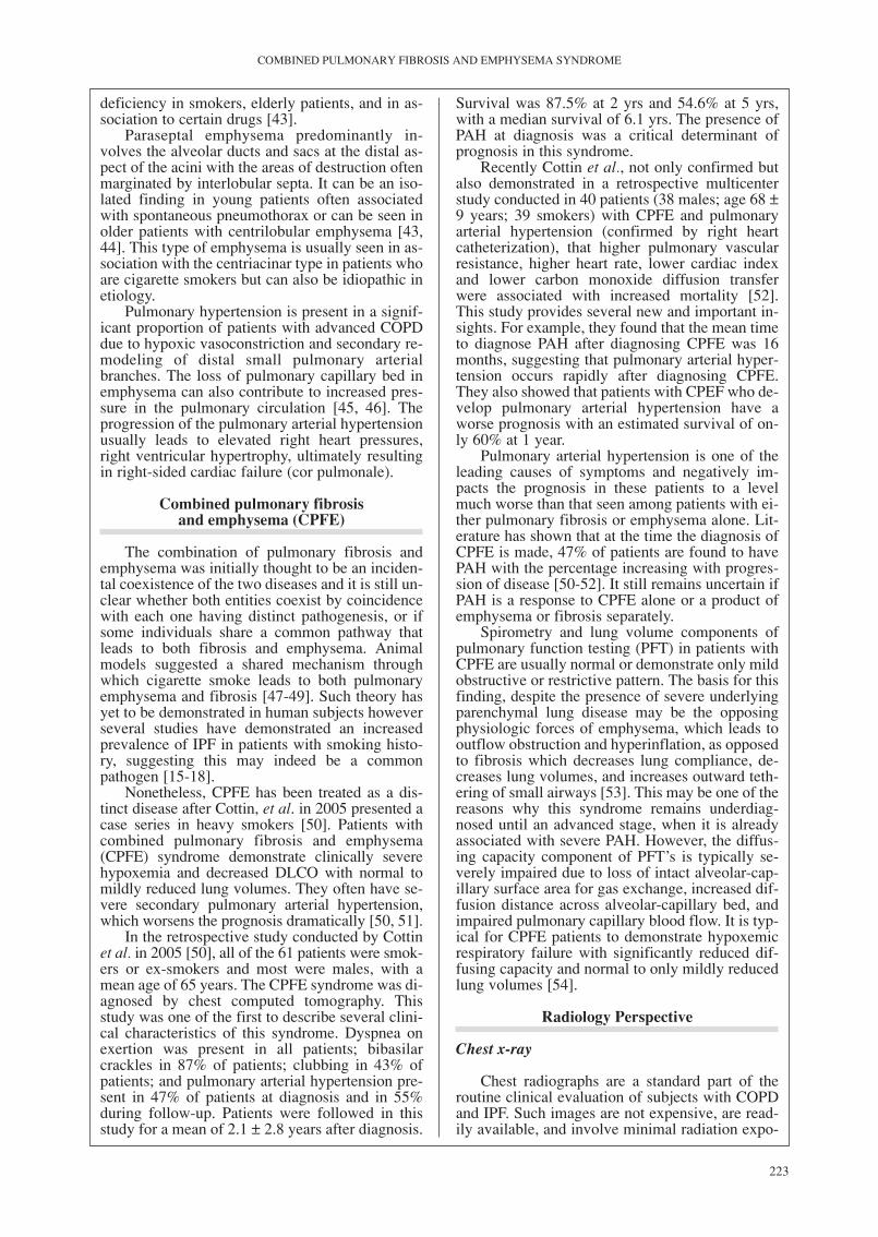
The chest x-ray of patients with pulmonaryemphysema usually demonstrates the combinationof increased lung volumes seen as flattening of thediaphragms with right diaphragm’s height measur-ing less than 2.7 cm on the lateral projection, in-creased sternodiaphragmatic angle measuringmore than 90 degrees, widening of the retrosternalclear space measuring more than 4.4 cm whenmeasured 3 cm below the manubrial-sternal junc-tion, increased AP diameter; and upper lobe pre-dominant destruction manifested by relative in-creased lucency and paucity of vessels in the lungapices and apparent increased interstitial markingsat the lower lung zones (fig. 1). Hyperinflation al-so results in a narrowed, elongated cardiomedisti-nal silhouette but this is a secondary sign that isneither specific nor sensitive for emphysema [56-60]. Visualization of associated bullae is diagnos-tic of emphysema but is seldom seen radiographi-cally. A sensitivity of 80% has been reported whenthese findings are used in diagnosis but the likeli-hood of diagnosis based on chest x-ray depends onthe severity of disease with mild forms being al-most impossible to detect [61] (fig. 2).
Approximately 10% of patients with IPF havenormal chest radiography [62, 63], particularly inearly stages of the disease (fig. 3). The radi-ographic appearance of IPF is nonspecific and cor-relates poorly with histological findings and sever-ity of disease [6]. When abnormal, chest x-ray mayshow lower lobe and peripherally predominant
reticular markings associated with decreased lungvolumes and relative sparing of the lung apices(fig. 4). With more advanced disease, lung vol-umes become even smaller and the reticular mark-ings progress from fine to coarse. Lower lobe pre-dominant cystic spaces, which represent tractionbronchiectasis and/or honeycombing, may also beapparent. The disease classically starts at the pos-terior costophrenic sulci, which are better assessedon the lateral radiograph.
While chest radiographs are neither sensitivenor specific in diagnosing emphysema or pul-monary fibrosis, when evaluating CPFE syndromethe sensitivity is even lower because the presenceof increased lung volumes seen in patients withemphysema is usually masked by the decreasedvolumes secondary to the concomitant pulmonaryfibrosis resulting in normal lung volumes. Otherfindings of each disease separately may help in thediagnosis but since the lung volumes play an im-portant role in diagnosing each entity separately,with the loss of this finding, accuracy decreaseseven more.
Some findings that may provide clues to thepresence of underlying CPFE include: increasedlucency of the upper lungs which is seen in pa-tients with emphysema, in conjunction with in-creased reticular markings in the lower lobes, thatis characteristic of patients with pulmonary fibro-sis (fig. 5). However, chest x-rays are of limitedvalue and insensitive in diagnosing this syndromewith computed tomography remaining the mostsensitive and specific method of diagnosis.
Fig. 1. - Frontal radiograph of the chest (a) shows upper lobe predominant pulmonary parenchyma destruction due to emphysema seen as lucen-cy and paucity of vessels in upper lung zones. The lateral projection demonstrates flattening of the diaphragms, widening of the retrosternal clearspace, and increased AP diameter of the thorax.
(a) (b)

225
COMBINED PULMONARY FIBROSIS AND EMPHYSEMA SYNDROME
Computed Tomography
Computed Tomography is much more expen-sive than radiographs and exposes the patients tosignificantly higher radiation doses with mean ef-fective dose using a 64-slice CT scanner of ap-proximately 19.9 mSv [64]. On the other hand,with the advancement of technology, the scannershave become faster and are now able to providethe same image quality with less radiation expo-sure. CT is as widely available as chest x-rays andremains the most accurate diagnostic modality forpatients with pulmonary emphysema and fibrosis.It is much more sensitive and specific in diagnos-ing and classifying pulmonary emphysema whencompared to chest radiography and was recentlyproven to reduce mortality when used for lungcancer screening [65], which these patients are atincreased risk of.
Emphysema is characterized on high resolu-tion computed tomography (HRCT) by areas ofabnormal low attenuation contrasted by the normal
surrounding lung parenchyma [66-68]. Areas ofcentriacinar emphysema are seen as focal lucencycentered in the middle of the secondary pulmonarylobule, surrounding the centrilobular artery, andwithout definable walls (fig. 6, 7). This type of em-physema is typically seen in cigarette smokers andhas an upper lobe predominance. Panacinar em-physema is seen as widespread abnormal low at-tenuation areas marginated by the interlobular sep-ta and also centered on the centrilobular artery. Itmaintains the polyhedric shape of the secondarypulmonary lobule and predominates in the lowerlobes (fig. 8). Paraseptal emphysema involves thedistal aspect of the secondary pulmonary lobuleand therefore has a subpleural distribution. It hasan elongated shape with perceivable thin walls,which generally correspond to the interlobular sep-ta. With paraseptal emphysema, there is a singlerow of subpleural cystic spaces (bullae). This is incontrast to honeycombing which is secondary topulmonary fibrosis in which, by definition, thereare at least 2 rows of subpleural cysts as well as
Fig. 2. - Normal frontal (a) and lateral (b) radiographs of the chest in a patient with mild pulmonary emphysema. Two axial images from the chestCT of the same patient demonstrate upper lobe predominant emphysema.
(a) (b)
(c) (d)

226
I.B. OLIVA ET AL.
other associated findings of fibrosis (ie: architec-tural distortion and traction bronchiectasis).Paraseptal emphysema predominates in the upperlobe. When the involved area is larger than 1 cm indiameter, it is termed a bulla (fig. 9). Bullae are di-agnostic of emphysema but are seldom seen onchest radiograph or CT.
Computed tomography scanning of the chestallows quantitative assessment of the extent andseverity of pulmonary emphysema as well as elu-cidating additional features such as the distribution(eg, apical, basal, diffuse) and subtype (eg, cen-triacinar, panacinar, paraseptal) cof emphysema[69]. Visual scoring by one or more observers [70]was the initial approach utilized when evaluatingCT scan data in patients with pulmonary emphyse-ma. As with any subjective form of evaluation, in-terobserver variability remains a concern. In astudy of severe COPD patients [71], interobserveragreement was good for the overall severity of em-physema but was poor in the determination of lo-bar predominance of emphysema.
Many studies have used CT of the chest toquantify the extent and severity of emphysemaand to document progression, but this ability de-pends on several technical factors including: scan-ner calibration, collimation, threshold values,window settings, radiation dose, phase of respira-tory cycle, reconstruction algorithm, and use ofintravenous contrast [72-77]. Subjective quantifi-cation of emphysema is the simplest method andis based on visual assessment of the CT images[78-80]. Each part of the lung that appears em-physematous is graded from 1 to 4 (1=1% to 25%,2=26% to 50%, 3=51% to 75%, and 4=76% to100% of the area) with the total score expressed aspercentage of total lung involved at that assignedlevel [81, 82].
Conventional chest CT and HRCT are alsomuch more sensitive than routine chest radiogra-phy when assessing patients with IPF, and theirfindings correlate well with symptoms and pul-monary function test results [83]. It has also beenproven that the extent of ground glass opacity cor-
Fig. 3. - Normal chest radiograph (a, b) of a patient with early stage of pulmonary fibrosis. Two axial images from the chest CT of the same pa-tient demonstrating lower lobe predominant reticular markings.
(a) (b)
(c) (d)

227
COMBINED PULMONARY FIBROSIS AND EMPHYSEMA SYNDROME
(a) (b)
(a) (b)
Fig. 4. - Frontal (a) and lateral (b) radiographs of the chest in a patient with idiopathic pulmonary fibrosis demonstrating decreased lung volumesand lower lobe predominant increased reticular markings with relative sparing of the upper lobes. Axial (c) and coronal reformatted (d) CT im-ages of the same patient better depict the peripheral and lower lobe predominant involvement by fibrosis with honeycombing formation.
(c) (d)
Fig. 5. - Chest x-rays of a patient with combined pulmonary emphysema and fibrosis syndrome. The lung volumes are normal but the frontal ra-diograph (a) shows lower lobe predominant reticular markings in conjunction with increased lucency of the upper lobes. The lateral projection (b)demonstrates relative flattening of diaphragms and mildly increased retrosternal clear space.

228
I.B. OLIVA ET AL.
relates well with the severity of dyspnea and re-duction in carbon monoxide diffusing capacity(DLCO), with patients having lower DLCOdemonstrating more extensive ground glass opaci-ties by CT [84].
Computed tomography of patients with IPFdemonstrates lower lobe and peripherally predomi-nant intra and inter-lobular septal thickening, trac-tion bronchiectasis, honeycombing and architectur-al distortion, all reflecting underlying parenchymalfibrosis (fig. 10). Honeycombing is the most reli-able CT finding of fibrosis. Ground glass opacitiesmay also be appreciated, especially early in thecourse of the disease, or during acute exacerbations,but are never the dominant finding in patients withidiopathic pulmonary fibrosis in the chronic pro-gressive stage. The ground glass opacities can eitherrepresent active alveolitis or very fine fibrosis. Thinsection CT images can sometimes help distinguishbetween the two. In about 70-95% of patients, retic-ulations involve mainly the subpleural areas andhave the classic apicobasal gradient of severity withthe abnormality being worse at the lung bases, par-ticularly at the costophrenic angles [19, 85, 86]. Be-cause this is a restrictive lung disease, decreasedlung volumes result in elevation of the hemidi-aphragms with decreased craniocaudal dimension, atypical finding in patients with lung fibrosis.
Fig. 6. - Axial CT images (a, b) of a patient with centriacinar emphysema demonstrating multiple areas of increased lucency involving the sec-ondary pulmonary lobule, centered around the centrilobular artery (white arrows). Note the absence of perceivable walls in the lucent areas.
(a) (b)
Fig. 7. - Single CT image of a patient with centriacinar emphysemashows the central involvement of the secondary pulmonary lobulewith preservation of its polyhedral shape. The interlobular septa ap-pear as walls surrounding the lucent area (arrow) and the centrilobu-lar artery is seen in the middle of it.
Fig. 8. - Axial (a) and coronal reformatted (b) CT images of a patient with alpha one antitrypsin deficiency showing lower lobe predominantpanacinar emphysema. Note that the parenchymal destruction involves multiple secondary pulmonary lobules entirely. The centrilobular artery isalso seen in middle of a few of these lucent areas (white arrows).
(a) (b)

229
COMBINED PULMONARY FIBROSIS AND EMPHYSEMA SYNDROME
The interpretation of HRCT scans in patientswith diffuse parenchymal lung diseases (DPLD)can be difficult, as findings may be nonspecific in50% of cases [87, 88]. While difficulty existswith diagnosing DPLDs overall, specialized tho-racic radiologists can become quite good at inter-preting HRCT scans of patients with IPF and oth-er idiopathic interstitial pneumonias [19, 89, 90].The positive predictive value of a HRCT diagno-sis of UIP is 90-100% in several studies [91-93].
A multidisciplinary approach with review ofthe case by pulmonologists, pulmonary patholo-gists, and thoracic radiologists is most beneficialwhen evaluating these often times complicated pa-tients. As discussed above, HRCT plays a key rolein distinguishing a UIP pattern from other IIPs.The HRCT criteria for diagnosing UIP pattern isbased in the presence of basal and subpleural pre-dominant reticular abnormalities in conjunctionwith honeycombing and absence of findings that
Fig. 9. - Axial (a) and coronal reformatted (b) CT images show upper lobeparaseptal emphysema. Note the subpleural location and elongated shape ofthe lucent areas with thin visible walls (arrow) that represent the surroundinginterlobular septa. A few of these areas measure more than 1 cm in diameter,and are therefore termed bulla.
(a)
(b)
suggest an alternative diagnosis such as peribron-chovascular predominance, extensive ground glassabnormality, discrete cysts, segmental/lobar con-solidations, and diffuse mosaic attenuation [12].
In clinical practice, the visual assessment ofdisease extent by HRCT can be used in associationwith pulmonary function test results to monitorprogression of IPF, as well as to evaluate responseto therapy and aid with prognosis. This measure-ment should be made with intermediate intervalfollow-ups rather than short-term interval follow-up since longitudinal changes in HRCT scans dur-ing short-term follow-up are less predictive of sur-vival when compared to physiologic measure-ments [94, 95].
Given that CT is accurate in diagnosing bothpulmonary emphysema and fibrosis alone and thateach distinct disease demonstrates different imag-ing characteristics, it is not difficult to make the di-agnosis of CPFE using computed tomography ofthe chest. Patients demonstrate lucent areas of lungdestruction in the upper lobes associated with low-er lobe predominant reticular markings, honey-combing, and traction bronchiectasis that reflectunderlying pulmonary fibrosis (fig. 11).
As discussed above, pulmonary arterial hyper-tension is seen in patients with emphysema andpulmonary fibrosis. Chest radiography of patientswith PAH classically demonstrates the “prunedtree” appearance made by the enlargement of thecentral pulmonary arteries and abrupt decrease incaliber of the pulmonary vasculature peripherally(fig. 12). Chest CT of these patients can demon-strate enlargement of the central pulmonary arter-ies but this finding is neither specific nor sensitive.It may also show abrupt narrowing and tapering ofthe peripheral pulmonary vessels, right ventricularhypertrophy, right ventricular and right atrial en-largement, dilated bronchial arteries, and a mosaicpattern of attenuation due to variable lung perfu-sion (fig. 13) [96].
Fig. 10. - 71 year old male with biopsy proven idiopathic pulmonaryfibrosis. Axial CT image shows peripheral and lower lobe predomi-nant end-stage fibrotic changes with architectural distortion, tractionbronchiectasis, and subpleural honeycombing.

230
I.B. OLIVA ET AL.
IPF patients are also at increased risk of pul-monary embolism, which is likely secondary to en-dothelial damage. Chest CT, when tailored to eval-uate the pulmonary arteries, is very sensitive andspecific in diagnosing pulmonary embolism, espe-cially when using multidetector CT scanners [97,98]. Patients with pulmonary fibrosis are also at in-creased risk of developing bronchogenic carcino-ma. CT is more sensitive than chest radiography atdetecting lung cancer, and can pick them up whenthey are smaller and at an earlier stage [99-102].
Radiation Considerations
It is important to keep in mind that patientswho undergo multiple serial CT scans are exposedto a higher cumulative radiation dose over time.Therefore the benefits and risks related to this ex-am should always be weighed prior to imaging. Inthe early days of CT, the radiation exposure couldnot be reduced, since the technology available atthat time did not allow for a reduction in the dose
without increasing image noise. A large amount ofimage noise degrades image contrast and quality,thus impairing diagnostic accuracy [77]. With ad-vancement of technology, the CT scanners are nowable to produce the same image quality with amuch lower radiation dose to the patient. While therisk of radiation is still dependent mostly on thepatient’s age, the body part that is being scanned,and how much of the patient’s body is covered(length of the scan), the effective radiation dose re-ceived during a routine computed tomography ofthe chest is about 8 mSv, which is equivalent to ap-proximately 100 chest x-rays [103].
In conclusion, combined pulmonary fibrosisand emphysema is an important but still under-diagnosed syndrome that has imaging findings ofboth pulmonary emphysema and fibrosis but is as-sociated with a worse prognosis when compared toeach of these diseases alone. Since radiology playsa key role in making this diagnosis, it is importantthat radiologists be familiar with the existence andappearance of this syndrome, which will facilitateearly and accurate diagnosis to potentially improveprognosis.
References
1. Lopez AD, Shibuya K, Rao C, Mathers CD, HansellAL, Held LS, et al. Chronic obstructive pulmonary dis-ease: current burden and future projections. Eur RespirJ 2006; 27: 397-412.
2. Murray CJ, Lopez AD. Alternative projections of mor-tality and disability by cause 1990-2020: Global Burdenof Disease Study. Lancet 1997; 349 (9065): 1498-504.
3. King TE, Jr., Tooze JA, Schwarz MI, Brown KR, Cher-niack RM. Predicting survival in idiopathic pulmonaryfibrosis: scoring system and survival model. Am JRespir Crit Care Med 2001; 164: 1171-81.
4. Wiggins J, Strickland B, Turner-Warwick M. Com-bined cryptogenic fibrosing alveolitis and emphysema:the value of high resolution computed tomography inassessment. Respir Med 1990; 84: 365-9.
5. American Thoracic Society/European Respiratory So-ciety International Multidisciplinary Consensus Classi-fication of the Idiopathic Interstitial Pneumonias. Thisjoint statement of the American Thoracic Society(ATS), and the European Respiratory Society (ERS)
Fig. 11. - 60 year old female with smoking history, worsening short-ness of breath and dry cough. Axial (a, b) and coronal reformatted (c)CT images showing the concomitant presence of upper lobe predom-inant emphysema with lucent areas of lung destrucion as well as pe-ripheral and lower lobe predominant fibrotic changes seen as in-creased reticular markings and traction bronchiectasis.
(c)
(a) (b)

231
COMBINED PULMONARY FIBROSIS AND EMPHYSEMA SYNDROME
was adopted by the ATS board of directors, June 2001and by the ERS Executive Committee, June 2001. Am JRespir Crit Care Med 2002; 165: 277-304.
6. Kim DS, Collard HR, King TE, Jr. Classification andnatural history of the idiopathic interstitial pneumonias.Proc Am Thorac Soc 2006; 3: 285-92.
7. Visscher DW, Myers JL. Histologic spectrum of idio-pathic interstitial pneumonias. Proc Am Thorac Soc2006; 3: 322-9.
8. Katzenstein AL, Myers JL. Idiopathic pulmonary fibro-sis: clinical relevance of pathologic classification. Am JRespir Crit Care Med 1998; 157 (4 Pt 1): 1301-15.
9. Keane MP, Strieter RM, Lynch JP, 3rd, Belperio JA. In-flammation and angiogenesis in fibrotic lung disease.Semin Respir Crit Care Med 2006; 27: 589-99.
10. Schwartz DA, Helmers RA, Galvin JR, Van Fossen DS,Frees KL, Dayton CS, et al. Determinants of survival in
idiopathic pulmonary fibrosis. Am J Respir Crit CareMed 1994; 149 (2 Pt 1): 450-4.
11. Selman M, King TE, Pardo A. Idiopathic pulmonary fi-brosis: prevailing and evolving hypotheses about itspathogenesis and implications for therapy. Ann InternMed 2001; 134: 136-51.
12. Raghu G, Collard HR, Egan JJ, Martinez FJ, Behr J,Brown KK, et al. An Official ATS/ERS/JRS/ALATStatement: Idiopathic Pulmonary Fibrosis: Evidence-based Guidelines for Diagnosis and Management. Am JRespir Crit Care Med 2010; 183: 788-824.
13. Coultas DB, Zumwalt RE, Black WC, Sobonya RE.The epidemiology of interstitial lung diseases. Am JRespir Crit Care Med 1994; 150: 967-72.
14. American Thoracic Society. Idiopathic pulmonary fi-brosis: diagnosis and treatment. International consensusstatement. American Thoracic Society (ATS), and the
Fig. 13. - 87 years old male with history of chronic pulmonary arterial hypertension. Axial (a) and coronal reformatted (b) images from a contrast-enhanced chest CT show the enlargement of the pulmonary arteries (white arrows).
(a) (b)
Fig. 12. - 87 years old male with history of chronic pulmonary arterial hypertension. Frontal (a) and lateral (b) radiographs of the chest demon-strate enlargement of the main pulmonary arterial trunk as well as the right and left pulmonary arteries (white and black arrows). Also note the“pruned-tree” appearance due to abrupt reduction in the caliber of the vessels in the peripheral two thirds of the lung, classically seen in this clin-ical setting.
(a) (b)

232
I.B. OLIVA ET AL.
European Respiratory Society (ERS). Am J Respir CritCare Med 2000; 161 (2 Pt 1): 646-64.
15. Raghu G, Weycker D, Edelsberg J, Bradford WZ, OsterG. Incidence and prevalence of idiopathic pulmonaryfibrosis. Am J Respir Crit Care Med 2006; 174: 810-6.
16. Nadrous HF, Myers JL, Decker PA, Ryu JH. Idiopath-ic pulmonary fibrosis in patients younger than 50 years.Mayo Clin Proc 2005; 80: 37-40.
17. Iwai K, Mori T, Yamada N, Yamaguchi M, Hosoda Y.Idiopathic pulmonary fibrosis. Epidemiologic ap-proaches to occupational exposure. Am J Respir CritCare Med 1994; 150: 670-5.
18. Gribbin J, Hubbard RB, Le Jeune I, Smith CJ, West J, TataLJ. Incidence and mortality of idiopathic pulmonary fibro-sis and sarcoidosis in the UK. Thorax 2006; 61: 980-5.
19. Hunninghake GW, Lynch DA, Galvin JR, Gross BH,Muller N, Schwartz DA, et al. Radiologic findings arestrongly associated with a pathologic diagnosis of usu-al interstitial pneumonia. Chest 2003; 124: 1215-23.
20. Martinez FJ, Safrin S, Weycker D, Starko KM, Brad-ford WZ, King TE, Jr., et al. The clinical course of pa-tients with idiopathic pulmonary fibrosis. Ann InternMed 2005; 142 (12 Pt 1): 963-7.
21. Horowitz JC, Thannickal VJ. Idiopathic pulmonary fi-brosis: new concepts in pathogenesis and implicationsfor drug therapy. Treat Respir Med 2006; 5: 325-42.
22. Weitzenblum E, Ehrhart M, Rasaholinjanahary J, HirthC. Pulmonary hemodynamics in idiopathic pulmonaryfibrosis and other interstitial pulmonary diseases. Res-piration 1983; 44: 118-27.
23. Newman JH. Pulmonary hypertension. Am J RespirCrit Care Med 2005; 172: 1072-7.
24. Panos RJ, Mortenson RL, Niccoli SA, King TE, Jr.Clinical deterioration in patients with idiopathic pul-monary fibrosis: causes and assessment. Am J Med1990; 88: 396-404.
25. Murray CJ, Lopez AD. Evidence-based health policy -lessons from the Global Burden of Disease Study. Sci-ence 1996; 274 (5288): 740-3.
26. Mathers CD, Loncar D. Projections of global mortalityand burden of disease from 2002 to 2030. PLoS Med2006; 3: e442.
27. Deaths from chronic obstructive pulmonary disease -United States, 2000-2005. MMWR Morb Mortal WklyRep 2008; 57: 1229-32.
28. Mannino DM, Homa DM, Akinbami LJ, Ford ES, ReddSC. Chronic obstructive pulmonary disease surveillance- United States, 1971-2000. MMWR Surveill Summ2002; 51: 1-16.
29. Celli BR, Halbert RJ, Isonaka S, Schau B. Populationimpact of different definitions of airway obstruction.Eur Respir J 2003 22: 268-73.
30. Menezes AM, Perez-Padilla R, Jardim JR, et al. Chron-ic obstructive pulmonary disease in five Latin Ameri-can cities (the PLATINO study): a prevalence study.Lancet 2005; 366 (9500): 1875-81.
31. Pena VS, Miravitlles M, Gabriel R, et al. Geographicvariations in prevalence and underdiagnosis of COPD:results of the IBERPOC multicentre epidemiologicalstudy. Chest 2000; 118: 981-9.
32. Tzanakis N, Anagnostopoulou U, Filaditaki V, Christa-ki P, Siafakas N. Prevalence of COPD in Greece. Chest2004; 125: 892-900.
33. de Marco R, Accordini S, Cerveri I, et al. An interna-tional survey of chronic obstructive pulmonary diseasein young adults according to GOLD stages. Thorax2004; 59: 120-5.
34. Talamo C, de Oca MM, Halbert R, et al. Diagnostic la-beling of COPD in five Latin American cities. Chest2007; 131: 60-7.
35. Damarla M, Celli BR, Mullerova HX, Pinto-Plata VM.Discrepancy in the use of confirmatory tests in patients
hospitalized with the diagnosis of chronic obstructivepulmonary disease or congestive heart failure. RespirCare 2006; 51: 1120-4.
36. Pauwels RA, Buist AS, Calverley PM, Jenkins CR,Hurd SS. Global strategy for the diagnosis, manage-ment, and prevention of chronic obstructive pulmonarydisease. NHLBI/WHO Global Initiative for ChronicObstructive Lung Disease (GOLD) Workshop summa-ry. Am J Respir Crit Care Med 2001; 163: 1256-76.
37. Prescott E. Tobacco-related diseases: the role of gender.An epidemiologic study based on data from the Copen-hagen Centre for Prospective Population Studies. DanMed Bull 2000; 47: 115-31.
38. Celli BR, MacNee W. Standards for the diagnosis andtreatment of patients with COPD: a summary of the ATS/ERS position paper. Eur Respir J 2004; 23: 932-46.
39. Rennard SI. Inflammation and repair processes inchronic obstructive pulmonary disease. Am J RespirCrit Care Med 1999; 160 (5 Pt 2): S12-6.
40. Peinado VI, Barbera JA, Abate P, et al. Inflammatoryreaction in pulmonary muscular arteries of patients withmild chronic obstructive pulmonary disease. Am JRespir Crit Care Med 1999; 159 (5 Pt 1): 1605-11.
41. Matsuba K, Wright JL, Wiggs BR, Pare PD, Hogg JC.The changes in airways structure associated with re-duced forced expiratory volume in one second. EurRespir J 1989; 2: 834-9.
42. Kitaguchi Y, Fujimoto K, Kubo K, Honda T. Charac-teristics of COPD phenotypes classified according tothe findings of HRCT. Respir Med 2006; 100: 1742-52.
43. Snider GL, Kleinerman J, Thurlbeck WM, Bengali ZH.The definition of emphysema. Report of a NationalHeart, Lung, and Blood Institute, Division of Lung Dis-eases workshop. Am Rev Respir Dis 1985; 132: 182-5.
44. Stern EJ, Frank MS, Schmutz JF, Glenny RW, SchmidtRA, Godwin JD. Panlobular pulmonary emphysemacaused by i.v. injection of methylphenidate (Ritalin):findings on chest radiographs and CT scans. Am JRoentgenol 1994; 162: 555-60.
45. Fisher MR, Criner GJ, Fishman AP, et al. Estimatingpulmonary artery pressures by echocardiography in pa-tients with emphysema. Eur Respir J 2007; 30: 914-21.
46. Scharf SM, Iqbal M, Keller C, Criner G, Lee S, FesslerHE. Hemodynamic characterization of patients with se-vere emphysema. Am J Respir Crit Care Med 2002;166: 314-22.
47. Hoyle GW, Li J, Finkelstein JB, et al. Emphysematouslesions, inflammation, and fibrosis in the lungs of trans-genic mice overexpressing platelet-derived growth fac-tor. Am J Pathol 1999; 154: 1763-75.
48. Lucattelli M, Bartalesi B, Cavarra E, et al. Is neutrophilelastase the missing link between emphysema and fi-brosis? Evidence from two mouse models. Respir Res2005; 6: 83.
49. Lundblad LK, Thompson-Figueroa J, Leclair T, et al.Tumor necrosis factor-alpha overexpression in lung dis-ease: a single cause behind a complex phenotype. Am JRespir Crit Care Med 2005; 171: 1363-70.
50. Cottin V, Nunes H, Brillet PY, et al. Combined pul-monary fibrosis and emphysema: a distinct underrecog-nised entity. Eur Respir J 2005; 26: 586-93.
51. Kosacka M, Brzecka A, Jankowska R, Lewczuk J,Mroczek E, Werynska B. Combined pulmonary fibrosisand emphysema - case report and literature review.Pneumonol Alergol Pol 2009; 77: 205-10.
52. Cottin V, Brillet PY, Nunes H, Cordier JF. Combinedpulmonary fibrosis and emphysema. Presse Med 2007;36 (6 Pt 2): 936-44.
53. Schwartz DA, Merchant RK, Helmers RA, Gilbert SR,Dayton CS, Hunninghake GW. The influence of ciga-rette smoking on lung function in patients with idio-
233
COMBINED PULMONARY FIBROSIS AND EMPHYSEMA SYNDROME
pathic pulmonary fibrosis. Am Rev Respir Dis 1991;144 (3 Pt 1): 504-6.
54. Hiwatari N, Shimura S, Takishima T. Pulmonary em-physema followed by pulmonary fibrosis of undeter-mined cause. Respiration 1993; 60: 354-8.
55. Parry RA, Glaze SA, Archer BR. The AAPM/RSNAphysics tutorial for residents. Typical patient radiationdoses in diagnostic radiology. Radiographics 1999; 19:1289-302.
56. Reich SB, Weinshelbaum A, Yee J. Correlation of radi-ographic measurements and pulmonary function tests inchronic obstructive pulmonary disease. Am J Roentgenol1985; 144: 695-9.
57. Standards for the diagnosis and care of patients withchronic obstructive pulmonary disease (COPD) andasthma. This official statement of the American Tho-racic Society was adopted by the ATS Board of Direc-tors, November 1986. Am Rev Respir Dis 1987; 136:225-44.
58. Burki NK. Roentgenologic diagnosis of emphysema.Accurate or not? Chest 1989; 95: 1178-9.
59. Foster WL, Jr., Gimenez EI, Roubidoux MA, et al. Theemphysemas: radiologic-pathologic correlations. Radi-ographics 1993; 13: 311-28.
60. Pratt PC. Role of conventional chest radiography in di-agnosis and exclusion of emphysema. Am J Med 1987;82: 998-1006.
61. Thurlbeck WM, Muller NL. Emphysema: definition,imaging, and quantification. Am J Roentgenol 1994;163: 1017-25.
62. Epler GR, McLoud TC, Gaensler EA, Mikus JP, Car-rington CB. Normal chest roentgenograms in chronicdiffuse infiltrative lung disease. N Engl J Med 1978;298: 934-9.
63. Carrington CB, Gaensler EA, Coutu RE, FitzGeraldMX, Gupta RG. Natural history and treated course ofusual and desquamative interstitial pneumonia. N EnglJ Med 1978; 298: 801-9.
64. Hurwitz LM, Reiman RE, Yoshizumi TT, et al. Radia-tion dose from contemporary cardiothoracic multidetec-tor CT protocols with an anthropomorphic female phan-tom: implications for cancer induction. Radiology 2007;245: 742-50.
65. Henschke CI, Boffetta P, Gorlova O, Yip R, DelanceyJ, Foy M. Assessment of lung-cancer mortality reduc-tion from CT Screening. Lung Cancer 2011; 71: 328-32.
66. Foster WL, Jr., Pratt PC, Roggli VL, Godwin JD, Halvors-en RA, Jr., Putman CE. Centrilobular emphysema: CT-pathologic correlation. Radiology 1986; 159: 27-32.
67. Hruban RH, Meziane MA, Zerhouni EA, et al. Highresolution computed tomography of inflation-fixedlungs. Pathologic-radiologic correlation of centrilobularemphysema. Am Rev Respir Dis 1987; 136: 935-40.
68. Webb WR, Stein MG, Finkbeiner WE, Im JG, Lynch D,Gamsu G. Normal and diseased isolated lungs: high-resolution CT. Radiology 1988; 166 (1 Pt 1): 81-7.
69. Webb WR. Thin-section CT of the secondary pul-monary lobule: anatomy and the image - the 2004 Fleis-chner lecture. Radiology 2006; 239: 322-38.
70. Goddard PR, Nicholson EM, Laszlo G, Watt I. Com-puted tomography in pulmonary emphysema. Clin Ra-diol 1982; 33: 379-87.
71. Hersh CP, Washko GR, Jacobson FL, et al. Interob-server variability in the determination of upper lobe-predominant emphysema. Chest 2007; 131: 424-31.
72. Boedeker KL, McNitt-Gray MF, Rogers SR, et al. Em-physema: effect of reconstruction algorithm on CTimaging measures. Radiology 2004; 232: 295-301.
73. Gevenois PA, de Maertelaer V, De Vuyst P, Zanen J,Yernault JC. Comparison of computed density andmacroscopic morphometry in pulmonary emphysema.Am J Respir Crit Care Med 1995; 152: 653-7.
74. Gevenois PA, Scillia P, de Maertelaer V, Michils A, DeVuyst P, Yernault JC. The effects of age, sex, lung size,and hyperinflation on CT lung densitometry. Am JRoentgenol 1996; 167: 1169-73.
75. Parr DG, Stoel BC, Stolk J, Nightingale PG, StockleyRA. Influence of calibration on densitometric studies ofemphysema progression using computed tomography.Am J Respir Crit Care Med 2004; 170: 883-90.
76. Stoel BC, Bakker ME, Stolk J, et al. Comparison of thesensitivities of 5 different computed tomography scan-ners for the assessment of the progression of pulmonaryemphysema: a phantom study. Invest Radiol 2004; 39:1-7.
77. Yuan R, Mayo JR, Hogg JC, et al. The effects of radia-tion dose and CT manufacturer on measurements oflung densitometry. Chest 2007; 132: 617-23.
78. Kuwano K, Matsuba K, Ikeda T, et al. The diagnosis ofmild emphysema. Correlation of computed tomographyand pathology scores. Am Rev Respir Dis 1990; 141:169-78.
79. Bergin CJ, Muller NL, Miller RR. CT in the qualitativeassessment of emphysema. J Thorac Imaging 1986; 1:94-103.
80. Kinsella M, Muller NL, Abboud RT, Morrison NJ, Dy-Buncio A. Quantitation of emphysema by computed to-mography using a “density mask” program and correla-tion with pulmonary function tests. Chest 1990; 97:315-21.
81. Sakai F, Gamsu G, Im JG, Ray CS. Pulmonary functionabnormalities in patients with CT-determined emphyse-ma. J Comput Assist Tomogr 1987; 11: 963-8.
82. Nishimura K, Murata K, Yamagishi M, et al. Compari-son of different computed tomography scanning meth-ods for quantifying emphysema. J Thorac Imaging1998; 13: 193-8.
83. Staples CA, Muller NL, Vedal S, Abboud R, Ostrow D,Miller RR. Usual interstitial pneumonia: correlation ofCT with clinical, functional, and radiologic findings.Radiology 1987; 162: 377-81.
84. Terriff BA, Kwan SY, Chan-Yeung MM, Muller NL.Fibrosing alveolitis: chest radiography and CT as pre-dictors of clinical and functional impairment at follow-up in 26 patients. Radiology 1992; 184: 445-9.
85. Lynch DA, Godwin JD, Safrin S, et al. High-resolutioncomputed tomography in idiopathic pulmonary fibrosis:diagnosis and prognosis. Am J Respir Crit Care Med2005; 172: 488-93.
86. MacDonald SL, Rubens MB, Hansell DM, et al. Non-specific interstitial pneumonia and usual interstitial pneu-monia: comparative appearances at and diagnostic accu-racy of thin-section CT. Radiology 2001; 221: 600-5.
87. Souza CA, Muller NL, Flint J, Wright JL, Churg A. Id-iopathic pulmonary fibrosis: spectrum of high-resolu-tion CT findings. Am J Roentgenol 2005; 185: 1531-9.
88. Johkoh T, Muller NL, Cartier Y, et al. Idiopathic inter-stitial pneumonias: diagnostic accuracy of thin-sectionCT in 129 patients. Radiology 1999; 211: 555-60.
89. Hunninghake GW, Zimmerman MB, Schwartz DA, etal. Utility of a lung biopsy for the diagnosis of idio-pathic pulmonary fibrosis. Am J Respir Crit Care Med2001; 164: 193-6.
90. Raghu G, Mageto YN, Lockhart D, Schmidt RA, WoodDE, Godwin JD. The accuracy of the clinical diagnosisof new-onset idiopathic pulmonary fibrosis and otherinterstitial lung disease: A prospective study. Chest1999; 116: 1168-74.
91. Mathieson JR, Mayo JR, Staples CA, Muller NL.Chronic diffuse infiltrative lung disease: comparison ofdiagnostic accuracy of CT and chest radiography. Radi-ology 1989; 171: 111-6.
92. Grenier P, Valeyre D, Cluzel P, Brauner MW, Lenoir S,Chastang C. Chronic diffuse interstitial lung disease:
234
I.B. OLIVA ET AL.
diagnostic value of chest radiography and high-resolu-tion CT. Radiology 1991; 179: 123-32.
93. Lee KS, Primack SL, Staples CA, Mayo JR, Aldrich JE,Muller NL. Chronic infiltrative lung disease: comparison ofdiagnostic accuracies of radiography and low- and conven-tional-dose thin-section CT. Radiology 1994; 191: 669-73.
94. Battista G, Zompatori M, Fasano L, Pacilli A, Basile B.Progressive worsening of idiopathic pulmonary fibro-sis. High resolution computed tomography (HRCT)study with functional correlations. Radiol Med 2003;105: 2-11.
95. Flaherty KR, Mumford JA, Murray S, et al. Prognosticimplications of physiologic and radiographic changes inidiopathic interstitial pneumonia. Am J Respir CritCare Med 2003; 168: 543-8.
96. Grosse C, Grosse A. CT findings in diseases associatedwith pulmonary hypertension: a current review. Radi-ographics 30: 1753-77.
97. Patel S, Kazerooni EA. Helical CT for the evaluation ofacute pulmonary embolism. AJR Am J Roentgenol 2005;185: 135-49.
98. Mamlouk MD, vanSonnenberg E, Gosalia R, et al. Pul-monary embolism at CT angiography: implications forappropriateness, cost, and radiation exposure in 2003patients. Radiology 256: 625-32.
99. Henschke CI, McCauley DI, Yankelevitz DF, et al. Ear-ly Lung Cancer Action Project: overall design and find-ings from baseline screening. Lancet 1999; 354 (9173):99-105.
100. Henschke CI, Yankelevitz DF, Libby DM, PasmantierMW, Smith JP, Miettinen OS. Survival of patients withstage I lung cancer detected on CT screening. N Engl JMed 2006; 355: 1763-71.
101. Foy M, Yip R, Chen X, Kimmel M, Gorlova OY, Hen-schke CI. Modeling the mortality reduction due to com-puted tomography screening for lung cancer. Cancer2011; 117: 2703-8.
102. http: //www.cancer.gov/newscenter/pressreleases/NL-STresultsRelease.
103. Brix G, Nagel HD, Stamm G, et al. Radiation exposurein multi-slice versus single-slice spiral CT: results of anationwide survey. Eur Radiol 2003; 13: 1979-91.
Pavia - Ponte Coperto