Combined Electron Transfer Dissociation–Collision-Induced Dissociation Fragmentation in the Mass...
8
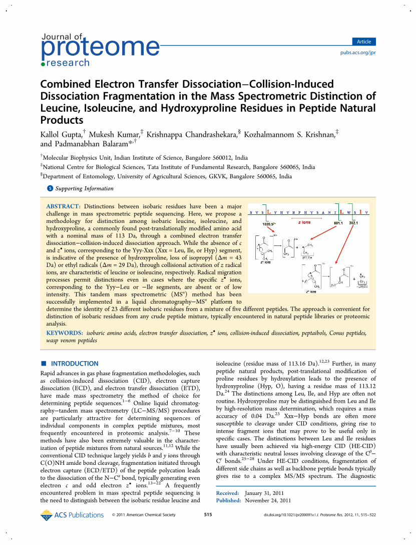
Combined Electron Transfer Dissociation−Collision-Induced Dissociation Fragmentation in the Mass Spectrometric Distinction of Leucine, Isoleucine, and Hydroxyproline Residues in Peptide Natural Products Kallol Gupta, † Mukesh Kumar, ‡ Krishnappa Chandrashekara, § Kozhalmannom S. Krishnan, ‡ and Padmanabhan Balaram* ,† † Molecular Biophysics Unit, Indian Institute of Science, Bangalore 560012, India ‡ National Centre for Biological Sciences, Tata Institute of Fundamental Research, Bangalore 560065, India § Department of Entomology, University of Agricultural Sciences, GKVK, Bangalore 560065, India * S Supporting Information ABSTRACT: Distinctions between isobaric residues have been a major challenge in mass spectrometric peptide sequencing. Here, we propose a methodology for distinction among isobaric leucine, isoleucine, and hydroxyproline, a commonly found post-translationally modified amino acid with a nominal mass of 113 Da, through a combined electron transfer dissociation−collision-induced dissociation approach. While the absence of c and z • ions, corresponding to the Yyy-Xxx (Xxx = Leu, Ile, or Hyp) segment, is indicative of the presence of hydroxyproline, loss of isopropyl (Δm = 43 Da) or ethyl radicals (Δm = 29 Da), through collisional activation of z radical ions, are characteristic of leucine or isoleucine, respectively. Radical migration processes permit distinctions even in cases where the specific z • ions, corresponding to the Yyy−Leu or −Ile segments, are absent or of low intensity. This tandem mass spectrometric (MS n ) method has been successfully implemented in a liquid chromatography−MS n platform to determine the identity of 23 different isobaric residues from a mixture of five different peptides. The approach is convenient for distinction of isobaric residues from any crude peptide mixture, typically encountered in natural peptide libraries or proteomic analysis. KEYWORDS: isobaric amino acids, electron transfer dissociation, z • ions, collision-induced dissociation, peptaibols, Conus peptides, wasp venom peptides ■ INTRODUCTION Rapid advances in gas phase fragmentation methodologies, such as collision-induced dissociation (CID), electron capture dissociation (ECD), and electron transfer dissociation (ETD), have made mass spectrometry the method of choice for determining peptide sequences. 1−6 Online liquid chromatog- raphy−tandem mass spectrometry (LC−MS/MS) procedures are particularly attractive for determining sequences of individual components in complex peptide mixtures, most frequently encountered in proteomic analysis. 7−10 These methods have also been extremely valuable in the character- ization of peptide mixtures from natural sources. 11,12 While the conventional CID technique largely yields b and y ions through C(O)NH amide bond cleavage, fragmentation initiated through electron capture (ECD/ETD) of the peptide polycation leads to the dissociation of the N−C α bond, typically generating even electron c and odd electron z • ions. 13−22 A frequently encountered problem in mass spectral peptide sequencing is the need to distinguish between the isobaric residue leucine and isoleucine (residue mass of 113.16 Da). 12,23 Further, in many peptide natural products, post-translational modification of proline residues by hydroxylation leads to the presence of hydroxyproline (Hyp, O), having a residue mass of 113.12 Da. 24 The distinctions among Leu, Ile, and Hyp are often not routine. Hydroxyproline may be distinguished from Leu and Ile by high-resolution mass determination, which requires a mass accuracy of 0.04 Da. 23 Xxx−Hyp bonds are often more susceptible to cleavage under CID conditions, giving rise to intense fragment ions that may prove to be useful only in specific cases. The distinctions between Leu and Ile residues have usually been achieved via high-energy CID (HE-CID) with characteristic neutral losses involving cleavage of the C β − C γ bonds. 25−28 Under HE-CID conditions, fragmentation of different side chains as well as backbone peptide bonds typically gives rise to a complex MS/MS spectrum. The diagnostic Received: January 31, 2011 Published: November 24, 2011 Article pubs.acs.org/jpr © 2011 American Chemical Society 515 dx.doi.org/10.1021/pr200091v | J. Proteome Res. 2012, 11, 515−522
-
Upload
padmanabhan -
Category
Documents
-
view
214 -
download
0
Transcript of Combined Electron Transfer Dissociation–Collision-Induced Dissociation Fragmentation in the Mass...
Combined Electron Transfer Dissociation−Collision-InducedDissociation Fragmentation in the Mass Spectrometric Distinction ofLeucine, Isoleucine, and Hydroxyproline Residues in Peptide NaturalProductsKallol Gupta,† Mukesh Kumar,‡ Krishnappa Chandrashekara,§ Kozhalmannom S. Krishnan,‡
and Padmanabhan Balaram*,†
†Molecular Biophysics Unit, Indian Institute of Science, Bangalore 560012, India‡National Centre for Biological Sciences, Tata Institute of Fundamental Research, Bangalore 560065, India§Department of Entomology, University of Agricultural Sciences, GKVK, Bangalore 560065, India
*S Supporting Information
ABSTRACT: Distinctions between isobaric residues have been a majorchallenge in mass spectrometric peptide sequencing. Here, we propose amethodology for distinction among isobaric leucine, isoleucine, andhydroxyproline, a commonly found post-translationally modified amino acidwith a nominal mass of 113 Da, through a combined electron transferdissociation−collision-induced dissociation approach. While the absence of cand z• ions, corresponding to the Yyy-Xxx (Xxx = Leu, Ile, or Hyp) segment,is indicative of the presence of hydroxyproline, loss of isopropyl (Δm = 43Da) or ethyl radicals (Δm = 29 Da), through collisional activation of z radicalions, are characteristic of leucine or isoleucine, respectively. Radical migrationprocesses permit distinctions even in cases where the specific z• ions,corresponding to the Yyy−Leu or −Ile segments, are absent or of lowintensity. This tandem mass spectrometric (MSn) method has beensuccessfully implemented in a liquid chromatography−MSn platform todetermine the identity of 23 different isobaric residues from a mixture of five different peptides. The approach is convenient fordistinction of isobaric residues from any crude peptide mixture, typically encountered in natural peptide libraries or proteomicanalysis.
KEYWORDS: isobaric amino acids, electron transfer dissociation, z• ions, collision-induced dissociation, peptaibols, Conus peptides,wasp venom peptides
■ INTRODUCTIONRapid advances in gas phase fragmentation methodologies, suchas collision-induced dissociation (CID), electron capturedissociation (ECD), and electron transfer dissociation (ETD),have made mass spectrometry the method of choice fordetermining peptide sequences.1−6 Online liquid chromatog-raphy−tandem mass spectrometry (LC−MS/MS) proceduresare particularly attractive for determining sequences ofindividual components in complex peptide mixtures, mostfrequently encountered in proteomic analysis.7−10 Thesemethods have also been extremely valuable in the character-ization of peptide mixtures from natural sources.11,12 While theconventional CID technique largely yields b and y ions throughC(O)NH amide bond cleavage, fragmentation initiated throughelectron capture (ECD/ETD) of the peptide polycation leadsto the dissociation of the N−Cα bond, typically generating evenelectron c and odd electron z• ions.13−22 A frequentlyencountered problem in mass spectral peptide sequencing isthe need to distinguish between the isobaric residue leucine and
isoleucine (residue mass of 113.16 Da).12,23 Further, in manypeptide natural products, post-translational modification ofproline residues by hydroxylation leads to the presence ofhydroxyproline (Hyp, O), having a residue mass of 113.12Da.24 The distinctions among Leu, Ile, and Hyp are often notroutine. Hydroxyproline may be distinguished from Leu and Ileby high-resolution mass determination, which requires a massaccuracy of 0.04 Da.23 Xxx−Hyp bonds are often moresusceptible to cleavage under CID conditions, giving rise tointense fragment ions that may prove to be useful only inspecific cases. The distinctions between Leu and Ile residueshave usually been achieved via high-energy CID (HE-CID)with characteristic neutral losses involving cleavage of the Cβ−Cγ bonds.25−28 Under HE-CID conditions, fragmentation ofdifferent side chains as well as backbone peptide bonds typicallygives rise to a complex MS/MS spectrum. The diagnostic
Received: January 31, 2011Published: November 24, 2011
Article
pubs.acs.org/jpr
© 2011 American Chemical Society 515 dx.doi.org/10.1021/pr200091v | J. Proteome Res. 2012, 11, 515−522

fragment ions are often found to be of low intensity, makingassignments ambiguous. Also, under HE-CID conditions, facileloss of any acid labile post-translational modification makes theprocess of assignment even more difficult. Marshall and co-workers have shown extensive characteristic side chain losses ofdifferent amino acids in ECD spectra, near the precursor m/zregion.29 Further, side chain losses from specific radical z• ionshave been shown by Zubarev and co-workers in the hotelectron capture dissociation (HECD) spectra of peptidepolycations.30 The processes of radical migration and radical-mediated secondary fragmentation have been probed byO’Connor and co-workers through ECD of diprotonated cyclicpeptides.31 Observation of extensive side chain losses andneutral amino acid residues has been explained in terms of a“cascade of free radical reaction” that is initiated from theprecursor where one radical center has been created throughthe transfer of one electron. Further studies with peptidescontaining α-deuterated glycine residues provide experimentalevidence of α-hydrogen abstraction during the process ofradical migration along the peptide backbone.32 A similarradical nature of the z• ions, formed during ETDfragmentation, has been established by McLuckey and co-workers through the observation of an oxygen adduct and morerecently through collisional activation of the z• ions.33,34 Usinghot electrons during electron capture dissociation (HECD),radical-mediated backbone cleavages have been shown to beuseful in effecting the distinction of Leu from Ile,35,36 but thetechnical difficulties in implementing ECD in mass spectrom-eters other than FT-ICR MS limit the application of thismethod. McLuckey and co-workers have established thatfragment ions obtained under ETD conditions may then besubjected to further CID (ETD−CID) fragmentation for theobservation of side chain specific losses through radical-inducedcleavages, in a QTRAP instrument, using synthetic modelsequences.34 Under conditions of electron transfer dissocia-tion/electron capture dissociation (ETD/ECD) fragmentation,cleavage of the N−Cα bond at the Pro residue does not lead tothe formation of characteristic c and z• ions due to theconstraints of side chain backbone cyclization in the pyrrolidinering.17 The absence of such diagnostic ions can be used tosignal the presence of Pro or Hyp residues. Herein, we presentan extension of these approaches to several peptides of naturalorigin containing multiple Leu, Ile, and Hyp residues andfurther demonstrate the applicability of this method in effectivedistinction among these isobaric amino acids from a peptidemixture.
■ EXPERIMENTAL SECTIONThe bee venom peptide melittin was a commercial sample fromSigma (St. Lous, MO). The fungal peptaibols, antiamoebin II,and zervamicin IIB were obtained as described previously.37,38
A novel peptide, Bt2328, was obtained from the venom of themarine cone snail Conus betulinus by reverse phase high-performance liquid chromatography (HPLC) fractionation ofthe mixture of venom peptides. Similarly, the wasp peptideVt1512 was obtained from the venom of social wasp speciesVespa tropica. The synthetic Bt2328 sample was synthesized viaa typical solid phase peptide synthesis protocol using Fmocchemistry.The ETD−CID experiments were conducted using a HCT
Ultra ETD II ion trap mass spectrometer (Bruker Daltonics,Bremen, Germany). The purified peptide samples (8 μM),dissolved in a 50:50 H2O/acetonirile mixture, containing 0.1%
formic acid (zervamicin IIB and antiameobin II were dissolvedin 100% MeOH, containing 0.1% formic acid, and the solutionswere directly infused into the mass spectrometer), wereinjected into the mass spectrometer typically at a flow rate of2 μL/min using an injector pump. The tandem massspectrometric (MSn) experiments in the direct infusion studywere performed manually. After observation of the desiredmultiply charged precursor in MS, the precursor ion wasmanually chosen for ETD fragmentation. The ETD reactiontime was set at 75 ms for zervamicin and antiamoebin, 85 msfor Bt2328 and Vt1512, and 100 ms for melittin. All the ETDMS2 experiments were performed using “smart decomposition”(supplemental activation) that provides a low-backgroundcollision gas to increase the product ion abundances throughprobable dissociation of the noncovalent bonds (see Dis-cussion). From the MS2 ETD spectra, desired precursor ionswere subsequently chosen for CID MS3 fragmentation insidethe ion trap through collision with He gas. The fragmentationamplitude (Vp−p) was kept between 1 and 3. All theexperiments were conducted at a scan speed of 8100 m/z s−1.The spectra were averaged over 6−10 scans. For LC−MSn
analysis, these peptides (5 μL each) were mixed together. TheLC−MS analysis was performed by coupling the mentioned iontrap mass spectrometer with an Agilent 1100 HPLC system andthe peptide samples were separated on a reverse phase C18analytical column (Porshell 120, SB-C18, particle size of 2.7μm, dimensions of 4.6 mm × 150 mm) using a H2O/acetonitrile mixture (0.1% formic acid) as the solvent system ata flow rate of 0.2 mL/min. The gradient was set from 90% H2Oto 100% ACN over 45 min in a linear gradient. The LC−MSn
experiments were performed in a data-independent manner.Before the start of the LC run, the total run time was dividedinto different segments as per the retention times of the fivepeptides used. In these individual segments, the m/z values ofthe precursor ions to be chosen for MS2/MS3 experiments weremanually inserted into the acquisition software. During theLC−MSn study, the scan speed was set to 26000 m/z s−1, andtypically, the spectra were averaged over three to five scans. TheETD reaction time was set at 75 ms for zervamicin andantiamoebin, 85 ms for Bt2328 and Vt1512, and 100 ms formelittin. In this case also, smart decomposition (supplementalactivation) was used while acquiring the ETD MS2 spectra.Subsequent CID fragmentation was conducted inside the iontrap through the collision of He gas with the ions of interest.The HE-CID experiments were performed on an UltraFlextreme MALDI system (Bruker Daltonics) equipped witha 337 nm laser operated at 50 Hz. Ar was used as the collisiongas at ∼4 × 10⁻⁶ to 1 × 10⁻⁵ mbar of pressure.
■ RESULTSThe application of the combined ETD−CID procedure, fordistinction among isoleucine (I), leucine (L), and hydroxypro-line (O), is illustrated using examples of diverse, naturallyoccurring peptides. This approach is based on the fate of theinitially formed aminoketyl radical under ETD conditions andsubsequent side chain fragmentations from z• ions under CIDconditions, as illustrated in Scheme 1. The z• ions, formed witha radical at the Cα position of the Ile or Leu residue, canundergo radical-induced cleavage of the Cβ−Cγ bond. In thecase of Leu, this results in a neutral loss of 43 Da, while for Ile,a loss of 29 Da is observed.32,34 If Hyp residues are present inthe sequence, the formation of aminoketyl radicals does notlead to further fragmentation of the Xxx−Hyp linkage. It should
Journal of Proteome Research Article
dx.doi.org/10.1021/pr200091v | J. Proteome Res. 2012, 11, 515−522516

be noted that the absence of fragment ions corresponding toN−Cα bond cleavage in ETD spectra provides support for thepresence of a Hyp residue but does not rule out the presence ofXxx−Ile or −Leu fragments, which may be resistant tofragmentation. However, unlike CID experiments in whichbackbone cleavage is strongly sequence-dependent, in ETDMS/MS spectra of peptides, N−Cα bond fragmentation doesnot appear to be strongly influenced by the amino acidsequence. Indeed, in our study with diverse peptide sequences,of 20 Xxx−Ile or −Leu bonds, we have not encountered anyexample of any Xxx−Ile or −Leu bond that is recalcitrant tocleavage under electron transfer conditions. In specific cases,any further ambiguity can also be resolved through ETDfragmentation of other possible charge states. A consistentabsence of c and z ions, corresponding to the Xxx−Ile, −Leu, or−Hyp segment, in the ETD MS/MS spectra of all the chargestates would strongly signal a presence of the Hyp residue.
Peptaibol Antibiotics
The peptaibols make up a microheterogeneous group ofpolypeptides of fungal origin that have been widelyinvestigated.37,38 They are characterized by a high proportionof the nonprotein amino acid α-aminoisobutyric acid (Aib, U)and the presence of a C-terminal aminoalcohol residue. Severalclasses of peptaibols also contain Hyp residues. Figure 1 showsthe ETD spectrum of antiamoebin II [Ac-FUUUJGLUUOQ-JOUPF*, where Ac is acetyl, U is α-aminoisobutyric acid (Aib),J is isovaline (Iva), O is hydroxyproline (Hyp), and F* isphenylalaninol (Phol)] by directly infusing the peptide andfragmenting the [M + H + K]2+ species at m/z 855.0. Some ofthe low-intensity fragment ions are shown as insets. Thevariation in the isotopic patterns for some of the very lowintensity peaks may be attributed to their extremely low signalintensity. Under these conditions, all observed c and z• ionscorresponded to their potassiated species. This indicates aselective capture of a proton by the reduced carbonyl groups ascompared to potassium. This feature has also been noted inearlier studies and substantiated by the calculation ofrecombination energy at the two positively charged sites.39
All the observed m/z values of these c and z• ions are found to
be greater than that of the doubly charged precursor. Theobserved fragment ions are consistent with the previouslydetermined sequence, with three residues having a residue massof 113 Da. Of these, two are Hyp residues, at positions 10 and13, while Leu is present at position 7. Here, the potassiateddoubly charged species was chosen because of its higherabundance. Typically, for these classes of extremely hydro-phobic peptides, a very high intensity sodiated and potassiatedspecies is observed under electrospray conditions. In this case,similar results were obtained for the [M + H + Na]2+ species(data not shown). Inspection of the ETD fragmentation patternestablishes the absence of cn/z
•n ions corresponding to
cleavages at Aib9−Hyp10 and Iva (isovaline)12−Hyp13 bonds.Figure 1a shows the CID fragmentation of the z• ion at m/z1092.6. The appearance of the product ion at m/z 1049.6,corresponding to a neutral loss of 43 Da, confirms the presenceof a Leu residue at position 7. Here the ion at m/z 1074.6corresponds to a loss of H2O, most feasibly from the side chainof Hyp residues. The related 16-residue peptaibol zervamicinIIB (Ac-WIQJITULUOQUOUPF*) possesses five residueshaving a residue mass of 113 Da: Hyp residues at positions 10and 13, Ile residues at positions 2 and 5, and Leu at position 8.Figure 2 shows the distribution of product ions obtainedthrough ETD fragmentation of the (M + H + K)2+ species, bydirectly infusing the peptide. The distribution of c and z• ions iscompletely in agreement with the previously determinedsequence. As anticipated, fragment ions corresponding tocleavage at the Xxx−Pro or −Hyp segments are not observed.The insets to Figure 2 show the key product ions obtained bysubjecting the specifically selected z• ions to furtherfragmentation under CID conditions. To confirm the presenceof Leu8, the choice of the z
•9 ion at m/z 993.6 would have been
desirable. However, the low intensity of this ion precludedfurther CID fragmentation. Similarly, the z•10 ion at m/z 1078.6was also of low intensity. Consequently, an intense z•11 ion atm/z 1179.6 was further fragmented under CID conditions. Asone can see in Figure 2a, a neutral loss of 43 Da is observed,confirming the presence of Leu at position 8. As noted by thegroups of McLuckey and O’Connor, radical migration along the
Scheme 1. Mechanistic Pathways for Characteristic SideChain Losses for (a) Isoleucine and (b) Leucine from theNascent Aminoketyl Radicals Leading to the Formation of z•
Ions That Are Subsequently Collisionally Activated and (c)Hydroxyproline, for Which the Initially Formed AminoketylRadical Cannot Lead to N−Cα Bond Cleavage
Figure 1. ETD MS/MS spectrum of (M + H + K)2+ species (m/z855.0) of antiamoebin II. Inset a shows the CID fragmentationspectrum of the potassiated z•10 ion. The schematic summarizes theobserved c and z ions. The symbol c* stands for the potassiated evenelectron c ion and z* the potassiated odd electron z ion.
Journal of Proteome Research Article
dx.doi.org/10.1021/pr200091v | J. Proteome Res. 2012, 11, 515−522517

backbone can induce cleavages, at positions remote from the N-terminus of the fragment ion.31,34 In our study, thisphenomenon has been judiciously used to distinguish betweenLeu and Ile, in cases where the specific z• ions were of notsufficient intensity. In Figure 2a, a neutral loss of 17 Da isconsistent with the elimination of a hydroxyl radical (•OH)from the N-terminal threonine residue. Similarly in Figure 2b,observation of a neutral loss of 29 Da, upon selection andsubsequent CID fragmentation of the z•13 ion, confirms thepresence of Ile at position 5. In this case, too, the z•12 ion,having Ile at the N-terminus, could not be fragmented becauseof its low intensity. While the migration of the radical from theinitial site of generation is possible, the observed side chaincleavages appear to occur preferentially at proximal Leu or Ileresidues. An intense loss of 29 Da, as shown in Figure 2c, uponCID fragmentation of the z•15 ion at m/z 1632.7, confirms thepresence of Ile at position 2. It is to be noted that z•15 containsa Leu residue in addition to an Ile. The absence of the productions corresponding to the Leu side chain fragment may beattributed to the remoteness of the Leu residue from theinitially formed radical centers. Interestingly, McLuckey and co-workers have shown that when both Ile and Leu are remotefrom the initially formed radical center, radical migrationprocesses appear to favor the side chain fragmentation of Leu,34
presumably as a consequence of the higher stability of theeliminated tertiary radical of Leu than the secondary radical ofIle. A similar result has also been observed by us. It should bestressed that when the radical center is initially generated ateither Ile or Leu, a very high abundance of the correspondingside chain fragmentation is exclusively observed. The CID MS3
spectra of z•15 of zervamicin IIb is an example of such behavior.Bt2328
The venom of marine cone snails has been extensively studied,and a large number of disulfide-bonded peptides (conotoxins)have been characterized.40 Cone snail venoms also containacyclic sequences that have been less studied.41,42 During thecourse of our work, aimed at sequencing conus peptide libraries,we encountered a linear peptide with a molecular mass of 2328Da (Bt2328), produced by C. betulinus. The sequence of thispeptide was determined by mass spectrometry, using CID and
ETD fragmentation. Figures S1a and S2 of the SupportingInformation show CID and ETD MS2 spectra of Bt2328. Denovo sequencing from mass spectral data yielded a 20-residuesequence, SVSXYHVRPHYSANTXWSXV (X is Leu, Ile, orHyp) with residues having a mass of 113 Da at position 4, 16,and 19, which could be assigned to leucine, isoleucine, orhydroxyproline. The presence of z•5, z
•17, and c18 ions negates
the possibility of Hyp at these positions. Initial HE-CIDexperiments, performed in MALDI, yielded w2, w5, and d4 ionsthat were indicative of Ile at position 19 and Leu at positions 4and 16 (data not shown). Subsequently, these peptides weresubjected to ETD−CID experiments. Figure S2 of theSupporting Information shows the ETD spectrum obtainedfor the (M + 4H)4+ species at m/z 583.1 through directinfusion of the peptide of interest. Here also, the low intensityof the z•2 ion precluded a direct assignment of the residue atposition 19. Hence, the assignment of Ile at position 19 wasmade, exploiting the process of radical migration, through theselection and subsequent CID fragmentation of the z•3 ion atm/z 302.1, which results in a neutral loss of 29 Da (Figure 3).
The z•5 ion at m/z 601.3 upon further CID fragmentationyields a product ion at m/z 558.3, which corresponds to aneutral loss of 43 Da, establishing Leu at position 16. Theneutral loss of 17 Da leading to a product ion at m/z 583.3 maybe assigned as a loss of hydroxyl radical (•OH) from Ser atposition 18. Selection of the doubly charged z•17 species at m/z1020.5 yielded the doubly charged product ion at m/z 999.0,corresponding to a neutral loss of 43 Da. This confirms thepresence of Leu at position 4. A low-intensity ion at m/z 1003.5may be assigned to a doubly charged species arising from a lossof 34 Da, corresponding to an elimination of a hydroxyl radicaland ammonia. Indeed, the sequence contains hydroxylatedresidues at positions 12 (Ser), 15 (Thr), and 18 (Ser) and anArg at position 8 that is capable of losing NH3. The correctnessof the sequence was confirmed by comparison with achemically synthesized peptide. Panels a and b of Figure S1of the Supporting Information show the comparative CID MS2
spectra of the natural and synthetic peptide. The examples ofzervamicin IIB and Bt2328 illustrate, in spite of the absence ofspecific z ions, how the distinction between the isobaricresidues can be made by fragmenting the larger z• ionscontaining the ambiguous residue, exploiting the radicalmigration phenomenon.
Vt1512
The venom of social wasps also contains a complex mixture oflinear peptides. During the course of investigation directed
Figure 2. ETD MS/MS spectrum of (M + H + K)2+ species (m/z939.1) of zervamicin IIB. Insets show the CID fragmentation spectraof (a) z•11, (b) z
•13, and (c) z•15 ions. The schematic summarizes the
observed c and z ions. The symbol c* stands for the potassiated evenelectron c ion and z* the potassiated odd electron z ion.
Figure 3. CID fragmentation spectra of (a) z•3, (b) z•5, and (c) z•17
ions of Bt2328.
Journal of Proteome Research Article
dx.doi.org/10.1021/pr200091v | J. Proteome Res. 2012, 11, 515−522518

towards characterization of the natural peptide library fromwasp venom, we encountered sequences rich in Leu and Ileresidues. In such cases, conventional de novo sequencing usingCID methods yielded ambiguous results at several sequencepositions that were assigned residue masses of 113 Da. FigureS3 of the Supporting Information shows the ETD spectrum ofone of the wasp venom peptides, Vt1512 (XNXKAXAAX-AKKXF*, where X is Leu or Ile and the asterisk denotes a C-terminal amidation), isolated from the crude venom of V.tropica, obtained by the transfer of electron to (M + 3H)3+
species through a direct infusion experiment. Figure 4 illustrates
the observed neutral losses upon fragmentation of the z• ions atm/z 262.1, 702.5, 957.7, 635.32+, and 748.72+. The loss of 43 Dafrom z•2 (m/z 262.1) and z•6 (m/z 702.5) establishes thepresence of a Leu residue at positions 13 and 9, respectively.Interestingly, the neutral loss of 17 Da is observed from boththe parent and product ion at m/z 262.1 and 219.1,respectively. This z•2 ion corresponds to the C-terminaldipeptide Leu-Phe amide, with 17 Da losses being attributableto elimination of NH3. In addition, the neutral loss of 58 Dafrom the ion at m/z 702.5 to yield the product ion at m/z 644.1may be attributed to the Lys side chain. The z•9 ion at m/z957.7 upon CID shows a neutral loss of 29 Da, establishing Ileat position 6. Doubly charged ETD MS2 ions at m/z 635.3 and749.0 are assigned to fragment ions z•12 and z•14, respectively.The observed product ion at m/z 613.82+, shown in Figure 4,upon CID of the z•12 ion at m/z 635.32+, is consistent with aLeu at position 3. In the case of the z•14 ion at m/z 749.02+,neutral losses of 29 Da (Ile), 44 Da (Asn/C-terminal CONH2),and 58 Da (Lys) are consistent with the determined sequence.In addition, the 56 Da loss may be assigned to the even electronloss of a four-carbon unit (CH3−CHCH−CH3) from Ile,generated via Cα−Cβ bond cleavage. In the case of Vt1512, theuse of conventional CID MS/MS, together with ETD−CIDMS3 experiments, permitted determination of the sequence of apreviously uncharacterized peptide.
Melittin
The 26-residue highly basic peptide melittin (molecular mass of2846 Da; GIGAVLKVLTTGLPALISWIKRKRQQ*, where theasterisk denotes a C-terminal amidation), isolated from beevenom, is used to illustrate a situation that may lead toambiguity in application of the ETD−CID method. Figure S4of the Supporting Information shows the fragmentation patternobtained by the transfer of electron to the (M + 5H)5+ speciesthrough a direct infusion experiment. Figure 5 shows the
neutral losses obtained upon subjecting specific z• ions to CIDconditions. The sequence contains six residues with a residuemass of 113 Da, assigned to Leu or Ile. Selection andfragmentation of the z•7 ion at m/z 939.6 yields a neutral loss of29 Da (product ion at m/z 910.6), confirming an Ile at position20. The loss of 87 and 71 Da can be attributed to the loss ofCH3CH2NHC(NH)NH2 and H2CCH(CH2)2NH2 groupsfrom the side chains of Arg and Lys residues, respectively.Fragmentation of the z•11 ion at m/z 1438.9 reveals a neutralloss of 43 Da consistent with Leu at position 16, which is theN-terminal end of the z• ion. Fragmentation of the doublycharged z•14 ion (m/z 860.6) yielded the anticipated neutralloss of 43 Da, confirming Leu at position 13. Similarly,fragmentation of the doubly charged z•18 and triply charged z
•21
ion at m/z 1046.6 and 811.2 yielded a neutral loss of 43 Da,establishing the presence of Leu at positions 9 and 6,respectively. The doubly charged z•25 ion at m/z 1387.3upon further fragmentation reveals a neutral loss of 29 Da,consistent with an Ile at position 2. In our case, the lowintensity of the z•10 ion at m/z 1326.8 precluded confirmationof Ile at position 17. Similar problems, encountered in theprevious sequences, were resolved utilizing the phenomenon ofradical migration, where larger z• ions, containing theambiguous residue, were chosen for fragmentation. However,in this case, the presence of an Ile at position 6 restricts the useof such an approach, as z• ions containing Ile17 would alsocontain Ile6. Hence, upon CID fragmentation of these z ions,any observed neutral loss of 29 Da cannot be unambiguouslyassigned to one of these Ile residues. The results obtained withmelittin suggest that in proteomic identification of unknownsequences, ambiguity may arise because of the low intensity ofspecifically useful z• ions, especially in cases where successivemultiple Leu or Ile residues occur in the sequence. This
Figure 4. CID fragmentation spectra of (a) z•2, (b) z•6, (c) z•9, (d)z•12, and (e) z•14 ions of Vt1512.
Figure 5. CID fragmentation spectra of (a) z•7, (b) z•11, (c) z
•14, (d)
z•18, (e) z•21, and (f) z•11 ions of melittin.
Journal of Proteome Research Article
dx.doi.org/10.1021/pr200091v | J. Proteome Res. 2012, 11, 515−522519

problem may be overcome by the collection of ETD−CID datafrom a different charge state or by turning to shorter peptidesequences generated by selective proteolysis. Alternatively, onemay use the proton transfer strategy of McLuckey and co-workers or “supercharging” reagents, as shown by Williams andco-workers,43,44 to increase the abundance of lower or highercharge states, permitting their analysis.
Direct Analysis of the Peptide Mixture
To evaluate whether the ETD−CID protocol could beconveniently extended to peptide mixtures, we examined asystem consisting of the five components described above;namely, antiamoebin II, zervamicin IIB, Bt2328, V1512, andmelittin. All five components separate readily under reversephase HPLC conditions, permitting LC−MSn analysis. Furtherfragmentation of the selective z• ions, of the components of themixture, yielded results identical to those obtained with thepure peptides (Figures S5−S9 of the Supporting Information).The compatibility of this approach with LC−MS methodsensures that the combination of CID and ETD methods can beeffective in the convenient characterization of individualcomponents of peptide mixtures, typically encountered innatural peptide libraries and in proteomic analysis. It should bestressed that the focus of this work has been on analysis ofpeptides whose sequences are not related to those in genomeor proteome databases. Such mixtures are frequentlyencountered in the secretions and venoms of a large numberof organisms. They are often the products of nonribosomalpeptide synthesis or gene products that have been subsequentlyheavily post-translationally modified. Interestingly, in a recentreport by Chait and co-workers, a methodology for rapidsequencing of venom peptides, involving chemical modificationto provide good ETD MS2 spectra, has been described.12 In thiscase, Leu versus Ile ambiguities were not resolved.
■ DISCUSSION
A further point that emerges from this study is that while Cβ−Cγ
bond cleavages are facile, leading to formation of product ionsthat are diagnostic for specific side chains, other fragmentationmodes are also detected. For example, product ions producedby Cα−Cβ bond cleavage are also observed. The losses of 71 Dafrom the Lys side chain and 56 Da from Leu and Ile side chainsare generally observed. The loss of hydroxyl radical from theThr side chain is also observed in Bt2328 and melittin.Interestingly, no such loss of hydroxyl radical (M = 17 Da) isobserved in the case of γ-hydroxyproline, where N−Cα bondcleavage must result in a charge-reduced species that has thesame mass as that of the parent ion. This is presumably becauseof the distance between the Cα and Cγ atoms. It should also benoted that the ETD spectra were obtained under conditions ofsupplemental activation (termed smart decomposition) thatprovides a low-energy background collision to separate [c + z]pairs, increasing product ion abundances.45 Although the workof Coon and co-workers shows increasing c•/c′ ratios anddecreasing z•/z′ ratios under supplemental activation con-ditions,45 the extensive radical-mediated secondary fragmenta-tions observed upon CID activation of all the studied z• ionssuggest that the use of supplemental activation, at least in thiscase, does not lead to complete electron parity reversal of the z•
ions. Interestingly, a greater abundance of the radical c and evenelectron z ions was observed upon CID fragmentation of thecharge-reduced precursors (CRCID).46 In ECD experiments,with an increase in the ion internal energy through vibrational
activation, a decreasing c•/c′ ratio and an increasing z•/z′ ratiowere detected.47 In a separate study, using oxygen along withHe as a buffer gas, McLuckey and co-workers observedextensive O2 adducts with the z• ions, proving its radicalcharacter.33 No such adduct was found with any c ions. Inseveral cases, secondary fragmentation has been shown to occurat a site remote from the initially formed radical center. Thisprocess of migration of the radical along the peptide backbonemay proceed through the transfer of an H atom between twocenters that are in spatial proximity, as previously described byO’Connor and co-workers.31 Detailed studies of the sequencedependence of radical migration in z• ions, generated byelectron transfer dissociation, may provide further mechanisticinsights. It is also noteworthy that prior to the MS3 collisionalactivation of the z• ions, there is a probability that the target ionmay lose its radical nature during the isolation step. Figure S10of the Supporting Information shows the isolation spectra of allthe z• ions of Vt1512. It is evident from the figure that, at leastin this case, all the isolated z• ions retained their radicalcharacter during the isolation step. Marshall and co-workershave previously shown the presence of characteristic neutrallosses from the side chains of different amino acids in ECDspectra, near the m/z region of the precursor or charge-reducedprecursors.29 Very recently, Coon and co-workers have shownsimilar results produced in ETD spectra of several proteolyticpeptides.48 Although such neutral losses enhance theconfidence in amino acid assignments, for peptides withmultiple isobaric residues, such as those used here, theobservation of a characteristic side chain loss from theprecursor ion cannot alone assign the position of the residuein the peptide chain. The definitive precursor−product ionrelationship established in the MS3 experiments makes it themethod of choice.
■ ASSOCIATED CONTENT*S Supporting Information
Figures S1−S10 showing mass spectrometric data. Thismaterial is available free of charge via the Internet at http://pubs.acs.org.
■ AUTHOR INFORMATIONCorresponding Author
*Molecular Biophysics Unit, Indian Institute of Science,Bangalore 560012, India. Fax: 91-80-236060683/91-23600535. Phone: 91-80-22932741. E-mail: [email protected].
■ ACKNOWLEDGMENTSThis research was supported by grants from the Department ofBiotechnology (DBT) and Department of Science andTechnology (DST), Government of India. K.G. acknowledgesthe receipt of a senior research fellowship from the Council ofScientific and Industrial Research (CSIR), India. The massspectrometric facility was supported by a program grant fromthe Department of Biotechnology (DBT), Government ofIndia.
■ REFERENCES(1) Chait, B. T.; Wang, R.; Beavis, R. C.; Kent, S. B. Protein laddersequencing. Science 1993, 262 (5130), 89−92.(2) Zhang, N.; Wu, G.; Wu, H.; Chalmers, M. J.; Gaskell, S. J.Purification, characterization and sequence determination of BmKK4,
Journal of Proteome Research Article
dx.doi.org/10.1021/pr200091v | J. Proteome Res. 2012, 11, 515−522520

a novel potassium channel blocker from Chinese scorpion Buthusmartensi Karsch. Peptides 2004, 25 (6), 951−957.(3) Nair, S. S.; Nilsson, C. L.; Emmett, M. R.; Schaub, T. M.; Gowd,K. H.; Thakur, S. S.; Krishnan, K. S.; Balaram, P.; Marshall, A. G. DeNovo Sequencing and Disulfide Mapping of a BromotryptophanContaining Conotoxin by Fourier Transform Ion CyclotronResonance Mass Spectrometry. Anal. Chem. 2006, 78 (23), 8082−8088.(4) Maselli, V. M.; Bilusich, D.; Bowie, J. H.; Tyler, M. J. Host-defence skin peptides of the Australian Streambank Froglet Criniariparia: Isolation and sequence determination by positive and negativeion electrospray mass spectrometry. Rapid Commun. Mass Spectrom.2006, 20 (5), 797−803.(5) Jackway, R. J.; Maselli, V. M.; Musgrave, I. F.; Maclean, M. J.;Tyler, M. J.; Bowie, J. H. Skin peptides from anurans of the Litoriarubella Group: Sequence determination using electrospray massspectrometry. Opioid activity of two major peptides. Rapid Commun.Mass Spectrom. 2009, 23 (8), 1189−1195.(6) Ma, M.; Chen, R.; Ge, Y.; He, H.; Marshall, A. G.; Li, L.Combining Bottom-Up and Top-Down Mass Spectrometric Strategiesfor De Novo Sequencing of the Crustacean Hyperglycemic Hormonefrom Cancer borealis. Anal. Chem. 2009, 81 (1), 240−247.(7) Olsen, J. V.; Blagoev, B.; Gnad, F.; Macek, B.; Kumar, C.;Mortensen, P.; Mann, M. Global In Vivo, and Site-SpecificPhosphorylation Dynamics in Signaling Networks. Cell 2006, 127(3), 635−648.(8) Choudhary, C.; Kumar, C.; Gnad, F.; Nielsen, M. L.; Rehman,M.; Walther, T. C.; Olsen, J. V.; Mann, M. Lysine Acetylation TargetsProtein Complexes and Co-Regulates Major Cellular Functions.Science 2009, 325 (5942), 834−840.(9) Doherty, M. K.; Hammond, D. E.; Clague, M. J.; Gaskell, S. J.;Beynon, R. J. Turnover of the Human Proteome: Determination ofProtein Intracellular Stability by Dynamic SILAC. J. Proteome Res.2009, 8 (1), 104−112.(10) Aguiar, M.; Haas, W.; Beausoleil, S. A.; Rush, J.; Gygi, S. P. Gas-Phase Rearrangements Do Not Affect Site Localization Reliability inPhosphoproteomics Data Sets. J. Proteome Res. 2010, 9 (6), 3103−3107.(11) Quinton, L.; Caer, J.-P. L.; Vinh, J.; Gilles, N.; Chamot-Rooke, J.Fourier transform mass spectrometry: A powerful tool for toxinanalysis. Toxicon 2006, 47 (6), 715−726.(12) Ueberheide, B. M.; Fenyo, D.; Alewood, P. F.; Chait, B. T.Rapid sensitive analysis of cysteine rich peptide venom components.Proc. Natl. Acad. Sci. U.S.A. 2009, 106 (17), 6910−6915.(13) Roepstorff, P.; Fohlman, J. Proposal for a CommonNomenclature for Sequence Ions in Mass Spectra of Peptides. Biomed.Mass Spectrom. 1984, 11 (11), 601.(14) Biemann, K. Mass Spectrometry of Peptides and Proteins. Annu.Rev. Biochem. 1992, 61 (1), 977−1010.(15) Papayannopoulos, I. A. The interpretation of collision-induceddissociation tandem mass spectra of peptides. Mass Spectrom. Rev.1995, 14 (1), 49−73.(16) Mitchell Wells, J.; McLuckey, S. A.; Burlingame, A. L.Collision−induced dissociation (CID) of peptides and proteins.Methods Enzymol. 2005, 402, 148−185.(17) Zubarev, R. A.; Kelleher, N. L.; McLafferty, F. W. ElectronCapture Dissociation of Multiply Charged Protein Cations. ANonergodic Process. J. Am. Chem. Soc. 1998, 120 (13), 3265−3266.(18) Zubarev, R. A.; Kruger, N. A.; Fridriksson, E. K.; Lewis, M. A.;Horn, D. M.; Carpenter, B. K.; McLafferty, F. W. Electron CaptureDissociation of Gaseous Multiply-Charged Proteins Is Favored atDisulfide Bonds and Other Sites of High Hydrogen Atom Affinity. J.Am. Chem. Soc. 1999, 121 (12), 2857−2862.(19) Syka, J. E. P.; Coon, J. J.; Schroeder, M. J.; Shabanowitz, J.;Hunt, D. F. Peptide and protein sequence analysis by electron transferdissociation mass spectrometry. Proc. Natl. Acad. Sci. U.S.A. 2004, 101(26), 9528−9533.(20) Coon, J. J.; Ueberheide, B.; Syka, J. E. P.; Dryhurst, D. D.; Ausio,J.; Shabanowitz, J.; Hunt, D. F. Protein identification using sequential
ion/ion reactions and tandem mass spectrometry. Proc. Natl. Acad. Sci.U.S.A. 2005, 102 (27), 9463−9468.(21) Pitteri, S. J.; Chrisman, P. A.; Hogan, J. M.; McLuckey, S. A.Electron Transfer Ion/Ion Reactions in a Three-DimensionalQuadrupole Ion Trap: Reactions of Doubly and Triply ProtonatedPeptides with SO2−. Anal. Chem. 2005, 77 (6), 1831−1839.(22) Pitteri, S. J.; Chrisman, P. A.; McLuckey, S. A. Electron-TransferIon/Ion Reactions of Doubly Protonated Peptides: Effect of ElevatedBath Gas Temperature. Anal. Chem. 2005, 77 (17), 5662−5669.(23) Steen, H.; Mann, M. Analysis of Bromotryptophan andHydroxyproline Modifications by High-Resolution, High-AccuracyPrecursor Ion Scanning Utilizing Fragment Ions with Mass-DeficientMass Tags. Anal. Chem. 2002, 74 (24), 6230−6236.(24) Stone, B. L.; Gray, W. R. Occurrence of hydroxyproline in atoxin from the marine snail Conus geographus. Arch. Biochem. Biophys.1982, 216 (2), 765−767.(25) Johnson, R. S.; Martin, S. A.; Biemann, K.; Stults, J. T.; Watson,J. T. Novel fragmentation process of peptides by collision-induceddecomposition in a tandem mass spectrometer: Differentiation ofleucine and isoleucine. Anal. Chem. 1987, 59 (21), 2621−2625.(26) Bean, M. F.; Carr, S. A.; Thorne, G. C.; Reilly, M. H.; Gaskell, S.J. Tandem mass spectrometry of peptides using hybrid and four-sectorinstruments: A comparative study. Anal. Chem. 1991, 63 (14), 1473−1481.(27) Mandal, A. K.; Ramasamy, M. R. S.; Sabareesh, V.; Openshaw,M. E.; Krishnan, K. S.; Balaram, P. Sequencing of T-SuperfamilyConotoxins from Conus virgo: Pyroglutamic Acid Identification andDisulfide Arrangement by MALDI Mass Spectrometry. J. Am. Soc.Mass Spectrom. 2007, 18 (8), 1396−1404.(28) Medzihradszky, K. F. Peptide sequence analysis. MethodsEnzymol. 2005, 402, 209−244.(29) Cooper, H.; Hudgins, R.; Hakansson, K.; Marshall, A. G.Characterization of amino acid side chain losses in electron capturedissociation. J. Am. Soc. Mass Spectrom. 2002, 13 (3), 241−249.(30) Kjeldsen, F.; Haselmann, K. F.; Budnik, B. A.; Jensen, F.;Zubarev, R. A. Dissociative capture of hot (3−13 eV) electrons bypolypeptide polycations: An efficient process accompanied bysecondary fragmentation. Chem. Phys. Lett. 2002, 356, 201−206.(31) Leymarie, N.; Costello, C. E.; O’Connor, P. B. Electron CaptureDissociation Initiates a Free Radical Reaction Cascade. J. Am. Chem.Soc. 2003, 125 (29), 8949−8958.(32) O’Connor, P. B.; Lin, C.; Cournoyer, J. J.; Pittman, J. L.;Belyayev, M.; Budnik, B. A. Long-Lived Electron Capture DissociationProduct Ions Experience Radical Migration via Hydrogen Abstraction.J. Am. Soc. Mass Spectrom. 2006, 17 (4), 576−585.(33) Xia, Y.; Chrisman, P. A.; Pitteri, S. J.; Erickson, D. E.; McLuckey,S. A. Ion/Molecule Reactions of Cation Radicals Formed fromProtonated Polypeptides via Gas-Phase Ion/Ion Electron Transfer. J.Am. Chem. Soc. 2006, 128 (36), 11792−11798.(34) Han, H.; Xia, Y.; McLuckey, S. A. Ion Trap CollisionalActivation of c and z• Ions Formed via Gas-Phase Ion/Ion Electron-Transfer Dissociation. J. Proteome Res. 2007, 6 (8), 3062−3069.(35) Kjeldsen, F.; Haselmann, K. F.; Sorensen, E. S.; Zubarev, R. A.Distinguishing of Ile/Leu Amino Acid Residues in the PP3 Protein by(Hot) Electron Capture Dissociation in Fourier Transform IonCyclotron Resonance Mass Spectrometry. Anal. Chem. 2003, 75 (6),1267−1274.(36) Savitski, M. M.; Nielsen, M. L.; Zubarev, R. A. Side-ChainLosses in Electron Capture Dissociation To Improve PeptideIdentification. Anal. Chem. 2007, 79 (6), 2296−2302.(37) Das, M. K.; Raghothama, S.; Balaram, P. Membrane channelforming polypeptides. Molecular conformation and mitochondrialuncoupling activity of antiamoebin, an α-aminoisobutyric acidcontaining peptide. Biochemistry 1986, 25 (22), 7110−7117.(38) Karle, I. L.; Flippen-Anderson, J. L.; Agarwalla, S.; Balaram, P.Crystal structure of [Leu1]zervamicin, a membrane ion-channelpeptide: Implications for gating mechanisms. Proc. Natl. Acad. Sci.U.S.A. 1991, 88 (12), 5307−5311.
Journal of Proteome Research Article
dx.doi.org/10.1021/pr200091v | J. Proteome Res. 2012, 11, 515−522521

(39) Zubarev, R. A.; Haselmann, K. F.; Budnik, B. A.; Kjeldsen, F.;Jensen, F. Towards an understanding of the mechanism of electron-capture dissociation: A historical perspective and modern ideas. Eur. J.Mass Spectrom. 2002, 8, 337−349.(40) Terlau, H.; Olivera, B. M. Conus Venoms: A Rich Source ofNovel Ion Channel-Targeted Peptides. Physiol. Rev. 2004, 84 (1), 41−68.(41) Sudarslal, S.; Singaravadivelan, G.; Ramasamy, P.; Ananda, K.;Sarma, S. P.; Sikdar, S. K.; Krishnan, K. S.; Balaram, P. A novel 13residue acyclic peptide from the marine snail, Conus monile, targetspotassium channels. Biochem. Biophys. Res. Commun. 2004, 317 (3),682−688.(42) Aguilar, M. B.; Luna-Ramírez, K. S.; Echeverría, D.; Falcon, A.;Olivera, B. M.; Heimer de la Cotera, E. P.; Maillo, M. Conorfamide-Sr2, a γ-carboxyglutamate-containing FMRFamide-related peptidefrom the venom of Conus spurius with activity in mice and mollusks.Peptides 2008, 29 (2), 186−195.(43) Stephenson, J. L. J.; McLuckey, S. A. Ion/Ion Proton TransferReactions for Protein Mixture Analysis. Anal. Chem. 1996, 68 (22),4026−4032.(44) Iavarone, A. T.; Jurchen, J. C.; Williams, E. R. SuperchargedProtein and Peptide Ions Formed by Electrospray Ionization. Anal.Chem. 2001, 73 (7), 1455−1460.(45) Swaney, D. L.; McAlister, G. C.; Wirtala, M.; Schwartz, J. C.;Syka, J. E. P.; Coon, J. J. Supplemental Activation Method for High-Efficiency Electron-Transfer Dissociation of Doubly ProtonatedPeptide Precursors. Anal. Chem. 2007, 79 (2), 477−485.(46) Hamidane, H. B.; Chiappe, D.; Hartmer, R.; Vorobyev, A.;Moniatte, M.; Tsybin, Y. O. Electron Capture and TransferDissociation: Peptide Structure Analysis at Different Ion InternalEnergy Levels. J. Am. Soc. Mass Spectrom. 2009, 20 (4), 567−575.(47) Tsybin, Y. O.; Huan, H.; Emmett, M. R.; Hendrickson, C. L.;Marshall, A. G. Ion Activation in Electron Capture Dissociation ToDistinguish Between N-Terminal and C-Terminal Product Ions. Anal.Chem. 2007, 79 (20), 7596−7602.(48) Xia, Q.; Lee, M. V.; Rose, C. M.; Marsh, A. J.; Hubler, S. H.;Wenger, C. D.; Coon, J. J. Characterization and Diagnostic Value ofAmino Acid Side Chain Neutral Losses Following Electron-TransferDissociation. J. Am. Soc. Mass Spectrom. 2011, 22 (2), 255−264.
Journal of Proteome Research Article
dx.doi.org/10.1021/pr200091v | J. Proteome Res. 2012, 11, 515−522522