Color Rapid Prototyping for Di usion-Tensor MRI...
3
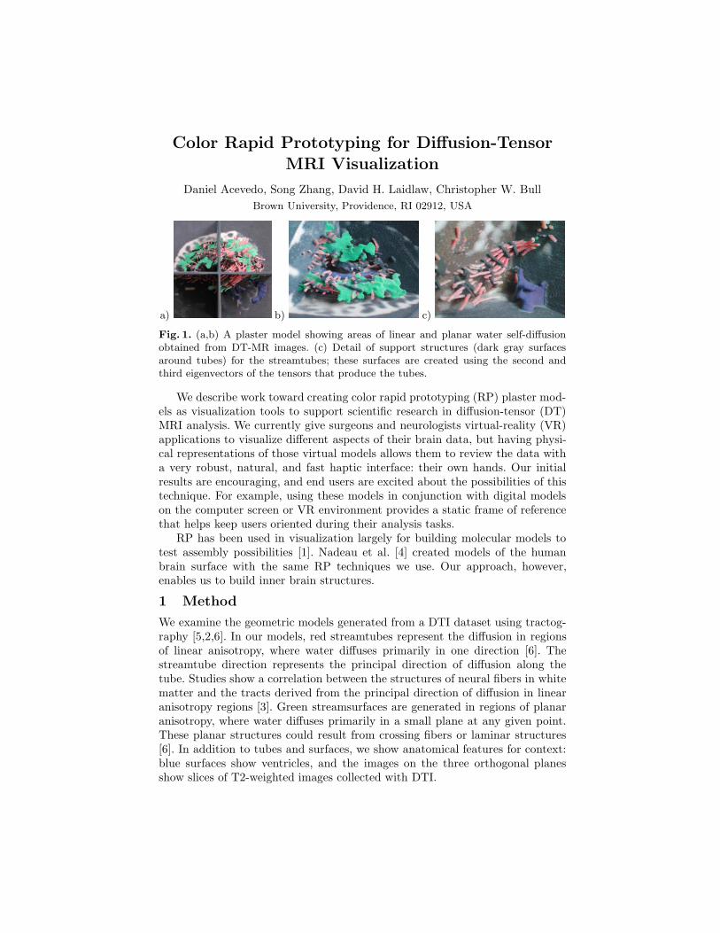
Color Rapid Prototyping for Diffusion-Tensor MRI Visualization Daniel Acevedo, Song Zhang, David H. Laidlaw, Christopher W. Bull Brown University, Providence, RI 02912, USA a) b) c) Fig. 1. (a,b) A plaster model showing areas of linear and planar water self-diffusion obtained from DT-MR images. (c) Detail of support structures (dark gray surfaces around tubes) for the streamtubes; these surfaces are created using the second and third eigenvectors of the tensors that produce the tubes. We describe work toward creating color rapid prototyping (RP) plaster mod- els as visualization tools to support scientific research in diffusion-tensor (DT) MRI analysis. We currently give surgeons and neurologists virtual-reality (VR) applications to visualize different aspects of their brain data, but having physi- cal representations of those virtual models allows them to review the data with a very robust, natural, and fast haptic interface: their own hands. Our initial results are encouraging, and end users are excited about the possibilities of this technique. For example, using these models in conjunction with digital models on the computer screen or VR environment provides a static frame of reference that helps keep users oriented during their analysis tasks. RP has been used in visualization largely for building molecular models to test assembly possibilities [1]. Nadeau et al. [4] created models of the human brain surface with the same RP techniques we use. Our approach, however, enables us to build inner brain structures. 1 Method We examine the geometric models generated from a DTI dataset using tractog- raphy [5,2,6]. In our models, red streamtubes represent the diffusion in regions of linear anisotropy, where water diffuses primarily in one direction [6]. The streamtube direction represents the principal direction of diffusion along the tube. Studies show a correlation between the structures of neural fibers in white matter and the tracts derived from the principal direction of diffusion in linear anisotropy regions [3]. Green streamsurfaces are generated in regions of planar anisotropy, where water diffuses primarily in a small plane at any given point. These planar structures could result from crossing fibers or laminar structures [6]. In addition to tubes and surfaces, we show anatomical features for context: blue surfaces show ventricles, and the images on the three orthogonal planes show slices of T2-weighted images collected with DTI.
Transcript of Color Rapid Prototyping for Di usion-Tensor MRI...
Color Rapid Prototyping for Diffusion-Tensor
MRI Visualization
Daniel Acevedo, Song Zhang, David H. Laidlaw, Christopher W. BullBrown University, Providence, RI 02912, USA
a) b) c)
Fig. 1. (a,b) A plaster model showing areas of linear and planar water self-diffusionobtained from DT-MR images. (c) Detail of support structures (dark gray surfacesaround tubes) for the streamtubes; these surfaces are created using the second andthird eigenvectors of the tensors that produce the tubes.
We describe work toward creating color rapid prototyping (RP) plaster mod-els as visualization tools to support scientific research in diffusion-tensor (DT)MRI analysis. We currently give surgeons and neurologists virtual-reality (VR)applications to visualize different aspects of their brain data, but having physi-cal representations of those virtual models allows them to review the data witha very robust, natural, and fast haptic interface: their own hands. Our initialresults are encouraging, and end users are excited about the possibilities of thistechnique. For example, using these models in conjunction with digital modelson the computer screen or VR environment provides a static frame of referencethat helps keep users oriented during their analysis tasks.
RP has been used in visualization largely for building molecular models totest assembly possibilities [1]. Nadeau et al. [4] created models of the humanbrain surface with the same RP techniques we use. Our approach, however,enables us to build inner brain structures.
1 Method
We examine the geometric models generated from a DTI dataset using tractog-raphy [5,2,6]. In our models, red streamtubes represent the diffusion in regionsof linear anisotropy, where water diffuses primarily in one direction [6]. Thestreamtube direction represents the principal direction of diffusion along thetube. Studies show a correlation between the structures of neural fibers in whitematter and the tracts derived from the principal direction of diffusion in linearanisotropy regions [3]. Green streamsurfaces are generated in regions of planaranisotropy, where water diffuses primarily in a small plane at any given point.These planar structures could result from crossing fibers or laminar structures[6]. In addition to tubes and surfaces, we show anatomical features for context:blue surfaces show ventricles, and the images on the three orthogonal planesshow slices of T2-weighted images collected with DTI.
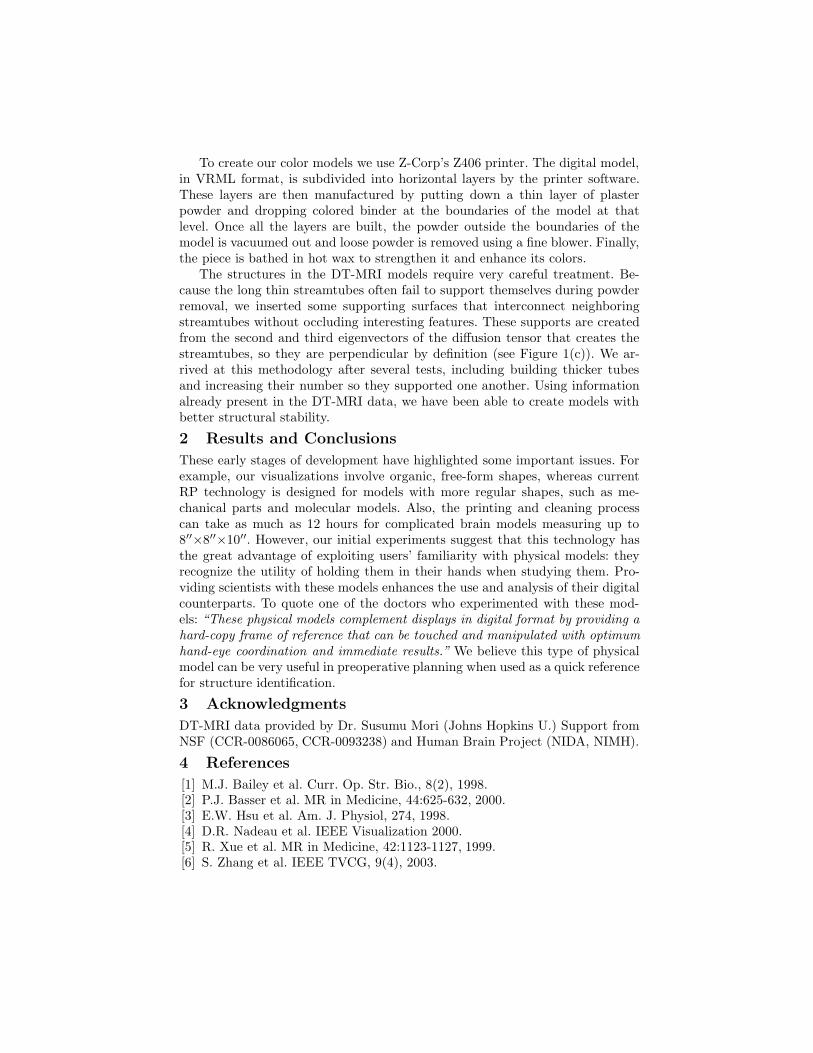
To create our color models we use Z-Corp’s Z406 printer. The digital model,in VRML format, is subdivided into horizontal layers by the printer software.These layers are then manufactured by putting down a thin layer of plasterpowder and dropping colored binder at the boundaries of the model at thatlevel. Once all the layers are built, the powder outside the boundaries of themodel is vacuumed out and loose powder is removed using a fine blower. Finally,the piece is bathed in hot wax to strengthen it and enhance its colors.
The structures in the DT-MRI models require very careful treatment. Be-cause the long thin streamtubes often fail to support themselves during powderremoval, we inserted some supporting surfaces that interconnect neighboringstreamtubes without occluding interesting features. These supports are createdfrom the second and third eigenvectors of the diffusion tensor that creates thestreamtubes, so they are perpendicular by definition (see Figure 1(c)). We ar-rived at this methodology after several tests, including building thicker tubesand increasing their number so they supported one another. Using informationalready present in the DT-MRI data, we have been able to create models withbetter structural stability.
2 Results and Conclusions
These early stages of development have highlighted some important issues. Forexample, our visualizations involve organic, free-form shapes, whereas currentRP technology is designed for models with more regular shapes, such as me-chanical parts and molecular models. Also, the printing and cleaning processcan take as much as 12 hours for complicated brain models measuring up to8′′×8′′×10′′. However, our initial experiments suggest that this technology hasthe great advantage of exploiting users’ familiarity with physical models: theyrecognize the utility of holding them in their hands when studying them. Pro-viding scientists with these models enhances the use and analysis of their digitalcounterparts. To quote one of the doctors who experimented with these mod-els: “These physical models complement displays in digital format by providing ahard-copy frame of reference that can be touched and manipulated with optimumhand-eye coordination and immediate results.” We believe this type of physicalmodel can be very useful in preoperative planning when used as a quick referencefor structure identification.
3 Acknowledgments
DT-MRI data provided by Dr. Susumu Mori (Johns Hopkins U.) Support fromNSF (CCR-0086065, CCR-0093238) and Human Brain Project (NIDA, NIMH).
4 References
[1] M.J. Bailey et al. Curr. Op. Str. Bio., 8(2), 1998.[2] P.J. Basser et al. MR in Medicine, 44:625-632, 2000.[3] E.W. Hsu et al. Am. J. Physiol, 274, 1998.[4] D.R. Nadeau et al. IEEE Visualization 2000.[5] R. Xue et al. MR in Medicine, 42:1123-1127, 1999.[6] S. Zhang et al. IEEE TVCG, 9(4), 2003.
Color Rapid Prototyping for Diffusion-TensorMRI Visualization
Daniel Acevedo, Song Zhang, David H. Laidlaw and Christopher W. BullBrown University Providence, RI, USA http://vis.cs.brown.edu
Users are familiar with physical models and recognize the usefulness of being able to use the sense of touch in studying them.
The data visualization is not dependent on computing power. The frame rate does not drop for more complex models.
The use of color in these models enables us to provide scientists with more complete representations of their multi-variate data, compared with more common monochrome rapid prototyping.
Providing scientists with these models leads to a better usage and analysis of their digital counterparts. One researcher said: “These physical models complement displays in digital format by providing a hard-copy frame of reference that can be touched and manipulated with optimum hand-eye coordination and immediate results.”
We are using machines designed for other types of models, and we are building more organic, free-form shapes. The technology was not developed for these models and current materials don't support fragile structures very well [1,4].
Since the pieces need to be structurally sound, the extra support needed might obstruct the view of the real data being visualized. Balancing these two factors takes many iterations of the printing process.
One printout takes up to 12 hours for a complicated model of the brain.
Texture mapping is not well implemented in the printing software.
The maximum size of the models can only be 8''x8''x10''. Larger models require tiling and assembling the pieces afterwards.
Scaled models require extra cues to convey the correct scale.
Changes in the data being visualized require a whole new model to be created.
Color reproducibility is very hard with current technology. It does not always provide the same results when creating multiple copies of the same model.
Color Rapid Prototyping Strengths
Issues using Rapid Prototyping
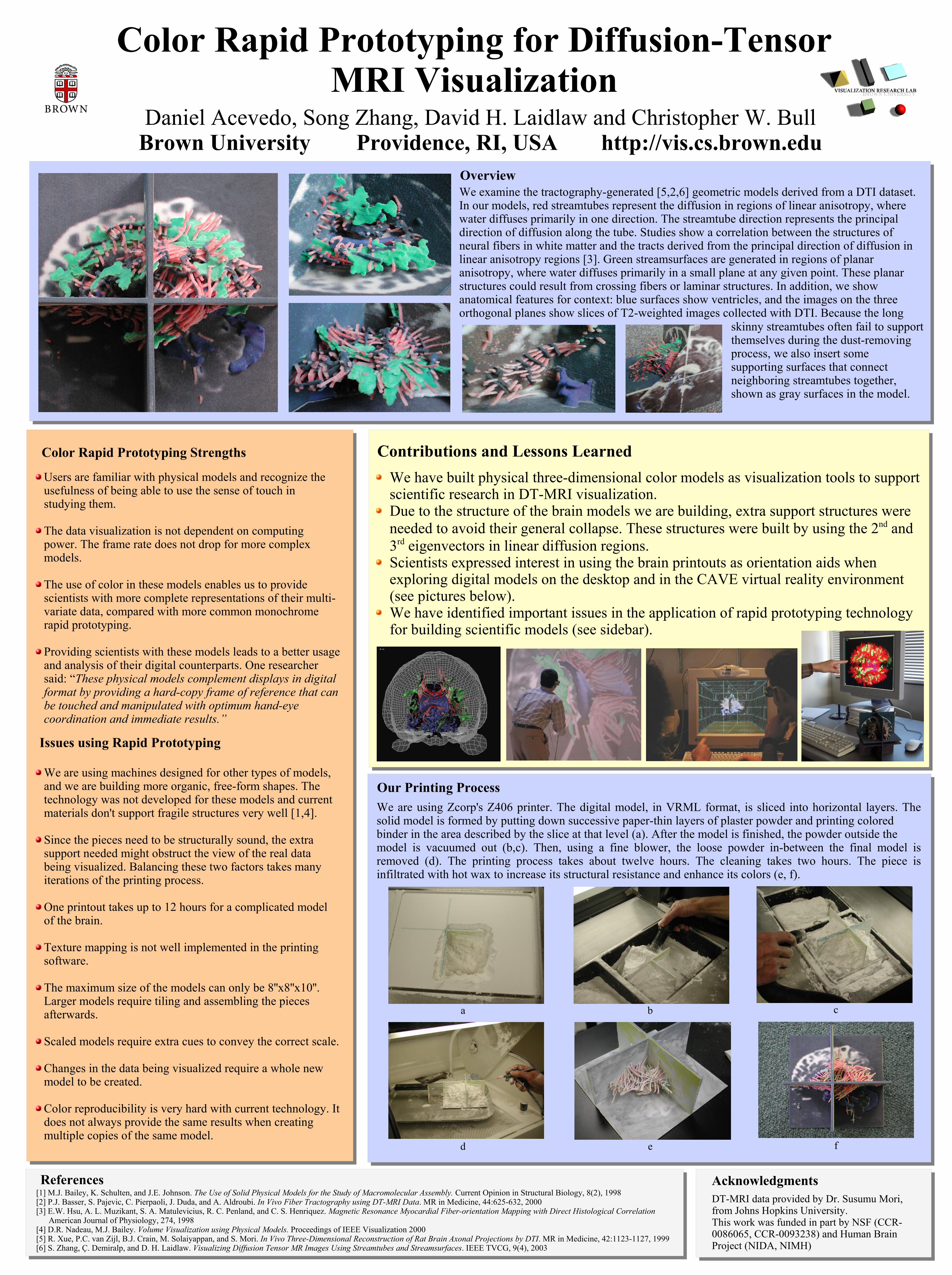
We examine the tractography-generated [5,2,6] geometric models derived from a DTI dataset. In our models, red streamtubes represent the diffusion in regions of linear anisotropy, where water diffuses primarily in one direction. The streamtube direction represents the principal direction of diffusion along the tube. Studies show a correlation between the structures of neural fibers in white matter and the tracts derived from the principal direction of diffusion in linear anisotropy regions [3]. Green streamsurfaces are generated in regions of planar anisotropy, where water diffuses primarily in a small plane at any given point. These planar structures could result from crossing fibers or laminar structures. In addition, we show anatomical features for context: blue surfaces show ventricles, and the images on the three orthogonal planes show slices of T2-weighted images collected with DTI. Because the long
skinny streamtubes often fail to support themselves during the dust-removing process, we also insert some supporting surfaces that connect neighboring streamtubes together, shown as gray surfaces in the model.
DT-MRI data provided by Dr. Susumu Mori, from Johns Hopkins University.This work was funded in part by NSF (CCR-0086065, CCR-0093238) and Human Brain Project (NIDA, NIMH)
Acknowledgments
We are using Zcorp's Z406 printer. The digital model, in VRML format, is sliced into horizontal layers. The solid model is formed by putting down successive paper-thin layers of plaster powder and printing coloredbinder in the area described by the slice at that level (a). After the model is finished, the powder outside themodel is vacuumed out (b,c). Then, using a fine blower, the loose powder in-between the final model is removed (d). The printing process takes about twelve hours. The cleaning takes two hours. The piece is infiltrated with hot wax to increase its structural resistance and enhance its colors (e, f).
c
f
a
d
Our Printing Process
References[1] M.J. Bailey, K. Schulten, and J.E. Johnson. The Use of Solid Physical Models for the Study of Macromolecular Assembly. Current Opinion in Structural Biology, 8(2), 1998[2] P.J. Basser, S. Pajevic, C. Pierpaoli, J. Duda, and A. Aldroubi. In Vivo Fiber Tractography using DT-MRI Data. MR in Medicine, 44:625-632, 2000[3] E.W. Hsu, A. L. Muzikant, S. A. Matulevicius, R. C. Penland, and C. S. Henriquez. Magnetic Resonance Myocardial Fiber-orientation Mapping with Direct Histological Correlation American Journal of Physiology, 274, 1998[4] D.R. Nadeau, M.J. Bailey. Volume Visualization using Physical Models. Proceedings of IEEE Visualization 2000[5] R. Xue, P.C. van Zijl, B.J. Crain, M. Solaiyappan, and S. Mori. In Vivo Three-Dimensional Reconstruction of Rat Brain Axonal Projections by DTI. MR in Medicine, 42:1123-1127, 1999[6] S. Zhang, Ç. Demiralp, and D. H. Laidlaw. Visualizing Diffusion Tensor MR Images Using Streamtubes and Streamsurfaces. IEEE TVCG, 9(4), 2003
We have built physical three-dimensional color models as visualization tools to support scientific research in DT-MRI visualization.Due to the structure of the brain models we are building, extra support structures were needed to avoid their general collapse. These structures were built by using the 2nd and 3rd eigenvectors in linear diffusion regions.Scientists expressed interest in using the brain printouts as orientation aids when exploring digital models on the desktop and in the CAVE virtual reality environment (see pictures below).We have identified important issues in the application of rapid prototyping technology for building scientific models (see sidebar).
b
e
Overview
Contributions and Lessons Learned