Colloids and Surfaces B: Biointerfaces - Harvard...
10
Colloids and Surfaces B: Biointerfaces 158 (2017) 203–212 Contents lists available at ScienceDirect Colloids and Surfaces B: Biointerfaces j o ur nal ho me pa ge: www.elsevier.com/locate/colsurfb Full Length Article Comparative analysis of poly-glycolic acid-based hybrid polymer starter matrices for in vitro tissue engineering Melanie Generali a,1 , Debora Kehl a,1 , Andrew K. Capulli b , Kevin K. Parker b , Simon P. Hoerstrup a,c,d,2 , Benedikt Weber a,c,d,∗,2 a Institute for Regenerative Medicine (IREM), Center for Therapy Development and Good Manufacturing Practice, University of Zurich, Zurich, Switzerland b Disease Biophysics Group, Wyss Institute for Biologically Inspired Engineering, John A. Paulson School of Engineering and Applied Sciences, Harvard University, Cambridge, USA c Center for Applied Biotechnology and Molecular Medicine (CABMM), University of Zurich, Zurich, Switzerland d Zurich Center for Integrative Human Physiology (ZIHP), University of Zurich, Zurich, Switzerland a r t i c l e i n f o Article history: Received 4 April 2017 Received in revised form 23 June 2017 Accepted 28 June 2017 Available online 1 July 2017 Keywords: Poly-glycolic acid Poly-lactic acid Poly-4-hydroxybutyrate Poly–caprolactone Polymers Tissue engineering a b s t r a c t Biodegradable scaffold matrixes form the basis of any in vitro tissue engineering approach by acting as a temporary matrix for cell proliferation and extracellular matrix deposition until the scaffold is replaced by neo-tissue. In this context several synthetic polymers have been investigated, however a concise sys- tematic comparative analyses is missing. Therefore, the present study systematically compares three frequently used polymers for the in vitro engineering of extracellular matrix based on poly-glycolic acid (PGA) under static as well as dynamic conditions. Ultra-structural analysis was used to examine the polymers structure. For tissue engineering (TE) three human fibroblast cell lines were seeded on either PGA-poly-4-hydroxybutyrate (P4HB), PGA-poly-lactic acid (PLA) or PGA-poly–caprolactone (PCL) patches. These patches were analyzed after 21 days of culture qualitative by histology and quantitative by determining the amount of DNA, glycosaminoglycan and hydroxyproline. We found that PGA-P4HB and PGA-PLA scaffolds enhance tissue formation significantly higher than PGA-PCL scaffolds (p < 0.05). Polymer remnants were visualized by polarization microscopy. In addition, biomechanical properties of the tissue engineered patches were determined in comparison to native tissue. This study may allow future studies to specifically select certain polymer starter matrices aiming at specific tissue properties of the bioengineered constructs in vitro. © 2017 Elsevier B.V. All rights reserved. 1. Introduction During the last years, the interdisciplinary field of tissue engi- neering (TE) has emerged as a platform for the development of biological substitutes. The overall goal is the repair and/or regeneration of tissues or organs to resolve major health related concerns in humans. Multiple disciplines, such as cell biology, bio- material research and biomedical engineering have contributed to the advances of tissue engineering technologies. Any tissue engi- ∗ Corresponding author at: Institute for Regenerative Medicine (IREM), University of Zurich, Moussonstrasse 13, 8091 Zurich, Switzerland. E-mail addresses: [email protected] (M. Generali), [email protected] (D. Kehl), [email protected] (A.K. Capulli), [email protected] (K.K. Parker), simon [email protected] (S.P. Hoerstrup), [email protected] (B. Weber). 1 These authors contributed equally to the study. 2 These senior authors contributed equally to the study. neering approach is composed of three major components: (1) cells, (2) biocompatible scaffolds, and (3) suitable biochemical (e.g. growth factors) and physical (e.g. cyclic mechanical loading) stim- uli supporting tissue formation in vitro and in situ [1]. While the engineered living substitute develops, the biocompatible scaffold should degrade without leaving remnants in the body, requiring a so-called biodegradable starter matrix (scaffold). A variety of synthetic biodegradable polymers has been inves- tigated as TE scaffold materials, though the main disadvantage of these materials is their lack of functional groups [2,3]. This results in limited capacity to combine with bioactive elements to reinforce their cell affinity [3]. In general, functional synthetic polymers have unsaturated bonds [4], or functional groups such as hydroxyl [5], carboxyl [6], and amide [7], through which functional biomaterials can be chemically modified by biomolecules to improve their bioac- tivity [5]. Nonetheless, synthetic polymers have been extensively used for TE, given their high durability, flexibility, and mechan- ical strength [8]. In addition, production conditions of synthetic http://dx.doi.org/10.1016/j.colsurfb.2017.06.046 0927-7765/© 2017 Elsevier B.V. All rights reserved.
Transcript of Colloids and Surfaces B: Biointerfaces - Harvard...

F
Cs
MSa
b
Uc
d
a
ARRAA
KPPPPPT
1
norcmt
o
((b
h0
Colloids and Surfaces B: Biointerfaces 158 (2017) 203–212
Contents lists available at ScienceDirect
Colloids and Surfaces B: Biointerfaces
j o ur nal ho me pa ge: www.elsev ier .com/ locate /co lsur fb
ull Length Article
omparative analysis of poly-glycolic acid-based hybrid polymertarter matrices for in vitro tissue engineering
elanie Generali a,1, Debora Kehl a,1, Andrew K. Capulli b, Kevin K. Parker b,imon P. Hoerstrup a,c,d,2, Benedikt Weber a,c,d,∗,2
Institute for Regenerative Medicine (IREM), Center for Therapy Development and Good Manufacturing Practice, University of Zurich, Zurich, SwitzerlandDisease Biophysics Group, Wyss Institute for Biologically Inspired Engineering, John A. Paulson School of Engineering and Applied Sciences, Harvardniversity, Cambridge, USACenter for Applied Biotechnology and Molecular Medicine (CABMM), University of Zurich, Zurich, SwitzerlandZurich Center for Integrative Human Physiology (ZIHP), University of Zurich, Zurich, Switzerland
r t i c l e i n f o
rticle history:eceived 4 April 2017eceived in revised form 23 June 2017ccepted 28 June 2017vailable online 1 July 2017
eywords:oly-glycolic acidoly-lactic acidoly-4-hydroxybutyrateoly–caprolactone
a b s t r a c t
Biodegradable scaffold matrixes form the basis of any in vitro tissue engineering approach by acting as atemporary matrix for cell proliferation and extracellular matrix deposition until the scaffold is replacedby neo-tissue. In this context several synthetic polymers have been investigated, however a concise sys-tematic comparative analyses is missing. Therefore, the present study systematically compares threefrequently used polymers for the in vitro engineering of extracellular matrix based on poly-glycolicacid (PGA) under static as well as dynamic conditions. Ultra-structural analysis was used to examinethe polymers structure. For tissue engineering (TE) three human fibroblast cell lines were seeded oneither PGA-poly-4-hydroxybutyrate (P4HB), PGA-poly-lactic acid (PLA) or PGA-poly–caprolactone (PCL)patches. These patches were analyzed after 21 days of culture qualitative by histology and quantitativeby determining the amount of DNA, glycosaminoglycan and hydroxyproline. We found that PGA-P4HB
olymersissue engineering
and PGA-PLA scaffolds enhance tissue formation significantly higher than PGA-PCL scaffolds (p < 0.05).Polymer remnants were visualized by polarization microscopy. In addition, biomechanical properties ofthe tissue engineered patches were determined in comparison to native tissue. This study may allowfuture studies to specifically select certain polymer starter matrices aiming at specific tissue propertiesof the bioengineered constructs in vitro.
© 2017 Elsevier B.V. All rights reserved.
. Introduction
During the last years, the interdisciplinary field of tissue engi-eering (TE) has emerged as a platform for the developmentf biological substitutes. The overall goal is the repair and/oregeneration of tissues or organs to resolve major health related
oncerns in humans. Multiple disciplines, such as cell biology, bio-aterial research and biomedical engineering have contributed tohe advances of tissue engineering technologies. Any tissue engi-
∗ Corresponding author at: Institute for Regenerative Medicine (IREM), Universityf Zurich, Moussonstrasse 13, 8091 Zurich, Switzerland.
E-mail addresses: [email protected] (M. Generali), [email protected]. Kehl), [email protected] (A.K. Capulli), [email protected]. Parker), simon [email protected] (S.P. Hoerstrup),[email protected] (B. Weber).1 These authors contributed equally to the study.2 These senior authors contributed equally to the study.
ttp://dx.doi.org/10.1016/j.colsurfb.2017.06.046927-7765/© 2017 Elsevier B.V. All rights reserved.
neering approach is composed of three major components: (1)cells, (2) biocompatible scaffolds, and (3) suitable biochemical (e.g.growth factors) and physical (e.g. cyclic mechanical loading) stim-uli supporting tissue formation in vitro and in situ [1]. While theengineered living substitute develops, the biocompatible scaffoldshould degrade without leaving remnants in the body, requiring aso-called biodegradable starter matrix (scaffold).
A variety of synthetic biodegradable polymers has been inves-tigated as TE scaffold materials, though the main disadvantage ofthese materials is their lack of functional groups [2,3]. This resultsin limited capacity to combine with bioactive elements to reinforcetheir cell affinity [3]. In general, functional synthetic polymers haveunsaturated bonds [4], or functional groups such as hydroxyl [5],carboxyl [6], and amide [7], through which functional biomaterials
can be chemically modified by biomolecules to improve their bioac-tivity [5]. Nonetheless, synthetic polymers have been extensivelyused for TE, given their high durability, flexibility, and mechan-ical strength [8]. In addition, production conditions of synthetic
2 aces B
pppm
pdmvdpptwfertpP([atl
rrw[icewntmt
ew[P[lir5PidFPtmiveatatacsc
heated at 60 ◦C for 10 min. The absorbance was analyzed at 550 nmusing a standard ELISA reader Synergy HT (Bio TEK, USA). To obtain
04 M. Generali et al. / Colloids and Surf
olymers can be tightly controlled, hence making mechanical andhysical properties of the material predictable, reproducible andrecisely defined [9]. These characteristics make synthetic poly-ers to an interesting raw material for scaffold fabrication [8–10].
In particular, poly-glycolic acid (PGA), poly-lactic acid (PLA),oly-hydroxy alkanoate (PHA), poly -caprolactone (PCL) and theireriving copolymers have generated substantial interest as scaffoldaterials for the in vitro TE of bone, cartilage, as well as cardio-
ascular tissues [11–14]. PGA is most commonly used because itegrades at predictable time point and into (generally) biocom-atible components [8]. Besides, the high porosity of PGA meshesermits a good diffusion, neovascularization and cellular infil-ration [15]. Unfortunately, PGA meshes are biodegraded rapidlyithin few weeks and can therefore not withstand mechanical
orces exerted to the materials and guide the shape of the bio-ngineered construct over longer culturing periods [15,16]. As aesult, hybrid polymers have been designed in order to combinehe shape-memory and mechanical stability of slowly degradingolymers with the fast degrading properties of polymers, such asGA [17]. For instance, combinations of PGA with polymers such aspoly-4-hydroxybutyrate) P4HB or PLA have been widely explored8,13,14,18,19]. PLA is synthesized by polymerization of lactic acidnd can be eliminated through the citric acid cycle [15]. Due tohe chiral nature of PLA, several distinct forms are existing: poly--lactide (PLLA), poly-d-lactide (PDLA) and LD racemic (PDLLA),espectively [15]. PLA can easily be processed and its degradationates and physical/mechanical characteristics are adjustable over aide range by using different molecular weights and copolymers
19,20]. Also �-caprolactone and copolymers have been studiedntensely and are frequently investigated for biomedical appli-ations [15]. Interestingly, PCL degrades very slowly in-vivo vianzymatic degradation and hydrolysis [21]. Unlike PGA and PCL,hich are synthetized using chemical methods, P4HB is produced
aturally by microorganisms making it more challenging to be syn-hesized [22]. After implantation into the body, P4HB degrades
ainly by bulk hydrolysis producing 4HB, a normal component ofhe mammalian body [10].
In 1998, Shinoka et al. reported surgical implantation of tissuengineered vascular grafts (TEVGs) in lambs, in which scaffoldsere constructed from autologous cells seeded onto PGA grafts
23]. Further studies have been conducted, for instance, by usingGA-poly-l-lactic acid (PLLA) scaffolds for microvessels in mice24] or scaffolds composed of polyglycolide knitted fiber, and an l-actide and �-caprolactone copolymer sponge for TEVGs in a caninenferior vena cava model [25]. The hybrid polymeric scaffold fab-icated from either PGA or PLA fiber-based mesh coated with a0:50 copolymer of l-lactide and �-caprolactone (PCLA/PGA orCLA/PLA) are more elastic than the PGA scaffold [26]. This results
n an improved compliance match between the vessel and the con-uit and ultimately in better surgical handling characteristics [26].or both heart valve and vascular tissue engineering the use ofGA meshes coated with P4HB, meaning the combination of thehermoplastic characteristic of P4HB and the high porosity of PGA
eshes, has been investigated intensively with promising resultsn vitro and in preclinical studies [27–29]. In 2006, Mol et al. pro-ided the first evidence of living, functional pulmonary arteriesngineered from vascular cells seeded on PGA/P4HB scaffolds in
growing lamb model [13]. In contrast, PCL has been mainly inves-igated for biomedical applications in bone and cartilage repair,s surgical suture as well as for drug delivery systems, especiallyhose with longer working lifetimes [12,15]. Son et al. showed that
PCL/poly(methyl methacrylate) (PMMA) scaffold was suitable for
ell growth in vitro and for new bone formation in vivo [30]. Thistudy suggested that PCL/PMMA blends can be used for biopolymeromposite scaffolds in bone tissue engineering [30]. In general, all: Biointerfaces 158 (2017) 203–212
polyesters are biocompatible and have formed the bases of numer-ous FDA approved medical devices for clinical use [9,31].
However, in spite of their frequent use in biomedical researchand therapeutic products, there is still a lack of systematic com-parative analyses, such as qualitative and quantitative assays oftissue formation and biomechanics between different syntheticpolymers. Therefore, the present study aims at a systematic, mul-timodal comparison of three frequently used polymers (PGA, PLA,PCL) integrated into a co-polymer solution with P4HB for the in vitroengineering of extracellular matrix under static as well as dynamicconditions. These data might allow for a specific selection of a cer-tain polymer starter matrices aiming at specific tissue properties ofbioengineered materials in vitro.
2. Material and methods
2.1. Isolation and culture of human umbilical cord fibroblasts
Human umbilical cords (n = 3) were collected after full-termbirths with informed consent according to the cantonal ethics com-mission of Zurich, Switzerland [KEK-ZH-2009-0095] and processedfor isolation of venous fibroblasts according to established proto-cols [32]. Briefly, the umbilical cord vein was isolated surgicallyand small tissue pieces were cut out using a dissecting scissors.Tissue pieces were placed on a sterile petri dish and were leftto adhere to the bottom for 20 min. Culture medium contain-ing DMEM high glucose (Sigma-Aldrich, Switzerland), 10% fetalbovine serum (Biowest, USA) and an antibiotic/antimitotic solution(Sigma-Aldrich, Switzerland), was gently added and changed everythird day. Tissue pieces were removed after first cellular outgrowthafter approximately 1–2 weeks of incubation under humidifiedincubator conditions at 5% CO2 at 37 ◦C. Three different fibroblastcell lines (named B014, B015 and B020) were isolated.
2.2. Phenotyping of human fibroblasts
Isolated human cells (n = 3) were characterized usingimmunofluorescence staining for common myofibroblast markers.Therefore, cells were cultured on 3.5 cm2 cell culture dishes, fixedwith 4% paraformaldehyde (Sigma-Aldrich, Switzerland), andincubated over night at 4 ◦C with the following primary antibod-ies: alpha smooth muscle actin (1A4, Abcam, United Kingdom),Vimentin (Vim 3B4, Abcam, United Kingdom), CD90 (EPR3133,Abcam, United Kingdom), CD31 (JC70A, Dako, USA), and Phal-loidin (Life Technologie, Switzerland). The following secondaryantibodies were used: anti mouse Alexa Flour 488 (Invitrogen,USA) and anti rabbit Alexa Flour 488 (Invitrogen, USA) and Dapi(Sigma-Aldrich, Switzerland). Cells were analyzed with a DM6000Bfluorescence microscope (Leica, Germany). Image processing wasperformed using the Leica software (Leica, Germany).
2.3. Proliferation assay
Cellular proliferation was assessed by determining the numberof total cells based on the absorbance of crystal violet when culturedon a 24-well plate for up to 7 days. In brief, every day cells were fixedwith methanol (Sigma-Aldrich, Switzerland) for 10 min and stainedwith 0.1% crystal violet (Artechemis, Switzerland) for 5 min. The24-well plates were washed and air-dried. Cells were solubilizedwith 2% Na-deoxycholat (Sigma-Aldrich, Switzerland) while being
quantitative information a standard curve with serial dilutions wasperformed.

aces B
2
bTiii1mmapae
2
2
(Ch(bSoAtap
2
dSAotts(l
daci
2
gfsrswa
2
l((c
M. Generali et al. / Colloids and Surf
.4. Surface morphology of biomaterials
Samples were mounted on electron imagining stubs using car-on tape and subsequently sputter coated in 5 nm of Pt/Pd (Quorumechnologies, EMS 300TD, USA) to reduce fiber degradation dur-
ng imaging. A field emitting electron microscope was used tomage samples at 15 kV power (Zeiss, FESEM Ultra Plus) for clar-ty. For each sample, four regions of interest (ROI) measuring000 × 800 um were imaged from which five fibers per ROI wereeasured using Image J software (NIH, v1.48s, line tool) to deter-ine the average fiber diameter of an ROI. For scaffold porosity, an
dditional four ROIs measuring 3000 × 2250 um were imaged. Eachorosity ROI image was then automatically thresholded in Image Jnd porosity was defined as the percent area that was non-fiber (iempty/porous space).
.5. Tissue engineering
.5.1. Scaffold fabricationPatches were fabricated from non-woven polyglycolic acid
PGA) meshes (thickness 1.0 mm; specific gravity 70 mg/cm3;ellon, Luxembourg) and coated with either 1% poly-4-ydroxybutyrate (P4HB; TEPHA, Inc., USA) or 1% Poly(l-lactide)PLA, Carbion, USA) or 1% polycaprolactone (PCL, Carbion, USA)y dipping into a tetrahydrofuran solution (Sigma-Aldrich,witzerland). After solvent evaporation and vacuum dryingvernight, the scaffolds were placed into a 70% EtOH (Sigma-ldrich, Switzerland) for 30 min to obtain sterility, followed by
wo washing cycles with PBS (Sigma-Aldrich, Switzerland). There-fter, scaffolds were pre-incubated in DMEM culture mediumreviously described for 12–24 h to facilitate cell attachment.
.5.2. Cell seedingHuman fibroblasts (n = 3) were seeded onto scaffolds using a
ensity of 1.5*106 cells/cm2. Therefore, fibrinogen (Sigma-Aldrich,witzerland) (10 mg/mL of active protein) and thrombin (Sigma-ldrich, Switzerland) were prepared, used and titrated to anptimal clotting time of approximately 30 s by adapting the concen-ration of fibrinogen. The cells were resuspended in a fibrinogen-hrombin co-solution and subsequently seeded onto the sterilecaffolds. After static incubation of seeded constructs in DMEM10% fetal bovine serum; penicillin/streptomycin and 0.9 mM of-ascorbic acid-2-phosphate (Sigma-Aldrich, Switzerland)) for 7ays, they were kept either under static conditions or placed onto
shaker for additional mechanical stimulation via shear stress. Theonstructs were harvested after 21 days of culture and processedmmediately for subsequent analyses.
.6. Qualitative tissue analysis
For immunohistochemical analysis of TE patches (n = 3 perroup), sections with 5 �m thickness derived from blocks oformalin-fixed, paraffin-embedded tissue were mounted on glasslides (SuperFrost Plus, Menzel Gläser,Germany), deparaffinized,ehydrated and stained with hematoxylin and eosin (H&E) or Mas-on Trichrome using standard histological techniques. All sectionsere analyzed using a Mirax Midi BF slide scanner (Zeiss, Germany)
nd processed using MIRAX viewer (Zeiss, Germany).
.7. Quantitative tissue analysis
TE patches (n = 6 per group) were minced, lyophilized, and ana-
yzed using biochemical assays for total deoxyribonucleic acidDNA) content as an indicator for cell number, hydroxyprolineHYP) content as an indicator for collagen, as well as for gly-osaminoglycan (GAG) content. All TE patches were digested in: Biointerfaces 158 (2017) 203–212 205
papain (Sigma-Aldrich, Switzerland) solution (300 �g/mL in PBSwith 5 mM EDTA (Sigma-Aldrich, Switzerland)) and 5 mM cys-teine (Sigma-Aldrich, Switzerland), at 65 ◦C for 16 h. For measuringthe cellularity of the constructs, the DNA amount was quan-tified according to manufacturer’s protocol (Life Technologies,Switzerland, No. P11496). The GAG content was determined usinga modified version of the protocol described by Ref. [33], anda standard curve prepared from chondroitin sulfate from sharkcartilage (Sigma-Aldrich, Switzerland). Hydroxyproline was deter-mined with a modified version of the protocol provided by Ref. [34].Remnants of the hybrid polymers matrices were visualized usingpolarization microscopy (Zeiss, Germany).
2.8. Biomechanics
After 0 (only for the unseeded group) and 21 days of cul-ture, the mechanical properties of the TE constructs (n = 4 pergroup) were assessed using a uniaxial tensile tester (Instron 5864,Boston, MA, USA) equipped with a 100-N load cell. For com-parison, native tissues (cartilage, skin and vein) were harvestedfrom sheep post mortem provided by the local slaughterhouseZurich/Hinwil, Switzerland (n = 4). The crosshead speed was set tocorrespond to an initial strain rate of 7 mm/min and the tester oper-ated at 5 bars of air pressure. Patches had a cross-sectional area of14 mm × 4 mm × 1 mm (length × width × thickness) and were fixedwith hydraulic clamps. Stress–strain curves were obtained andYoung’s modulus was determined as the slope of the curve at astrain of 10%, as a measure for tissue stiffness.
2.9. Statistic
Quantitative data are presented as mean ± standard devia-tion. For statistical comparison of the proliferation rate of thethree different cell lines a one-way ANOVA was performed andp-values < 0.05 were considered statistically significant. Surfacemorphology measurements were statistically analyzed by non-parametric Mann-Whitney-U test and p-values < 0.05 (correctedpost hoc according to Bonferroni) were considered statistically sig-nificant. In addition, quantitative tissue analysis and biomechanicswere statistically evaluated by an unpaired students-t-test and p-values p < 0.05 (corrected post hoc according to Bonferroni) wereconsidered statistically significant. KS normality test was used toconfirm normal distribution of the dataset (p > 0.05). All statisti-cal analyses were performed using the GraphPad Prism 5 software(GraphPad Software Inc., USA).
3. Results
3.1. Phenotype and proliferation of umbilical cord derivedfibroblasts
Immunofluorescence performed on fibroblasts derived fromthree different patients revealed that all cell types displayedsimilar immunophenotypic patterns. Isolated human fibroblastsexpressed myogenic markers, such as alpha smooth muscle actinand vimentin, and common fibroblast marker CD90 (Fig. 1a).
In contrast, the cells were negative for the endothelial cell sur-face marker CD31 excluding non-fibroblastic contamination duringcell isolation procedures. In the proliferation analysis based oncrystal violet staining (Fig. 1b), no significant differences in pro-
liferation parameters were detected between the three fibroblastlines when considering cell numbers over 7 days (p < 0.05). Thesefindings exclude potential significant inter-individual proliferationdifferences.
206 M. Generali et al. / Colloids and Surfaces B: Biointerfaces 158 (2017) 203–212
Fig. 1. Phenotypic characterization and proliferation of isolated fibroblasts. a) Immunohistochemical analyses of three different human fibroblast cell lines (B014, B015 andB020) revealed positive expression of alpha smooth muscle actin, Vimentin and CD90 (Phalloidin in red, Dapi in blue and respective antibody in green). All three markerss theliat rencei
3
(pc
i(msosUtTtgsbh
formation for respective hybrid polymer conditions excluding a
howed a homogenous expression pattern. Fibroblasts were negative for the endohree cell lines using the crystal violet-based proliferation assay. No significant diffen this figure legend, the reader is referred to the web version of this article.)
.2. Surface morphology of biomaterials
Microstructural analysis using scanning electron microscopySEM) was used to analyze the fiber size and porosity of unseededolymers before and after coating (Fig. 2a–d and f–i) (n = 4 perondition).
The PGA-PCL matrix (Fig. 2a and f) was less porous in compar-son to the hybrid polymers PGA-P4HB (Fig. 2b and g), PGA-PLAFig. 2c and h), and PGA (Fig. 2d and i). Quantitative measure-
ents of the porosity confirmed these differences (Fig. 2j). PGA-PCLcaffolds were significantly less porous than PGA-P4HB (p < 0.05)r PGA-PLA (p < 0.05) scaffolds. PGA-P4HB and PGA-PLA scaffoldshowed a similar porosity with no significant difference (p = 0.69).ncoated PGA mesh was significant more porous in comparison
o PGA-P4HB (p < 0.05), PGA-PLA (p < 0.05), and PGA-PCL (p < 0.05).he fiber diameter was uniform among all biomaterials with no sta-istically significant differences between different hybrid polymerroups or uncoated PGA (Fig. 2g, p < 0.05) (Fig. 2e). Imaging of the
urface (Fig. 2a–d) and cross section (Fig. 2f–i) display the distri-ution of the coating compared to PGA scaffold only. The differentybrid polymer present a similar distribution of the coating. In gen-l cell surface marker CD31. b) Cell number was determined over one week for alls were detected. (Scale bars: 50 �m). (For interpretation of the references to colour
eral, there was more coating visible on the outer scaffold regions,while in the central part of the scaffold less was detected.
3.3. Qualitative tissue analyses
Microstructural features of human fibroblast-derived TEpatches were analyzed by histological staining procedures (Fig. 3)as well as polarization microscopy (Fig. 4).
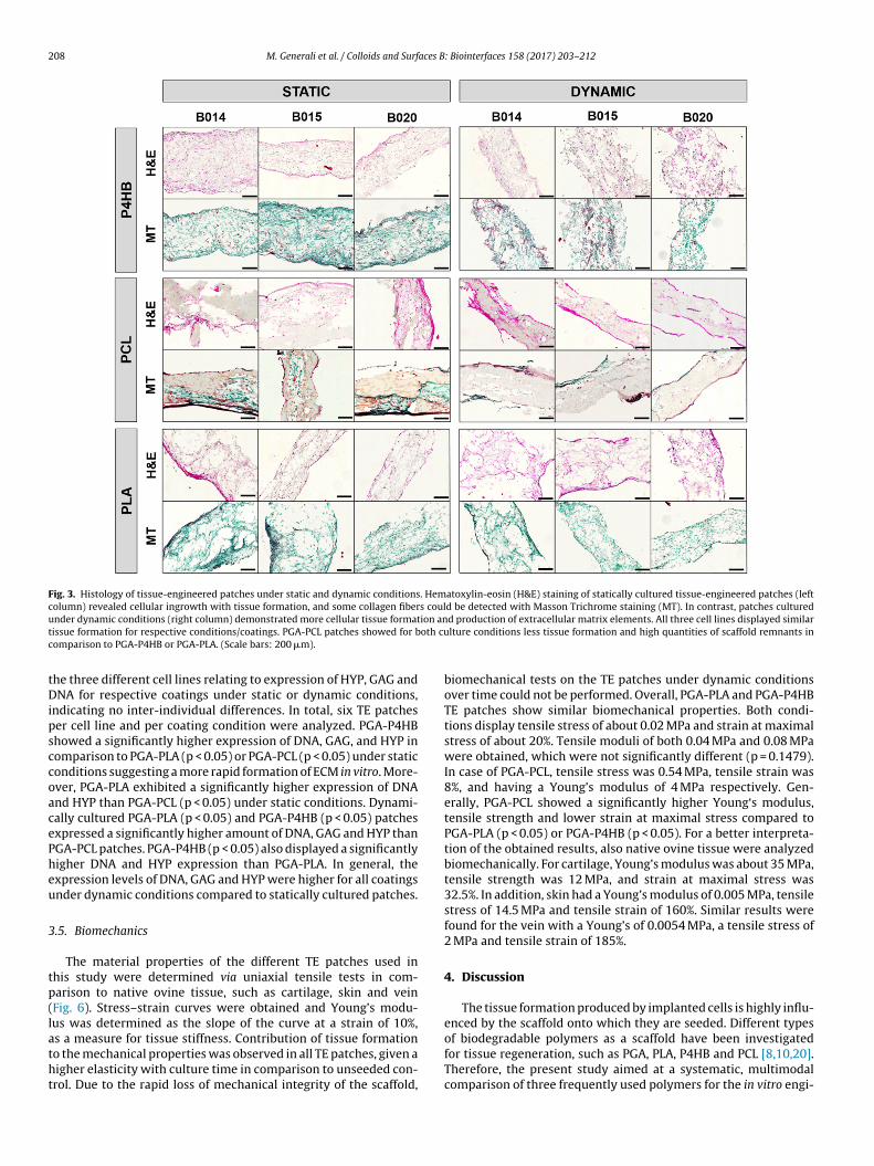
H&E staining demonstrated formation of extracellular matrix(ECM) in vitro with high cellularity and layered tissue on the outerscaffold regions, while in the central part of the scaffold low cellu-larity and no significant formation of ECM were present. In orderto investigate the deposition of collagen fibers Masson Trichomstaining was used. In general, TE patches under dynamic condi-tions (Fig. 3, right column) revealed more tissue formation and ECMdeposition compared to static conditions.
Importantly, all three cell lines exhibited comparable tissue
potential bias due to inter-individual differences. PGA-PCL-basedpatches showed for both culture conditions (dynamic and static)less tissue formation and high quantities of scaffold remnants in

M. Generali et al. / Colloids and Surfaces B: Biointerfaces 158 (2017) 203–212 207
F crograo ong aP 0 �m)
cfii
tgfiwobs(
ig. 2. Surface and intersection morphology of biomaterials. Scanning electron mif the surface f–i) SEM of the cross section e) the fiber diameter was consistent amGA-PLA. PGA-P4HB and PGA-PLA showed a similar porosity. (n = 4) (Scale bars: 10
omparison to the PGA-P4HB or PGA-PLA. In order to confirm thesendings polarization microscopy was performed to further visual-
ze different polymer components (Fig. 4).The starter matrices showed no major remodeling in the cen-
ral part of the constructs in static as well as dynamic conditionsiven the lack of tissue formation in this area of the constructs. PGAbers were visible as elongated ellipses (Fig. 4, high magnification),hereas the webbings between PGA fibers represented remnants
f the coating (Fig. 4, low magnification). In dynamic conditions theiomaterial was more degraded (Fig. 4d–f) when compared to thetatic cultures (Fig. 4a–c). In particular, the PGA-PCL starter matrixFig. 4b and e) showed strong preservation of the polymer compo-
phs (SEM) show the fiber alignment and size of unseeded biomaterials. a–d) SEMll biomaterials. (n = 4) j) PGA-PCL was significantly less porous than PGA-P4HB or.
nents compared to PGA-P4HB (Fig. 4a and d) or PGA-PLA (Fig. 4cand f).
3.4. Quantitative tissue analysis
The composition of the ECM of the human fibroblast-derived TEconstructs was biochemically analyzed using assays for HYP, GAG,and the cell number (DNA) (Fig. 5).
The expression level of DNA, GAG, and HYP is for all 3 coat-ings higher under dynamic conditions than under static conditions.Analyses were performed after three weeks of static or dynamic cul-ture. In general, no significant differences were detected between
208 M. Generali et al. / Colloids and Surfaces B: Biointerfaces 158 (2017) 203–212
Fig. 3. Histology of tissue-engineered patches under static and dynamic conditions. Hematoxylin-eosin (H&E) staining of statically cultured tissue-engineered patches (leftcolumn) revealed cellular ingrowth with tissue formation, and some collagen fibers could be detected with Masson Trichrome staining (MT). In contrast, patches culturedu ion ant oth cuc
tDipsccoacePheu
3
tp(latht
nder dynamic conditions (right column) demonstrated more cellular tissue formatissue formation for respective conditions/coatings. PGA-PCL patches showed for bomparison to PGA-P4HB or PGA-PLA. (Scale bars: 200 �m).
he three different cell lines relating to expression of HYP, GAG andNA for respective coatings under static or dynamic conditions,
ndicating no inter-individual differences. In total, six TE patcheser cell line and per coating condition were analyzed. PGA-P4HBhowed a significantly higher expression of DNA, GAG, and HYP inomparison to PGA-PLA (p < 0.05) or PGA-PCL (p < 0.05) under staticonditions suggesting a more rapid formation of ECM in vitro. More-ver, PGA-PLA exhibited a significantly higher expression of DNAnd HYP than PGA-PCL (p < 0.05) under static conditions. Dynami-ally cultured PGA-PLA (p < 0.05) and PGA-P4HB (p < 0.05) patchesxpressed a significantly higher amount of DNA, GAG and HYP thanGA-PCL patches. PGA-P4HB (p < 0.05) also displayed a significantlyigher DNA and HYP expression than PGA-PLA. In general, thexpression levels of DNA, GAG and HYP were higher for all coatingsnder dynamic conditions compared to statically cultured patches.
.5. Biomechanics
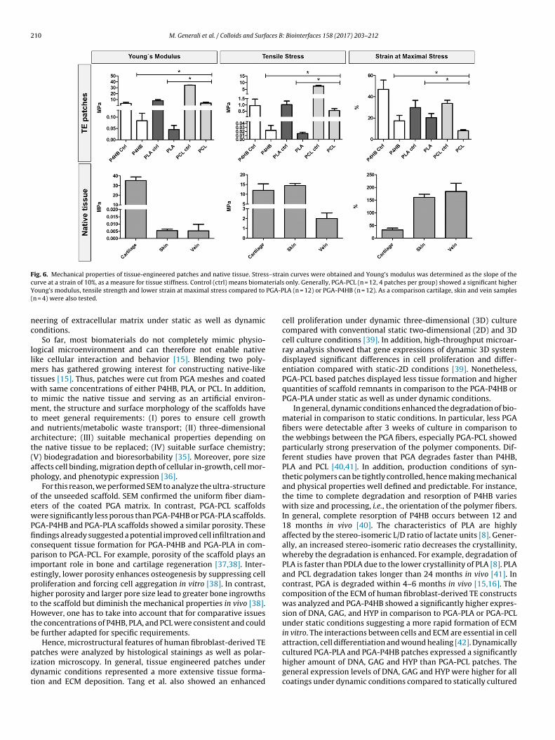
The material properties of the different TE patches used inhis study were determined via uniaxial tensile tests in com-arison to native ovine tissue, such as cartilage, skin and veinFig. 6). Stress–strain curves were obtained and Young’s modu-us was determined as the slope of the curve at a strain of 10%,
s a measure for tissue stiffness. Contribution of tissue formationo the mechanical properties was observed in all TE patches, given aigher elasticity with culture time in comparison to unseeded con-rol. Due to the rapid loss of mechanical integrity of the scaffold,d production of extracellular matrix elements. All three cell lines displayed similarlture conditions less tissue formation and high quantities of scaffold remnants in
biomechanical tests on the TE patches under dynamic conditionsover time could not be performed. Overall, PGA-PLA and PGA-P4HBTE patches show similar biomechanical properties. Both condi-tions display tensile stress of about 0.02 MPa and strain at maximalstress of about 20%. Tensile moduli of both 0.04 MPa and 0.08 MPawere obtained, which were not significantly different (p = 0.1479).In case of PGA-PCL, tensile stress was 0.54 MPa, tensile strain was8%, and having a Young’s modulus of 4 MPa respectively. Gen-erally, PGA-PCL showed a significantly higher Young‘s modulus,tensile strength and lower strain at maximal stress compared toPGA-PLA (p < 0.05) or PGA-P4HB (p < 0.05). For a better interpreta-tion of the obtained results, also native ovine tissue were analyzedbiomechanically. For cartilage, Young’s modulus was about 35 MPa,tensile strength was 12 MPa, and strain at maximal stress was32.5%. In addition, skin had a Young’s modulus of 0.005 MPa, tensilestress of 14.5 MPa and tensile strain of 160%. Similar results werefound for the vein with a Young’s of 0.0054 MPa, a tensile stress of2 MPa and tensile strain of 185%.
4. Discussion
The tissue formation produced by implanted cells is highly influ-enced by the scaffold onto which they are seeded. Different types
of biodegradable polymers as a scaffold have been investigatedfor tissue regeneration, such as PGA, PLA, P4HB and PCL [8,10,20].Therefore, the present study aimed at a systematic, multimodalcomparison of three frequently used polymers for the in vitro engi-
M. Generali et al. / Colloids and Surfaces B: Biointerfaces 158 (2017) 203–212 209
Fig. 4. Polarization microscopy of tissue-engineered patches under static and dynamic conditions. Polarization microscopy revealed the presence of the initial scaffold matrixin particular in the central part of the constructs. PGA fibers are shown as elongated ellipses (high magnification). Webbings between PGA fibers represent coating remnants(low magnification). However, in the dynamic fibroblast-based constructs the biomaterial remnants were more degraded (d–f) compared with static cultures (a–c). PGA-PCL(b and e) starter matrix showed a very strong preservation of the co-polymer when compared to PGA-P4HB (a and d) or PGA-PLA (c and f) scaffolds (scale bars: 400 �m lowmagnification, 100 �m high magnification).
Fig. 5. Extracellular matrix analysis of tissue-engineered patches under static and dynamic conditions. Extracellular matrix analysis shows the amount of hydroxyproline(HYP), glycosaminoglycans (GAG), and the cell number (deoxyribonucleic acid, DNA) of tissue-engineered patches, which were statically or dynamically cultured (all valuesrelative to control samples (ctrl) indicating biomaterials only). Generally, all three cell lines displayed similar amounts of HYP, GAG and DNA for respective conditions/coatings,no significant differences were detected. Overall PGA-P4HB (n = 6) showed a higher expression of DNA, GAG, and HYP in comparison to PGA-PLA (n = 6) or PGA-PCL (n = 6).
210 M. Generali et al. / Colloids and Surfaces B: Biointerfaces 158 (2017) 203–212
Fig. 6. Mechanical properties of tissue-engineered patches and native tissue. Stress–strain curves were obtained and Young’s modulus was determined as the slope of thec terialsY PGA-P(
nc
llmtwtmtaat(ap
oewPficpiephtHtb
pidt
urve at a strain of 10%, as a measure for tissue stiffness. Control (ctrl) means biomaoung’s modulus, tensile strength and lower strain at maximal stress compared to
n = 4) were also tested.
eering of extracellular matrix under static as well as dynamiconditions.
So far, most biomaterials do not completely mimic physio-ogical microenvironment and can therefore not enable nativeike cellular interaction and behavior [15]. Blending two poly-
ers has gathered growing interest for constructing native-likeissues [15]. Thus, patches were cut from PGA meshes and coatedith same concentrations of either P4HB, PLA, or PCL. In addition,
o mimic the native tissue and serving as an artificial environ-ent, the structure and surface morphology of the scaffolds have
o meet general requirements: (I) pores to ensure cell growthnd nutrients/metabolic waste transport; (II) three-dimensionalrchitecture; (III) suitable mechanical properties depending onhe native tissue to be replaced; (IV) suitable surface chemistry;V) biodegradation and bioresorbability [35]. Moreover, pore sizeffects cell binding, migration depth of cellular in-growth, cell mor-hology, and phenotypic expression [36].
For this reason, we performed SEM to analyze the ultra-structuref the unseeded scaffold. SEM confirmed the uniform fiber diam-ters of the coated PGA matrix. In contrast, PGA-PCL scaffoldsere significantly less porous than PGA-P4HB or PGA-PLA scaffolds.
GA-P4HB and PGA-PLA scaffolds showed a similar porosity. Thesendings already suggested a potential improved cell infiltration andonsequent tissue formation for PGA-P4HB and PGA-PLA in com-arison to PGA-PCL. For example, porosity of the scaffold plays an
mportant role in bone and cartilage regeneration [37,38]. Inter-stingly, lower porosity enhances osteogenesis by suppressing cellroliferation and forcing cell aggregation in vitro [38]. In contrast,igher porosity and larger pore size lead to greater bone ingrowthso the scaffold but diminish the mechanical properties in vivo [38].owever, one has to take into account that for comparative issues
he concentrations of P4HB, PLA, and PCL were consistent and coulde further adapted for specific requirements.
Hence, microstructural features of human fibroblast-derived TEatches were analyzed by histological stainings as well as polar-
zation microscopy. In general, tissue engineered patches underynamic conditions represented a more extensive tissue forma-ion and ECM deposition. Tang et al. also showed an enhanced
only. Generally, PGA-PCL (n = 12, 4 patches per group) showed a significant higherLA (n = 12) or PGA-P4HB (n = 12). As a comparison cartilage, skin and vein samples
cell proliferation under dynamic three-dimensional (3D) culturecompared with conventional static two-dimensional (2D) and 3Dcell culture conditions [39]. In addition, high-throughput microar-ray analysis showed that gene expressions of dynamic 3D systemdisplayed significant differences in cell proliferation and differ-entiation compared with static-2D conditions [39]. Nonetheless,PGA-PCL based patches displayed less tissue formation and higherquantities of scaffold remnants in comparison to the PGA-P4HB orPGA-PLA under static as well as under dynamic conditions.
In general, dynamic conditions enhanced the degradation of bio-material in comparison to static conditions. In particular, less PGAfibers were detectable after 3 weeks of culture in comparison tothe webbings between the PGA fibers, especially PGA-PCL showedparticularly strong preservation of the polymer components. Dif-ferent studies have proven that PGA degrades faster than P4HB,PLA and PCL [40,41]. In addition, production conditions of syn-thetic polymers can be tightly controlled, hence making mechanicaland physical properties well defined and predictable. For instance,the time to complete degradation and resorption of P4HB varieswith size and processing, i.e., the orientation of the polymer fibers.In general, complete resorption of P4HB occurs between 12 and18 months in vivo [40]. The characteristics of PLA are highlyaffected by the stereo-isomeric L/D ratio of lactate units [8]. Gener-ally, an increased stereo-isomeric ratio decreases the crystallinity,whereby the degradation is enhanced. For example, degradation ofPLA is faster than PDLA due to the lower crystallinity of PLA [8]. PLAand PCL degradation takes longer than 24 months in vivo [41]. Incontrast, PGA is degraded within 4–6 months in vivo [15,16]. Thecomposition of the ECM of human fibroblast-derived TE constructswas analyzed and PGA-P4HB showed a significantly higher expres-sion of DNA, GAG, and HYP in comparison to PGA-PLA or PGA-PCLunder static conditions suggesting a more rapid formation of ECMin vitro. The interactions between cells and ECM are essential in cellattraction, cell differentiation and wound healing [42]. Dynamicallycultured PGA-PLA and PGA-P4HB patches expressed a significantly
higher amount of DNA, GAG and HYP than PGA-PCL patches. Thegeneral expression levels of DNA, GAG and HYP were higher for allcoatings under dynamic conditions compared to statically cultured
aces B
ptcFbccmiwtmtft
aosaspmvticTPaPhtfltfcb
5
rfTcpeceat
un
A
e
A
h
[
[
[
[
[
[
[
[
[
[
[
[
[
[
[
[
M. Generali et al. / Colloids and Surf
atches. These findings confirm and underline the findings in his-ology. It is proven that mechanobiological interactions betweenells and scaffolds can crucially influence cell behavior [43–45].or example, the mechanobiologic regulation of cartilage matrixiosynthesis was successfully exploited by the application of cyclicompression to constructs formed by encapsulating primary bovinehondrocytes in agarose hydrogels [45,46]. By 28 days, deter-ined improvements in the compressive stiffness were observed
n groups that had been exposed to mechanical loading comparedith free swelling controls [46]. Even though relations between
hese forces and cell responses are exhibited, elucidation of theechanotransduction pathways is far from revealed. The transduc-
ion of external forces to signaling pathways involves intracellularorces and associated molecular deformations, such as gap junc-ions on the cell membrane, the cytoskeleton, and DNA [44,45].
Importantly, a balance between the rate of scaffold degradationnd tissue formation is crucial for maintaining mechanical integrityf the replaced tissues [47]. The biodegradable scaffold should haveufficient mechanical properties (such as strength and stiffness)pproximating those of the host tissue until the biodegradablecaffold matrix is substituted by the new tissue [47]. The materialroperties of different TE patches used in this study were deter-ined via uniaxial tensile tests. In addition, we compared these
alues with native ovine cartilage, skin and vein tissue. Contribu-ion of tissue formation to the mechanical properties was observedn all TE patches, as samples became less stiff with culture time inomparison to unseeded controls. Overall, PGA-PLA and PGA-P4HBE patches show similar biomechanical properties. Generally, PGA-CL showed a significant higher Young’s modulus, tensile strengthnd lower strain at maximal stress compared to PGA-PLA or PGA-4HB. Meaning that on one hand PGA-PCL is stiffer and on the otherand is more robust than PGA-P4HB and PGA-PLA. Notably, in con-rast to PGA-PCL, PGA-P4HB and PGA-PLA are more ductile andexible. The values are still not in the range of native tissue buthis may rely on the polymer remnants and the incomplete tissueormation in vitro. However, as with many polymers, the mechani-al properties of the material depend not only on the basal material,ut also on its processing history [40].
. Conclusion
Biodegradable synthetic polymers are an interesting raw mate-ial for scaffold fabrication and have been intensively investigatedor TE. As the scaffold plays a crucial role in the successful design ofE constructs, the choice of material directly influences the out-ome. Our study may allow for a specific selection of a certainolymer starter matrices aiming at specific tissue properties of bio-ngineered materials in vitro. In general, we showed that PGA-P4HBoating display better tissue formation and a significant higherxpression level of DNA, GAG and HYP in comparison to PGA-PLAnd PGA-PCL. However, PGA-PCL is under biomechanical condi-ions more robust than PGA-P4HB and PGA-PLA.
Future studies are required to evaluate, which combinationnder which conditions allow for the best tissue formation andative-like biomechanical properties.
uthor disclosure statement
The authors declare no competing or conflicting financial inter-sts.
cknowledgments
The authors would like to thank Nicole Gampp, Ursula Steck-olzer, Nicole Gross and Ulrich Bleul for their technical help and
[
[
: Biointerfaces 158 (2017) 203–212 211
assistance. Furthermore, we would like to thank Kirill Feldmann forthe biomechanical advice and assistance. The research leading tothese results has received funding from the Swiss National ScienceFoundation (Project 310030 143992), Forschungskredit Candoc ofthe University of Zurich, Forschungskredit Postdoc of the Univer-sity of Zurich, the Foundation for Research in Science and theHumanities at the University of Zurich and the Alfred and AnnelieseSutter-Stöttner-Foundation.
References
[1] R. Langer, J.P. Vacanti, Tissue engineering, Science 260 (5110) (1993) 920–926.[2] P. Yuan, et al., Design, development and clinical validation of computer-aided
surgical simulation system for streamlined orthognathic surgical planning,Int. J. Comput. Assist. Radiol. Surg. 12 (2017) 1–15.
[3] Q. Li, L. Ma, C.Y. Gao, Biomaterials for in situ tissue regeneration: developmentand perspectives, J. Mater. Chem. B 3 (46) (2015) 8921–8938.
[4] B. Hu, C. Ye, C.Y. Gao, Synthesis and characterization of biodegradablepolyurethanes with unsaturated carbon bonds based on poly(propylenefumarate), J. Appl. Polym. Sci. 132 (24) (2015).
[5] X.P. Bi, et al., A functional polyester carrying free hydroxyl groups promotesthe mineralization of osteoblast and human mesenchymal stem cellextracellular matrix, Acta Biomater. 10 (6) (2014) 2814–2823.
[6] W.Y. Cai, et al., Carboxyl-ebselen-based layer-by-layer films as potentialantithrombotic and antimicrobial coatings, Biomaterials 32 (31) (2011)7774–7784.
[7] A. Philipp, et al., Functional modification of amide-crosslinkedoligoethylenimine for improved siRNA delivery, React. Funct. Polym. 71 (3)(2011) 288–293.
[8] P.E. Dijkman, et al., Polymeric starter matrices for cardiovascular tissueengineering, in: Encyclopedia of Biomedical Polymers and PolymericBiomaterials, CRC Press, 2015, pp. 1–25.
[9] P. Sensharma, et al., Biomaterials and cells for neural tissue engineering:current choices, Mater. Sci. Eng. C Mater. Biol. Appl. 77 (2017) 1302–1315.
10] G.Q. Chen, Q. Wu, The application of polyhydroxyalkanoates as tissueengineering materials, Biomaterials 26 (33) (2005) 6565–6578.
11] P. Nooeaid, et al., Osteochondral tissue engineering: scaffolds, stem cells andapplications, J. Cell. Mol. Med. 16 (10) (2012) 2247–2270.
12] B. Rentsch, et al., Embroidered and surface coatedpolycaprolactone-co-lactide scaffolds: a potential graft for bone tissueengineering, Biomatter 2 (3) (2012) 158–165.
13] A. Mol, et al., Autologous human tissue-engineered heart valves: prospects forsystemic application, Circulation 114 (Suppl. 1) (2006) I152–8.
14] B. Weber, et al., Injectable living marrow stromal cell-based autologous tissueengineered heart valves: first experiences with a one-step intervention inprimates, Eur. Heart J. 32 (22) (2011) 2830–2840.
15] A. Asti, L. Gioglio, Natural and synthetic biodegradable polymers: differentscaffolds for cell expansion and tissue formation, Int. J. Artif. Organs 37 (3)(2014) 187–205.
16] A.S. Dunn, P.G. Campbell, K.G. Marra, The influence of polymer blendcomposition on the degradation of polymer/hydroxyapatite biomaterials, J.Mater. Sci. Mater. Med. 12 (8) (2001) 673–677.
17] T. Sugiura, et al., Tropoelastin inhibits intimal hyperplasia of mousebioresorbable arterial vascular grafts, Circulation 134 (2016).
18] Q. Wu, Y. Wang, G.Q. Chen, Medical application of microbial biopolyesterspolyhydroxyalkanoates, Artif. Cells Blood Substit. Immobil. Biotechnol. 37 (1)(2009) 1–12.
19] P. Saini, M. Arora, M.N. Kumar, Poly(lactic acid) blends in biomedicalapplications, Adv. Drug Deliv. Rev. 107 (2016) 47–59.
20] I. Engelberg, J. Kohn, Physico-mechanical properties of degradable polymersused in medical applications: a comparative study, Biomaterials 12 (3) (1991)292–304.
21] F. Couet, N. Rajan, D. Mantovani, Macromolecular biomaterials forscaffold-based vascular tissue engineering, Macromol. Biosci. 7 (5) (2007)701–718.
22] H. Li, R. Du, J. Chang, Fabrication, characterization, and in vitro degradation ofcomposite scaffolds based on PHBV and bioactive glass, J. Biomater. Appl. 20(2) (2005) 137–155.
23] T. Shinoka, et al., Creation of viable pulmonary artery autografts throughtissue engineering, J. Thorac. Cardiovasc. Surg. 115 (3) (1998) 545–546,discussion 545–6.
24] X. Wu, et al., Tissue-engineered microvessels on three-dimensionalbiodegradable scaffolds using human endothelial progenitor cells, Am. J.Physiol. Heart Circ. Physiol. 287 (2) (2004) H480–7.
25] G. Matsumura, et al., Long-term results of cell-free biodegradable scaffolds forin situ tissue-engineering vasculature: in a canine inferior vena cava model,PLoS One 7 (4) (2012) e35760.
26] M. Watanabe, et al., Tissue-engineered vascular autograft: inferior vena cavareplacement in a dog model, Tissue Eng. 7 (4) (2001) 429–439.
27] D. Schmidt, U.A. Stock, S.P. Hoerstrup, Tissue engineering of heart valves usingdecellularized xenogeneic or polymeric starter matrices, Philos. Trans. R. Soc.Lond. B Biol. Sci. 362 (1484) (2007) 1505–1512.

2 aces B
[
[
[
[
[
[
[
[
[
[
[
[
[
[
[
[
[
[
[dynamic loading of chondrocyte-seeded agarose gels, J. Biomech. Eng. 122 (3)
12 M. Generali et al. / Colloids and Surf
28] S.P. Hoerstrup, et al., Functional living trileaflet heart valves grown in vitro,Circulation 102 (19 Suppl 3) (2000), III44-9.
29] B. Weber, et al., Off-the-shelf human decellularized tissue-engineered heartvalves in a non-human primate model, Biomaterials 34 (30) (2013)7269–7280.
30] S.R. Son, et al., In vitro and in vivo evaluation of electrospun PCL/PMMAfibrous scaffolds for bone regeneration, Sci. Technol. Adv. Mater. 14 (1) (2013).
31] L.V. Thomas, V. Lekshmi, P.D. Nair, Tissue engineered vasculargrafts–preclinical aspects, Int. J. Cardiol. 167 (4) (2013) 1091–1100.
32] S.P. Hoerstrup, et al., Living, autologous pulmonary artery conduits tissueengineered from human umbilical cord cells, Ann. Thorac. Surg. 74 (1) (2002)46–52, discussion 52.
33] R.W. Farndale, D.J. Buttle, A.J. Barrett, Improved quantitation anddiscrimination of sulphated glycosaminoglycans by use ofdimethylmethylene blue, Biochim. Biophys. Acta 883 (2) (1986) 173–177.
34] G. Huszar, J. Maiocco, F. Naftolin, Monitoring of collagen and collagenfragments in chromatography of protein mixtures, Anal. Biochem. 105 (2)(1980) 424–429.
35] L. Olah, L. Borbas, Properties of calcium carbonate-containing compositescaffolds, Acta Bioeng. Biomech. 10 (1) (2008) 61–66.
36] F.J. O’Brien, et al., The effect of pore size on cell adhesion in collagen-GAGscaffolds, Biomaterials 26 (4) (2005) 433–441.
37] S.R. Frenkel, et al., Regeneration of articular cartilage – evaluation ofosteochondral defect repair in the rabbit using multiphasic implants,Osteoarthr. Cartil. 13 (9) (2005) 798–807.
[
: Biointerfaces 158 (2017) 203–212
38] V. Karageorgiou, D. Kaplan, Porosity of 3D biomaterial scaffolds andosteogenesis, Biomaterials 26 (27) (2005) 5474–5491.
39] Y. Tang, et al., The combination of three-dimensional and rotary cell culturesystem promotes the proliferation and maintains the differentiation potentialof rat BMSCs, Sci. Rep. 7 (1) (2017) 192.
40] S.F. Williams, S. Rizk, D.P. Martin, Poly-4-hydroxybutyrate (P4HB): a newgeneration of resorbable medical devices for tissue repair and regeneration,Biomed. Tech. (Berl.) 58 (5) (2013) 439–452.
41] P.A. Gunatillake, R. Adhikari, Biodegradable synthetic polymers for tissueengineering, Eur. Cell Mater. 5 (2003) 1–16, discussion 16.
42] A. Phadke, C.W. Chang, S. Varghese, Functional biomaterials for controllingstem cell differentiation, Biomater. Stem Cell Niche 2 (2010) 19–44.
43] P.A. Janmey, C.A. McCulloch, Cell mechanics: integrating cell responses tomechanical stimuli, Annu. Rev. Biomed. Eng. 9 (2007) 1–34.
44] F. Guilak, et al., Biomechanics and mechanobiology in functional tissueengineering, J. Biomech. 47 (9) (2014) 1933–1940.
45] D.L. Butler, et al., The impact of biomechanics in tissue engineering andregenerative medicine, Tissue Eng. Part B Rev. 15 (4) (2009) 477–484.
46] R.L. Mauck, et al., Functional tissue engineering of articular cartilage through
(2000) 252–260.47] H.J. Chung, T.G. Park, Surface engineered and drug releasing pre-fabricated
scaffolds for tissue engineering, Adv. Drug Deliv. Rev. 59 (4–5) (2007)249–262.

![Colloids and Surfaces B: Biointerfaces · Colloids and Surfaces B: Biointerfaces 88 (2011) 279–286 Contents lists available at ScienceDirect Colloids ... [26,27]. Other researchers](https://static.fdocuments.in/doc/165x107/5fc50395d8208315bc08a19b/colloids-and-surfaces-b-colloids-and-surfaces-b-biointerfaces-88-2011-279a286.jpg)

![Colloids and Surfaces B: Biointerfaces Colloids Surfaces B... · Colloids and Surfaces B: Biointerfaces 116 (2014) ... antibiotics [3–6]. Their broad ... Alamethicin is most effective](https://static.fdocuments.in/doc/165x107/5a94ecce7f8b9a9c5b8c50e4/colloids-and-surfaces-b-colloids-surfaces-bcolloids-and-surfaces-b-biointerfaces.jpg)