Colletotrichum Spp
43
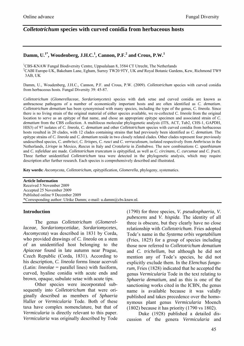
Online advance Fungal Diversity 45 Colletotrichum species with curved conidia from herbaceous hosts Damm, U. 1* , Woudenberg, J.H.C. 1 , Cannon, P.F. 2 and Crous, P.W. 1 1 CBS-KNAW Fungal Biodiversity Centre, Uppsalalaan 8, 3584 CT Utrecht, The Netherlands 2 CABI Europe-UK, Bakeham Lane, Egham, Surrey TW20 9TY, UK and Royal Botanic Gardens, Kew, Richmond TW9 3AB, UK Damm, U., Woudenberg, J.H.C., Cannon, P.F. and Crous, P.W. (2009). Colletotrichum species with curved conidia from herbaceous hosts. Fungal Diversity 39: 45-87. Colletotrichum (Glomerellaceae, Sordariomycetes) species with dark setae and curved conidia are known as anthracnose pathogens of a number of economically important hosts and are often identified as C. dematium. Colletotrichum dematium has been synonymised with many species, including the type of the genus, C. lineola. Since there is no living strain of the original material of either species available, we re-collected C. lineola from the original location to serve as an epitype of that name, and chose an appropriate epitype specimen and associated strain of C. dematium from the CBS collection. A multilocus molecular phylogenetic analysis (ITS, ACT, Tub2, CHS-1, GAPDH, HIS3) of 97 isolates of C. lineola, C. dematium and other Colletotrichum species with curved conidia from herbaceous hosts resulted in 20 clades, with 12 clades containing strains that had previously been identified as C. dematium. The epitype strains of C. lineola and C. dematium reside in two closely related clades. Other clades represent four previously undescribed species, C. anthrisci, C. liriopes, C. rusci and C. verruculosum, isolated respectively from Anthriscus in the Netherlands, Liriope in Mexico, Ruscus in Italy and Crotalaria in Zimbabwe. The new combinations C. spaethianum and C. tofieldiae are made. Colletotrichum truncatum is epitypified, as well as C. circinans, C. curcumae and C. fructi. Three further unidentified Colletotrichum taxa were detected in the phylogenetic analysis, which may require description after further research. Each species is comprehensively described and illustrated. Key words: Ascomycota, Colletotrichum, epitypification, Glomerella, phylogeny, systematics. Article Information Received 5 November 2009 Accepted 25 November 2009 Published online 9 December 2009 *Corresponding author: Ulrike Damm; e-mail: [email protected]. Introduction The genus Colletotrichum (Glomerel- laceae, Sordariomycetidae, Sordariomycetes, Ascomycota) was described in 1831 by Corda, who provided drawings of C. lineola on a stem of an unidentified host belonging to the Apiaceae found in late autumn near Prague, Czech Republic (Corda, 1831). According to his description, C. lineola forms linear acervuli (Latin: lineolae = parallel lines) with fusiform, curved, hyaline conidia with acute ends and brown, opaque, subulate setae with acute tips. Other species were incorporated sub- sequently into Colletotrichum that were ori- ginally described as members of Sphaeria Haller or Vermicularia Tode. Both of these taxa have complex nomenclature, but that of Vermicularia is directly relevant to this paper. Vermicularia was originally described by Tode (1790) for three species, V. pseudosphaeria, V. pubescens and V. hispida. The identity of all three is obscure, but they clearly have no close relationship with Colletotrichum. Fries adopted Tode’s name in the Systema orbis vegetabilium (Fries, 1825) for a group of species including those now referred to Colletotrichum dematium and C. trichellum, but although he did not mention any of Tode’s species, he did not explicitly exclude them. In the Elenchus fungo- rum, Fries (1828) indicated that he accepted the genus Vermicularia Tode in the text relating to Sphaeria dematium, and as this is one of the sanctioning works cited in the ICBN, the genus name is available because it was validly published and takes precedence over the homo- nymous plant genus Vermicularia Moench (1802) because it has priority (1790 vs 1802). Duke (1928) published a detailed dis- cussion of the genera Vermicularia and
Transcript of Colletotrichum Spp
Online advance Fungal Diversity
45
Colletotrichum species with curved conidia from herbaceous hosts Damm, U.1*, Woudenberg, J.H.C.1, Cannon, P.F.2 and Crous, P.W.1 1CBS-KNAW Fungal Biodiversity Centre, Uppsalalaan 8, 3584 CT Utrecht, The Netherlands 2CABI Europe-UK, Bakeham Lane, Egham, Surrey TW20 9TY, UK and Royal Botanic Gardens, Kew, Richmond TW9 3AB, UK
Damm, U., Woudenberg, J.H.C., Cannon, P.F. and Crous, P.W. (2009). Colletotrichum species with curved conidia from herbaceous hosts. Fungal Diversity 39: 45-87. Colletotrichum (Glomerellaceae, Sordariomycetes) species with dark setae and curved conidia are known as anthracnose pathogens of a number of economically important hosts and are often identified as C. dematium. Colletotrichum dematium has been synonymised with many species, including the type of the genus, C. lineola. Since there is no living strain of the original material of either species available, we re-collected C. lineola from the original location to serve as an epitype of that name, and chose an appropriate epitype specimen and associated strain of C. dematium from the CBS collection. A multilocus molecular phylogenetic analysis (ITS, ACT, Tub2, CHS-1, GAPDH, HIS3) of 97 isolates of C. lineola, C. dematium and other Colletotrichum species with curved conidia from herbaceous hosts resulted in 20 clades, with 12 clades containing strains that had previously been identified as C. dematium. The epitype strains of C. lineola and C. dematium reside in two closely related clades. Other clades represent four previously undescribed species, C. anthrisci, C. liriopes, C. rusci and C. verruculosum, isolated respectively from Anthriscus in the Netherlands, Liriope in Mexico, Ruscus in Italy and Crotalaria in Zimbabwe. The new combinations C. spaethianum and C. tofieldiae are made. Colletotrichum truncatum is epitypified, as well as C. circinans, C. curcumae and C. fructi. Three further unidentified Colletotrichum taxa were detected in the phylogenetic analysis, which may require description after further research. Each species is comprehensively described and illustrated. Key words: Ascomycota, Colletotrichum, epitypification, Glomerella, phylogeny, systematics.
Article Information Received 5 November 2009 Accepted 25 November 2009 Published online 9 December 2009 *Corresponding author: Ulrike Damm; e-mail: [email protected]. Introduction
The genus Colletotrichum (Glomerel-laceae, Sordariomycetidae, Sordariomycetes, Ascomycota) was described in 1831 by Corda, who provided drawings of C. lineola on a stem of an unidentified host belonging to the Apiaceae found in late autumn near Prague, Czech Republic (Corda, 1831). According to his description, C. lineola forms linear acervuli (Latin: lineolae = parallel lines) with fusiform, curved, hyaline conidia with acute ends and brown, opaque, subulate setae with acute tips.
Other species were incorporated sub-sequently into Colletotrichum that were ori-ginally described as members of Sphaeria Haller or Vermicularia Tode. Both of these taxa have complex nomenclature, but that of Vermicularia is directly relevant to this paper. Vermicularia was originally described by Tode
(1790) for three species, V. pseudosphaeria, V. pubescens and V. hispida. The identity of all three is obscure, but they clearly have no close relationship with Colletotrichum. Fries adopted Tode’s name in the Systema orbis vegetabilium (Fries, 1825) for a group of species including those now referred to Colletotrichum dematium and C. trichellum, but although he did not mention any of Tode’s species, he did not explicitly exclude them. In the Elenchus fungo-rum, Fries (1828) indicated that he accepted the genus Vermicularia Tode in the text relating to Sphaeria dematium, and as this is one of the sanctioning works cited in the ICBN, the genus name is available because it was validly published and takes precedence over the homo-nymous plant genus Vermicularia Moench (1802) because it has priority (1790 vs 1802).
Duke (1928) published a detailed dis-cussion of the genera Vermicularia and
46
Colletotrichum, in the course of which she typified Vermicularia with Sphaeria dematium Pers. This is now considered to be acceptable practice even though it was not one of the original species included in Vermicularia by Tode, as the genus was sanctioned by Fries. Duke appreciated the close similarity of the two genera, and observed that technically the name Vermicularia should be adopted if they are considered synonyms. She indicated that it would be advisable to conserve the name Colletotrichum over Vermicularia, but this appears never to have been carried out. The name Vermicularia was used subsequently by some authors for species with curved spores (Wollenweber and Hochapfel, 1949; Vassil-jevski and Karakulin, 1950), but the name fell out of use following von Arx’s revision (von Arx, 1957). In this seminal work, von Arx substantially reduced the number of species accepted in Colletotrichum, and synonymised C. lineola and many other Colletotrichum species with curved conidia with C. dematium. Sutton (1980) largely followed von Arx’s synonymy, but there has not been a modern assessment of that arrangement.
The original description of Sphaeria dematium by Persoon (1801) comprises only a few observations: slightly flattened spheres on grey spots, in the centre with erect, stiff, diverging, monochromatic hairs/setae. The fungus was stated to be common on dead, dry herbaceous stems, especially on Solanum tuberosum (Persoon 1801). While later descrip-tions of Colletotrichum dematium all include characters such as the dark, stiff setae, the curved/falcate conidia and the circular or elliptical appressoria, there are considerable differences concerning size and shape of conidia (von Arx, 1957; Sutton, 1980, 1992; Baxter et al., 1983). According to Sutton (1980) conidia of C. dematium are strongly curved and less than 3 µm wide, features used to dis-tinguish the species from C. capsici. However, in drawings of Baxter et al. (1983) one side of the conidia of C. dematium is nearly straight. The figures of two different strains exhibit different conidium shapes, which was regarded as an indication of the variability of the species. Drawings in Wollenweber and Hochapfel (1949) display a diverse range of variation for C. dematium on various host plants and media.
Von Arx (1957) lists 88 synonyms of C. dema-tium. For most he did not study the original material, including C. capsici, C. lineola and C. trichellum. The last-named species had been shown to be different from C. dematium by Sutton (1962), while C. capsici has been epity-pified recently (Shenoy et al., 2007). Many species have never been recollected and few have living cultures available that are derived from type material.
While C. lineola was described by Corda from a specimen on an “Umbelliferen” (=Apiaceae) stem, Grove (1937) combined that species name into Vermicularia with a des-cription based on a specimen from Dactylis glomerata (Poaceae), though he indicated that V. dematium occurred on all kinds of herbaceous stems, including Heracleum (Apia-ceae). That probably led to many grass-inhabiting collections being identified as C. dematium (e.g. Farr et al. 2009), which are now mostly if not all accommodated elsewhere. Wollenweber and Hochapfel (1949) and Feige and Ale-Agha (2004) mention C. dematium on Heracleum sphondylium, H. pubescens and H. mantegazzianum in Germany.
Colletotrichum dematium is now consi-dered to be polyphagous, occuring on stems of various herbaceous hosts, but with a number of host-restricted parasitic forms. According to von Arx (1957), C. dematium is a widespread saprobe on dead leaves, onion peel, twigs and rotting fruits, only occasionally found as a parasite causing fruit rots, leaf spots and anthracnose, for example of Fragaria (Rosa-ceae), Raphanus sativus var. hortensis (Brassi-caceae) Rhododendron (Ericaceae), Morus (Moraceae), Goniolimon tataricum (Plumba-ginaceae), Vigna unguiculata (Fabaceae) and Polygonatum falcatum (Liliaceae) (Beraha and Wright, 1973; Smith et al., 1999; Yoshida and Shirata, 1999; Vinnere et al., 2002; Sato et al., 2005; Babu et al., 2008; Tomioka et al., 2008; Bobev et al., 2009). The species can also be associated with infections of humans, most often as keratitis (Mendiratta et al., 2005). While there is, as far as we know, no authentic strain of C. lineola available in any culture collection, there are numerous C. dematium strains from many hosts available, including on Eryngium campestre (Apiaceae).
Fungal Diversity
47
Apart from C. lineola and C. dematium, many other Colletotrichum species with curved conidia are known as pathogens of different herbaceous plants, for example C. truncatum on Glycine max (Backman et al., 1982), C. capsici on Capsicum (Solanaceae) (Than et al., 2008) and C. lilii on Lilium longiflorum (Plakidas, 1944). Colletotrichum species with curved conidia on grass hosts have been studied recently with the addition of seven new species (Crouch et al., 2009a,b). A group of species that are sometimes mentioned as slightly curved, such as C. fuscum, C. higgin-sianum and C. lini (Sutton, 1980), seem to be closely related to each other according to preliminary phylogenies, and are excluded here. Colletotrichum trichellum is excluded in the morphological analysis, and will be the subject of a separate paper.
The C. dematium group has been largely overlooked in modern phylogenetic studies. The first rDNA-based studies (Sherriff et al., 1994; Sreenivasaprasad et al., 1996) included strains identified as C. capsici (treated as a synonym of C. truncatum in this paper), C. dematium, C. trichellum and C. truncatum, although the identification of some of the strains is doubtful. Both papers suggested that C. capsici was related to the C. gloeosporioides aggregate rather than the C. dematium group, and Sreenivasaprasad and co-workers detected a close relationship between C. dematium, C. trichellum and C. truncatum, though C. coc-codes was also found in that clade. Moriwaki et al. (2002) came to broadly similar conclusions, and established that C. circinans also belonged in that aggregate based on studies of rDNA. More recent studies (e.g. Crouch et al., 2009c) concur that rDNA data alone are inadequate to detect relationships between Colletotrichum spp. except at the species aggregate level. Some studies (e.g. Lubbe et al., 2004; Cannon et al., 2008) have included strains from the C. dematium aggregate to place other studies in a phylogenetic context. A number of strains of C. capsici were included in the study that led to epitypification of that name (Shenoy et al., 2007), but no attempt was made to elucidate relationships between that taxon and other non-graminicolous falcate-spored species. Ford et al. (2004) included strains identified as C. trun-catum from a range of host plants in their study
of populations of that species on lentil in Canada. They detected a number of clades based on rDNA data including those accepted in this paper as C. spaethianum and C. tofiel-diae, but the strains from lentil seem to belong to a separate taxon. We have not studied cultures from this source, but ITS sequences (see also Latunde-Dada and Lucas 2007) indicate strongly that they belong to the C. destructivum rather than the C. dematium clade. This is of particular concern as the teleomorph name Glomerella truncata (Armstrong-Cho and Banniza 2006) is based on a cross between two of the lentil strains.
In preliminary phylogenies using ITS sequence data (unpublished data), strains from the CBS culture collection that had been previously identified as C. dematium formed several clades, suggesting that C. dematium is polyphyletic in its current circumscription. The scope of this paper is therefore to clarify the identity of C. dematium and to epitypify this species, and to reveal the phylogenetic relation-ships of C. dematium and other allied species with curved conidia from herbaceous hosts. Materials and methods Isolates
Decayed or recently dead stems of Apiaceae were collected near Prague (Czech Republic), in Utrecht (Netherlands) and Han-nover (Germany). Type specimens of the species studied were located in the Herbarium of the Royal Botanic Gardens in Kew, UK (K), Corda’s herbarium in the Mycological Depart-ment of the National Museum in Prague, Czech Republic (PRM), herbarium of the Botanische Staatssammlung München (M), and the Farlow Herbarium, Harvard University, Cambridge, MA, USA (FH). The lectotype of Sphaeria dematium was chosen from original material in Persoon’s herbarium, specimens from which are preserved in the National Herbarium in Leiden (L), the Netherlands. The epitype specimens of C. circinans, C. curcumae, C. dematium, C. fructi, C. lilii, C. spaethianum, C. spinaciae, C. tofieldiae and C. truncatum were selected from the culture collections of the Centraalbureau voor Schimmelcultures (CBS) Utrecht, The Netherlands and CABI Europe-UK, Egham, Surrey, UK (IMI) and are
48
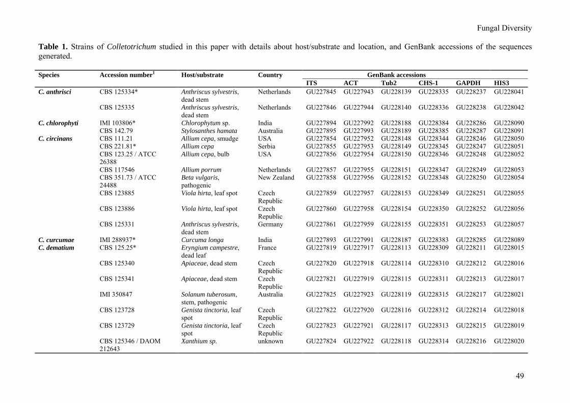
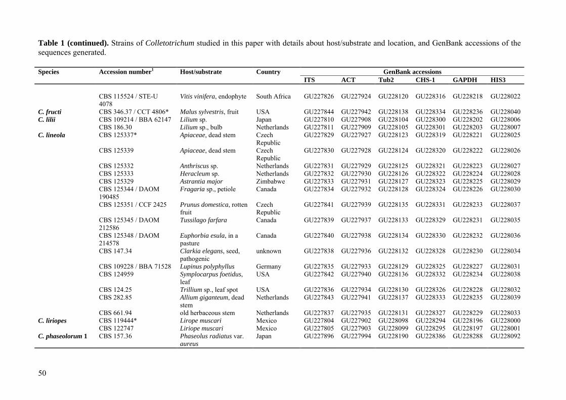
preserved as dried cultures in the CBS herbarium. All descriptions are based on the ex-type, ex-epitype or ex-neotype culture as appropriate. Features of other strains are added if deviant. Subcultures of the types and epi-types, respectively, as well as all other isolates used for morphological and sequence analyses are maintained in the culture collection of CBS and/or CABI (IMI) and presented in Table 1. Morphological analysis
To enhance sporulation, autoclaved filter paper and double-autoclaved stems of An-thriscus sylvestris were placed onto the surface of synthetic nutrient-poor agar medium (SNA; Nirenberg 1976). SNA, OA, PDA and MEA cultures incubated at 20 °C under near UV light with 12 h photoperiod or permanent near UV light for 10 d. Measurements and photographs of characteristic structures were made accor-ding to Damm et al. (2007). Appressoria on hyphae were observed on the undersurface of the SNA cultures. Microscopic preparations were made in clear lactic acid or water, with 30 measurements per structure and observed with a Nikon SMZ1000 dissecting microscope (DM) or with a Nikon Eclipse 80i microscope using differential interference contrast (DIC) illumi-nation. Colony characters and pigment produc-tion on malt extract agar (MEA, 2 % malt extract, Oxoid Ltd., England; 1.5 % agar, Difco, USA) and 2 % potato-dextrose agar (PDA; Crous et al., 2009) incubated at 20°C were noted after 1 wk. Colony colours were rated according to Rayner (1970). Growth rates were measured after 5, 7 and 10d. Phylogenetic analysis
Genomic DNA of the isolates was extracted using the method of Damm et al. (2008). The 5.8S nuclear ribosomal gene with the two flanking internal transcribed spacers (ITS), a 200-bp intron of the glyceraldehyde-3-phosphate dehydrogenase (GAPDH), a partial sequence of the actin (ACT), chitin synthase 1 (CHS-1), beta-tubulin (Tub2) and of the histone3 (HIS3) gene were amplified and sequenced using the primer pairs V9G (de Hoog and Gerrits van den Ende 1998) + ITS-4 (White et al., 1990), GDF1 + GDR1 (Guerber et al., 2003), ACT-512F + ACT-783R (Carbone and Kohn, 1999), CHS-354R + CHS-
79F (Carbone and Kohn 1999), BT2Fd + BT4R (Woudenberg et al., 2009) or T1 (O'Donnell and Cigelnik, 1997) + Bt-2b (Glass and Do-naldson 1995) and CYLH3F + CYLH3R (Crous et al., 2004b), respectively. The PCRs were per-formed in a 2720 Thermal Cycler (Applied Biosystems, Foster City, California) in a total volume of 12.5 μl. The GAPDH, ACT, CHS-1, Tub2 and HIS3 PCR mixture contained 1 μl 20x diluted genomic DNA, 0.2 μM of each primer, 1x PCR buffer (Bioline, Luckenwalde, Germany), 2 mM MgCl2, 20 μM of each dNTP, 0.7 μl DMSO and 0.25 U Taq DNA poly-merase (Bioline). Conditions for amplification were an initial denaturation step of 5 min at 94oC, followed by 40 cycles of 30 s at 94oC, 30 s at 52oC and 30 s at 72oC, and a final denaturation step of 7 min at 72oC. The ITS PCR was performed as described by Wouden-berg et al. (2009). The DNA se-quences ob-tained from forward and reverse primers were used to obtain consensus sequences using Bionumeris v. 4.60 (Applied Maths, St-Marthens-Lathem, Belgium) which were added to the outgroup (C. lindemuthia-num CBS 315.28) and the alignment assembled and manually adjusted using Sequence Alignment Editor v. 2.0a11 (Rambaut, 2002).
A maximum parsimony analysis was performed on the multilocus alignment (ITS, ACT, Tub2, CHS-1, GAPDH, HIS3) with PAUP (Phylogenetic Analysis Using Parsi-mony) v. 4.0b10 (Swofford, 2000) using the heuristic search option with 100 random sequence additions and tree bisection and reconstruction (TBR) as the branch-swapping algorithm. Alignment gaps were treated as missing and all characters were unordered and of equal weight. The robustness of the trees obtained was evaluated by 500 bootstrap replications with 2 random sequence additions (Hillis and Bull 1993). Tree length, consistency index (CI), retention index (RI), rescaled consistency index (RC) and homoplasy index (HI) were calculated for the resulting tree. A maximum likelihood phylogenetic analyses of the dataset was performed with RAxML on the Cipres Web Portal (http://www.phylo.org/sub _sections/portal/), with a GTR model of molecular evolution (selected by the program) and 1000 bootstrap replicates using RAxML VI-HPC (Stamatakis et al., 2008). Sequences
Fungal Diversity
49
Table 1. Strains of Colletotrichum studied in this paper with details about host/substrate and location, and GenBank accessions of the sequences generated. Species Accession number1 Host/substrate Country GenBank accessions ITS ACT Tub2 CHS-1 GAPDH HIS3 C. anthrisci CBS 125334* Anthriscus sylvestris,
dead stem Netherlands GU227845 GU227943 GU228139 GU228335 GU228237 GU228041
CBS 125335 Anthriscus sylvestris, dead stem
Netherlands GU227846 GU227944 GU228140 GU228336 GU228238 GU228042
C. chlorophyti IMI 103806* Chlorophytum sp. India GU227894 GU227992 GU228188 GU228384 GU228286 GU228090 CBS 142.79 Stylosanthes hamata Australia GU227895 GU227993 GU228189 GU228385 GU228287 GU228091 C. circinans CBS 111.21 Allium cepa, smudge USA GU227854 GU227952 GU228148 GU228344 GU228246 GU228050 CBS 221.81* Allium cepa Serbia GU227855 GU227953 GU228149 GU228345 GU228247 GU228051 CBS 123.25 / ATCC
26388 Allium cepa, bulb USA GU227856 GU227954 GU228150 GU228346 GU228248 GU228052
CBS 117546 Allium porrum Netherlands GU227857 GU227955 GU228151 GU228347 GU228249 GU228053 CBS 351.73 / ATCC
24488 Beta vulgaris, pathogenic
New Zealand GU227858 GU227956 GU228152 GU228348 GU228250 GU228054
CBS 123885 Viola hirta, leaf spot Czech Republic
GU227859 GU227957 GU228153 GU228349 GU228251 GU228055
CBS 123886 Viola hirta, leaf spot Czech Republic
GU227860 GU227958 GU228154 GU228350 GU228252 GU228056
CBS 125331 Anthriscus sylvestris, dead stem
Germany GU227861 GU227959 GU228155 GU228351 GU228253 GU228057
C. curcumae IMI 288937* Curcuma longa India GU227893 GU227991 GU228187 GU228383 GU228285 GU228089 C. dematium CBS 125.25* Eryngium campestre,
dead leaf France GU227819 GU227917 GU228113 GU228309 GU228211 GU228015
CBS 125340 Apiaceae, dead stem Czech Republic
GU227820 GU227918 GU228114 GU228310 GU228212 GU228016
CBS 125341 Apiaceae, dead stem Czech Republic
GU227821 GU227919 GU228115 GU228311 GU228213 GU228017
IMI 350847 Solanum tuberosum, stem, pathogenic
Australia GU227825 GU227923 GU228119 GU228315 GU228217 GU228021
CBS 123728 Genista tinctoria, leaf spot
Czech Republic
GU227822 GU227920 GU228116 GU228312 GU228214 GU228018
CBS 123729 Genista tinctoria, leaf spot
Czech Republic
GU227823 GU227921 GU228117 GU228313 GU228215 GU228019
CBS 125346 / DAOM 212643
Xanthium sp. unknown GU227824 GU227922 GU228118 GU228314 GU228216 GU228020
50
Table 1 (continued). Strains of Colletotrichum studied in this paper with details about host/substrate and location, and GenBank accessions of the sequences generated.
Species Accession number1 Host/substrate Country GenBank accessions ITS ACT Tub2 CHS-1 GAPDH HIS3 CBS 115524 / STE-U
4078 Vitis vinifera, endophyte South Africa GU227826 GU227924 GU228120 GU228316 GU228218 GU228022
C. fructi CBS 346.37 / CCT 4806* Malus sylvestris, fruit USA GU227844 GU227942 GU228138 GU228334 GU228236 GU228040 C. lilii CBS 109214 / BBA 62147 Lilium sp. Japan GU227810 GU227908 GU228104 GU228300 GU228202 GU228006 CBS 186.30 Lilium sp., bulb Netherlands GU227811 GU227909 GU228105 GU228301 GU228203 GU228007 C. lineola CBS 125337* Apiaceae, dead stem Czech
Republic GU227829 GU227927 GU228123 GU228319 GU228221 GU228025
CBS 125339 Apiaceae, dead stem Czech Republic
GU227830 GU227928 GU228124 GU228320 GU228222 GU228026
CBS 125332 Anthriscus sp. Netherlands GU227831 GU227929 GU228125 GU228321 GU228223 GU228027 CBS 125333 Heracleum sp. Netherlands GU227832 GU227930 GU228126 GU228322 GU228224 GU228028 CBS 125329 Astrantia major Zimbabwe GU227833 GU227931 GU228127 GU228323 GU228225 GU228029 CBS 125344 / DAOM
190485 Fragaria sp., petiole Canada GU227834 GU227932 GU228128 GU228324 GU228226 GU228030
CBS 125351 / CCF 2425 Prunus domestica, rotten fruit
Czech Republic
GU227841 GU227939 GU228135 GU228331 GU228233 GU228037
CBS 125345 / DAOM 212586
Tussilago farfara Canada GU227839 GU227937 GU228133 GU228329 GU228231 GU228035
CBS 125348 / DAOM 214578
Euphorbia esula, in a pasture
Canada GU227840 GU227938 GU228134 GU228330 GU228232 GU228036
CBS 147.34 Clarkia elegans, seed, pathogenic
unknown GU227838 GU227936 GU228132 GU228328 GU228230 GU228034
CBS 109228 / BBA 71528 Lupinus polyphyllus Germany GU227835 GU227933 GU228129 GU228325 GU228227 GU228031 CBS 124959 Symplocarpus foetidus,
leaf USA GU227842 GU227940 GU228136 GU228332 GU228234 GU228038
CBS 124.25 Trillium sp., leaf spot USA GU227836 GU227934 GU228130 GU228326 GU228228 GU228032 CBS 282.85 Allium giganteum, dead
stem Netherlands GU227843 GU227941 GU228137 GU228333 GU228235 GU228039
CBS 661.94 old herbaceous stem Netherlands GU227837 GU227935 GU228131 GU228327 GU228229 GU228033 C. liriopes CBS 119444* Lirope muscari Mexico GU227804 GU227902 GU228098 GU228294 GU228196 GU228000 CBS 122747 Liriope muscari Mexico GU227805 GU227903 GU228099 GU228295 GU228197 GU228001 C. phaseolorum 1 CBS 157.36 Phaseolus radiatus var.
aureus Japan GU227896 GU227994 GU228190 GU228386 GU228288 GU228092
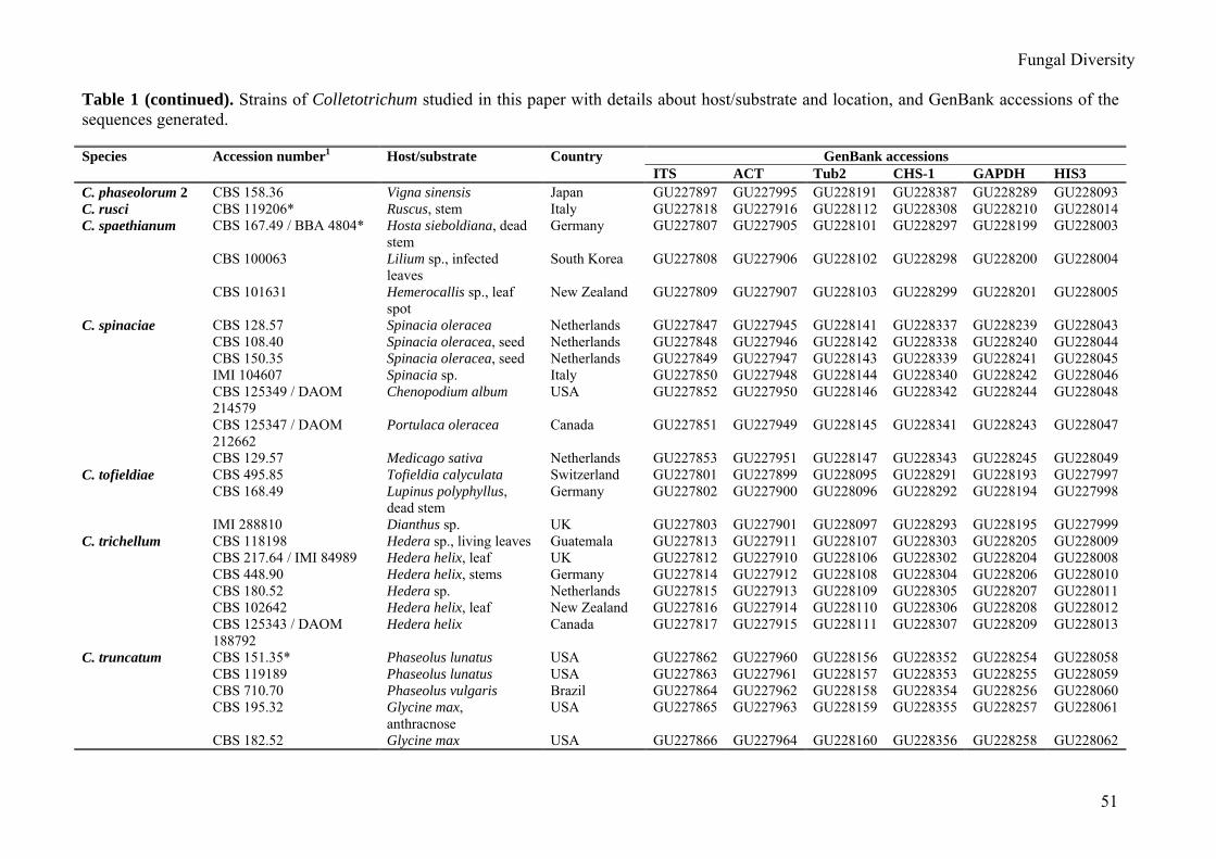
Fungal Diversity
51
Table 1 (continued). Strains of Colletotrichum studied in this paper with details about host/substrate and location, and GenBank accessions of the sequences generated. Species Accession number1 Host/substrate Country GenBank accessions ITS ACT Tub2 CHS-1 GAPDH HIS3 C. phaseolorum 2 CBS 158.36 Vigna sinensis Japan GU227897 GU227995 GU228191 GU228387 GU228289 GU228093 C. rusci CBS 119206* Ruscus, stem Italy GU227818 GU227916 GU228112 GU228308 GU228210 GU228014 C. spaethianum CBS 167.49 / BBA 4804* Hosta sieboldiana, dead
stem Germany GU227807 GU227905 GU228101 GU228297 GU228199 GU228003
CBS 100063 Lilium sp., infected leaves
South Korea GU227808 GU227906 GU228102 GU228298 GU228200 GU228004
CBS 101631 Hemerocallis sp., leaf spot
New Zealand GU227809 GU227907 GU228103 GU228299 GU228201 GU228005
C. spinaciae CBS 128.57 Spinacia oleracea Netherlands GU227847 GU227945 GU228141 GU228337 GU228239 GU228043 CBS 108.40 Spinacia oleracea, seed Netherlands GU227848 GU227946 GU228142 GU228338 GU228240 GU228044 CBS 150.35 Spinacia oleracea, seed Netherlands GU227849 GU227947 GU228143 GU228339 GU228241 GU228045 IMI 104607 Spinacia sp. Italy GU227850 GU227948 GU228144 GU228340 GU228242 GU228046 CBS 125349 / DAOM
214579 Chenopodium album USA GU227852 GU227950 GU228146 GU228342 GU228244 GU228048
CBS 125347 / DAOM 212662
Portulaca oleracea Canada GU227851 GU227949 GU228145 GU228341 GU228243 GU228047
CBS 129.57 Medicago sativa Netherlands GU227853 GU227951 GU228147 GU228343 GU228245 GU228049 C. tofieldiae CBS 495.85 Tofieldia calyculata Switzerland GU227801 GU227899 GU228095 GU228291 GU228193 GU227997 CBS 168.49 Lupinus polyphyllus,
dead stem Germany GU227802 GU227900 GU228096 GU228292 GU228194 GU227998
IMI 288810 Dianthus sp. UK GU227803 GU227901 GU228097 GU228293 GU228195 GU227999 C. trichellum CBS 118198 Hedera sp., living leaves Guatemala GU227813 GU227911 GU228107 GU228303 GU228205 GU228009 CBS 217.64 / IMI 84989 Hedera helix, leaf UK GU227812 GU227910 GU228106 GU228302 GU228204 GU228008 CBS 448.90 Hedera helix, stems Germany GU227814 GU227912 GU228108 GU228304 GU228206 GU228010 CBS 180.52 Hedera sp. Netherlands GU227815 GU227913 GU228109 GU228305 GU228207 GU228011 CBS 102642 Hedera helix, leaf New Zealand GU227816 GU227914 GU228110 GU228306 GU228208 GU228012 CBS 125343 / DAOM
188792 Hedera helix Canada GU227817 GU227915 GU228111 GU228307 GU228209 GU228013
C. truncatum CBS 151.35* Phaseolus lunatus USA GU227862 GU227960 GU228156 GU228352 GU228254 GU228058 CBS 119189 Phaseolus lunatus USA GU227863 GU227961 GU228157 GU228353 GU228255 GU228059 CBS 710.70 Phaseolus vulgaris Brazil GU227864 GU227962 GU228158 GU228354 GU228256 GU228060 CBS 195.32 Glycine max,
anthracnose USA GU227865 GU227963 GU228159 GU228355 GU228257 GU228061
CBS 182.52 Glycine max USA GU227866 GU227964 GU228160 GU228356 GU228258 GU228062
52
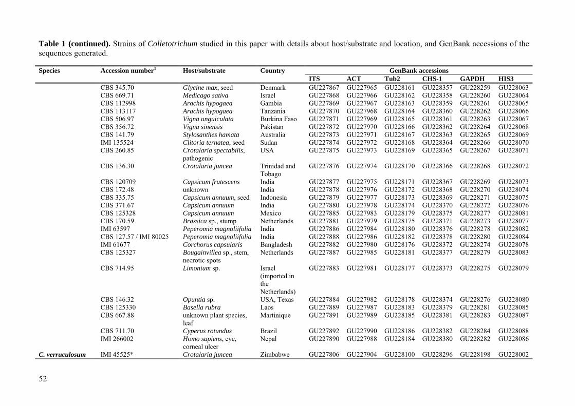
Table 1 (continued). Strains of Colletotrichum studied in this paper with details about host/substrate and location, and GenBank accessions of the sequences generated. Species Accession number1 Host/substrate Country GenBank accessions ITS ACT Tub2 CHS-1 GAPDH HIS3 CBS 345.70 Glycine max, seed Denmark GU227867 GU227965 GU228161 GU228357 GU228259 GU228063 CBS 669.71 Medicago sativa Israel GU227868 GU227966 GU228162 GU228358 GU228260 GU228064 CBS 112998 Arachis hypogaea Gambia GU227869 GU227967 GU228163 GU228359 GU228261 GU228065 CBS 113117 Arachis hypogaea Tanzania GU227870 GU227968 GU228164 GU228360 GU228262 GU228066 CBS 506.97 Vigna unguiculata Burkina Faso GU227871 GU227969 GU228165 GU228361 GU228263 GU228067 CBS 356.72 Vigna sinensis Pakistan GU227872 GU227970 GU228166 GU228362 GU228264 GU228068 CBS 141.79 Stylosanthes hamata Australia GU227873 GU227971 GU228167 GU228363 GU228265 GU228069 IMI 135524 Clitoria ternatea, seed Sudan GU227874 GU227972 GU228168 GU228364 GU228266 GU228070 CBS 260.85 Crotalaria spectabilis,
pathogenic USA GU227875 GU227973 GU228169 GU228365 GU228267 GU228071
CBS 136.30 Crotalaria juncea Trinidad and Tobago
GU227876 GU227974 GU228170 GU228366 GU228268 GU228072
CBS 120709 Capsicum frutescens India GU227877 GU227975 GU228171 GU228367 GU228269 GU228073 CBS 172.48 unknown India GU227878 GU227976 GU228172 GU228368 GU228270 GU228074 CBS 335.75 Capsicum annuum, seed Indonesia GU227879 GU227977 GU228173 GU228369 GU228271 GU228075 CBS 371.67 Capsicum annuum India GU227880 GU227978 GU228174 GU228370 GU228272 GU228076 CBS 125328 Capsicum annuum Mexico GU227885 GU227983 GU228179 GU228375 GU228277 GU228081 CBS 170.59 Brassica sp., stump Netherlands GU227881 GU227979 GU228175 GU228371 GU228273 GU228077 IMI 63597 Peperomia magnoliifolia India GU227886 GU227984 GU228180 GU228376 GU228278 GU228082 CBS 127.57 / IMI 80025 Peperomia magnoliifolia India GU227888 GU227986 GU228182 GU228378 GU228280 GU228084 IMI 61677 Corchorus capsularis Bangladesh GU227882 GU227980 GU228176 GU228372 GU228274 GU228078 CBS 125327 Bougainvillea sp., stem,
necrotic spots Netherlands GU227887 GU227985 GU228181 GU228377 GU228279 GU228083
CBS 714.95 Limonium sp. Israel (imported in the Netherlands)
GU227883 GU227981 GU228177 GU228373 GU228275 GU228079
CBS 146.32 Opuntia sp. USA, Texas GU227884 GU227982 GU228178 GU228374 GU228276 GU228080 CBS 125330 Basella rubra Laos GU227889 GU227987 GU228183 GU228379 GU228281 GU228085 CBS 667.88 unknown plant species,
leaf Martinique GU227891 GU227989 GU228185 GU228381 GU228283 GU228087
CBS 711.70 Cyperus rotundus Brazil GU227892 GU227990 GU228186 GU228382 GU228284 GU228088 IMI 266002 Homo sapiens, eye,
corneal ulcer Nepal GU227890 GU227988 GU228184 GU228380 GU228282 GU228086
C. verruculosum IMI 45525* Crotalaria juncea Zimbabwe GU227806 GU227904 GU228100 GU228296 GU228198 GU228002

Fungal Diversity
53
Table 1 (continued). Strains of Colletotrichum studied in this paper with details about host/substrate and location, and GenBank accessions of the sequences generated. Species Accession number1 Host/substrate Country GenBank accessions ITS ACT Tub2 CHS-1 GAPDH HIS3 Colletotrichum sp. 1 CBS 125326 Rubus idaeus Canada GU227827 GU227925 GU228121 GU228317 GU228219 GU228023 Colletotrichum sp. 2 CBS 125338 / DAOM
147549 Hemerocallis fulva, old flower stalk
Canada GU227828 GU227926 GU228122 GU228318 GU228220 GU228024
C. lindemuthianum (outgroup)
CBS 151.28 Phaseolus vulgaris UK GU227800 GU227898 GU228094 GU228290 GU228192 GU227996
1CBS: Culture collection of the Centraalbureau voor Schimmelcultures, Fungal Biodiversity Centre, Utrecht, The Netherlands; IMI: Culture collection of CABI Europe UK Centre, Egham, UK; ATCC: American Type culture collection; DAOM: National Mycological Herbarium, Ottawa, Canada; STE-U: Culture collection of the Department of Plant Pathology, University of Stellenbosch, South Africa; CCT: Colecao de Culturas Tropical, Sao Paulo, Brazil; BBA: Culture collection of the Biologische Bundesanstalt für Landund Forstwirtschaft, Berlin, Germany; CCF: Culture Collection of Fungi, Prague, Czech Republic;. * ex-type and ex-epitype cultures.
54
CBS 151.28
CBS 495.85CBS 168.49IMI 288810
IMI 45525CBS 119444CBS 122747CBS 167.49CBS 100063
CBS 101631CBS 109214CBS 186.30
CBS 217.64CBS 180.52CBS 448.90CBS 118198CBS 125343CBS 102642
CBS 119206CBS 125.25CBS 125341CBS 125340CBS 123728CBS 123729CBS 125346IMI 350847CBS 115524
CBS 125337CBS 125329CBS 125344CBS 109228CBS 124.25CBS 661.94CBS 125339CBS 125332CBS 125333CBS 147.34CBS 125345CBS 125348CBS 125351CBS 124959CBS 282.85CBS 125326CBS 125338
CBS 346.37CBS 125334CBS 125335
CBS 128.57CBS 108.40CBS 150.35IMI 104607CBS 125347CBS 125349CBS 129.57CBS 111.21CBS 221.81CBS 123.25CBS 117546CBS 351.73CBS 123885CBS 123886CBS 125331
IMI 103806CBS 142.79
CBS 157.36CBS 158.36
CBS 151.35 CBS 667.88 CBS 710.70 CBS 119189 CBS 136.30 IMI 61677
CBS 260.85 CBS 195.32 CBS 182.52 IMI 266002 CBS 714.95 CBS 345.70 CBS 356.72 CBS 120709 CBS 172.48 CBS 170.59 IMI 63597 CBS 127.57 CBS 125330 IMI 135524 CBS 335.75 CBS 371.67 CBS 125327 CBS 146.32 CBS 125328
CBS 669.71 CBS 112998 CBS 141.79 CBS 711.70
CBS 113117 CBS 506.97
IMI 288937 10 changes
C. tofieldiae
C. verruculosum C. liriopes
C. spaethianum
C. lilii
C. trichellum
Colletotrichum sp. 1
C. rusci
C. dematium
C. lineola
C. phaseolorum 2
Colletotrichum sp. 2 C. fructiC. anthrisci
C. spinaciae
C. circinans
C. chlorophyti C. phaseolorum 1
C. truncatum
C. curcumae
100
86
100
100
83
100
66
100100
100
100
100
100
100
100
100
96
64
80
100
59
100
85
100
100
64
63
77
59
100
83
63
67
62
100
52
87
100
100
50 55
77 91
89
79
70
53
56
89
59
73
96
77
Clade 1
Clade 2
Clade 3
Clade 4
Clade 5
Clade 6
Fungal Diversity
55
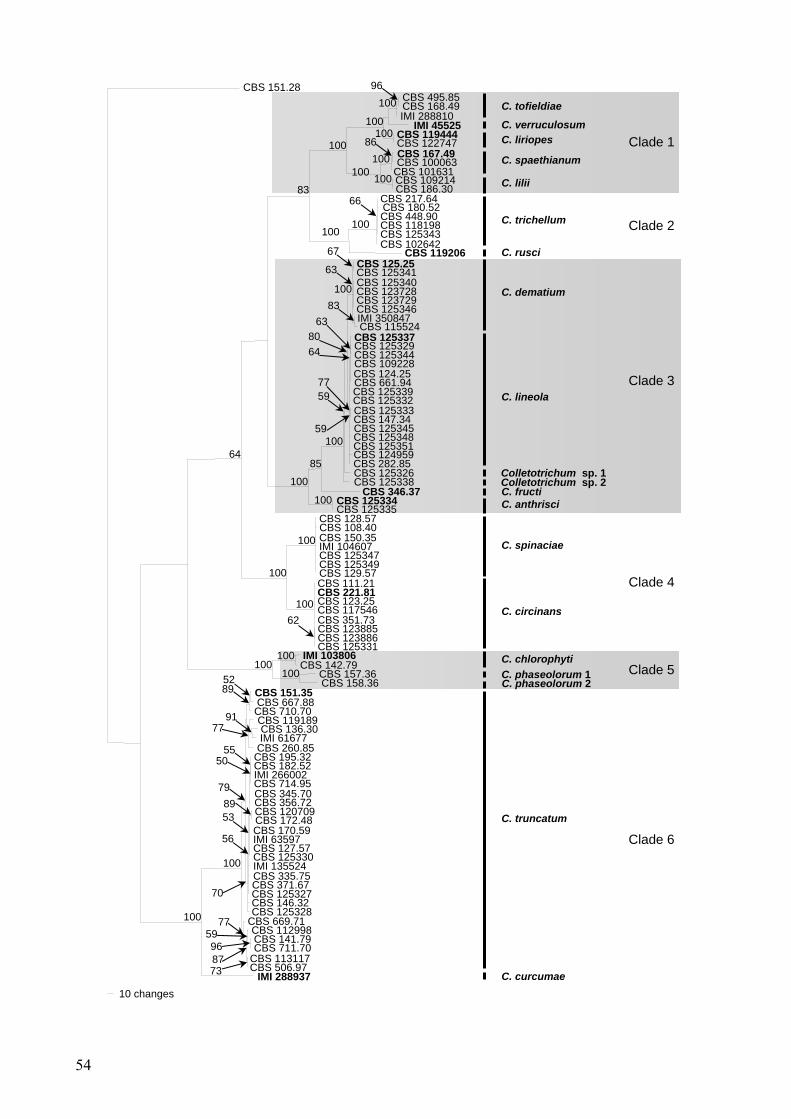
Fig. 1. One of 8735 most parsimonious trees obtained from heuristic searches of ITS, ACT, BT, CHS-1, GAPDH and HIS3 gene sequences of Colletotrichum species (length = 2141 steps, CI = 0.632, RI = 0.948, RC = 0.599, HI = 0.368). Bootstrap support values (500 replicates) above 50 % are shown at the nodes. Colletotrichum lindemuthianum CBS 151.28 is used as outgroup. Numbers of ex-type and ex-epitype strains are emphasised in bold. derived in this study were lodged at GenBank, the alignment in TreeBASE (http://www.tree base.org/treebase/index.html), and taxonomic novelties in MycoBank (Crous et al., 2004a). Results Phylogeny
In the multigene analyses (ITS, ACT, Tub2, CHS-1, HIS3, GAPDH) of 98 isolates of C. dematium and other Colletotrichum species with curved conidia including the outgroup, 2333 characters including the alignment gaps were processed, of which 740 characters were parsimony-informative, 157 parsomony-unin-formative and 1436 constant. For the individual alignments of the six genes, the obtained trees were compared by eye and the tree topology of the individual data sets was found to be similar to each other and to the tree obtained from the combined alignment. However some clades, e.g. C. circinans and C. spinaciae were very short-branched in the ITS phylogeny and some, e.g. C. dematium and C. lineola were only distinguished in three (Actin, HIS3 and GAPDH) of the six phylogenies. After a heu-ristic search using PAUP, 8735 most parsi-monious trees were retained (length = 2141 steps, CI = 0.632, RI = 0.948, RC = 0.599, HI = 0.368) of which one is shown in Fig. 1. The topology of the 8735 trees was similar, which was verified for a large selection of trees. They differed in the position of taxa within the subclades.
The analyses resulted in detection of 6 clades and 20 subclades, presumably repre-senting different Colletotrichum species. Clade 1 (100 % bootstrap support) is divided into five subclades, of which four subclades (C. tofiel-diae, C. liriopes, C. spaethianum and C. lilii) are well supported (100 %) and contain two or tree strains each, including at least one strain previously identified as C. dematium. The fifth subclade (C. verruculosum) is represented by a single strain, IMI 45525 that groups with the C. tofieldiae and the C. liriopes subclades (100 %), while C. spaethianum and C. lilii form a sisterclade (100 %). The second clade (100 %)
consists of two subclades, C. trichellum (100 %) and C. rusci (CBS 119206), a single-strain clade. The six strains of the C. trichellum clade, all from Hedera sp., have little variability. Clade 1 and clade 2 are sisterclades (83 %). Clade 3 (100 %) contains 6 subclades. Two of these clades, the C. dematium (100 %) and the C. lineola clade (77 %), are closely related to each other, contain both many strains from diverse host plants, and group with two single-strain clades belonging to unidentified taxa (100 %). These clades group with the subclade formed by one strain of C. fructi (85 %) of which C. anthrisci formes a sisterclade (100 %). The two subclades in clade 4 (100 %) represent C. spinaciae (100 %) and C. circinans (100 %), have little intraspecific variability and contain 4 strains from Spinacia and Allium, respec-tively. Clade 5 consists of C. chlorophyti (100 %) and C. phaseolorum is represented by two single strain clades, which cluster with each other (100 %). Clade 6 (100%) consists of one clade formed by a single strain (IMI 288937) representing C. curcumae and the C. truncatum clade (100 %), which is very heterogenous and contains strains from many different host plants, with the majority from Fabaceae and Cap-sicum spp., that had been identified as C. dematium, C. capsici (including the epitype strain), C. truncatum, C. curvatum (authentic material), Glomerella glycines, C. corchori and C. dematium f. sp. clitoriicola before. In the single gene phylogenies (not shown) there was, however, no consistency in subgrouping that would support distinguishing further taxa within this subclade.
The maximum likelihood phylogenetic analyses with RAxML resulted in a similar phylogeny, with the same 20 clades as in the parsimony analyses and similar bootstap sup-ports (not shown).
Taxonomy
The 97 strains studied (Table 1) could be assigned to 20 species based on DNA sequence data and morphology, including four species, C. anthrisci, C. liriopes, C. rusci and C. verru-culosum, that proved to be new to science, two
56
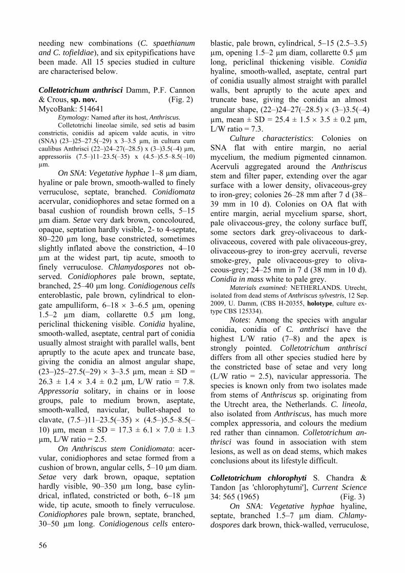
needing new combinations (C. spaethianum and C. tofieldiae), and six epitypifications have been made. All 15 species studied in culture are characterised below. Colletotrichum anthrisci Damm, P.F. Cannon & Crous, sp. nov. (Fig. 2) MycoBank: 514641
Etymology: Named after its host, Anthriscus. Colletotrichi lineolae simile, sed setis ad basim
constrictis, conidiis ad apicem valde acutis, in vitro (SNA) (23–)25–27.5(–29) x 3–3.5 µm, in cultura cum caulibus Anthrisci (22–)24–27(–28.5) x (3–)3.5(–4) µm, appressoriis (7.5–)11–23.5(–35) x (4.5–)5.5–8.5(–10) µm.
On SNA: Vegetative hyphae 1–8 µm diam, hyaline or pale brown, smooth-walled to finely verruculose, septate, branched. Conidiomata acervular, conidiophores and setae formed on a basal cushion of roundish brown cells, 5–15 µm diam. Setae very dark brown, concoloured, opaque, septation hardly visible, 2- to 4-septate, 80–220 µm long, base constricted, sometimes slightly inflated above the constriction, 4–10 µm at the widest part, tip acute, smooth to finely verruculose. Chlamydospores not ob-served. Conidiophores pale brown, septate, branched, 25–40 µm long. Conidiogenous cells enteroblastic, pale brown, cylindrical to elon-gate ampulliform, 6–18 × 3–6.5 µm, opening 1.5–2 µm diam, collarette 0.5 µm long, periclinal thickening visible. Conidia hyaline, smooth-walled, aseptate, central part of conidia usually almost straight with parallel walls, bent apruptly to the acute apex and truncate base, giving the conidia an almost angular shape, (23–)25–27.5(–29) × 3–3.5 µm, mean ± SD = 26.3 ± 1.4 × 3.4 ± 0.2 µm, L/W ratio = 7.8. Appressoria solitary, in chains or in loose groups, pale to medium brown, aseptate, smooth-walled, navicular, bullet-shaped to clavate, (7.5–)11–23.5(–35) × (4.5–)5.5–8.5(–10) µm, mean ± SD = 17.3 ± 6.1 × 7.0 ± 1.3 µm, L/W ratio = 2.5.
On Anthriscus stem Conidiomata: acer-vular, conidiophores and setae formed from a cushion of brown, angular cells, 5–10 µm diam. Setae very dark brown, opaque, septation hardly visible, 90–350 µm long, base cylin-drical, inflated, constricted or both, 6–18 µm wide, tip acute, smooth to finely verruculose. Conidiophores pale brown, septate, branched, 30–50 µm long. Conidiogenous cells entero-
blastic, pale brown, cylindrical, 5–15 (2.5–3.5) µm, opening 1.5–2 µm diam, collarette 0.5 µm long, periclinal thickening visible. Conidia hyaline, smooth-walled, aseptate, central part of conidia usually almost straight with parallel walls, bent apruptly to the acute apex and truncate base, giving the conidia an almost angular shape, (22–)24–27(–28.5) × (3–)3.5(–4) µm, mean ± SD = 25.4 ± 1.5 × 3.5 ± 0.2 µm, L/W ratio = 7.3.
Culture characteristics: Colonies on SNA flat with entire margin, no aerial mycelium, the medium pigmented cinnamon. Acervuli aggregated around the Anthriscus stem and filter paper, extending over the agar surface with a lower density, olivaceous-grey to iron-grey; colonies 26–28 mm after 7 d (38–39 mm in 10 d). Colonies on OA flat with entire margin, aerial mycelium sparse, short, pale olivaceous-grey, the colony surface buff, some sectors dark grey-olivaceous to dark-olivaceous, covered with pale olivaceous-grey, olivaceous-grey to iron-grey acervuli, reverse smoke-grey, pale olivaceous-grey to oliva-ceous-grey; 24–25 mm in 7 d (38 mm in 10 d). Conidia in mass white to pale grey.
Materials examined: NETHERLANDS. Utrecht, isolated from dead stems of Anthriscus sylvestris, 12 Sep. 2009, U. Damm, (CBS H-20355, holotype, culture ex-type CBS 125334).
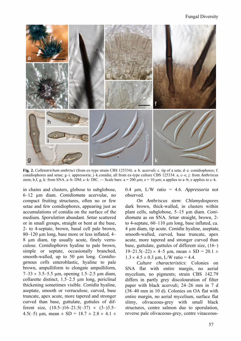
Notes: Among the species with angular conidia, conidia of C. anthrisci have the highest L/W ratio (7–8) and the apex is strongly pointed. Colletotrichum anthrisci differs from all other species studied here by the constricted base of setae and very long (L/W ratio = 2.5), navicular appressoria. The species is known only from two isolates made from stems of Anthriscus sp. originating from the Utrecht area, the Netherlands. C. lineola, also isolated from Anthriscus, has much more complex appressoria, and colours the medium red rather than cinnamon. Colletotrichum an-thrisci was found in association with stem lesions, as well as on dead stems, which makes conclusions about its lifestyle difficult. Colletotrichum chlorophyti S. Chandra & Tandon [as 'chlorophytumi'], Current Science 34: 565 (1965) (Fig. 3)
On SNA: Vegetative hyphae hyaline, septate, branched 1.5–7 µm diam. Chlamy- dospores dark brown, thick-walled, verruculose,
Fungal Diversity
57
Fig. 2. Colletotrichum anthrisci (from ex-type strain CBS 125334). a–b. acervuli; c. tip of a seta; d–e. conidiophores; f. conidiophores and setae; g–i. appressoria; j–k.conidia; all from ex-type culture CBS 125334. a, c–e, j: from Anthriscus stem; b,f, g, k: from SNA. a–b: DM; c–k: DIC. — Scale bars: a = 200 µm; e = 10 µm; a applies to a–b; e applies to c–k. in chains and clusters, globose to subglobose, 6–12 µm diam. Conidiomata acervular, no compact fruiting structures, often no or few setae and few conidiophores, appearing just as accumulations of conidia on the surface of the medium. Sporulation abundant. Setae scattered or in small groups, straight or bent at the base, 2- to 4-septate, brown, basal cell pale brown, 80–120 µm long, base more or less inflated, 4–8 µm diam, tip usually acute, finely verru-culose. Conidiophores hyaline to pale brown, simple or septate, occasionally branched, smooth-walled, up to 50 µm long. Conidio-genous cells enteroblastic, hyaline to pale brown, ampulliform to elongate ampulliform, 7–33 × 3.5–5.5 µm, opening 1.5–2.5 µm diam, collarette distinct, 1.5–2.5 µm long, periclinal thickening sometimes visible. Conidia hyaline, aseptate, smooth or verruculose, curved, base truncate, apex acute, more tapered and stronger curved than base, guttulate, guttules of dif-ferent size, (10.5–)16–21.5(–37) × (3–)3.5–4.5(–5) µm, mean ± SD = 18.7 ± 2.8 × 4.1 ±
0.4 µm, L/W ratio = 4.6. Appressoria not observed.
On Anthriscus stem: Chlamydospores dark brown, thick-walled, in clusters within plant cells, subglobose, 5–15 µm diam. Coni-diomata as on SNA. Setae straight, brown, 2- to 4-septate, 60–110 µm long, base inflated, ca. 4 µm diam, tip acute. Conidia hyaline, aseptate, smooth-walled, curved, base truncate, apex acute, more tapered and stronger curved than base, guttulate, guttules of different size, (16–) 19–21.5(–22) × 4–5 µm, mean ± SD = 20.1 ± 1.3 × 4.5 ± 0.3 µm, L/W ratio = 4.4.
Culture characteristics: Colonies on SNA flat with entire margin, no aerial mycelium, no pigments; strain CBS 142.79 differs in partly grey discolouration of filter paper with black acervuli; 24–26 mm in 7 d (38–40 mm in 10 d). Colonies on OA flat with entire margin, no aerial mycelium, surface flat slimy, olivaceous-grey with small black structures, centre salmon due to sporulation, reverse pale olivaceous-grey, centre vinaceous-
58
Fig. 3. Colletotrichum chlorophyti (from ex-type strain IMI 103806). a–b. acervuli; c. seta; d. conidiophores; e. conidiophores; f. conidiophores and setae; g–i. appressoria; j. conidia; k. conidia; all from ex-type culture CBS 125334. a, c–j: from SNA; b, k: from Anthriscus stem. a–b: DM; c–l: DIC. — Scale bars: a = 100 µm; d = 10 µm; a applies to a–b; d applies to c–l. buff. Conidia in masse white to salmon; strain CBS 142.79 olivaceous-grey to iron-grey and with greyish white conidia masses; 24–27 mm in 7 d (35–40 mm in 10 d).
Materials examined: INDIA. Allahabad, Alfred Park, on leaves of Chlorophytum sp., Oct. 1963, S. Chandra (IMI 103806 – holotype; K(M) – isotype, culture ex-type IMI 103806); AUSTRALIA. Queensland, Townsville, on Stylosanthes hamata, isolated 1978 by W.A. Shipton (living culture CBS 142.79).
Notes: Colletotrichum chlorophyti was described as causing a leaf spot of Chloro-phytum sp. from India (Chandra and Tandon, 1965). Strain CBS 142.79 from Stylosanthes hamata, originally identified as C. truncatum, has only a few bp differences in the sequences and is therefore regarded as C. chlorophyti as well. Chandra and Tandon (1965) gave conidial measurements on host tissue as 16.4–26.2 × 3.5 µm (av. 20.4 × 3.1 µm), and in (unknown medium) culture 20.8–30.2 × 3.2–5.6 µm (av. 24.2 × 4.1 µm), which correspond well with those from our studies.
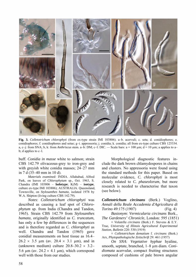
Morphological diagnostic features in-clude the dark brown chlamydospores in chains and clusters. No appressoria were found using the standard methods for this paper. Based on molecular evidence, C. chlorophyti is most closely related to C. phaseolorum, but more research is needed to characterise that taxon (see below). Colletotrichum circinans (Berk.) Voglino, Annali della Reale Accademia d'Agricoltura di Torino 49:175 (1907) (Fig. 4)
Basionym: Vermicularia circinans Berk., The Gardeners’ Chronicle, London: 595 (1851)
≡ Volutella circinans (Berk.) F. Stevens & E.Y. True, University of Illinois Agricultural Experimental Station, Bulletin 220: 530 (1919)
≡ Colletotrichum dematium f. circinans (Berk.) Arx, Phytopathologische Zeitschrift 29: 461 (1957)
On SNA: Vegetative hyphae hyaline, smooth, septate, branched, 1–8 µm diam. Coni-diomata acervular, compact fruiting structures composed of cushions of pale brown angular
Fungal Diversity
59
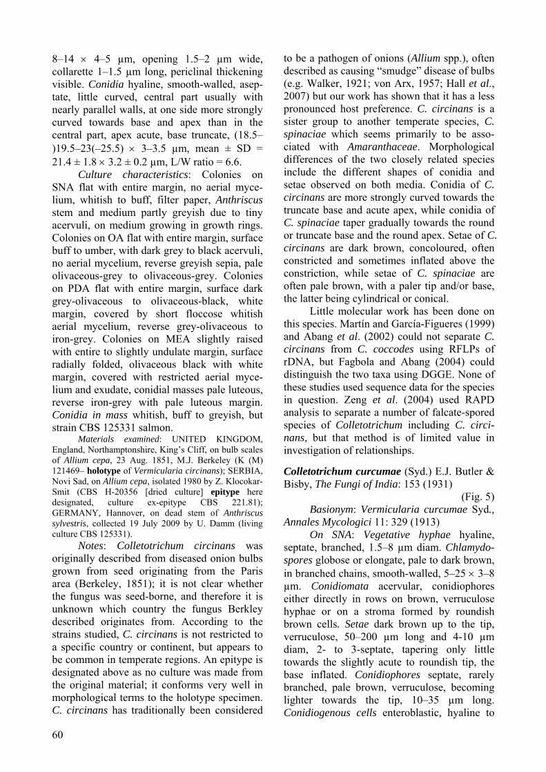
Fig. 4. Colletotrichum circinans (from ex-epitype strain CBS 221.81). a–b. acervuli; c. conidiophores with basal parts of setae; d–f. conidiophores; g. basal parts of setae; h. tips of setae; i–m. appressoria; n–o. conidia; a, c, e, f, n: from Anthriscus stem; b, d, g–m, o: from SNA. a–b: DM; c–o: DIC. — Scale bars: a = 200 µm; b = 100 µm; c = 10 µm; c applies to c–o. cells from which setae and conidiophores are produced. Setae dark brown, concoloured, smooth-walled to finely verruculose, 2- to 4-(5-) septate, (70–)100–180(–290) µm long, irregu-lar in length within acervulus, often one or few long setae with the rest much shorter, base constricted, sometimes slightly inflated above the constriction or cylindrical, 3.5–6(–9) µm diam, tip somewhat acute. Chlamydospores not observed. Conidiophores hyaline to pale brown, septate, branched, to 80 µm long. Conidioge-nous cells enteroblastic, hyaline to pale brown, cylindrical to clavate, 5–16 × 3–5 µm, opening 1–2 µm wide, collarette distinct, 1–1.5 µm long, periclinal thickening visible. Conidia hyaline, smooth-walled, aseptate, little curved, the central part often with nearly parallel walls, at one side more strongly curved towards base and apex than in the central part, apex acute, base truncate, (15–)19–23(–23.5) × (2.5–)3–
3.5(–4) µm, mean ± SD = 21.0 ± 1.8 × 3.4 ± 0.3 µm, L/W ratio = 6.2. Appressoria solitary, elongate elliptical to clavate, sometimes cre-nate or slightly lobed, smooth-walled, one- or two-celled, pale to mid brown, (7–)7–16(–24) × (3.5–)5–7.5(–11) µm, mean ± SD = 11.6 ± 4.4 × 6.1 ± 1.3 µm, L/W ratio = 1.9.
On Anthriscus stem: Conidiomata acer-vular, compact fruiting structures composed of cushions of pale brown angular cells from which setae and conidiophores are produced. Setae dark brown, concoloured, smooth to verruculose, 1- to 6-septate, 45–340 µm long, setae with very variable lengths within acervulus, base cylindrical or constricted, often inflated shortly above the constriction, 4–8 µm wide, tip somewhat acute. Conidiophores pale brown, simple to 2-septate, usually not branched, 10–30 µm long. Conidiogenous cells enteroblastic, pale brown, cylindrical to clavate,
60
8–14 × 4–5 µm, opening 1.5–2 µm wide, collarette 1–1.5 µm long, periclinal thickening visible. Conidia hyaline, smooth-walled, asep-tate, little curved, central part usually with nearly parallel walls, at one side more strongly curved towards base and apex than in the central part, apex acute, base truncate, (18.5–)19.5–23(–25.5) × 3–3.5 µm, mean ± SD = 21.4 ± 1.8 × 3.2 ± 0.2 µm, L/W ratio = 6.6.
Culture characteristics: Colonies on SNA flat with entire margin, no aerial myce-lium, whitish to buff, filter paper, Anthriscus stem and medium partly greyish due to tiny acervuli, on medium growing in growth rings. Colonies on OA flat with entire margin, surface buff to umber, with dark grey to black acervuli, no aerial mycelium, reverse greyish sepia, pale olivaceous-grey to olivaceous-grey. Colonies on PDA flat with entire margin, surface dark grey-olivaceous to olivaceous-black, white margin, covered by short floccose whitish aerial mycelium, reverse grey-olivaceous to iron-grey. Colonies on MEA slightly raised with entire to slightly undulate margin, surface radially folded, olivaceous black with white margin, covered with restricted aerial myce-lium and exudate, conidial masses pale luteous, reverse iron-grey with pale luteous margin. Conidia in mass whitish, buff to greyish, but strain CBS 125331 salmon.
Materials examined: UNITED KINGDOM, England, Northamptonshire, King’s Cliff, on bulb scales of Allium cepa, 23 Aug. 1851, M.J. Berkeley (K (M) 121469– holotype of Vermicularia circinans); SERBIA, Novi Sad, on Allium cepa, isolated 1980 by Z. Klocokar-Smit (CBS H-20356 [dried culture] epitype here designated, culture ex-epitype CBS 221.81); GERMANY, Hannover, on dead stem of Anthriscus sylvestris, collected 19 July 2009 by U. Damm (living culture CBS 125331).
Notes: Colletotrichum circinans was originally described from diseased onion bulbs grown from seed originating from the Paris area (Berkeley, 1851); it is not clear whether the fungus was seed-borne, and therefore it is unknown which country the fungus Berkley described originates from. According to the strains studied, C. circinans is not restricted to a specific country or continent, but appears to be common in temperate regions. An epitype is designated above as no culture was made from the original material; it conforms very well in morphological terms to the holotype specimen. C. circinans has traditionally been considered
to be a pathogen of onions (Allium spp.), often described as causing “smudge” disease of bulbs (e.g. Walker, 1921; von Arx, 1957; Hall et al., 2007) but our work has shown that it has a less pronounced host preference. C. circinans is a sister group to another temperate species, C. spinaciae which seems primarily to be asso-ciated with Amaranthaceae. Morphological differences of the two closely related species include the different shapes of conidia and setae observed on both media. Conidia of C. circinans are more strongly curved towards the truncate base and acute apex, while conidia of C. spinaciae taper gradually towards the round or truncate base and the round apex. Setae of C. circinans are dark brown, concoloured, often constricted and sometimes inflated above the constriction, while setae of C. spinaciae are often pale brown, with a paler tip and/or base, the latter being cylindrical or conical.
Little molecular work has been done on this species. Martín and García-Figueres (1999) and Abang et al. (2002) could not separate C. circinans from C. coccodes using RFLPs of rDNA, but Fagbola and Abang (2004) could distinguish the two taxa using DGGE. None of these studies used sequence data for the species in question. Zeng et al. (2004) used RAPD analysis to separate a number of falcate-spored species of Colletotrichum including C. circi-nans, but that method is of limited value in investigation of relationships. Colletotrichum curcumae (Syd.) E.J. Butler & Bisby, The Fungi of India: 153 (1931)
(Fig. 5) Basionym: Vermicularia curcumae Syd.,
Annales Mycologici 11: 329 (1913) On SNA: Vegetative hyphae hyaline,
septate, branched, 1.5–8 µm diam. Chlamydo-spores globose or elongate, pale to dark brown, in branched chains, smooth-walled, 5–25 × 3–8 µm. Conidiomata acervular, conidiophores either directly in rows on brown, verruculose hyphae or on a stroma formed by roundish brown cells. Setae dark brown up to the tip, verruculose, 50–200 µm long and 4-10 µm diam, 2- to 3-septate, tapering only little towards the slightly acute to roundish tip, the base inflated. Conidiophores septate, rarely branched, pale brown, verruculose, becoming lighter towards the tip, 10–35 µm long. Conidiogenous cells enteroblastic, hyaline to
Fungal Diversity
61
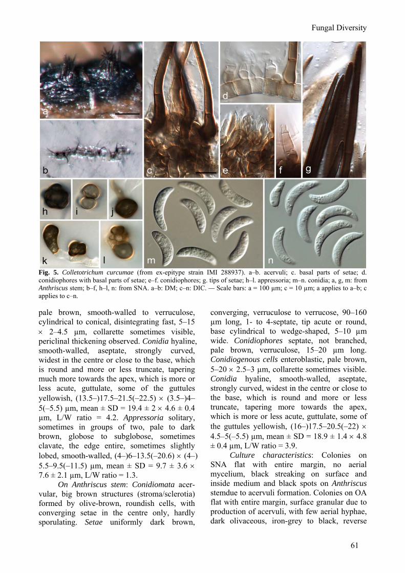
Fig. 5. Colletotrichum curcumae (from ex-epitype strain IMI 288937). a–b. acervuli; c. basal parts of setae; d. conidiophores with basal parts of setae; e–f. conidiophores; g. tips of setae; h–l. appressoria; m–n. conidia; a, g, m: from Anthriscus stem; b–f, h–l, n: from SNA. a–b: DM; c–n: DIC. — Scale bars: a = 100 µm; c = 10 µm; a applies to a–b; c applies to c–n. pale brown, smooth-walled to verruculose, cylindrical to conical, disintegrating fast, 5–15 × 2–4.5 µm, collarette sometimes visible, periclinal thickening observed. Conidia hyaline, smooth-walled, aseptate, strongly curved, widest in the centre or close to the base, which is round and more or less truncate, tapering much more towards the apex, which is more or less acute, guttulate, some of the guttules yellowish, (13.5–)17.5–21.5(–22.5) × (3.5–)4–5(–5.5) µm, mean ± SD = 19.4 ± 2 × 4.6 ± 0.4 µm, L/W ratio = 4.2. Appressoria solitary, sometimes in groups of two, pale to dark brown, globose to subglobose, sometimes clavate, the edge entire, sometimes slightly lobed, smooth-walled, (4–)6–13.5(–20.6) × (4–) 5.5–9.5(–11.5) µm, mean ± SD = 9.7 ± 3.6 × 7.6 ± 2.1 µm, L/W ratio = 1.3.
On Anthriscus stem: Conidiomata acer-vular, big brown structures (stroma/sclerotia) formed by olive-brown, roundish cells, with converging setae in the centre only, hardly sporulating. Setae uniformly dark brown,
converging, verruculose to verrucose, 90–160 µm long, 1- to 4-septate, tip acute or round, base cylindrical to wedge-shaped, 5–10 µm wide. Conidiophores septate, not branched, pale brown, verruculose, 15–20 µm long. Conidiogenous cells enteroblastic, pale brown, 5–20 × 2.5–3 µm, collarette sometimes visible. Conidia hyaline, smooth-walled, aseptate, strongly curved, widest in the centre or close to the base, which is round and more or less truncate, tapering more towards the apex, which is more or less acute, guttulate, some of the guttules yellowish, (16–)17.5–20.5(–22) × 4.5–5(–5.5) µm, mean ± SD = 18.9 ± 1.4 × 4.8 ± 0.4 µm, L/W ratio = 3.9.
Culture characteristics: Colonies on SNA flat with entire margin, no aerial mycelium, black streaking on surface and inside medium and black spots on Anthriscus stemdue to acervuli formation. Colonies on OA flat with entire margin, surface granular due to production of acervuli, with few aerial hyphae, dark olivaceous, iron-grey to black, reverse
62
pale olivaceous-grey to iron-grey. Conidia in mass white, greyish, yellowish to pale salmon.
Materials examined: INDIA. Tamil Nadu, Kistna, Angalur, on leaves of Curcuma longa, 24 Dec. 1912, W. McRae 24 (IMI 20994, K(M) – isotypes of Vermicularia curcumae Syd.); INDIA. Maharashtra, Warora, isol. ex Curcuma longa, 22 Aug. 1984, M.Y. Palarpawar 1 (IMI 288937 [dried culture], epitype here designated, culture ex-epitype IMI 288937). There is further material in IMI identified as this species isolated from leaves of Curcuma longa from Bangladesh and India (Uttar Pradesh, West Bengal).
Notes: Colletotrichum curcumae differs from all other species studied here by forming big brown flattened stromata on Anthriscus stems with straight setae that are aggregated in the centre and with little sporulation; in other species setae are diverging and formed all over the acervulus/stroma, and usually sporulation is abundant on that host material. The species appears to be at least largely confined to turmeric (Curcuma longa) but few strains have been sequenced of the C. dematium aggregate from South Asia. Palarpawar and Ghurde (1988) isolated strains that confirmed to C. curcumae in morphological features from plants surrounding turmeric fields including Brachiaria reptans, Cynodon dactylon, Sola-num xanthocarpum and Colocasia esculenta, and demonstrated their pathogenicity to Cur-cuma. Colletotrichum dematium (Pers.) Grove, Journal of Botany, British and Foreign, London 56: 341 (1918)
(Fig. 6) Basionym: Sphaeria dematium Pers.,
Synopsis methodica fungorum (Göttingen) 88 (1801)
≡ Exosporium dematium (Pers.) Link, in Willdenow, Willd., Sp. pl., Edn 4 6(2): 122 (1825)
≡ Vermicularia dematium (Pers.) Fr., Summa Vegetabilium Scandinaviae, Sectio Posterior: 420 (1849)
≡ Lasiella dematium (Pers.) Quél. Mémoires de la Société d’Émulation de Montbéliard, 2e Série, 5: 518 (1875)
= Vermicularia eryngii Desm., Plantes Cryptogames du Nord de la France, fasc. 11: 542 (1831)
≡ Colletotrichum eryngii (Desm.) Duke, Transactions of the British Mycological Society 13: 170 (1928)
On SNA: Vegetative hyphae <1–7 µm diam, hyaline, smooth-walled, septate, bran-ched. Chlamydospores in old cultures observed, in branched chains, dark brown, verrucose, single cells 6–13 × 5–8 µm, but not observed in
other strains. Conidiomata acervular, conidio-phores formed directly on hyphae. Setae not observed. Conidiophores hyaline to pale brown, septate, branched. Conidiogenous cells entero-blastic, hyaline to pale brown, cylindrical, 7–17 × 3–4 µm, opening 1–1.5 µm wide, collarette 0.5 µm long, periclinal thickening not observed. Conidia hyaline, smooth-walled, sometimes finely verruculose, aseptate, central part of conidia usually almost straight with parallel walls, bent apruptly to the roundish to acute apex and truncate base, giving the conidia an almost angular shape, (18–)20–23(–24) × 3–4(–5.5) µm, mean ± SD = 21.3 ± 1.5 × 3.5 ± 0.4 µm, L/W ratio = 6.1; other isolates form longer conidia, e.g. IMI 350847: 22.5–27.5 × 3–3.5 µm, while CBS 125340 did not sporulate on SNA. Appressoria solitary, elliptical to clavate or slightly lobed, brown, smooth-walled, aseptate, rarely septate, (2.5–)5–12(–18.5) × (2–)3–6.5(–8.5) µm, mean ± SD = 8.5 ± 3.5 × 4.8 ± 1.5 µm, L/W ratio = 1.8.
On Anthriscus stem: Conidiomata acer-vular, consisting of dark brown roundish cells from which setae (usually one seta per acervulus) and conidiophores develop. Setae straight, dark brown, 30-140 µm long, 3- to 8-septate, base cylindrical, conical or slightly inflated, 7–12 µm diam, tip acute. Conidio-phores hyaline to pale brown, septate, up to 20 µm long. Conidiogenous cells enteroblastic, hyaline to pale brown, cylindrical to elongate ampulliform, 4–15 × 3–5 µm, opening 0.5–1 µm wide, collarette or periclinal thickening not observed. Conidia hyaline, smooth-walled, aseptate, central part of conidia usually almost straight with parallel walls, bent apruptly to the roundish to acute apex and truncate base, giving the conidia an almost angular shape, (18.5–)20–22.5(–23.5) × 3–4 µm, mean ± SD = 21.3 ± 1.3 × 3.6 ± 0.3 µm, L/W ratio = 6.0; other isolates form longer conidia, e.g. IMI 350847: 21–26.5 × 3–4 µm and CBS 125340 12.5–26.5 × 3–4 µm.
Culture characteristics: Colonies on SNA flat with entire margin, surface of An-thriscus stem and filter paper partly covered by floccose white aerial mycelium, medium close to stem stained pale honey, margins of filter paper grey, 27–29 mm in 7 d at 20 °C. OA flat with entire margin, no aerial mycelium, surface
Fungal Diversity
63
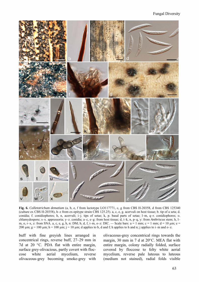
Fig. 6. Colletotrichum dematium (a, b, e, f from lectotype LO117771, c, g from CBS H-20358, d from CBS 125340 (culture ex CBS H-20358), h–z from ex-epitype strain CBS 125.25). a, c, e, g. acervuli on host tissue; b. tip of a seta; d. conidia; f. conidiophores; h, n, acervuli; i–j. tips of setae; k, p. basal parts of setae; l–m, q–r. conidiophores; o. chlamydospore; s–x. appressoria; y–z. conidia; a–c, e–g: from host tissue; d, i–k, n, p–q, y: from Anthriscus stem; h, l–m, o, r–x, z: from SNA. a, c, e, g, h, n: DM; b, d, f, i–m, o–z: DIC. — Scale bars: a = 1 mm; c = 1 mm; d = 10 µm; e = 200 µm; g = 100 µm; h = 100 µm; j = 10 µm; d applies to b, d and f; h applies to h and n; j applies to i–m and o–z. buff with fine greyish lines arranged in concentrical rings, reverse buff, 27–29 mm in 7d at 20 °C. PDA flat with entire margin, surface grey-olivacious, partly covert with floc-cose white aerial mycelium, reverse olivaceous-grey becoming smoke-grey with
olivaceous-grey concentrical rings towards the margin, 30 mm in 7 d at 20°C. MEA flat with entire margin, colony radially folded, surface covered by floccose to felty white aerial mycelium, reverse pale luteous to luteous (medium not stained), radial folds visible
64
delimited by whitish lines, 25 mm in 7 d at 20 °C. Conidia in mass greyish white.
Materials examined: FRANCE, on stem of Eryngium sp. (L 0117771 – syntype of Sphaeria dematium, here designated as lectotype); FRANCE, from dead leaf of Eryngium campestre, deposited in CBS by C. Killian in Dec. 1925 (CBS H-20357 [dried culture] epitype here designated, culture ex-epitype CBS 125.25); ITALY, Piemonte, unknown host (L 011772 syntype of Sphaeria dematium); UNKNOWN LOCA-TION, on stems of Solanum tuberosum (L 011772 syntype of Sphaeria dematium); UNKNOWN LOCA-TION, unknown host (K(M) syntype of Sphaeria dematium), CZECH REPUBLIC, Central Bohemia, Celakovice ca 30 km E of Prague, sandpits Malviny, on dead stem of Apiaceae, 20 Sept. 2009, M. Reblová (CBS H-20358, living culture CBS 125340); CZECH REPUBLIC, Central Bohemia, Celakovice ca 30 km E of Prague, sandpits Malviny, on dead stem of Apiaceae, 20 Sept. 2009, M. Reblová (CBS H-20359, living culture CBS 125341). AUSTRALIA, Northern Tasmania, from stem of Solanum tuberosum, deposited 1991 by L. Ransom (living culture IMI 350847); UNKNOWN LOCATION, unknown host (K(M) isotype of Vermicularia eryngii Desm.).
Notes: The original description of Sphaeria dematium by Persoon (1801) com-prises only a few observations: tiny, slightly flattened spheres on grey spots covered in the centre with erect, stiff, diverging, homochro-matic hairs/setae. The fungus is common on dead dry herbaceous stems, especially on Solanum tuberosum, while variety capreae occurs on Salix caprea. No type specimen was designated by Persoon. There are 20 specimens of Sphaeria dematium in the Persoon Herbarium in Leiden, 15 of them with Per-soon's handwriting, and two of them annotated with Syn. Fung. (= Synopsis Methodica Fungo-rum), where the description was published. With one of these two collections, specimen L0117771, host and locality was mentioned, Eryngium Gallia (Persoon wrote: Sphaeria dematium Syn. Fung., Eryngium Gallia, Exosporium dematium Link). There were no conidia found on the holotype material of Sphaeria dematium, but all structures observed, e.g. setae and conidiogenous cells, resemble those of the epitype, which is from the same host and location, and new collections of C. dematium from other Apiaceae from the Czech Republic. The symptoms found on the type material, on other herbarium specimens of S. dematium and of the new collections is similar (Fig. 6a, c) and differ from the symptoms caused by C. lineola (Fig. 9a, h). A further
specimen labelled as Sphaeria dematium in Persoon’s handwriting is stored in K(M) and could be part of the type material, but a note in another hand states that it contains “no fruit”. Examination of isotype material of Colletotri-chum eryngii (Desm.) Duke (Vermicularia eryngii Desm.) from K shows a very similar fungus to C. dematium and the two taxa are almost certainly synonymous. However, there is no living culture associated with authentic material of C. eryngii and epitypification would serve little purpose.
Typical features include the angular conidia, the production in many fresh cultures of red pigment, and the well-developed sclerotium-like conidiomata. There has been much confusion in the past regarding the separation of this species from Colletotrichum capsici, with differential characters cited by some authors (e.g. Sutton 1980) including conidial width but with others (e.g. von Arx 1957; Baxter et al., 1983) accepting a broader species concept. Mordue (1971) separated the two taxa using presence or absence of sclerotial structures. Its distribution is difficult to assess due to differing species concepts, but there is some suggestion that it occurs primarily in temperate rather than tropical zones. Some authors (e.g. Sutton 1962) maintain that the species is not a pathogen, developing exclusively on dead plant material from a wide range of species.
Colletotrichum dematium is claimed to cause several economically important diseases, such as leaf blight of Japanese radish (Raphanus sativus var. hortensis) seedlings (Sato et al., 2005), mulberry (Morus spp.) and cowpea (Vigna unguiculata) anthracnose (Smith et al., 1999; Yoshida and Shirata, 1999; Babu et al., 2008), spotting, blight and drop of leaves on potted plants of Polygonatum falcatum (Tomioka et al., 2008), and anthrac-nose of statice (Goniolimon tataricum) (Bobev et al., 2009). However, comparisons of ITS sequences of the causal organisms with sequences generated in this study (not shown) revealed that at least most of them do not belong to C. dematium as defined here. ITS sequences from a strain associated with Raphanus sativus var. hortensis (AB196295-AB196301) are identical to those of C. spae-thianum; ITS sequences of a fungus associated
Fungal Diversity
65
with Polygonatum falcatum (AB334523) differ in two nucleotides from C. spaethianum sequences; ITS sequences of a fungus associa-ted with Goniolimon tataricum (FJ236461- FJ236463) are similar to those of C. tofieldiae, while sequences of a fungus associated with Morus spp. (EU554165, EU4173) are different from the species studied here. One Canadian strain from strawberry (CBS 125344) belongs to C. lineola. However, it still needs to be confirmed that the causal organisms of straw-berry anthracnose in the USA and India (Beraha and Wright, 1973; Singh et al., 2003) belong to the same species. There is no sequence from cowpea anthracnose from South Africa available, but strains from Vigna that were included in our study, belong either to C. truncatum or to C. phaseolorum as it is originally described.
Few molecular studies have been pub-lished that include strains identified as C. dematium. Vinnere et al. (2002) included two strains in their study of Colletotrichum diseases of Rhododendron in Sweden, using sequences from rDNA, mtDNA and β-tubulin genes, but no attempt was made to establish the precise phylogeny. Their ITS sequences submitted to GenBank (AF411770, AF411773) suggest that the species they were studying was either C. dematium sensu stricto or C. lineola in our interpretations. Cano et al. (2004) investigated the relationships of Colletotri-chum species associated with clinical cases, which included sequences from two strains initially identified as C. dematium. One of these clustered with a sequence from CBS 351.73, identified at that time as C. truncatum but re-determined in this paper as C. circinans. The other is derived from a strain that is here designated as epitype of C. spaethianum (CBS 167.49). While the C. dematium strains used in that study originated from plants, there was one strain from a corneal ulcer of a human eye included in our study, that belongs to C. truncatum. Figures from case studies (Joseph et al., 2004; Kaliamurthy et al., 2004) might suggest the same species, but this needs to be examined more carefully. A study of five Colletotrichum species from India using RAPDs included strains identified as C. dema-tium and C. capsici (Wijesekara et al., 2005). The two taxa clustered together in their study,
but the true identity of their strains needs confirmation and RAPDs is not a good method for assessing relationships.
Colletotrichum dematium sensu stricto comprises only a few of the strains originally identified as C. dematium in our study, which could be assigned to 12 different species, namely C. circinans, C. dematium, C. lilii, C. lineola, C. liriopes, C. spaethianum, C. spina-ciae, C. tofieldiae, C. trichellum, C. truncatum and two unidentified species. But even with the reduced number of strains that could be shown to represent C. dematium in this study, it can be confirmed that C. dematium has a wide host range and can have pathogenic, saprobic and endophytic lifestyles. Colletotrichum fructi (F. Stevens & J.G. Hall) Sacc. [as 'fructus'], Sylloge fungorum (Abellini) 22: 1201 (1913) (Fig. 7)
Basionym. Volutella fructi F. Stevens & J.G. Hall, Journal of Mycology 13: 97 (1907)
≡ Vermicularia fructi (F. Stevens & J.G. Hall) Vassiljevsky [as 'fructus'], Fungi Imperfecti Parasitici 2: 351 (1950)
On SNA: Vegetative hyphae hyaline, septate, branched, smooth, 1.5–6 µm diam. Conidiomata acervular, with small clusters of hyaline to pale brown, roundish to angular cells, 3–6 µm diam, from which conidiophores and conidia are produced. Setae not (or rarely) formed on SNA. Chlamydospores not observed. Conidiophores hyaline, simple or septate, rare-ly branched, up to 30 µm. Conidiogenous cells enteroblastic, hyaline, cylindrical, occasionally ampulliform, 5–15 × 2–4(–10) µm, opening 0.5–1 µm diam, with collarette 1-2 µm long, periclinal thickening not observed. Conidia hyaline, aseptate, smooth-walled, central part of conidium almost straight with parallel walls, often bent apruptly to the apex giving the conidia an almost angular shape, apex narrow and acute, base usually broader and truncate (16.5–)20.5–24(–24.5) × (3–)3.5–4(–4.5) µm, mean ± SD = 22.3 ± 1.8 × 3.7 ± 0.3 µm, L/W ratio = 6.0. Appressoria solitary, elliptical to clavate, pale brown, smooth-walled, aseptate, (3.5–)5.5–8.5(–10.5) × (2–)3–4.5(–5) µm, mean ± SD = 6.9 ± 1.5 × 3.8 ± 0.7 µm, L/W ratio = 1.8.
On Anthriscus stem: Conidiomata acer-vular, forming roundish cushions of pale brown
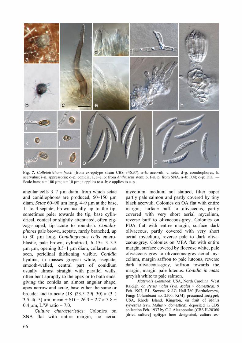
66
Fig. 7. Colletotrichum fructi (from ex-epitype strain CBS 346.37). a–b. acervuli; c. seta; d–g. conidiophores; h. acervulus; i–n. appressoria; o–p. conidia; a, c–e, o: from Anthriscus stem; b, f–n, p: from SNA. a–b: DM; c–p: DIC. — Scale bars: a = 100 µm; c = 10 µm; a applies to a–b; c applies to c–p. angular cells 3–7 µm diam, from which setae and conidiophores are produced, 50–150 µm diam. Setae 60–90 µm long, 4–9 µm at the base, 1- to 4-septate, brown usually up to the tip, sometimes paler towards the tip, base cylin-drical, conical or slightly attenuated, often zig-zag-shaped, tip acute to roundish. Conidio-phores pale brown, septate, rarely branched, up to 30 µm long. Conidiogenous cells entero-blastic, pale brown, cylindrical, 6–15× 3–3.5 µm µm, opening 0.5–1 µm diam, collarette not seen, periclinal thickening visible. Conidia hyaline, in masses greyish white, aseptate, smooth-walled, central part of conidium usually almost straight with parallel walls, often bent apruptly to the apex or to both ends, giving the conidia an almost angular shape, apex narrow and acute, base either the same or broader and truncate (18–)23.5–29(–30) × (3–) 3.5–4(–5) µm, mean ± SD = 26.3 ± 2.7 × 3.8 ± 0.4 µm, L/W ratio = 7.0.
Culture characteristics: Colonies on SNA flat with entire margin, no aerial
mycelium, medium not stained, filter paper partly pale salmon and partly covered by tiny black acervuli. Colonies on OA flat with entire margin, surface buff to olivaceous, partly covered with very short aerial mycelium, reverse buff to olivaceous-grey. Colonies on PDA flat with entire margin, surface dark olivaceous, partly covered with very short aerial mycelium, reverse pale to dark oliva-ceous-grey. Colonies on MEA flat with entire margin, surface covered by floccose white, pale olivaceous grey to olivaceous-grey aerial my-celium, margin saffron to pale luteous, reverse dark olivaceous-grey, saffron towards the margin, margin pale luteous. Conidia in mass greyish white to pale salmon.
Materials examined: USA, North Carolina, West Raleigh, on Pyrus malus (syn. Malus × domestica), 9 Feb. 1907, F.L. Stevens & J.G. Hall 780 (Bartholomew, Fungi Columbiani no. 2500; K(M), presumed isotype); USA, Rhode Island, Kingston, on fruit of Malus sylvestris (syn. Malus × domestica), deposited in CBS collection Feb. 1937 by C.J. Alexopoulos (CBS H-20360 [dried culture] epitype here designated, culture ex-
Fungal Diversity
67
epitype CBS 346.37 = CCT 4806). Notes: Walker (1925) noticed the
difference in conidium shape between C. circi-nans and C. fructi, which forms slightly angular conidia. The conidium shape is similar to that of C. dematium, however C. fructi is slower growing. The species has rarely been referred to in the literature, and it seems likely that any disease caused is of little economic importance. It was not investigated by Gonza-lez et al. (2006) in their study of Colleto-trichum species causing leaf spot and fruit rot of apple in North and South America.
Probable type material of C. fructi is stored in BPI; its status is doubtful as although the collection number is correctly cited, the collection date is after that given on the original publication (Stevens and Hall, 1907). No living culture is associated with that speci-men, and according to WFCC World Federa-tion for Culture Collections, (http://www .wfcc.info/datacenter.html) strain CBS 346.37 is the only strain of C. fructi in any public culture collection. The morphological charac-teristics of that strain are in concord with the illustration given in the original publication, and it originates from a closely related species in the same geographical region as the type. It is therefore an appropriate choice as epitype. Colletotrichum lilii Plakidas ex Boerema & Hamers, Netherlands Journal of Plant Pathology 94(suppl. 1): 12 (1988) (Fig. 8)
“Colletotrichum lilii” Plakidas, Phytopa-thology 34: 568 (1944), nom. inval. (Vienna Code, Art. 36.1).
≡. Vermicularia lilii (Plakidas ex Boerema & Hamers) Vassiljevsky, Fungi Imperfecti Parasitici 2: 346 (1950)
On SNA: Vegetative hyphae 1.5–5 µm diam, hyaline or pale brown, smooth-walled, septate, branched. Conidiomata acervular, conidiophores loosely arranged, no compact fruiting structures formed, with masses of pale salmon conidia. Setae smooth to finely verru-culose, 1- to 3-septate, 20–70 µm long, base cylindrical to conical, 3–5 µm diam, pale to medium brown up to the tip, tip acute to roundish. Chlamydospores not observed. Coni-diophores brown or very pale brown, septate, branched, filiform, up to 50 µm long. Conidio-genous cells enteroblastic, long cylindrical to elongate ampulliform, 7.5–20 × 2–3.5 µm,
opening 1.5–2 µm diam, collarette distinct, 1.5 (–2) µm long, periclinal thickening visible. Conidia hyaline, smooth-walled or verruculose, aseptate, very variable in size and shape: some strongly curved, more strongly curved towards the (often broadly) rounded apex than towards the truncate base, some small conidia almost straight, (9.5–)13–19.5(–33.5) × 3–4(–4.5) µm, mean ± SD = 16.2 ± 4.6 × 3.4 ± 0.4 µm, L/W ratio = 5.6. Appressoria solitary or in loose groups, 1-, sometimes 2-celled, dark brown, irregularly shaped, but often with clavate to somewhat triangular outline, strongly lobed, smooth-walled, (7.5–)10.5–19(–28.5) × (4.5–) 6–10(–14) µm, mean ± SD = 14.7 ± 4.4 × 8 ± 2.2 µm, L/W ratio = 1.8.
On Anthriscus stem: Conidiomata few, composed of pale brown angular cells, 4–7 µm diam. Setae pale to medium brown up to the tip, but often paler basal cell, verruculose, 30–150 µm, (mostly 50–100 µm) long, 1- to 4- (to 5-) septate, base cylindrical, conical, sometimes slightly inflated, 3–7 µm wide, tip more or less acute to roundish. Conidiophores pale brown, septate, branched, up to 50 (–80) µm long, smooth to verruculose. Conidiogenous cells enteroblastic, pale brown, cylindrical, occa-tionally ellipsoidal, smooth to finely verru-culose, 9–20 (–38) × 3.5–6 µm, opening 1.5–2 µm wide, collarette 0.5–1 µm long, periclinal thickening visible. Conidia hyaline, aseptate, smooth-walled curved, more strongly curved towards the more or less acute apex than towards the truncate base, (14.5–)16.5–19(–20) × 3–3.5(–4) µm, mean ± SD = 17.6 ± 1.3 ( 3.4 ± 0.3 µm, L/W ratio = 5.1.
Culture characteristics: Colonies on SNA flat with entire margin, short hyaline aerial mycelium on filter paper, Anthriscus stem, medium close to stem and under filter paper yellowish brown. Colonies on OA flat with entire margin, surface moist, no aerial mycelium, honey to isabelline with tiny darker brown dots, reverse hazel. Conidia in mass salmon.
Materials examined: JAPAN, unlocalised, on Lilium sp., deposited in CBS collection Jan. 2001 by H. Nirenberg (CBS H-20361 [dried culture], living culture CBS 109214 = BBA 62147).
Notes: This appears to be a host-specific pathogen of Lilium species, causing black scale disease of bulbs. It was originally described
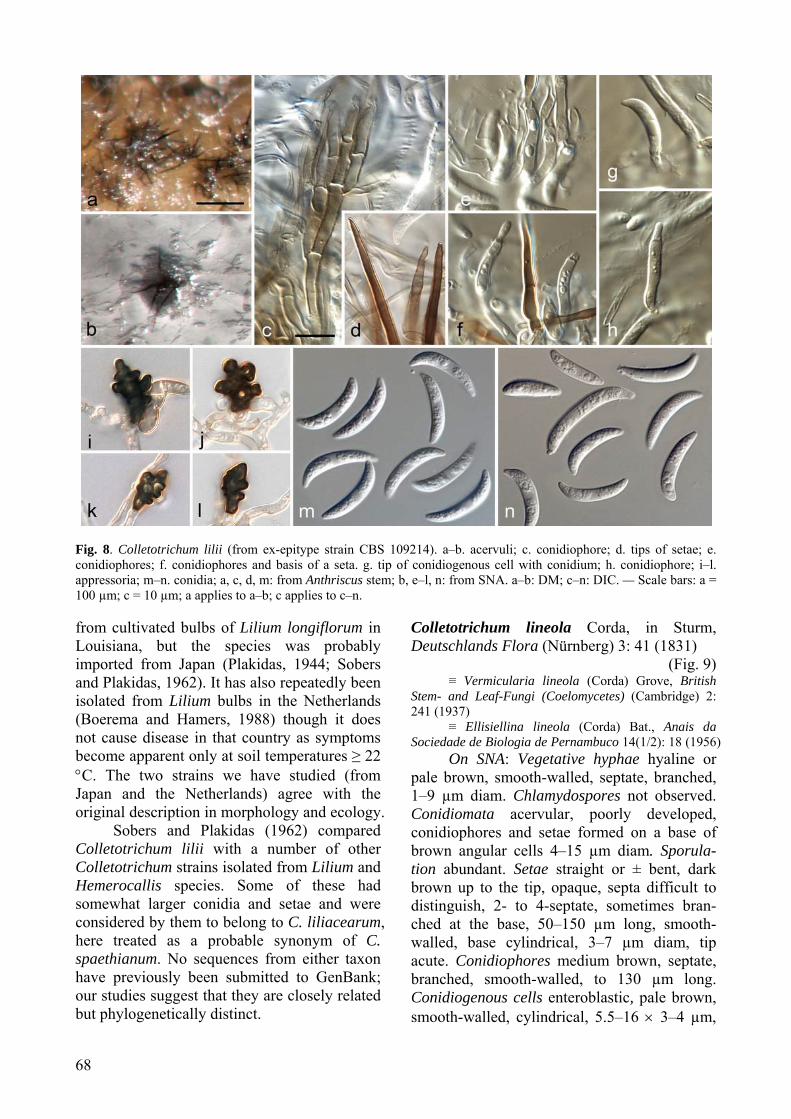
68
Fig. 8. Colletotrichum lilii (from ex-epitype strain CBS 109214). a–b. acervuli; c. conidiophore; d. tips of setae; e. conidiophores; f. conidiophores and basis of a seta. g. tip of conidiogenous cell with conidium; h. conidiophore; i–l. appressoria; m–n. conidia; a, c, d, m: from Anthriscus stem; b, e–l, n: from SNA. a–b: DM; c–n: DIC. — Scale bars: a = 100 µm; c = 10 µm; a applies to a–b; c applies to c–n. from cultivated bulbs of Lilium longiflorum in Louisiana, but the species was probably imported from Japan (Plakidas, 1944; Sobers and Plakidas, 1962). It has also repeatedly been isolated from Lilium bulbs in the Netherlands (Boerema and Hamers, 1988) though it does not cause disease in that country as symptoms become apparent only at soil temperatures ≥ 22 °C. The two strains we have studied (from Japan and the Netherlands) agree with the original description in morphology and ecology.
Sobers and Plakidas (1962) compared Colletotrichum lilii with a number of other Colletotrichum strains isolated from Lilium and Hemerocallis species. Some of these had somewhat larger conidia and setae and were considered by them to belong to C. liliacearum, here treated as a probable synonym of C. spaethianum. No sequences from either taxon have previously been submitted to GenBank; our studies suggest that they are closely related but phylogenetically distinct.
Colletotrichum lineola Corda, in Sturm, Deutschlands Flora (Nürnberg) 3: 41 (1831)
(Fig. 9) ≡ Vermicularia lineola (Corda) Grove, British
Stem- and Leaf-Fungi (Coelomycetes) (Cambridge) 2: 241 (1937)
≡ Ellisiellina lineola (Corda) Bat., Anais da Sociedade de Biologia de Pernambuco 14(1/2): 18 (1956)
On SNA: Vegetative hyphae hyaline or pale brown, smooth-walled, septate, branched, 1–9 µm diam. Chlamydospores not observed. Conidiomata acervular, poorly developed, conidiophores and setae formed on a base of brown angular cells 4–15 µm diam. Sporula-tion abundant. Setae straight or ± bent, dark brown up to the tip, opaque, septa difficult to distinguish, 2- to 4-septate, sometimes bran-ched at the base, 50–150 µm long, smooth-walled, base cylindrical, 3–7 µm diam, tip acute. Conidiophores medium brown, septate, branched, smooth-walled, to 130 µm long. Conidiogenous cells enteroblastic, pale brown, smooth-walled, cylindrical, 5.5–16 × 3–4 µm,
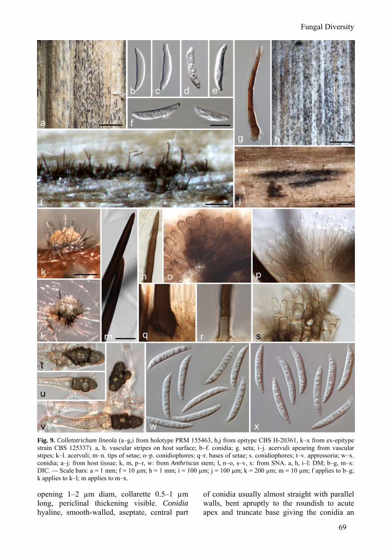
Fungal Diversity
69
Fig. 9. Colletotrichum lineola (a–g,i from holotype PRM 155463, h,j from epitype CBS H-20361, k–x from ex-epitype strain CBS 125337). a, h. vascular stripes on host surface; b–f. conidia; g. seta; i–j. acervuli apearing from vascular stipes; k–l. acervuli; m–n. tips of setae; o–p. conidiophores; q–r. bases of setae; s. conidiophores; t–v. appressoria; w–x. conidia; a–j: from host tissue; k, m, p–r, w: from Anthriscus stem; l, n–o, s–v, x: from SNA. a, h, i–l: DM; b–g, m–x: DIC. — Scale bars: a = 1 mm; f = 10 µm; h = 1 mm; i = 100 µm; j = 100 µm; k = 200 µm; m = 10 µm; f applies to b–g; k applies to k–l; m applies to m–x. opening 1–2 µm diam, collarette 0.5–1 µm long, periclinal thickening visible. Conidia hyaline, smooth-walled, aseptate, central part
of conidia usually almost straight with parallel walls, bent apruptly to the roundish to acute apex and truncate base giving the conidia an
70
almost angular shape, (21.5–)22.5–24.5(–25.5) × 3–3.5 (–4) µm, mean ± SD = 23.4 ± 0.9 × 3.4 ± 0.2 µm, L/W ratio = 6.8; strain CBS 125351 forms shorter conidia: 19–23 × 3–3.5 µm. Appressoria solitary, in small groups or short chains, medium to dark brown, smooth-walled, ellipsoidal to clavate, sometimes crenate or slightly lobed, (7.5–)7.5–16.5(–26) × (4–)5–9.5(–14) µm, mean ± SD = 12.0 ± 4.3 × 7.3 ± 2.1 µm, L/W ratio = 1.6.
On Anthriscus stem: Conidiomata acer-vular, conidiophores and setae formed on cushions of brown, angular cells, 3–9 µm diam. Setae straight, hyaline, pale to medium brown, hyaline towards the tip, smooth-walled or verruculose, 1- to 3-septate, often only septate at the base, 50–160 µm long, base cylindrical, conical or slightly inflated, 4–8 µm diam, tips of brown setae ± acute, tips of hyaline setae rounded. Conidiophores brown, septate, bran-ched, smooth-walled, to 30 µm long. Conidio-genous cells enteroblastic, pale brown, smooth-walled, cylindrical, 10–16 × 3–4 µm, opening 1.5–2 µm diam, collarette distinct, 0.5 µm long, periclinal thickening visible. Conidia hyaline, smooth-walled, aseptate, central part of conidia usually almost straight with parallel walls, bent apruptly to the acute apex and truncate base giving the conidia an almost angular shape, (21.5–)23–25(–25.5) × (3–)3.5–4 µm, mean ± SD = 23.9 ± 1.1 × 3.6 ± 0.2 µm, L/W ratio = 6.6; strain CBS 125351 forms shorter conidia: 18–23 × 3–4 µm.
Culture characteristics: Colonies on SNA flat with entire margin, medium hyaline slightly pale olivaceous to cinnamon, filter paper and Anthriscus stem covered with short olivaceous grey aerial mycelium and oliva-ceous grey to iron grey acervuli; 36–37 mm in 7 d. Colonies on OA flat with entire margin, surface smoke-grey to olivaceous-buff, covert with short, felty, pale olivaceous-grey aerial mycelium and iron-grey acervuli, reverse smoke-grey to olivaceous-buff; 33 mm in 7 d. Conidia in mass white, greyish white to salmon.
Materials examined: CZECH REPUBLIC, close to Prague, from stem of Apiaceae plant, late autum/winter 1829, A. J. Corda (PRM 155463, holotype of Colletotrichum lineola); CZECH REPUBLIC, Central Bohemia, Lazne Tousen (ca 25 km E of Prague), left bank of river Labe, from dead stem of Apiaceae plant, 20 Sep. 2009, M. Reblová (CBS H-20362 [dried specimen] epitype here designated, culture ex-epitype CBS
125337); CZECH REPUBLIC, same location, from dead stem of Apiaceae plant, 20 Sep. 2009, M. Reblová (CBS H-20363, living culture CBS 115339).
Notes: The structures found on the newly collected material designated as epitype and the ex-epitype strain (described above) resemble those on the holotype material, including a few conidia, measuring 15–23 × 3–4(–4.5) µm, setae and conidiogenous cells (Fig. 9a–g, i).
Based on our research, C. lineola is a widespread, primarily temperate species asso-ciated with a very wide range of plant species. It is characterised by small compressed acervuli emerging in rows/lines (“lineola”) on stems of the type host plant associated with short brown vascular stripes, while C. dema-tium forms big black spherical (“Sphaeria dematium”) stromatic acervuli in irregular groups on the host surface. Apart from these host-related differences we did not find consistent morphological distinctions between these two species, though red pigmentation in culture was only observed in C. lineola. Several strains (e.g. CBS 125339, CBS 109228, CBS 124959) released an apricot to coral pigment into the medium. Wollenweber and Hochapfel (1949) isolated single-spore strains of C. dematium s.l. from Heracleum pubescens with and without a red pigment that corres-ponded in all other features; these are likely to belong to C. lineola. The two species are however clearly divergent in sequence and occupy separate clades. Therefore we prefer to retain them as separate taxa, awaiting more detailed population studies verify if they represent two populations or two distinct species.
Colletotrichum lineola is the type species of the genus Colletotrichum, collected in late autumn 1829 on stems of a species of Apiaceae near Prague (Corda, 1831). The publication was issued in parts, and despite the title page of the volume bearing the date of 1837, the part containing Colletotrichum had already been indexed in the journal Flora in November 1831 (Stafleu and Cowan, 1986). The genus name was actually cited as Colletothrichum, and as the name is spelled in this way in the index and the following genus (Aseimothrichum) is formed in a similar manner, it would not be appropriate to assume that the spelling results from a misprint. There is an overwhelming
Fungal Diversity
71
case for conservation, and we do not recom-mend adoption of the original spelling.
Corda indicated that C. lineola had conidiomata in groups in a linear arrangement, setae and conidia in slime. No measurements were given. Grove (1937) transferred the species name to Vermicularia, based on a collection from sheaths and culms of Dactylis glomerata in Warwickshire, England. Although the name V. lineola is nomenclaturally an authentic homotypic synonym of C. lineola, Grove’s description of the Dactylis fungus is ambiguous in some respects and its taxonomic identity is in doubt.Batista (1956) transferred C. lineola to the genus Ellisiellina Sousa de Câmara, which is based on the species now commonly treated as Colletotrichum caudatum.
Colletotrichum liriopes Damm, P.F. Cannon & Crous, sp. nov. (Fig. 10) MycoBank: 514642
Etymology: Named after its host, Liriope. Colletotrichi tofieldiae simile, sed cellulis conidiogenis saepe valde inflatis, celeriter fatiscentibus, conidiis maioribus, in vitro (SNA) (10.5–)16–23.5(–25.5) x (2.5–) 3.5–4.5(–5) µm, in cultura cum caulibus Anthrisci (19–) 21.5–24.5(–27) x 3.5–4.5(–5) µm, appressoriis crenatioribus et lobatioribus, (9.5–)10.5–15(–17.5) x (6–) 7.5–11.5(–16) µm.
On SNA: Vegetative hyphae 1.5–5 µm diam, hyaline, smooth-walled, septate, bran-ched. Conidiomata acervular, conidiophores and rarely setae formed directly on hyphae. Setae brown up to the tip, 2- to 3-septate, 50–80 µm long, base conical to slightly inflated, 4–7 µm diam, tip acute. Chlamydospores not observed. Conidiophores hyaline to pale brown, septate, branched. Conidiogenous cells entero-blastic, hyaline, ampulliform or cylindrical, the cells often strongly inflated, disintegrating quickly, 8–15 × 3.5–5.5 µm, collarette 0.5–1.5 µm long, opening 1–2 µm diam, periclinal thickening distinct. Conidia hyaline, smooth-walled, rarely finely verruculose, aseptate, slightly curved, both sides gradually tapering towards the round to slightly acute apex and truncate base, (10.5–)16–23.5(–25.5) × (2.5–) 3.5–4.5(–5) µm, mean ± SD = 19.7 ± 3.6 × 4 ± 0.5 µm, L/W ratio = 5.0. Appressoria solitary or in loose groups, irregularly shaped, but often with a somewhat circular to elliptical outline, crenate to lobed, smooth-walled, aseptate, dark brown, (9.5–)10.5–15(–17.5) × (6–)7.5–11.5(–
16) µm, mean ± SD = 12.9 ± 2.3 × 9.4 ± 2.1 µm, L/W ratio = 1.4.
On Anthriscus stem. Conidiomata acer-vular, hyaline to pale brown cells, 3–7 µm diam, roundish to more or less globose, from which setae (few setae per acervulus) and conidiophores are produced. Setae light to medium brown, finely verruculose, 70–110 µm long, 2- to 4- septate, base conical to inflated, 4–7 µm diam, tip acute. Conidiophores hyaline to pale brown, not differentiated from basal cells. Conidiogenous cells enteroblastic, hya-line to pale brown, conical, subglobose to ellipsoidal, 3–10 × 2.5–5 µm, opening 1–2 µm diam, collarette 0.5–1 µm long, periclinal thickening visible. Conidia hyaline, smooth-walled, aseptate, slightly curved, both sides gradually tapering towards the round to slightly acute apex and truncate base, (19–)21.5–24.5(–27) × 3.5–4.5(–5) µm, mean ± SD = 23.1 ± 1.6 × 4.1 ± 0.4 µm, L/W ratio = 5.6.
Culture characteristics: Colonies on SNA flat with entire margin, no aerial my-celium, no pigmentation. Colonies on OA flat with entire margin, surface moist, no aerial mycelium, smoke-grey, honey to rosy-buff, reverse buff to very pale smoke-grey. Conidia in mass salmon.
Materials examined: MEXICO, APHIS interception Houstan 057263, on Liriope muscari, collected 29 Nov. 2000 by M.J. Segall, isolated 2000 by A.Y. Rossman (CBS H-20364, holotype, culture ex-type CBS 119444 = AR 3563).
Notes: This species is known only from two duplicate strains isolated from Liriope muscari, originating from Mexico and the result of a quarantine interception in Houston, USA. It belongs to a major clade that is almost completely confined to petaloid monoco-tyledon plants from the Liliales, primarily characterised morphologically by its appres-soria with complex outlines that are similar to those of C. lilii, but differs from it by the often strongly inflated conidiogenous cells. The same strain was included in a phylogenetic analysis of Colletotrichum species from Agavaceae by Farr et al. (2006), identified there as C. dematium. Colletotrichum phaseolorum S. Takim., An-nals of the Phytopathological Society of Japan 5: 21 (1934).
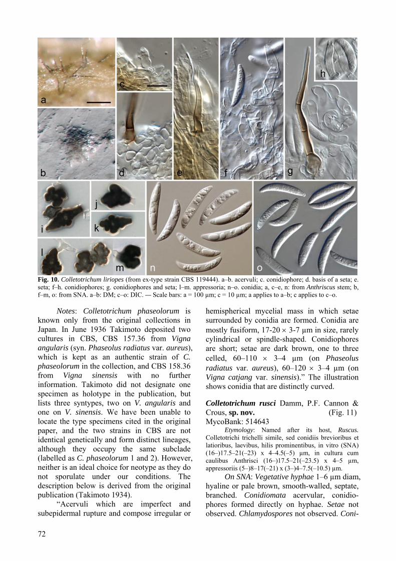
72
Fig. 10. Colletotrichum liriopes (from ex-type strain CBS 119444). a–b. acervuli; c. conidiophore; d. basis of a seta; e. seta; f–h. conidiophores; g. conidiophores and seta; l–m. appressoria; n–o. conidia; a, c–e, n: from Anthriscus stem; b, f–m, o: from SNA. a–b: DM; c–o: DIC. — Scale bars: a = 100 µm; c = 10 µm; a applies to a–b; c applies to c–o.
Notes: Colletotrichum phaseolorum is known only from the original collections in Japan. In June 1936 Takimoto deposited two cultures in CBS, CBS 157.36 from Vigna angularis (syn. Phaseolus radiatus var. aureus), which is kept as an authentic strain of C. phaseolorum in the collection, and CBS 158.36 from Vigna sinensis with no further information. Takimoto did not designate one specimen as holotype in the publication, but lists three syntypes, two on V. angularis and one on V. sinensis. We have been unable to locate the type specimens cited in the original paper, and the two strains in CBS are not identical genetically and form distinct lineages, although they occupy the same subclade (labelled as C. phaseolorum 1 and 2). However, neither is an ideal choice for neotype as they do not sporulate under our conditions. The description below is derived from the original publication (Takimoto 1934).
“Acervuli which are imperfect and subepidermal rupture and compose irregular or
hemispherical mycelial mass in which setae surrounded by conidia are formed. Conidia are mostly fusiform, 17-20 × 3-7 µm in size, rarely cylindrical or spindle-shaped. Conidiophores are short; setae are dark brown, one to three celled, 60–110 × 3–4 µm (on Phaseolus radiatus var. aureus), 60–120 × 3–4 µm (on Vigna catjang var. sinensis).” The illustration shows conidia that are distinctly curved. Colletotrichum rusci Damm, P.F. Cannon & Crous, sp. nov. (Fig. 11) MycoBank: 514643
Etymology: Named after its host, Ruscus. Colletotrichi trichelli simile, sed conidiis brevioribus et latioribus, laevibus, hilis prominentibus, in vitro (SNA) (16–)17.5–21(–23) x 4–4.5(–5) µm, in cultura cum caulibus Anthrisci (16–)17.5–21(–23.5) x 4–5 µm, appressoriis (5–)8–17(–21) x (3–)4–7.5(–10.5) µm.
On SNA: Vegetative hyphae 1–6 µm diam, hyaline or pale brown, smooth-walled, septate, branched. Conidiomata acervular, conidio-phores formed directly on hyphae. Setae not observed. Chlamydospores not observed. Coni-
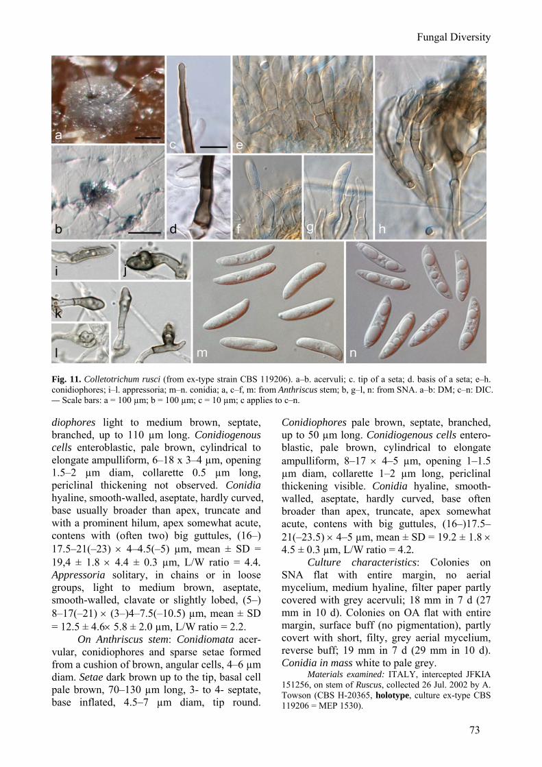
Fungal Diversity
73
Fig. 11. Colletotrichum rusci (from ex-type strain CBS 119206). a–b. acervuli; c. tip of a seta; d. basis of a seta; e–h. conidiophores; i–l. appressoria; m–n. conidia; a, c–f, m: from Anthriscus stem; b, g–l, n: from SNA. a–b: DM; c–n: DIC. — Scale bars: a = 100 µm; b = 100 µm; c = 10 µm; c applies to c–n. diophores light to medium brown, septate, branched, up to 110 µm long. Conidiogenous cells enteroblastic, pale brown, cylindrical to elongate ampulliform, 6–18 x 3–4 µm, opening 1.5–2 µm diam, collarette 0.5 µm long, periclinal thickening not observed. Conidia hyaline, smooth-walled, aseptate, hardly curved, base usually broader than apex, truncate and with a prominent hilum, apex somewhat acute, contens with (often two) big guttules, (16–) 17.5–21(–23) × 4–4.5(–5) µm, mean ± SD = 19,4 ± 1.8 × 4.4 ± 0.3 µm, L/W ratio = 4.4. Appressoria solitary, in chains or in loose groups, light to medium brown, aseptate, smooth-walled, clavate or slightly lobed, (5–) 8–17(–21) × (3–)4–7.5(–10.5) µm, mean ± SD = 12.5 ± 4.6× 5.8 ± 2.0 µm, L/W ratio = 2.2.
On Anthriscus stem: Conidiomata acer-vular, conidiophores and sparse setae formed from a cushion of brown, angular cells, 4–6 µm diam. Setae dark brown up to the tip, basal cell pale brown, 70–130 µm long, 3- to 4- septate, base inflated, 4.5–7 µm diam, tip round.
Conidiophores pale brown, septate, branched, up to 50 µm long. Conidiogenous cells entero-blastic, pale brown, cylindrical to elongate ampulliform, 8–17 × 4–5 µm, opening 1–1.5 µm diam, collarette 1–2 µm long, periclinal thickening visible. Conidia hyaline, smooth-walled, aseptate, hardly curved, base often broader than apex, truncate, apex somewhat acute, contens with big guttules, (16–)17.5–21(–23.5) × 4–5 µm, mean ± SD = 19.2 ± 1.8 × 4.5 ± 0.3 µm, L/W ratio = 4.2.
Culture characteristics: Colonies on SNA flat with entire margin, no aerial mycelium, medium hyaline, filter paper partly covered with grey acervuli; 18 mm in 7 d (27 mm in 10 d). Colonies on OA flat with entire margin, surface buff (no pigmentation), partly covert with short, filty, grey aerial mycelium, reverse buff; 19 mm in 7 d (29 mm in 10 d). Conidia in mass white to pale grey.
Materials examined: ITALY, intercepted JFKIA 151256, on stem of Ruscus, collected 26 Jul. 2002 by A. Towson (CBS H-20365, holotype, culture ex-type CBS 119206 = MEP 1530).
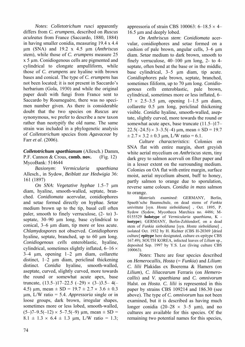
74
Notes: Colletotrichum rusci apparently differs from C. erumpens, described on Ruscus aculeatus from France (Saccardo, 1880, 1884) in having smaller conidia, measuring 19.4 x 4.4 µm (SNA) and 19.2 x 4.5 µm (Anthriscus stem), while those of C. erumpens measure 25 x 5 µm. Conidiogenous cells are pigmented and cylindrical to elongate ampulliform, while those of C. erumpens are hyaline with brown bases and conical. The type of C. erumpens has not been located; it is not present in Saccardo’s herbarium (Gola, 1930) and while the original paper dealt with fungi from France sent to Saccardo by Roumeguère, there was no speci-men number given. As there is considerable doubt that the two species on Ruscus are synonymous, we prefer to describe a new taxon rather than neotypify the old name. The same strain was included in a phylogenetic analysis of Colletotrichum species from Agavaceae by Farr et al. (2006). Colletotrichum spaethianum (Allesch.) Damm, P.F. Cannon & Crous, comb. nov. (Fig. 12) MycoBank: 514644
Basionym: Vermicularia spaethiana Allesch., in Sydow, Beiblatt zur Hedwigia 36: 161 (1897)
On SNA: Vegetative hyphae 1.5–7 µm diam, hyaline, smooth-walled, septate, bran-ched. Conidiomata acervular, conidiophores and setae formed directly on hyphae. Setae medium brown up to the tip, basal cell often paler, smooth to finely verruculose, (2- to) 3-septate, 30–90 µm long, base cylindrical to conical, 3–6 µm diam, tip more or less acute. Chlamydospores not observed. Conidiophores hyaline, septate, branched, up to 60 µm long. Conidiogenous cells enteroblastic, hyaline, cylindrical, sometimes slightly inflated, 6–16 × 3–4 µm, opening 1–2 µm diam, collarette distinct, 1–2 µm diam, periclinal thickening distinct. Conidia hyaline, smooth-walled, aseptate, curved, slightly curved, more towards the round or somewhat acute apex, base truncate, (13.5–)17–22.5 (–29) × (3–)3.5– 4(–4.5) µm, mean ± SD = 19.7 ± 2.7 × 3.6 ± 0.3 µm, L/W ratio = 5.4. Appressoria single or in loose groups, dark brown, irregular shapes, sometimes more or less lobed, smooth-walled, (5–)7–9.5(–12) × 5–7.5(–9) µm, mean ± SD = 8.1 ± 1.3 × 6.4 ± 1.3 µm, L/W ratio = 1.3;
appressoria of strain CBS 100063: 6–18.5 × 4–16.5 µm and deeply lobed.
On Anthriscus stem: Conidiomata acer-vular, conidiophores and setae formed on a cushion of pale brown, angular cells, 3–6 µm diam. Setae medium to dark brown, smooth to finely verruculose, 40–100 µm long, 2- to 4- septate, often bend at the base or in the middle, base cylindrical, 3–5 µm diam, tip acute. Conidiophores pale brown, septate, branched, sometimes filiform, up to 70 µm long. Conidio-genous cells enteroblastic, pale brown, cylindrical, sometimes more or less inflated, 6–17 × 2.5–3.5 µm, opening 1–1.5 µm diam, collarette 0.5 µm long, periclinal thickening visible. Conidia hyaline, smooth-walled, asep-tate, slightly curved, more towards the round or somewhat acute apex, base truncate (11.5–)17–22.5(–24.5) × 3–3.5(–4) µm, mean ± SD = 19.7 ± 2.7 × 3.2 ± 0.3 µm, L/W ratio = 6.1.
Culture characteristics: Colonies on SNA flat with entire margin, short greyish white aerial mycelium on Anthriscus stem, tiny dark grey to salmon acervuli on filter paper and in a lesser extent on the surrounding medium. Colonies on OA flat with entire margin, surface moist, aerial mycelium absent, buff to honey, partly salmon to orange due to sporulation, reverse same colours. Conidia in mass salmon to orange.
Materials examined: GERMANY, Berlin, Spaeth’sche Baumschule, on dead stems of Funkia univittata [syn. Hosta sieboldiana] , Oct. 1895, P. Sydow (Sydow, Mycotheca Marchica no. 4486; M-0155529 holotype of Vermicularia spaethiana, K - isotype); GERMANY, Berlin-Zehlendorf, on a dead stem of Funkia sieboldiana [syn. Hosta sieboldiana] , isolated Oct. 1932 by H. Richter (CBS H-20369 [dried culture] epitype here designated, culture ex-epitype CBS 167.49); SOUTH KOREA, infected leaves of Lilium sp., deposited Sep. 1997 by Y.S. Lee (living culture CBS 100063).
Notes: There are four species described on Hemerocallis, Hosta (= Funkia) and Lilium: C. lilii Plakidas ex Boerema & Hamers (on Lilium), C. liliacearum Ferraris (on Hemero-callis) and V. spaethiana and C. omnivorum Halst. on Hosta. C. lilii is represented in this paper by strains CBS 109214 and 186.30 (see above). The type of C. omnivorum has not been examined, but it is described as having much longer conidia (20–28 × 3–5 µm), and no cultures are available for this species. Of the remaining two potential names for this species,
Fungal Diversity
75
Fig. 12. Colletotrichum spaethianum (b, n–o. from ex-epitype strain CBS 167.49, a, c–n. from CBS 1100063). a–b. acervuli; c. seta; d–g. conidiophores; h. tip of a seta: i–m; appressoria; n–o. conidia; a, c–f, n: from Anthriscus stem; b, g–m, o: from SNA. a–b: DM; c–o: DIC. — Scale bars: a = 100 µm; c = 10 µm; a applies to a–b; c applies to c–o. V. spaethiana (Sydow 1897) was described before C. liliacearum Ferraris (1902). There is in addition a homonym of C. liliacearum Ferraris; C. liliacearum Duke, nom. nov., nom. illegit. (Duke, 1928), and V. liliacearum Schwein. (as “liliaceorum”; Schweinitz, 1832) could also be synonymous with C. spaethia-num. C. liliacearum Duke, nom. nov., nom. illegit. has been used quite widely for species on Liliaceae but it has not been studied in any detail in modern times and no type material has been examined.
The characters of the type of Vermi-cularia spaethiana, including conidia, agree in most respects with those of the material illustrated here. As far as we can tell, V. spae-thiana represents the earliest legitimate name for this species, so we make the necessary new combination and epitypify the name with a dried specimen for which a living culture is available. C. spaethianum was described from dead stems of Hosta sieboldiana in Berlin, Germany; strain 167.49 was collected from dead stems of the same host in the same city.
This species differs from the other four closely related species, which have also similar conidia shapes, mainly in setae that usually have an acute tip and cylindrical to conical base and appressoria with irregular outline that are more or less lobed but not crenate. Colletotrichum spinaciae Ellis & Halst., Journal of Mycology 6: 34 (1890) (Fig. 13)
≡ Vermicularia spinaciae (Ellis & Halst.) Vassiljevsky, Fungi imperfecti Parasitici 2: 339 (1950)
≡ Colletotrichum dematium f. spinaciae (Ellis & Halst.) Arx, Phytopathologische Zeitschrift 29(4): 460 (1957)
On SNA: Vegetative hyphae hyaline, smooth or verrucose, septate, branched, 1–8 µm diam. Conidiomata acervular, forming irregular masses of pale brown angular cells from which setae and conidiophores are produced, only a few acervuli on surface of medium, usually with no setae or only one seta per acervulus. Setae pale to medium brown, sometimes dark brown, base and tip sometimes lighter, finely verruculose, 30–90(–200) µm long, 2- to 3- septate, base cylindrical to
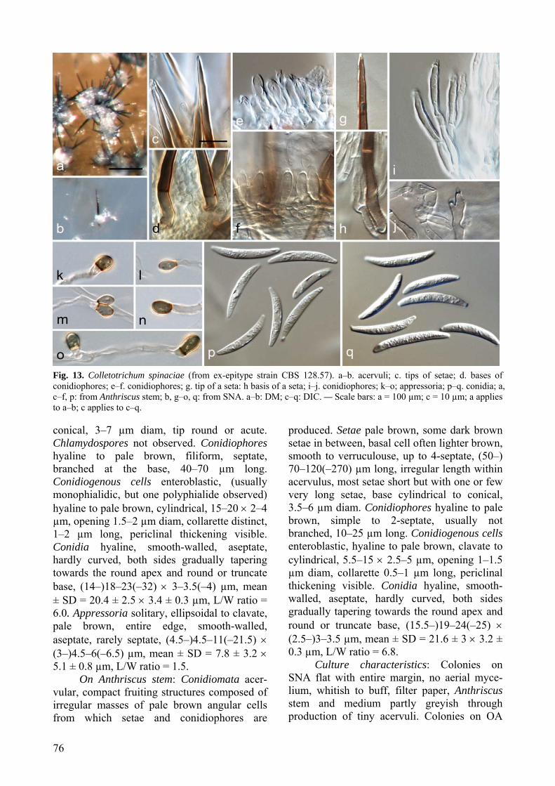
76
Fig. 13. Colletotrichum spinaciae (from ex-epitype strain CBS 128.57). a–b. acervuli; c. tips of setae; d. bases of conidiophores; e–f. conidiophores; g. tip of a seta: h basis of a seta; i–j. conidiophores; k–o; appressoria; p–q. conidia; a, c–f, p: from Anthriscus stem; b, g–o, q: from SNA. a–b: DM; c–q: DIC. — Scale bars: a = 100 µm; c = 10 µm; a applies to a–b; c applies to c–q. conical, 3–7 µm diam, tip round or acute. Chlamydospores not observed. Conidiophores hyaline to pale brown, filiform, septate, branched at the base, 40–70 µm long. Conidiogenous cells enteroblastic, (usually monophialidic, but one polyphialide observed) hyaline to pale brown, cylindrical, 15–20 × 2–4 µm, opening 1.5–2 µm diam, collarette distinct, 1–2 µm long, periclinal thickening visible. Conidia hyaline, smooth-walled, aseptate, hardly curved, both sides gradually tapering towards the round apex and round or truncate base, (14–)18–23(–32) × 3–3.5(–4) µm, mean ± SD = 20.4 ± 2.5 × 3.4 ± 0.3 µm, L/W ratio = 6.0. Appressoria solitary, ellipsoidal to clavate, pale brown, entire edge, smooth-walled, aseptate, rarely septate, (4.5–)4.5–11(–21.5) × (3–)4.5–6(–6.5) µm, mean ± SD = 7.8 ± 3.2 × 5.1 ± 0.8 µm, L/W ratio = 1.5.
On Anthriscus stem: Conidiomata acer-vular, compact fruiting structures composed of irregular masses of pale brown angular cells from which setae and conidiophores are
produced. Setae pale brown, some dark brown setae in between, basal cell often lighter brown, smooth to verruculouse, up to 4-septate, (50–) 70–120(–270) µm long, irregular length within acervulus, most setae short but with one or few very long setae, base cylindrical to conical, 3.5–6 µm diam. Conidiophores hyaline to pale brown, simple to 2-septate, usually not branched, 10–25 µm long. Conidiogenous cells enteroblastic, hyaline to pale brown, clavate to cylindrical, 5.5–15 × 2.5–5 µm, opening 1–1.5 µm diam, collarette 0.5–1 µm long, periclinal thickening visible. Conidia hyaline, smooth-walled, aseptate, hardly curved, both sides gradually tapering towards the round apex and round or truncate base, (15.5–)19–24(–25) × (2.5–)3–3.5 µm, mean ± SD = 21.6 ± 3 × 3.2 ± 0.3 µm, L/W ratio = 6.8.
Culture characteristics: Colonies on SNA flat with entire margin, no aerial myce-lium, whitish to buff, filter paper, Anthriscus stem and medium partly greyish through production of tiny acervuli. Colonies on OA
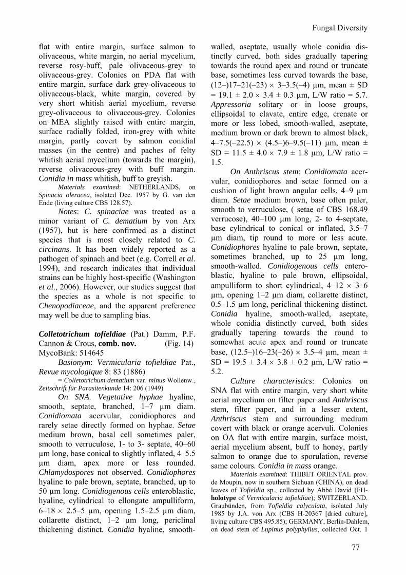
Fungal Diversity
77
flat with entire margin, surface salmon to olivaceous, white margin, no aerial mycelium, reverse rosy-buff, pale olivaceous-grey to olivaceous-grey. Colonies on PDA flat with entire margin, surface dark grey-olivaceous to olivaceous-black, white margin, covered by very short whitish aerial mycelium, reverse grey-olivaceous to olivaceous-grey. Colonies on MEA slightly raised with entire margin, surface radially folded, iron-grey with white margin, partly covert by salmon conidial masses (in the centre) and paches of felty whitish aerial mycelium (towards the margin), reverse olivaceous-grey with buff margin. Conidia in mass whitish, buff to greyish.
Materials examined: NETHERLANDS, on Spinacia oleracea, isolated Dec. 1957 by G. van den Ende (living culture CBS 128.57).
Notes: C. spinaciae was treated as a minor variant of C. dematium by von Arx (1957), but is here confirmed as a distinct species that is most closely related to C. circinans. It has been widely reported as a pathogen of spinach and beet (e.g. Correll et al. 1994), and research indicates that individual strains can be highly host-specific (Washington et al., 2006). However, our studies suggest that the species as a whole is not specific to Chenopodiaceae, and the apparent preference may well be due to sampling bias. Colletotrichum tofieldiae (Pat.) Damm, P.F. Cannon & Crous, comb. nov. (Fig. 14) MycoBank: 514645
Basionym: Vermicularia tofieldiae Pat., Revue mycologique 8: 83 (1886)
= Colletotrichum dematium var. minus Wollenw., Zeitschrift für Parasitenkunde 14: 206 (1949)
On SNA. Vegetative hyphae hyaline, smooth, septate, branched, 1–7 µm diam. Conidiomata acervular, conidiophores and rarely setae directly formed on hyphae. Setae medium brown, basal cell sometimes paler, smooth to verruculose, 1- to 3- septate, 40–60 µm long, base conical to slightly inflated, 4–5.5 µm diam, apex more or less rounded. Chlamydospores not observed. Conidiophores hyaline to pale brown, septate, branched, up to 50 µm long. Conidiogenous cells enteroblastic, hyaline, cylindrical to ellongate ampulliform, 6–18 × 2.5–5 µm, opening 1.5–2.5 µm diam, collarette distinct, 1–2 µm long, periclinal thickening distinct. Conidia hyaline, smooth-
walled, aseptate, usually whole conidia dis-tinctly curved, both sides gradually tapering towards the round apex and round or truncate base, sometimes less curved towards the base, (12–)17–21(–23) × 3–3.5(–4) µm, mean ± SD = 19.1 ± 2.0 × 3.4 ± 0.3 µm, L/W ratio = 5.7. Appressoria solitary or in loose groups, ellipsoidal to clavate, entire edge, crenate or more or less lobed, smooth-walled, aseptate, medium brown or dark brown to almost black, 4–7.5(–22.5) × (4.5–)6–9.5(–11) µm, mean ± SD = 11.5 ± 4.0 × 7.9 ± 1.8 µm, L/W ratio = 1.5.
On Anthriscus stem: Conidiomata acer-vular, conidiophores and setae formed on a cushion of light brown angular cells, 4–9 µm diam. Setae medium brown, base often paler, smooth to verruculose, ( setae of CBS 168.49 verrucose), 40–100 µm long, 2- to 4-septate, base cylindrical to conical or inflated, 3.5–7 µm diam, tip round to more or less acute. Conidiophores hyaline to pale brown, septate, sometimes branched, up to 25 µm long, smooth-walled. Conidiogenous cells entero-blastic, hyaline to pale brown, ellipsoidal, ampulliform to short cylindrical, 4–12 × 3–6 µm, opening 1–2 µm diam, collarette distinct, 0.5–1.5 µm long, periclinal thickening distinct. Conidia hyaline, smooth-walled, aseptate, whole conidia distinctly curved, both sides gradually tapering towards the round to somewhat acute apex and round or truncate base, (12.5–)16–23(–26) × 3.5–4 µm, mean ± SD = 19.5 ± 3.4 × 3.8 ± 0.2 µm, L/W ratio = 5.2.
Culture characteristics: Colonies on SNA flat with entire margin, very short white aerial mycelium on filter paper and Anthriscus stem, filter paper, and in a lesser extent, Anthriscus stem and surrounding medium covert with black or orange acervuli. Colonies on OA flat with entire margin, surface moist, aerial mycelium absent, buff to honey, partly salmon to orange due to sporulation, reverse same colours. Conidia in mass orange.
Materials examined: THIBET ORIENTAL prov. de Moupin, now in southern Sichuan (CHINA), on dead leaves of Tofieldia sp., collected by Abbé David (FH-holotype of Vermicularia tofieldiae); SWITZERLAND. Graubünden, from Tofieldia calyculata, isolated July 1985 by J.A. von Arx (CBS H-20367 [dried culture], living culture CBS 495.85); GERMANY, Berlin-Dahlem, on dead stem of Lupinus polyphyllus, collected Oct. 1
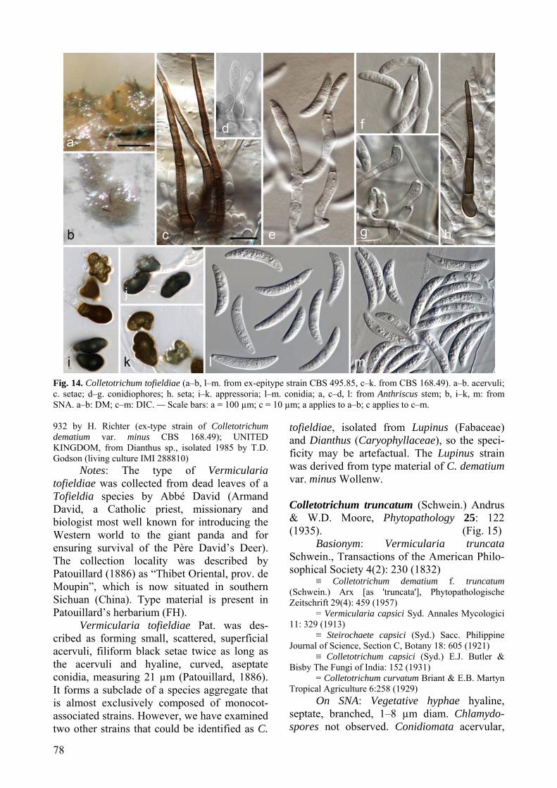
78
Fig. 14. Colletotrichum tofieldiae (a–b, l–m. from ex-epitype strain CBS 495.85, c–k. from CBS 168.49). a–b. acervuli; c. setae; d–g. conidiophores; h. seta; i–k. appressoria; l–m. conidia; a, c–d, l: from Anthriscus stem; b, i–k, m: from SNA. a–b: DM; c–m: DIC. — Scale bars: a = 100 µm; c = 10 µm; a applies to a–b; c applies to c–m. 932 by H. Richter (ex-type strain of Colletotrichum dematium var. minus CBS 168.49); UNITED KINGDOM, from Dianthus sp., isolated 1985 by T.D. Godson (living culture IMI 288810)
Notes: The type of Vermicularia tofieldiae was collected from dead leaves of a Tofieldia species by Abbé David (Armand David, a Catholic priest, missionary and biologist most well known for introducing the Western world to the giant panda and for ensuring survival of the Père David’s Deer). The collection locality was described by Patouillard (1886) as “Thibet Oriental, prov. de Moupin”, which is now situated in southern Sichuan (China). Type material is present in Patouillard’s herbarium (FH).
Vermicularia tofieldiae Pat. was des-cribed as forming small, scattered, superficial acervuli, filiform black setae twice as long as the acervuli and hyaline, curved, aseptate conidia, measuring 21 µm (Patouillard, 1886). It forms a subclade of a species aggregate that is almost exclusively composed of monocot-associated strains. However, we have examined two other strains that could be identified as C.
tofieldiae, isolated from Lupinus (Fabaceae) and Dianthus (Caryophyllaceae), so the speci-ficity may be artefactual. The Lupinus strain was derived from type material of C. dematium var. minus Wollenw. Colletotrichum truncatum (Schwein.) Andrus & W.D. Moore, Phytopathology 25: 122 (1935). (Fig. 15)
Basionym: Vermicularia truncata Schwein., Transactions of the American Philo-sophical Society 4(2): 230 (1832)
≡ Colletotrichum dematium f. truncatum (Schwein.) Arx [as 'truncata'], Phytopathologische Zeitschrift 29(4): 459 (1957)
= Vermicularia capsici Syd. Annales Mycologici 11: 329 (1913)
≡ Steirochaete capsici (Syd.) Sacc. Philippine Journal of Science, Section C, Botany 18: 605 (1921)
≡ Colletotrichum capsici (Syd.) E.J. Butler & Bisby The Fungi of India: 152 (1931)
= Colletotrichum curvatum Briant & E.B. Martyn Tropical Agriculture 6:258 (1929)
On SNA: Vegetative hyphae hyaline, septate, branched, 1–8 µm diam. Chlamydo-spores not observed. Conidiomata acervular,
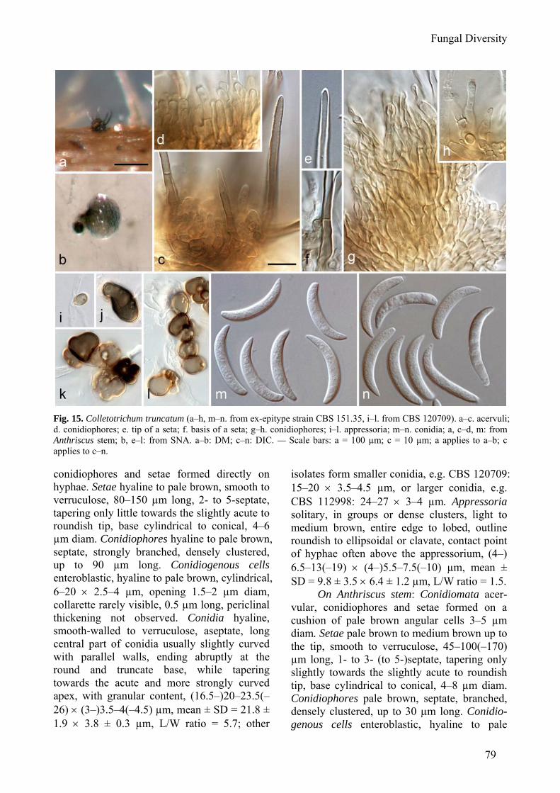
Fungal Diversity
79
Fig. 15. Colletotrichum truncatum (a–h, m–n. from ex-epitype strain CBS 151.35, i–l. from CBS 120709). a–c. acervuli; d. conidiophores; e. tip of a seta; f. basis of a seta; g–h. conidiophores; i–l. appressoria; m–n. conidia; a, c–d, m: from Anthriscus stem; b, e–l: from SNA. a–b: DM; c–n: DIC. — Scale bars: a = 100 µm; c = 10 µm; a applies to a–b; c applies to c–n. conidiophores and setae formed directly on hyphae. Setae hyaline to pale brown, smooth to verruculose, 80–150 µm long, 2- to 5-septate, tapering only little towards the slightly acute to roundish tip, base cylindrical to conical, 4–6 µm diam. Conidiophores hyaline to pale brown, septate, strongly branched, densely clustered, up to 90 µm long. Conidiogenous cells enteroblastic, hyaline to pale brown, cylindrical, 6–20 × 2.5–4 µm, opening 1.5–2 µm diam, collarette rarely visible, 0.5 µm long, periclinal thickening not observed. Conidia hyaline, smooth-walled to verruculose, aseptate, long central part of conidia usually slightly curved with parallel walls, ending abruptly at the round and truncate base, while tapering towards the acute and more strongly curved apex, with granular content, (16.5–)20–23.5(–26) × (3–)3.5–4(–4.5) µm, mean ± SD = 21.8 ± 1.9 × 3.8 ± 0.3 µm, L/W ratio = 5.7; other
isolates form smaller conidia, e.g. CBS 120709: 15–20 × 3.5–4.5 µm, or larger conidia, e.g. CBS 112998: 24–27 × 3–4 µm. Appressoria solitary, in groups or dense clusters, light to medium brown, entire edge to lobed, outline roundish to ellipsoidal or clavate, contact point of hyphae often above the appressorium, (4–) 6.5–13(–19) × (4–)5.5–7.5(–10) µm, mean ± SD = 9.8 ± 3.5 × 6.4 ± 1.2 µm, L/W ratio = 1.5.
On Anthriscus stem: Conidiomata acer-vular, conidiophores and setae formed on a cushion of pale brown angular cells 3–5 µm diam. Setae pale brown to medium brown up to the tip, smooth to verruculose, 45–100(–170) µm long, 1- to 3- (to 5-)septate, tapering only slightly towards the slightly acute to roundish tip, base cylindrical to conical, 4–8 µm diam. Conidiophores pale brown, septate, branched, densely clustered, up to 30 µm long. Conidio-genous cells enteroblastic, hyaline to pale
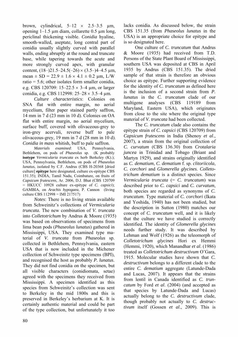
80
brown, cylindrical, 5–12 × 2.5–3.5 µm, opening 1–1.5 µm diam, collarette 0.5 µm long, periclinal thickening visible. Conidia hyaline, smooth-walled, aseptate, long central part of conidia usually slightly curved with parallel walls, ending abruptly at the round and truncate base, while tapering towards the acute and more strongly curved apex, with granular content, (18–)21.5–24.5(–26) × (3.5–)4–4.5 µm, mean ± SD = 22.9 ± 1.6 × 4.1 ± 0.2 µm, L/W ratio = 5.6; other isolates form smaller conidia, e.g. CBS 120709: 15–22.5 × 3–4 µm, or larger conidia, e.g. CBS 112998: 25–28 × 3.5–4 µm.
Culture characteristics: Colonies on SNA flat with entire margin, no aerial mycelium, filter paper stained partly saffron, 14 mm in 7 d (23 mm in 10 d). Colonies on OA flat with entire margin, no aerial mycelium, surface buff, covered with olivaceous-grey to iron-grey acervuli, reverse buff to pale olivaceous-grey, 19 mm in 7 d (28 mm in 10 d). Conidia in mass whitish, buff to pale saffron.
Materials examined: USA, Pennsylvania, Bethlehem, on pods of Phaseolus sp. (K probably isotype Vermicularia truncata ex herb Berkeley (K).); USA, Pennsylvania, Bethlehem, on pods of Phaseolus lunatus, isolated by C.F. Andrus (CBS H-20368 [dried culture] epitype here designated, culture ex-epitype CBS 151.35); INDIA, Tamil Nadu, Coimbature, on fruits of Capsicum frutescens, Ar. 2006, D.J. Bhat (CBS 120709 = HKUCC 10928 culture ex-epitype of C. capsici); GAMBIA, on Arachis hypogaea, P. Cannon (living culture CBS 112998 = IMI 217517)
Notes: There is no living strain available from Schweinitz’s collections of Vermicularia truncata. The new combination of V. truncata into Colletotrichum by Andrus & Moore (1935) was based on observations of specimens from lima bean pods (Phaseolus lunatus) gathered in Mississippi, USA. They examined type ma-terial of V. truncata from Phaseolus sp. collected in Bethlehem, Pennsylvania, eastern USA that is now included in the Michener collection of Schweinitz type specimens (BPI), and recognised the host as probably P. lunatus. They did not find conidia on the specimen, but all visible characters (conidiomata, setae) agreed with the specimens they received from Mississippi. A specimen identified as this species from Schweinitz’s collection was sent to Berkeley in the mid 1800s and this is preserved in Berkeley’s herbarium at K. It is certainly authentic material and could be part of the type collection, but unfortunately it too
lacks conidia. As discussed below, the strain CBS 151.35 (from Phaseolus lunatus in the USA) is an appropriate choice for epitype and is so designated here.
One culture of C. truncatum that Andrus & Moore (1935) had received from T.D. Persons of the State Plant Board of Mississippi, southern USA was deposited at CBS in April 1935 by Andrus (CBS 151.35). The dried sample of that strain is therefore an obvious choice as epitype. Further supporting evidence for the identity of C. truncatum as defined here is the inclusion of a second strain from P. lunatus in the C. truncatum clade of our multigene analyses (CBS 119189 from Maryland, Eastern USA), which originates from close to the site where the original type material of V. truncata had been collected.
The C. truncatum clade also contains the epitype strain of C. capsici (CBS 120709) from Capsicum frutescens in India (Shenoy et al., 2007), a strain from the original collection of C. curvatum (CBS 136.30) from Crotalaria juncea in Trinidad and Tobago (Briant and Martyn 1929), and strains originally identified as C. dematium, C. dematium f. sp. clitoriicola, C. corchori and Glomerella glycines. Colleto-trichum dematium is a distinct species. Since Vermicularia truncata (= C. truncatum) was described prior to C. capsici and C. curvatum, both species are regarded as synonyms of C. truncatum. Type material of C. corchori (Ikata and Yoshida, 1940) has not been studied, but the description in Sutton (1980) matches our concept of C. truncatum well, and it is likely that the culture we have studied is correctly identified. The identity of Glomerella glycines needs further study. It was described by Lehman and Wolf (1926) as the teleomorph of Colletotrichum glycines Hori ex Hemmi (Hemmi, 1920), which Manandhar et al. (1986) treated as Colletotrichum destructivum O’Gara, 1915. Molecular studies have shown that C. destructivum belongs to a different clade to the entire C. dematium aggregate (Latunde-Dada and Lucas, 2007). It appears that the strains from lentil in Canada identified as C. trun-catum by Ford et al. (2004) (and accepted as that species by Latunde-Dada and Lucas) actually belong to the C. destructivum clade, though probably not actually to C. destruc-tivum itself (Gossen et al., 2009). This is
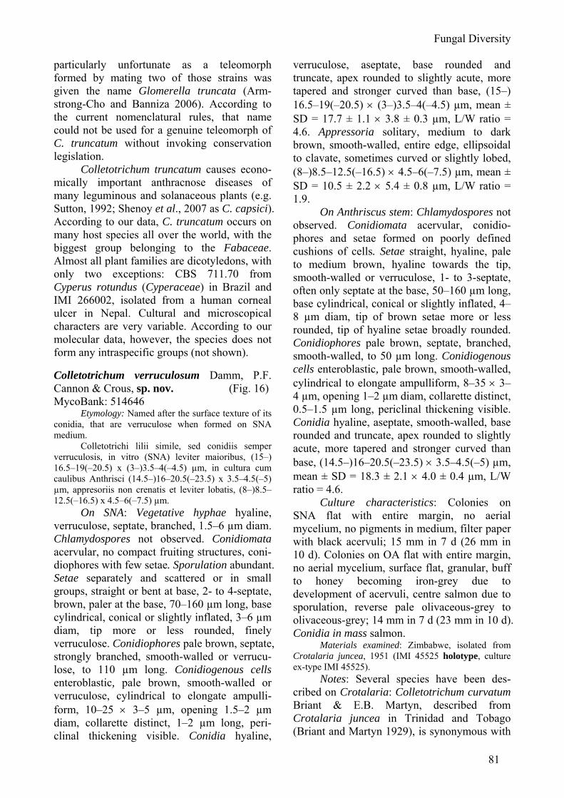
Fungal Diversity
81
particularly unfortunate as a teleomorph formed by mating two of those strains was given the name Glomerella truncata (Arm-strong-Cho and Banniza 2006). According to the current nomenclatural rules, that name could not be used for a genuine teleomorph of C. truncatum without invoking conservation legislation.
Colletotrichum truncatum causes econo-mically important anthracnose diseases of many leguminous and solanaceous plants (e.g. Sutton, 1992; Shenoy et al., 2007 as C. capsici). According to our data, C. truncatum occurs on many host species all over the world, with the biggest group belonging to the Fabaceae. Almost all plant families are dicotyledons, with only two exceptions: CBS 711.70 from Cyperus rotundus (Cyperaceae) in Brazil and IMI 266002, isolated from a human corneal ulcer in Nepal. Cultural and microscopical characters are very variable. According to our molecular data, however, the species does not form any intraspecific groups (not shown). Colletotrichum verruculosum Damm, P.F. Cannon & Crous, sp. nov. (Fig. 16) MycoBank: 514646
Etymology: Named after the surface texture of its conidia, that are verruculose when formed on SNA medium.
Colletotrichi lilii simile, sed conidiis semper verruculosis, in vitro (SNA) leviter maioribus, (15–) 16.5–19(–20.5) x (3–)3.5–4(–4.5) µm, in cultura cum caulibus Anthrisci (14.5–)16–20.5(–23.5) x 3.5–4.5(–5) µm, appresoriis non crenatis et leviter lobatis, (8–)8.5–12.5(–16.5) x 4.5–6(–7.5) µm.
On SNA: Vegetative hyphae hyaline, verruculose, septate, branched, 1.5–6 µm diam. Chlamydospores not observed. Conidiomata acervular, no compact fruiting structures, coni-diophores with few setae. Sporulation abundant. Setae separately and scattered or in small groups, straight or bent at base, 2- to 4-septate, brown, paler at the base, 70–160 µm long, base cylindrical, conical or slightly inflated, 3–6 µm diam, tip more or less rounded, finely verruculose. Conidiophores pale brown, septate, strongly branched, smooth-walled or verrucu-lose, to 110 µm long. Conidiogenous cells enteroblastic, pale brown, smooth-walled or verruculose, cylindrical to elongate ampulli-form, 10–25 × 3–5 µm, opening 1.5–2 µm diam, collarette distinct, 1–2 µm long, peri-clinal thickening visible. Conidia hyaline,
verruculose, aseptate, base rounded and truncate, apex rounded to slightly acute, more tapered and stronger curved than base, (15–) 16.5–19(–20.5) × (3–)3.5–4(–4.5) µm, mean ± SD = 17.7 ± 1.1 × 3.8 ± 0.3 µm, L/W ratio = 4.6. Appressoria solitary, medium to dark brown, smooth-walled, entire edge, ellipsoidal to clavate, sometimes curved or slightly lobed, (8–)8.5–12.5(–16.5) × 4.5–6(–7.5) µm, mean ± SD = 10.5 ± 2.2 × 5.4 ± 0.8 µm, L/W ratio = 1.9.
On Anthriscus stem: Chlamydospores not observed. Conidiomata acervular, conidio-phores and setae formed on poorly defined cushions of cells. Setae straight, hyaline, pale to medium brown, hyaline towards the tip, smooth-walled or verruculose, 1- to 3-septate, often only septate at the base, 50–160 µm long, base cylindrical, conical or slightly inflated, 4–8 µm diam, tip of brown setae more or less rounded, tip of hyaline setae broadly rounded. Conidiophores pale brown, septate, branched, smooth-walled, to 50 µm long. Conidiogenous cells enteroblastic, pale brown, smooth-walled, cylindrical to elongate ampulliform, 8–35 × 3–4 µm, opening 1–2 µm diam, collarette distinct, 0.5–1.5 µm long, periclinal thickening visible. Conidia hyaline, aseptate, smooth-walled, base rounded and truncate, apex rounded to slightly acute, more tapered and stronger curved than base, (14.5–)16–20.5(–23.5) × 3.5–4.5(–5) µm, mean ± SD = 18.3 ± 2.1 × 4.0 ± 0.4 µm, L/W ratio = 4.6.
Culture characteristics: Colonies on SNA flat with entire margin, no aerial mycelium, no pigments in medium, filter paper with black acervuli; 15 mm in 7 d (26 mm in 10 d). Colonies on OA flat with entire margin, no aerial mycelium, surface flat, granular, buff to honey becoming iron-grey due to development of acervuli, centre salmon due to sporulation, reverse pale olivaceous-grey to olivaceous-grey; 14 mm in 7 d (23 mm in 10 d). Conidia in mass salmon.
Materials examined: Zimbabwe, isolated from Crotalaria juncea, 1951 (IMI 45525 holotype, culture ex-type IMI 45525).
Notes: Several species have been des-cribed on Crotalaria: Colletotrichum curvatum Briant & E.B. Martyn, described from Crotalaria juncea in Trinidad and Tobago (Briant and Martyn 1929), is synonymous with
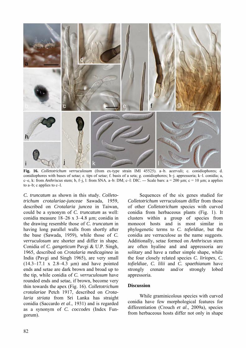
82
Fig. 16. Colletotrichum verruculosum (from ex-type strain IMI 45525). a–b. acervuli; c. conidiophores; d. conidiophores with bases of setae; e. tips of setae; f. basis of a seta; g. conidiophores; h–j. appressoria; k–l. conidia; a, c–e, k: from Anthriscus stem; b, f–j, l: from SNA. a–b: DM; c–l: DIC. — Scale bars: a = 200 µm; c = 10 µm; a applies to a–b; c applies to c–l. C. truncatum as shown in this study. Colleto-trichum crotalariae-junceae Sawada, 1959, described on Crotalaria juncea in Taiwan, could be a synonym of C. truncatum as well: conidia measure 18–26 x 3–4.8 µm; conidia in the drawing resemble those of C. truncatum in having long parallel walls from shortly after the base (Sawada, 1959), while those of C. verruculosum are shorter and differ in shape. Conidia of C. gangeticum Pavgi & U.P. Singh, 1965, described on Crotalaria medicaginea in India (Pavgi and Singh 1965), are very small (14.3–17.1 x 2.8–4.3 µm) and have pointed ends and setae are dark brown and broad up to the tip, while conidia of C. verruculosum have rounded ends and setae, if brown, become very thin towards the apex (Fig. 16). Colletotrichum crotalariae Petch 1917, described on Crota-laria striata from Sri Lanka has straight conidia (Saccardo et al., 1931) and is regarded as a synonym of C. coccodes (Index Fun-gorum).
Sequences of the six genes studied for Colletotrichum verruculosum differ from those of other Colletotrichum species with curved conidia from herbaceous plants (Fig. 1). It clusters within a group of species from monocot hosts and is most similar in phylogenetic terms to C. tofieldiae, but the conidia are verruculose as the name suggests. Additionally, setae formed on Anthriscus stem are often hyaline and and appressoria are solitary and have a rather simple shape, while the four closely related species C. liriopes, C. tofieldiae, C. lilii and C. spaethianum have strongly crenate and/or strongly lobed appressoria. Discussion
While graminicolous species with curved conidia have few morphological features for differentiation (Crouch et al., 2009a), species from herbaceous hosts differ not only in shape
Fungal Diversity
83
and size of appressoria, but also in shapes of conidia and conidiophores, chlamydospores, setae and colony growth. In some cases it is possible to identify species with some confi-dence based on morphological features and knowledge of the host plant, but many of the differential characters are subtle and the majority of the species studied only show limited host specificity. The apparent host preferences for species based on the strains studied may be artefactual and subject to sampling bias.
The large degree of infraspecific morpho-logical variation in comparison to differences between species may partially be due to strain variability (colony colour, aerial mycelium and sporulation, size of conidia) which result at least partly from degeneration during repeated subculturing; the strains used here range up to 88 years old with an unknown frequency of transferral especially in the early years. Even freshly collected strains differ significantly, and there is evidence (e.g. Guerber and Correll, 2001) in other Colletotrichum species of small genetic change resulting in large cultural differences. Because of the high variability of the morphological features, even in this group, the most certain identification is by sequencing of at least one of the genes used here in addition to ITS.
Since many previous descriptions of C. dematium and other Colletotrichum species with curved conidia are uninformative and sampling has rarely been adequate before the description of new taxa, the assumption by many plant pathologists has been that host identity is a key diagnostic feature. This means that the applied literature is peppered with accounts of diseases caused by Colletotrichum species with the causal organism identified only (or almost only) by the host. This lack of investment over the years in diagnostic systems has now led to a situation where a high proportion of published literature on Colleto-trichum must be interpreted with extreme caution. A good recent example of this is the confusion caused by misidentification of the Canadian strains from lentil described above in the account of C. truncatum, but this is only one of a whole series of situations where good science is let down by what is subsequently found to be inaccurate identification. Recent
molecular tools are of very substantial use in untangling these confusions.
The interpretation of host specificity in Colletotrichum may be more appropriately addressed using information on pathogenicity rather than simple occurrence; this is clearly the case for endophytic strains where the identity of the host may be incidental (e.g. Lu et al., 2004). Our studies indicate that all (or almost all) strains of Colletotrichum with curved conidia causing leaf spot of Hedera species are referable to C. trichellum, those causing disease of spinach and sugar beet are almost certainly C. spinaciae and those causing disease of onion bulbs are likely to be C. circinans. However, in many, if not most cases of apparent host specificity a proportion of strains are found associated with the “wrong” host. In some instances this may be linked to the ease with which Colletotrichum species survive in soil and may subsequently cause non-debilitating infections of subsequent crops, but we are not aware of research that documents this process in any detail. Other species of Colletotrichum (notably C. lineola, C. dematium, and C. truncatum) occur on many different hosts and at least in some cases include strains that are pathogenic to quite unrelated plants. We are now starting to understand species of Colletotrichum in phylogenetic terms, but much remains to be studied in terms of the variety of host-pathogen interactions. Acknowledgements
The authors are indebted to Dr Martina Réblová, Department of Plant Taxonomy, Institute of Botany, Academy of Sciences of the Czech Republic, 252 43 Průhonice, Czech Republic, for specially recollecting the material for C. lineola. Prof. dr Uwe Braun, Martin-Luther-Universität Halle-Wittenberg, Institut für Geobotanik und Botanischer Garten, is kindly thanked for providing the Latin diagnoses, Dr. Konstanze Schubert, Botanische Staatssammlung München, Germany, for her support in selecting the lectotype of S. dematium and Dr. Shaun Pennycook, Landcare Research, Auckland, New Zealand, for verifying the nomenclature. We kindly thank the Herbarium of the Royal Botanic Gardens in Kew, Herbarium of the Mycological Department of the National Museum in Prague, National Herbarium in Leiden, Herbarium of the Botanische Staatssammlung München and Farlow Herbarium, who all provided us with herbarium specimens and National Mycological Herbarium, Ottawa, Culture Collection of
84
Fungi, Prague and Stephen Rehner, Systematic Mycology and Microbiology Laboratory, Beltsville, USA for the supply of Colletotrichum strains. This research was supported by the Dutch Ministry of Agriculture, Nature and Food Quality through an endowment of the FES programme “Versterking infrastructuur Plantgezondheid”. References Abang, M.M., Winter, S., Green, K.R., Hoffmann, P.,
Mignouna, H.D. and Wolf, G.A. (2002). Mole-cular identification of Colletotrichum gloeo-sporioides causing yam anthracnose in Nigeria. Plant Pathology 51: 63-71.
Andrus, C.F. and Moore, W.D. (1935). Colletotrichum truncatum (Schw.), n. comb., on garden and lima beans. Phytopathology 25: 121-125.
Armstrong-Cho, C.L. and Banniza, S. (2006) Glomerella truncata sp. nov., the teleomorph of Colletotri-chum truncatum. Mycological Research 110: 951-956.
Arx, J.A. von. (1957). Die Arten der Gattung Colletotri-chum Cda. Phytopathologische Zeitschrift 29: 413-468.
Babu, A.M., Chowdary, N.B., Kumar, V., Rajan, M.V. and Dandin, S.B. (2008). Infection process of Colletotrichum dematium on mulberry leaves: An unusual method of sporulation. Archives of Phytopathology and Plant Protection 41: 290-295.
Backman, P.A., Williams J.C. and Crawford, M.A. (1982). Yield losses in soybeans from anthrac-nose caused by Colletotrichum truncatum. Plant Disease 66: 1032-1034.
Batista, A.C. (1956). Systematic revision of the genera Ellisiella Sacc. and Ellisiellina Camara, and the new genus Ellisiellopsis. Anais do Sociedade Biologico de Pernambuco 14: 16-25.
Baxter, A.P., Westhuizen, G.C.A. van der and Eicker, A. (1983). Morphology and taxonomy of South African isolates of Colletotrichum. South African Journal of Botany 2: 259-289.
Beraha, L. and Wright, W.R. (1973). A new anthracnose of strawberry caused by Colletotrichum dematium. Plant Disease Reporter 57: 445-448.
Berkeley, M.J. (1851). Unnamed paper. Gardeners’ Chronicle, September 20, 1851: 595.
Bobev, S.G., Jelev, Z.J., Zveibil, A., Maymon, M. and Freeman, S. (2009). First report of Anthracnose caused by Colletotrichum dematium on statice (Goniolimon tataricum, synonym Limonium tataricum) in Bulgaria. Plant Disease 93: 552.
Boerema, G.H. and Hamers, M.E.C. (1988). Check-list for scientific names of common parasitic fungi. Series 3a: Fungi on bulbs: Liliaceae. Netherlands Journal of Plant Pathology 94 (suppl.): 1-32.
Briant, A.K. and Martyn, E.B. (1929). Diseases of cover crops. Tropical Agriculture Trinidad 6: 258-260.
Cannon, P.F., Buddie, A.G. and Bridge, P.D. (2008). The typification of Colletotrichum gloeospor-ioides. Mycotaxon 104: 189-204.
Cano, J., Guarro, J. and Gené J. (2004). Molecular and morphological identification of Colletotrichum
species of clinical interest. Journal of Clinical Microbiology 42: 2450-2454.
Carbone, I. and Kohn, L.M. (1999). A method for designing primer sets for speciation studies in filamentous ascomycetes. Mycologia 91: 553-556.
Chandra, S. and Tandon, R.N. (1965). Two new leaf-spot fungi. Current Science 34: 565-566.
Corda, A.C.I. (1831). Die Pilze Deutschlands. In: Deutschlands Flora in Abbildungen nach der Natur mit Beschreibungen 3 (ed. J. Sturm). Abt., tab. 21-32. Nürnberg; Sturm 12: 33-64.
Correll, J.C., Morelock, T.E., Black, M.C., Koike, S.T., Brandenberger, L.P. and Dainello, F.J. (1994). Economically important diseases of spinach. Plant Disease 78: 653-660.
Crouch, J.A., Clarke, B.B., White, J.W. and Hillman, B.I. (2009a). Systematic analysis of the falcate-spored graminicolous Colletotrichum and a description of six new species of the fungus from warm-season grasses. Mycologia 101: 717-732.
Crouch, J.A., Beirn, L.A., Cortese, L.M., Bonos, S.A. and Clarke, B.B. (2009b). Anthracnose disease of switchgrass caused by the novel fungal species Colletotrichum navitas. Mycological Research 113: 1411-1421.
Crouch, J.A., Clark, B.B. and Hillman, B.I. (2009c). What is the value of ITS sequence data in Colletotrichum systematics and species diagnosis? A case study using the falcate-spored gramini-colous Colletotrichum group. Mycologia 101: 648-656.
Crous, P.W., Gams, W., Stalpers, J.A., Robert, V. and Stegehuis, G. (2004a). MycoBank: an online initiative to launch mycology into the 21st century. Studies in Mycology 50: 19-22.
Crous, P.W., Groenewald, J.Z., Risede, J.M. and Hywel-Jones, N.L. (2004b). Calonectria species and their Cylindrocladium anamorphs: species with sphaeropedunculate vesicles. Studies in Myco-logy 50: 415-430.
Crous, P.W., Verkley, G.J.M., Groenewald, J.Z. and Samson, R.A. (2009). Fungal Biodiversity. CBS Laboratory Manual Series 1. Centraalbureau voor Schimmelcultures, Utrecht, Netherlands.
Damm, U., Crous, P.W. and Fourie, P.H. (2007). Botryosphaeriaceae as potential pathogens of Prunus species in South Africa, with descriptions of Diplodia africana and Lasiodiplodia plurivora spp. nov. Mycologia 99: 664-680.
Damm, U., Mostert, L., Crous, P.W. and Fourie, P.H. (2008). Novel Phaeoacremonium species assoc-iated with necrotic wood of Prunus trees. Persoonia 20: 87-102.
Duke, M.M. (1928). The genera Vermicularia Fr. and Colletotrichum Cda. Transactions of the British Mycological Society 13: 156-184.
Fagbola, O. and Abang, M.M. (2004). Colletotrichum circinans and Colletotrichum coccodes can be distinguished by DGGE analysis of PCR-amplified 18S rDNA fragments. African Journal of Biotechnology 3: 195-198.
Farr, D.F., Aime, M.C., Rossman, A.Y. and Palm, M.E. (2006). Species of Colletotrichum on Agavaceae.
Fungal Diversity
85
Mycological Research 110: 1395-1408. Farr, D.F. and Rossman, A.Y. (2009). Fungal Databases,
Systematic Mycology and Microbiology Labora-tory, ARS, USDA. Retrieved November 9, 2009, from http://nt.ars-grin.gov/fungaldatabases/.
Feige, G.B. and Ale-Agha, N. (2004). Mycodiversity on a dead stem of the giant hogweed-Heracleum mantegazzianum Sommer et Levier. Commun-ications in Agricultural and Applied Biological Sciences 69: 479-487.
Ferraris, T. (1902). Materiali per una flora micologica del Piemonte. Miceti raccolti nei dintorni di Crescentino. Seconda contribuzione. Malpighia 16 (extr.): 1-46.
Ford, R., Banniza, S., Photita, W. and Taylor, P.W.J. (2004). Morphological and molecular discri-mination of Colletotrichum truncatum causing anthracnose on lentil in Canada. Australasian Plant Pathology 33: 559-569.
Fries, E.M. (1825). Systema Orbis Vegetabilis. Sweden, Lund; Typographica Academica 1:1- 369.
Fries, E.M. (1828). Elenchus Fungorum sistens Commentarium in Systema Mycologicum vol. 2. Gryphiswaldia.
Glass, N.L. and Donaldson, G. (1995). Development of primer sets designed for use with PCR to amplify conserved genes from filamentous ascomycetes. Applied and Environmental Microbiology 61: 1323-1330.
Gola, G. (1930) L’Erbario Micologico di P.A. Saccardo. Catalogo. Padova: Antoniana.
Gonzalez, E., Sutton, T.B. and Correll, J.C. (2006). Clarification of the etiology of Glomerella leaf spot and bitter rot of apple caused by Colletotri-chum spp. based on morphology and genetic, molecular and pathogenicity tests. Phytopatho-logy 96: 982-992.
Gossen, B.D., Anderson, K.L. and Buchwaldt, L. (2009). Host specificity of Colletotrichum truncatum from lentil. Canadian Journal of Plant Pathology 31: 65-73.
Grove, W.B. (1937). British Stem- and Leaf-Fungi (Coelomycetes) 2. Cambridge University Press, Cambridge, UK
Guerber, J.C. and Correll, J.C. (2001). Characterization of Glomerella acutata, the teleomorph of Colletotrichum acutatum. Mycologia 93: 216-229.
Guerber, J.C., Liu, B., Correll, J.C. and Johnston, P.R. (2003). Characterization of diversity in Colletotri-chum acutatum sensu lato by sequence analysis of two gene introns, mtDNA and intron RFLPs, and mating compatibility. Mycologia 95: 872-895.
Hall, B.H., Hitch, C.J., Oxspring, E.A. and Wicks, T.J. (2007). Leek diseases in Australia. Australasian Plant Pathology 36: 383-388.
Hemmi, T. (1920). Beiträge zur Kenntnis der Morpho-logie und Physiologie der japanischen Gloeo-sporien. Journal of the College of Agriculture, Hokkaido Imperial University 9: 1-159.
Hillis, D.M. and Bull, J.J. (1993). An empirical test of bootstrapping as a method for assessing confi-
dence in phylogenetic analysis. Systematic Biolo-gy 42: 182-192.
Hoog, G.S. de, Gerrits, van den and Ende, A.H.G. (1998). Molecular diagnostics of clinical strains of filamentous Basidiomycetes. Mycoses 41: 183-189.
Ikata, S. and Yoshida, M. (1940). A new anthracnose of jute-plant. Annals Phytopathological Society of Japan 10: 141-149.
Joseph, J., Fernandes, M. and Sharma, S. (2004). Colletotrichum dematium keratitis. Journal of Postgraduate Medicine 50: 309-310.
Kaliamurthy, J., Kalavathy, C.M. and Ramalingam, M.D.K. (2004) Keratitis due to a coelomycetous fungus: Case reports and review of the literature Cornea 23: 3-12.
Latunde-Dada, A.O. and Lucas, J.A. (2007). Localized hemibiotrophy in Colletotrichum: cytological and molecular taxonomic similarities among C. destructivum, C. linicola and C. truncatum. Plant Pathology 56: 437-447.
Lehman, S.G. and Wolf, F.A. (1926). Soy-bean anthracnose. Journal of Agricultural Research 33: 381-390.
Lu, G.Z., Cannon, P.F., Reid, A. and Simmons, C. (2004). Diversity and molecular relationships of endophytic Colletotrichum isolates from the Iwokrama Forest Reserve, Guyana. Mycological Research 108: 53-63.
Lubbe, C.M., Denman, S., Cannon, P.F., Groenewald, J.Z., Lamprecht, S.C. and Crous, P.W. (2004). Characterization of Colletotrichum species asso-ciated with diseases of Proteaceae. Mycologia 96: 1268-1279.
Manandhar, J.B., Hartman, G.L. and Sinclair, J.B. (1986). Colletotrichum destructivum, the ana-morph of Glomerella glycines. Phytopathology 76: 282-285.
Martín, M.P. and García-Figueres, F. (1999). Colletotri-chum acutatum and C. gloeosporioides cause anthracnose on olives. European Journal of Plant Pathology 105: 733-741.
Mendiratta, D.K., Thamke, D., Shukla, A.K. and Narang, P. (2005). Keratitis due to Colletotrichum dematium - a case report. Indian Journal of Medical Microbiology 23: 56-58.
Mordue, J.E.M. (1971) Colletotrichum capsici. IMI Descriptions of Fungi & Bacteria no. 317.
Moriwaki, J., Tsukiboshi, T. and Sato, T. (2002). Grouping of Colletotrichum species in Japan based on rDNA sequences. Journal of General Plant Pathology 68: 307-320.
Nirenberg, H.I. (1976). Untersuchungen über die morphologische und biologische Differenzierung in der Fusarium-Sektion Liseola. Mitteilungen aus der Biologischen Bundesanstalt für Land- und Forstwirtschaft Berlin-Dahlem 169: 1-117
O'Donnell, K. and Cigelnik, E. (1997). Two divergent intragenomic rDNA ITS2 types within a monophyletic lineage of the fungus Fusarium are nonorthologous. Molecular Phylogenetics and Evolution 7: 103-116.
86
Palarpawar, M.Y. and Ghurde, V.R. (1988). Some new hosts of Colletotrichum curcumae. Indian Journal of Mycology and Plant Pathology 18: 220-221.
Patouillard, N.T. (1886). Champignons parasites des phanérogames exotiques. Revue Mycologique 8: 80-85.
Pavgi, M.S. and Singh, U.P. (1965). Parasitic fungi from North India – IV. Mycopathologia et Mycologia Applicata 27: 81-88.
Persoon, C.H. (1801). Synopsis Methodica Fungorum 1. Germany, Göttingen; Henricus Dieterich.
Plakidas, A.G. (1944). Black Scale: a disease of Easter lily bulbs. Phytopathology 34: 556-571.
Rambaut, A. (2002) Sequence Alignment Editor. Version 2.0. University of Oxford, Oxford.
Rayner, R.W. (1970). A Mycological Colour Chart. Commonwealth Mycological Institute, Kew.
Saccardo, P.A. (1880). Fungi Gallici lecti a cl. viris P. Brunaud, Abb. Letendre, A. Malbranche, J. Therry vel editi in Mycotheca Gallica C. Roumeguèri. Series II. Michelia 2: 39-135.
Saccardo, P.A. (1884). Sylloge Fungorum 3. Padova. Saccardo, P.A., Saccardo, D., Traverso, G.B. and Trotter,
A. (1931). Sylloge Fungorum 25. Padova. Sato, T., Muta, T., Imamura, Y., Nojima, H., Moriwaki,
J. and Yaguchi, Y. (2005). Anthracnose of Japanese radish caused by Colletotrichum dematium. Journal of General Plant Pathology 71: 380-383.
Sawada, K. (1959). Discriptive Catalogue of Taiwan (Formosan) fungi. Part XI. Special Publications of the College Agriculture National Taiwan University 8: 1-268.
Schweinitz, L.D. von (1832). Synopsis fungorum in America boreali media degentium. Transactions of the American Philosophical Society of Philadelphia New series 4: 141-316.
Shenoy, B.D., Jeewon, R., Lam, W.H., Bhat, D.J., Than, P.P., Taylor, P.W.J. and Hyde, K.D. (2007). Morpho-molecular characterisation and epitypifi-cation of Colletotrichum capsici (Glomerellaceae, Sordariomycetes), the causative agent of anthrac-nose in chilli. Fungal Diversity 27: 197-211.
Sherriff, C., Whelan, M.J., Arnold, G.M., Lafay, J.F., Brygoo, Y. and Bailey, J.A. (1994). Ribosomal DNA sequence analysis reveals new species groupings in the genus Colletotrichum. Experi-mental Mycology 18: 121-138.
Singh, B., Singh, S.K. and Agarwal, P.C. (2003). Colletotrichum dematium causing anthracnose in hybrid strawberry (Fragaria x ananassa) - a new host record for India. Indian Journal of Agricultural Sciences 73: 238-239.
Smith, J.E., Korsten, L. and Aveling, T.A.S. (1999). Infection process of Colletotrichum dematium on cowpea stems. Mycological Research 103: 230-234.
Sobers, E.K. and Plakidas, A.G. (1962). Colletotrichum associated with lily bulbs. Phytopathology 52: 884-887.
Sreenivasaprasad, S., Mills, P.R., Meehan, B.M. and Brown, A.E. (1996). Phylogeny and systematics
of 18 Colletotrichum species based on ribosomal DNA spacer sequences. Genome 39: 499-512.
Stafleu, F.A. and Cowan, R.S. (1986). Taxonomic Literature 2ed, vol. VI: Sti-Vuy. Bohn, Scheltema & Holkema, Den Haag.
Stamatakis, A., Hoover, P. and Rougemont, J. (2008). A Rapid Bootstrap Algorithm for the RAxML Web-Servers. Systematic Biology 75: 758-771.
Stevens, F.L. and Hall, F.G. (1907). An apple rot due to Volutella. Journal of Mycology 13: 94-99.
Sutton, B.C. (1962). Colletotrichum dematium (Pers. ex Fr.) Grove and C. trichellum (Fr. ex Fr.) Duke. Transactions of the British Mycological Society 45: 222-232.
Sutton, B.C. (1980). The Coelomycetes. Fungi imperfecti with pycnidia, acervuli and stromata. Common-wealth Mycological Institute, Kew, England.
Sutton, B.C. (1992). The genus Glomerella and its anamorph Colletotrichum. In: Colletotrichum: Biology, Pathogenicity, and Control (eds. J.A. Bailey and M.J. Jeger). CAB International, Wallingford, UK.
Swofford, D.L. (2000). PAUP* 4.0: phylogenetic analysis using parsimony (* and other methods). Sinauer Associates, Sunderland, MA.
Sydow, P. (1897). Beiträge zur Kenntnis der Pilzflora der Mark Brandenburg. I. Hedwigia Beiblatt 36: 157-164.
Takimoto, S. (1934). A new anthracnose of Azuki bean. Annals Phytopathological Society of Japan 5: 21-24.
Than, P.P., Jeewon, R., Hyde, K.D., Pongsupasamit, S., Mongkolporn, O. and Taylor, P.W.J. (2008). Characterization and pathogenicity of Colletotri-chum species associated with anthracnose on chilli (Capsicum spp.) in Thailand. Plant Pathology 57: 562-572.
Tode, H.J. (1790). Fungi Mecklenburgenses Selecti. Fasc. 1. Nova Fungorum Genera Complectens.
Tomioka, K., Moriwaki, J. and Sato, T. (2008). Anthracnose of Polygonatum falcatum caused by Colletotrichum dematium. Journal of General Plant Pathology 74: 402-404.
Vassiljevski, N.I. and Karakulin, B.P. (1950). Fungi imperfecti Parasitici: Pars II. Melanconiales. Academiae Scientiarum URSS, Moscow and Leningrad.
Vinnere, O., Fatehi, J., Wright, S.A.I. and Gerhardson, B. (2002). The causal agent of anthracnose of Rhododendron in Sweden and Latvia. Mycologi-cal Research 106: 60-69.
Walker, J.C. (1921). Onion smudge. Journal of Agricultural Research 20: 685-721.
Walker, J.C. (1925). Studies on disease resistance in the onion. The Proceedings of the National Academy of Sciences 11: 183-189.
Washington, W.S., Irvine, G., Aldaoud, R., DeAlwis, S., Edwards, J. and Pascoe, I.G. (2006). First record of anthracnose of spinach caused by Colletotri-chum dematium in Australia. Australasian Plant Pathology 35: 89-91.
White, T.J., Bruns, T., Lee, S., Taylor, J. (1990). Amplification and direct sequencing of fungal
Fungal Diversity
87
ribosomal RNA genes for phylogenetics. In: PCR Protocols: A Guide to Methods and Applications (eds. M.A. Innis, D.H. Gelfand, J.J. Sninsky and T.J. White. Academic Press, San Diego: 315-322.
Wijesekara, H.T.R., Aggarwal, R. and Agarwal, D.K. (2005). Morphological and molecular character-isation of five Colletotrichum species from India. Indian Phytopathology 58: 448-453.
Wollenweber, H.W. and Hochapfel, H. (1949). Beiträge zur Kenntnis parasitärer und saprophytischer Pilze. VI. Vermicularia, Colletotrichum, Gloeo-sporium, Glomerella und ihre Beziehung zur Fruchtfäule. Zeitschrift für Parasitenkunde 14: 181-268.
Woudenberg, J.H.C., Aveskamp, M.M., Gruyter, J. de, Spiers, A.G. and Crous, P.W. (2009). Multiple Didymella teleomorphs are linked to the Phoma clematidina morphotype. Persoonia 22: 56-62.
Yoshida, S. and Shirata, A. (1999). Survival of Colletotrichum dematium in soil and infected mulberry leaves. Plant Disease 83: 465-468.
Zeng, D.X., Qi, P.K. and Jiang, Z.D. (2004). RAPD analysis and taxonomy of the falcate-spored species of Colletotrichum. Mycosystema 23: 188-194.