Collecting Cancer Data: Esophagus 6/3/2010 Stomach · Collecting Cancer Data: Esophagus and ... •...
36
Collecting Cancer Data: Esophagus and Stomach 6/3/2010 2009‐2010 NAACCR Webinar Series 1 NAACCR 2009‐2010 Webinar Series COLLECTING CANCER DATA: ESOPHAGUS AND STOMACH JUNE 3, 2010 Questions • Please use the Q&A panel to submit your questions • Send questions to “All Panelist” 2 Fabulous Prizes 3
Transcript of Collecting Cancer Data: Esophagus 6/3/2010 Stomach · Collecting Cancer Data: Esophagus and ... •...
Collecting Cancer Data: Esophagus and Stomach
6/3/2010
2009‐2010 NAACCR Webinar Series 1
NAACCR 2009‐2010 Webinar Series
COLLECTING CANCER DATA: ESOPHAGUS AND STOMACH
JUNE 3, 2010
Questions
• Please use the Q&A panel to submit your questions
• Send questions to “All Panelist”
2
Fabulous Prizes
3
Collecting Cancer Data: Esophagus and Stomach
6/3/2010
2009‐2010 NAACCR Webinar Series 2
Agenda
• Overview– Esophagus
– Stomach
• Quiz 1
• CSv2– Esophagus
– Stomach
• Quiz 2
• Break • Treatment– Esophagus
– Stomach
• Review of Exercises
4
Overview
Epidemiology‐Esophagus
• Estimated new cases and deaths from esophageal cancer in the United States in 2009:– New cases: 16 470New cases: 16,470
– Deaths: 14,530
• The incidence of esophageal cancer has risen in recent decades, with a shift in histologic type and primary tumor location.
6
Collecting Cancer Data: Esophagus and Stomach
6/3/2010
2009‐2010 NAACCR Webinar Series 3
Epidemiology‐Esophagus
• Fewer than 50% of esophageal cancers are squamous cell carcinomas.
• Adenocarcinomas typically arising in Barrett• Adenocarcinomas, typically arising in Barrett esophagus, account for at least 50% of malignant lesions, and the incidence of this histology appears to be rising.
7
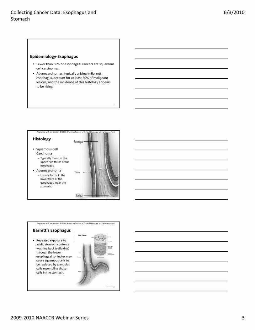
Histology
• Squamous Cell Carcinoma– Typically found in the upper two thirds of the
h
Reprinted with permission. © 2008 American Society of Clinical Oncology. All rights reserved.
esophagus.
• Adenocarcinoma– Usually forms in the lower third of the esophagus, near the stomach.
Z‐Line
8
Barrett’s Esophagus
• Repeated exposure to acidic stomach contents washing back (refluxing) through the lower esophageal sphincter may
Reprinted with permission. © 2008 American Society of Clinical Oncology. All rights reserved.
esophageal sphincter may cause squamous cells to be replaced by glandular cells resembling those cells in the stomach.
9
Collecting Cancer Data: Esophagus and Stomach
6/3/2010
2009‐2010 NAACCR Webinar Series 4
High Grade Dysplasia• The terminology preferred by pathologists for carcinoma
in situ of the esophagus is high grade dysplasia. • This terminology is not reportable to most cancer
registries. – Therefore, it may be a future issue that early/very low stage
esophageal cancer is under‐reported as a result of registry reporting terminology.
• If high grade dysplasia of the esophagus is a reportable cancer, it should be coded as 00 in CS Extension.
CS Manual Section I Part 2 Page 29 Version 02.00.01
10
Question
• The esophagus chapter in AJCC 7 specifically redefined the explanation of Tis (for the esophagus) to high grade dysplasia (formally called in situ). Sinceto high grade dysplasia (formally called in situ). Since it seems to be universal that pathologists will no longer use in situ when describing tissue in the esophagus, will high grade dysplasia of the esophagus be reportable?
11
Answer
• AJCC staging does not determine reportability. This is up to the standard setters, state and federal law. Please discuss with your facility's cancer committeePlease discuss with your facility s cancer committee regarding whether this should be reportable‐by‐agreement for your facility.
(I & R Team)
47292
4/8/2010
12
Collecting Cancer Data: Esophagus and Stomach
6/3/2010
2009‐2010 NAACCR Webinar Series 5
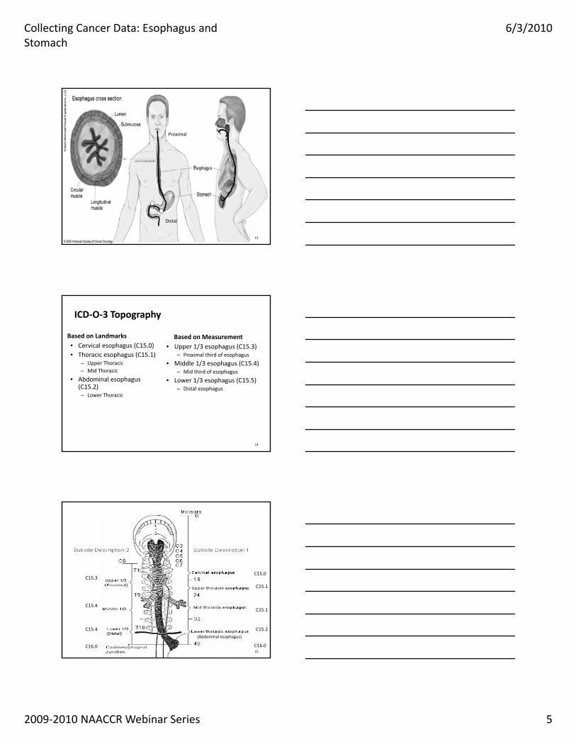
Proximal
13
Distal
ICD‐O‐3 Topography
Based on Measurement• Upper 1/3 esophagus (C15.3)
– Proximal third of esophagus
• Middle 1/3 esophagus (C15.4)– Mid third of esophagus
Based on Landmarks• Cervical esophagus (C15.0)• Thoracic esophagus (C15.1)
– Upper Thoracic– Mid Thoracic Mid third of esophagus
• Lower 1/3 esophagus (C15.5)– Distal esophagus
• Abdominal esophagus (C15.2)– Lower Thoracic
14
Esophagus Overview
• Anatomy
C15.3C15.0
C15.4
C15.4
C16.0 C16.0
C15.2
C15.1
C15.1
(Abdominal esophagus)
15
Collecting Cancer Data: Esophagus and Stomach
6/3/2010
2009‐2010 NAACCR Webinar Series 6
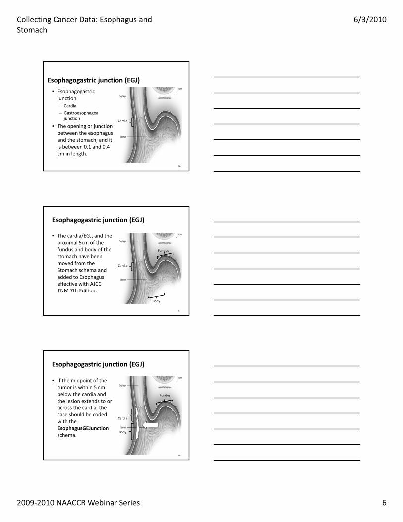
Esophagogastric junction (EGJ)
• Esophagogastric junction– Cardia
– Gastroesophageal junction
• The opening or junction between the esophagus and the stomach, and it is between 0.1 and 0.4 cm in length.
16
Cardia
Esophagogastric junction (EGJ)
• The cardia/EGJ, and the proximal 5cm of the fundus and body of the stomach have been
Fundus
17
moved from the Stomach schema and added to Esophagus effective with AJCC TNM 7th Edition.
Cardia
Body
Esophagogastric junction (EGJ)
• If the midpoint of the tumor is within 5 cm below the cardia and the lesion extends to or
Fundus
across the cardia, the case should be coded with the EsophagusGEJunction schema.
18
Cardia
Body
Collecting Cancer Data: Esophagus and Stomach
6/3/2010
2009‐2010 NAACCR Webinar Series 7
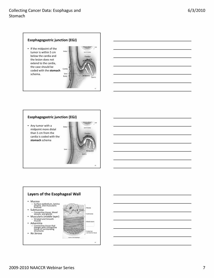
Esophagogastric junction (EGJ)
• If the midpoint of the tumor is within 5 cm below the cardia and the lesion does not
Fundus
extend to the cardia, the case should be coded with the stomachschema.
19
Cardia
Body Midpoint5cm’s
Esophagogastric junction (EGJ)
• Any tumor with a midpoint more distal than 5 cm from the cardia is coded with the
Fundus
stomach schema
20
Midpoint
5cm’s
Layers of the Esophageal Wall
• Mucosa – Surface epithelium, lamina
propria, and muscularis mucosa
• Submucosa – Connective tissue, blood
vessels, and glands M l i ( iddl l )• Muscularis (middle layer)– Striated and Smooth
muscle• Adventitia
– Connective tissue that merges with connective tissue of surrounding structures
• No Serosa
21
Collecting Cancer Data: Esophagus and Stomach
6/3/2010
2009‐2010 NAACCR Webinar Series 8
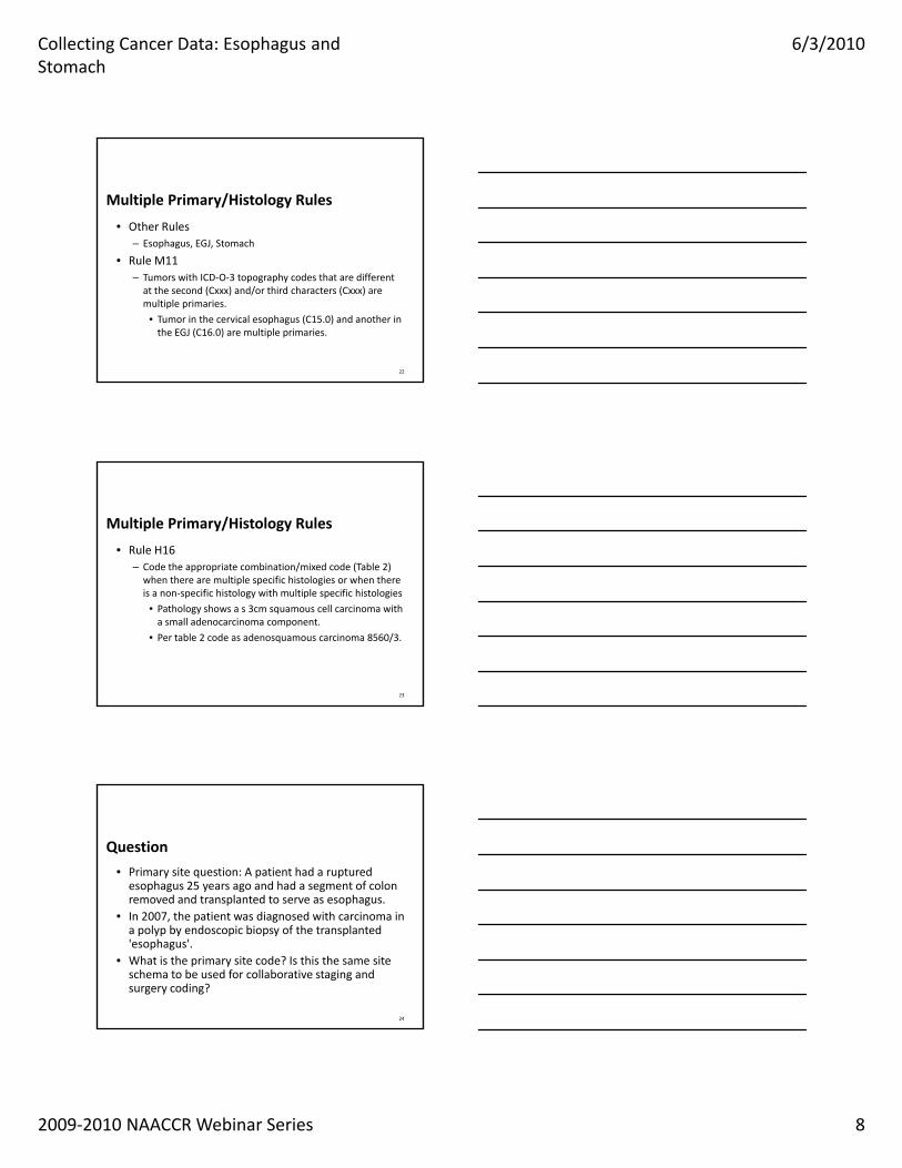
Multiple Primary/Histology Rules
• Other Rules– Esophagus, EGJ, Stomach
• Rule M11• Rule M11 – Tumors with ICD‐O‐3 topography codes that are different at the second (Cxxx) and/or third characters (Cxxx) are multiple primaries.
• Tumor in the cervical esophagus (C15.0) and another in the EGJ (C16.0) are multiple primaries.
22
Multiple Primary/Histology Rules
• Rule H16 – Code the appropriate combination/mixed code (Table 2) when there are multiple specific histologies or when there p p gis a non‐specific histology with multiple specific histologies
• Pathology shows a s 3cm squamous cell carcinoma with a small adenocarcinoma component.
• Per table 2 code as adenosquamous carcinoma 8560/3.
23
Question
• Primary site question: A patient had a ruptured esophagus 25 years ago and had a segment of colon removed and transplanted to serve as esophagus.
• In 2007, the patient was diagnosed with carcinoma in a polyp by endoscopic biopsy of the transplanted 'esophagus'.
• What is the primary site code? Is this the same site schema to be used for collaborative staging and surgery coding?
24
Collecting Cancer Data: Esophagus and Stomach
6/3/2010
2009‐2010 NAACCR Webinar Series 9
Answer
• Code the primary site esophagus, NOS (C15.9). Use the surgery codes and collaborative staging schema for esophagus. Document the unusual nature of thisfor esophagus. Document the unusual nature of this case in text fields. See also SINQ 20021078. – SINQ 20091017
– Last updated 4/13/2009
25
Grade
• For Esophagus and EGJ, grade is required to derive AJCC TNM stages 0‐IIA
• Standard four grade grading system• Standard four grade grading system– Well differentiated
– Moderately differentiated
– Poorly differentiated
– Undifferentiated
26
Grade
• C T1a N0 M0 G1 Stage IA– Treatment options include
• EsophagectomyEsophagectomy
• Endoscopic mucosal resection
• Other ablative technique
• C T1a N0 M0 G2‐3 Stage IB– Esophagectomy
27
Collecting Cancer Data: Esophagus and Stomach
6/3/2010
2009‐2010 NAACCR Webinar Series 10
Upper GI Diagnostic Testsg
Imaging• Upper GI Series (Barium Swallow)
• Computed tomography (CT or CAT) scan
• Magnetic resonance imaging (MRI)
• Positron emission tomography (PET) scan– PET‐CT
– FDG‐PET
29
Endoscopy
• Esophagogastroduodenoscopy (EGD)
• Endoscopic Ultrasound (EUS)
30
Collecting Cancer Data: Esophagus and Stomach
6/3/2010
2009‐2010 NAACCR Webinar Series 11
Overview
Stomach
Stomach: Epidemiology
• 2009 estimates in the United States– New cases: 21,130
– Deaths: 10 620Deaths: 10,620
• Environmental risk factors– Helicobacter pylori (H. pylori) infection
– Smoking
– High salt intake
32
Stomach: Epidemiology
• Adenocarcinoma is histology in 90‐95% of stomach malignancies
• Cancer originating in distal stomach has decreased in• Cancer originating in distal stomach has decreased in United States since the 1930’s
• Cancer originating in cardia or esophagogastric junction has rapidly increased during last 2 decades
33
Collecting Cancer Data: Esophagus and Stomach
6/3/2010
2009‐2010 NAACCR Webinar Series 12
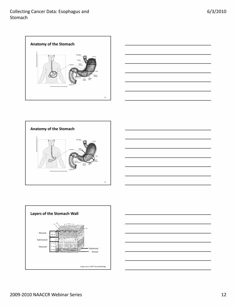
Anatomy of the Stomach
Cardia
34
Anatomy of the Stomach
35
Layers of the Stomach Wall
Mucosal
36
Serosal
SubserosalMuscular
Submucosal
Image source: SEER Training Website
Collecting Cancer Data: Esophagus and Stomach
6/3/2010
2009‐2010 NAACCR Webinar Series 13
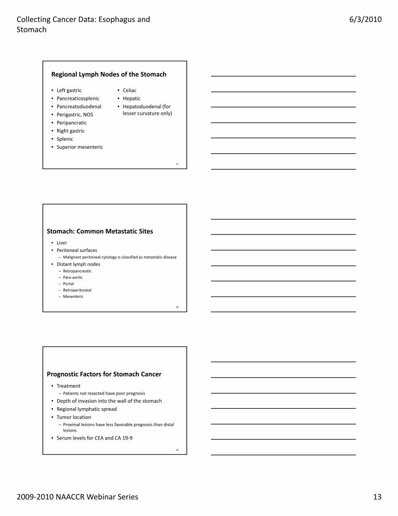
Regional Lymph Nodes of the Stomach
• Left gastric
• Pancreaticosplenic
• Pancreatoduodenal
• Perigastric NOS
• Celiac
• Hepatic
• Hepatoduodenal (for lesser curvature only)• Perigastric, NOS
• Peripancratic
• Right gastric
• Splenic
• Superior mesenteric
lesser curvature only)
37
Stomach: Common Metastatic Sites
• Liver
• Peritoneal surfaces– Malignant peritoneal cytology is classified as metastatic diseaseMalignant peritoneal cytology is classified as metastatic disease
• Distant lymph nodes– Retropancreatic
– Para‐aortic
– Portal
– Retroperitoneal
– Mesenteric
38
Prognostic Factors for Stomach Cancer
• Treatment– Patients not resected have poor prognosis
• Depth of invasion into the wall of the stomach• Depth of invasion into the wall of the stomach
• Regional lymphatic spread
• Tumor location– Proximal lesions have less favorable prognosis than distal lesions
• Serum levels for CEA and CA 19‐9
39
Collecting Cancer Data: Esophagus and Stomach
6/3/2010
2009‐2010 NAACCR Webinar Series 14
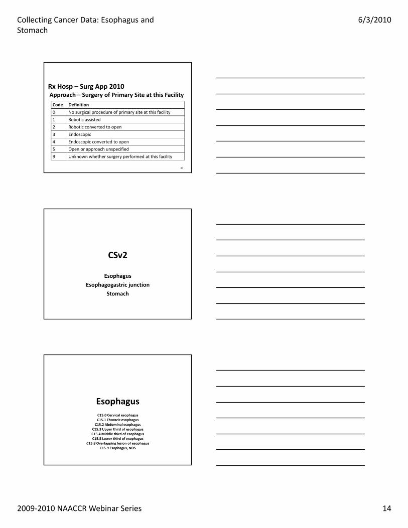
Rx Hosp – Surg App 2010Approach – Surgery of Primary Site at this Facility
Code Definition
0 No surgical procedure of primary site at this facility
1 Robotic assisted
2 Robotic converted to open
3 Endoscopic
4 Endoscopic converted to open
5 Open or approach unspecified
9 Unknown whether surgery performed at this facility
40
CSv2
Esophagus
Esophagogastric junction
Stomach
EsophagusC15.0 Cervical esophagus C15.1 Thoracic esophagus
C15.2 Abdominal esophagus C15.3 Upper third of esophagus C15.4 Middle third of esophagus C15.5 Lower third of esophagus
C15.8 Overlapping lesion of esophagus C15.9 Esophagus, NOS
Collecting Cancer Data: Esophagus and Stomach
6/3/2010
2009‐2010 NAACCR Webinar Series 15
Tumor Size
• For esophagus, this field is used for size of tumor/length of involved esophagus
43
Question
• For CS Tumor Size, the note says, "For esophagus, this field is used for size/length of involved esophagus."esophagus.
• Define the length of involved tumor and how is this determined?
44
Answer
• This is the longest dimension of the tumor or the measurement of the esophagus that has cancer in it, such as the tumor involved the esophagus from 18cm to 26cm, which would mean it was an 8cm tumor.
• It can be found on the pathology report for a resected tumor, and if the information is not available then it would be taken from imaging or other reports according to the priority order in Part I of the CS Manual.– (I & R Team) 24240
45
Collecting Cancer Data: Esophagus and Stomach
6/3/2010
2009‐2010 NAACCR Webinar Series 16
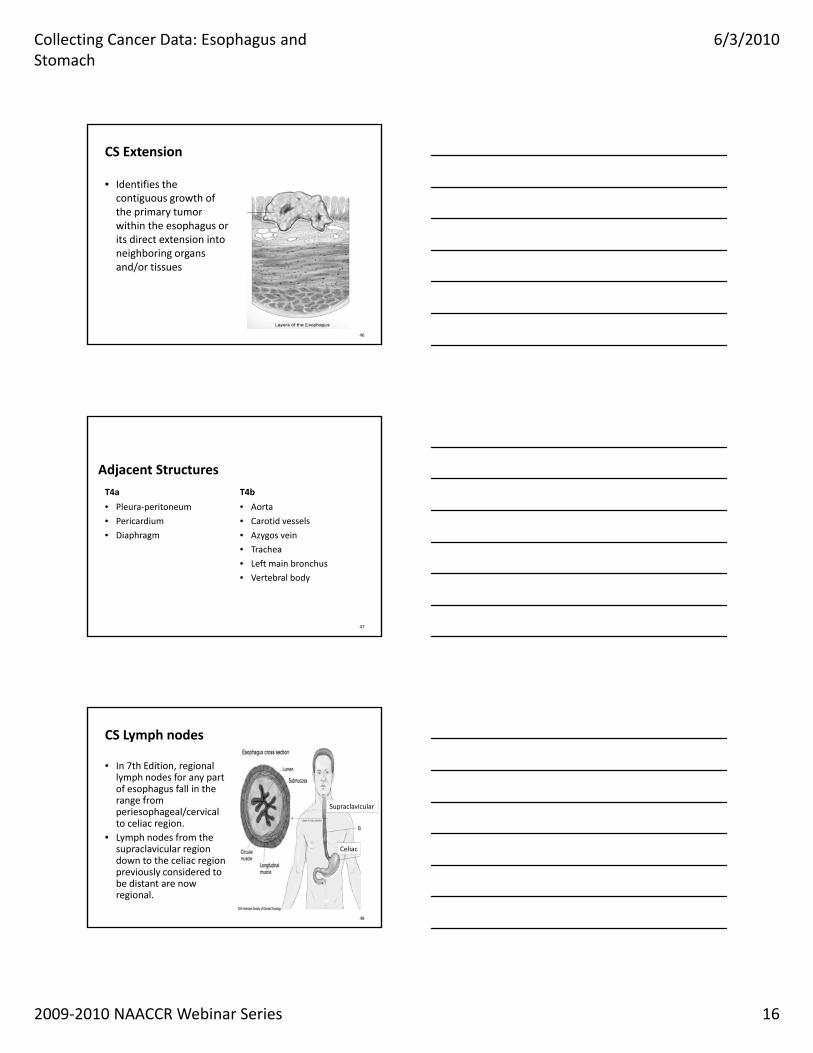
CS Extension
• Identifies the contiguous growth of the primary tumor within the esophagus or its direct extension into neighboring organs and/or tissues
46
Adjacent StructuresT4a
• Pleura‐peritoneum
• Pericardium
T4b
• Aorta
• Carotid vessels
• Diaphragm • Azygos vein
• Trachea
• Left main bronchus
• Vertebral body
47
CS Lymph nodes
• In 7th Edition, regional lymph nodes for any part of esophagus fall in the range from periesophageal/cervical
Supraclavicular
to celiac region. • Lymph nodes from the
supraclavicular region down to the celiac region previously considered to be distant are now regional.
48
Celiac
Collecting Cancer Data: Esophagus and Stomach
6/3/2010
2009‐2010 NAACCR Webinar Series 17
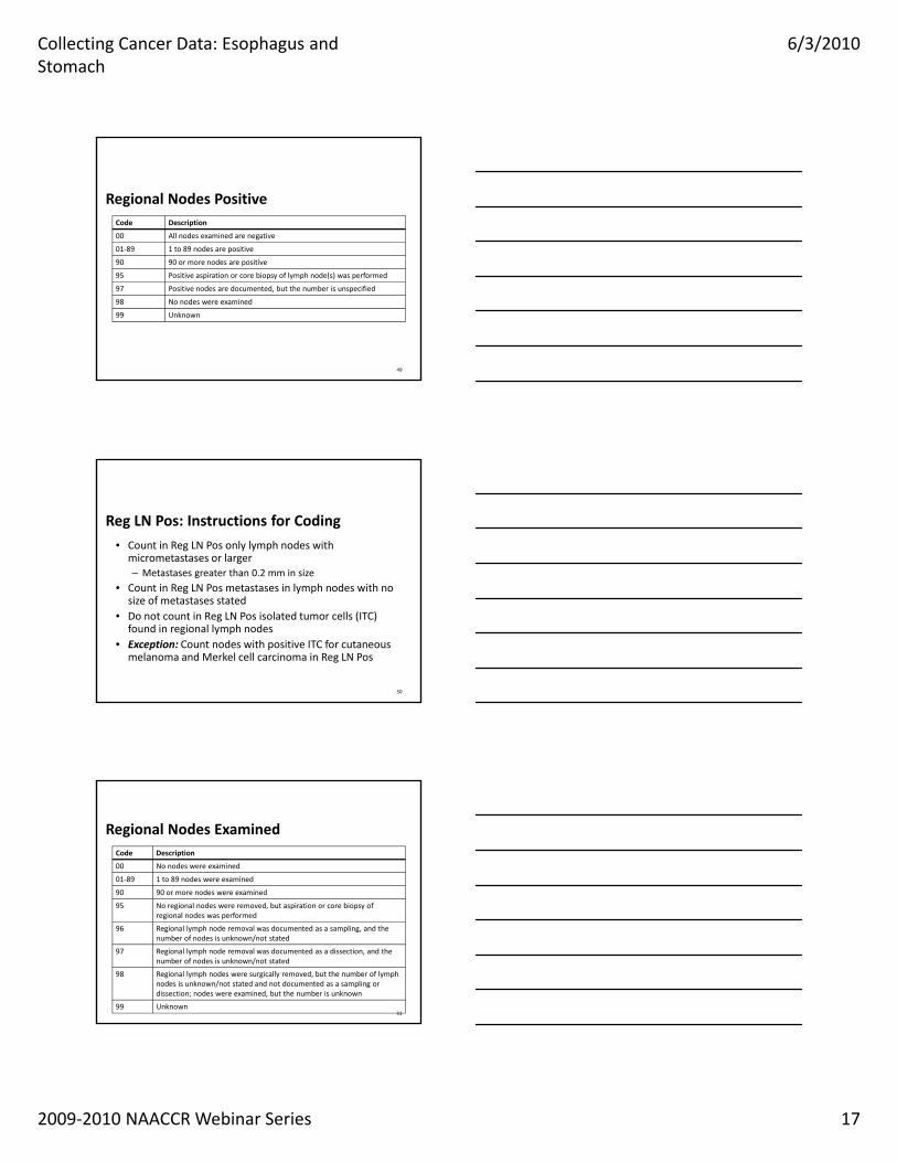
Regional Nodes PositiveCode Description
00 All nodes examined are negative
01‐89 1 to 89 nodes are positive
90 90 or more nodes are positive
95 Positive aspiration or core biopsy of lymph node(s) was performed
97 Positive nodes are documented, but the number is unspecified
98 No nodes were examined
99 Unknown
49
Reg LN Pos: Instructions for Coding• Count in Reg LN Pos only lymph nodes with
micrometastases or larger– Metastases greater than 0.2 mm in size
• Count in Reg LN Pos metastases in lymph nodes with no size of metastases stated
• Do not count in Reg LN Pos isolated tumor cells (ITC) found in regional lymph nodes
• Exception: Count nodes with positive ITC for cutaneous melanoma and Merkel cell carcinoma in Reg LN Pos
50
Regional Nodes ExaminedCode Description
00 No nodes were examined
01‐89 1 to 89 nodes were examined
90 90 or more nodes were examined
95 No regional nodes were removed, but aspiration or core biopsy of regional nodes was performed
96 Regional lymph node removal was documented as a sampling, and the number of nodes is unknown/not stated
97 Regional lymph node removal was documented as a dissection, and the number of nodes is unknown/not stated
98 Regional lymph nodes were surgically removed, but the number of lymph nodes is unknown/not stated and not documented as a sampling or dissection; nodes were examined, but the number is unknown
99 Unknown51
Collecting Cancer Data: Esophagus and Stomach
6/3/2010
2009‐2010 NAACCR Webinar Series 18
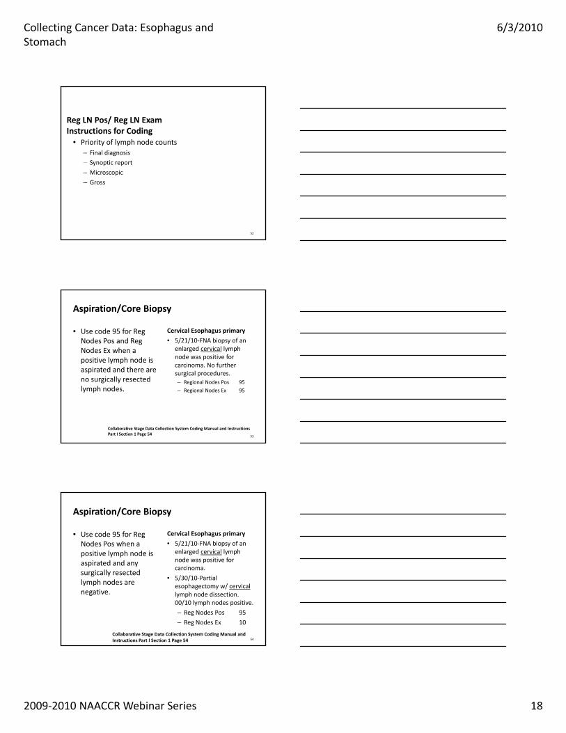
Reg LN Pos/ Reg LN ExamInstructions for Coding
• Priority of lymph node counts– Final diagnosis
– Synoptic reportSynoptic report
– Microscopic
– Gross
52
Aspiration/Core Biopsy
• Use code 95 for Reg Nodes Pos and Reg Nodes Ex when a positive lymph node is
Cervical Esophagus primary
• 5/21/10‐FNA biopsy of an enlarged cervical lymph node was positive for carcinoma No further
aspirated and there are no surgically resected lymph nodes.
carcinoma. No further surgical procedures.– Regional Nodes Pos 95
– Regional Nodes Ex 95
53
Collaborative Stage Data Collection System Coding Manual and Instructions Part I Section 1 Page 54
Aspiration/Core Biopsy
• Use code 95 for Reg Nodes Pos when a positive lymph node is aspirated and any
Cervical Esophagus primary
• 5/21/10‐FNA biopsy of an enlarged cervical lymph node was positive for carcinoma
surgically resected lymph nodes are negative.
54
carcinoma.
• 5/30/10‐Partial esophagectomy w/ cervicallymph node dissection. 00/10 lymph nodes positive.
– Reg Nodes Pos 95
– Reg Nodes Ex 10
Collaborative Stage Data Collection System Coding Manual and Instructions Part I Section 1 Page 54
Collecting Cancer Data: Esophagus and Stomach
6/3/2010
2009‐2010 NAACCR Webinar Series 19
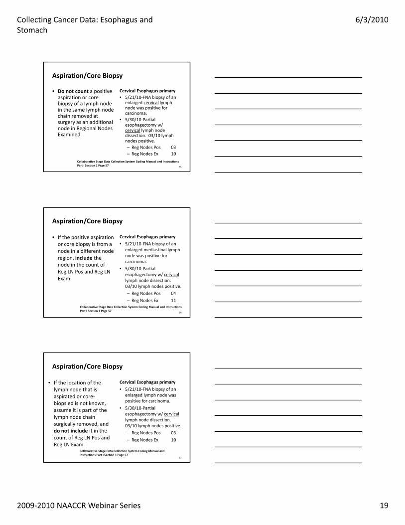
Aspiration/Core Biopsy
• Do not count a positive aspiration or core biopsy of a lymph node in the same lymph node chain removed at
Cervical Esophagus primary• 5/21/10‐FNA biopsy of an
enlarged cervical lymph node was positive for carcinoma.chain removed at
surgery as an additional node in Regional Nodes Examined
• 5/30/10‐Partial esophagectomy w/ cervical lymph node dissection. 03/10 lymph nodes positive.– Reg Nodes Pos 03– Reg Nodes Ex 10
55
Collaborative Stage Data Collection System Coding Manual and Instructions Part I Section 1 Page 57
Aspiration/Core Biopsy
• If the positive aspiration or core biopsy is from a node in a different node region, include the
Cervical Esophagus primary
• 5/21/10‐FNA biopsy of an enlarged mediastinal lymph node was positive for carcinoma
node in the count of Reg LN Pos and Reg LN Exam.
carcinoma.
• 5/30/10‐Partial esophagectomy w/ cervicallymph node dissection. 03/10 lymph nodes positive.
– Reg Nodes Pos 04
– Reg Nodes Ex 11
56
Collaborative Stage Data Collection System Coding Manual and Instructions Part I Section 1 Page 57
Aspiration/Core Biopsy
• If the location of the lymph node that is aspirated or core‐biopsied is not known,
Cervical Esophagus primary
• 5/21/10‐FNA biopsy of an enlarged lymph node was positive for carcinoma.
• 5/30/10 Partialassume it is part of the lymph node chain surgically removed, and do not include it in the count of Reg LN Pos and Reg LN Exam.
57
• 5/30/10‐Partial esophagectomy w/ cervicallymph node dissection. 03/10 lymph nodes positive.
– Reg Nodes Pos 03
– Reg Nodes Ex 10
Collaborative Stage Data Collection System Coding Manual and Instructions Part I Section 1 Page 57
Collecting Cancer Data: Esophagus and Stomach
6/3/2010
2009‐2010 NAACCR Webinar Series 20
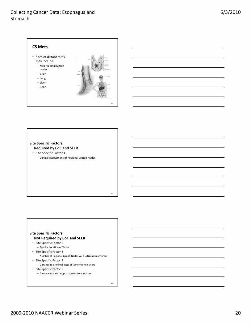
CS Mets
• Sites of distant mets may include– Non regional lymph nodes
– Brain
– Lung
– Liver
– Bone
58
Site Specific FactorsRequired by CoC and SEER
• Site Specific Factor 1– Clinical Assessment of Regional Lymph Nodes
59
Site Specific FactorsNot Required by CoC and SEER
• Site Specific Factor 2– Specific Location of Tumor
• Site Specific Factor 3Site Specific Factor 3– Number of Regional Lymph Nodes with Extracapsular tumor
• Site Specific Factor 4– Distance to proximal edge of tumor from incisors
• Site Specific Factor 5– Distance to distal edge of tumor from incisors
60
Collecting Cancer Data: Esophagus and Stomach
6/3/2010
2009‐2010 NAACCR Webinar Series 21
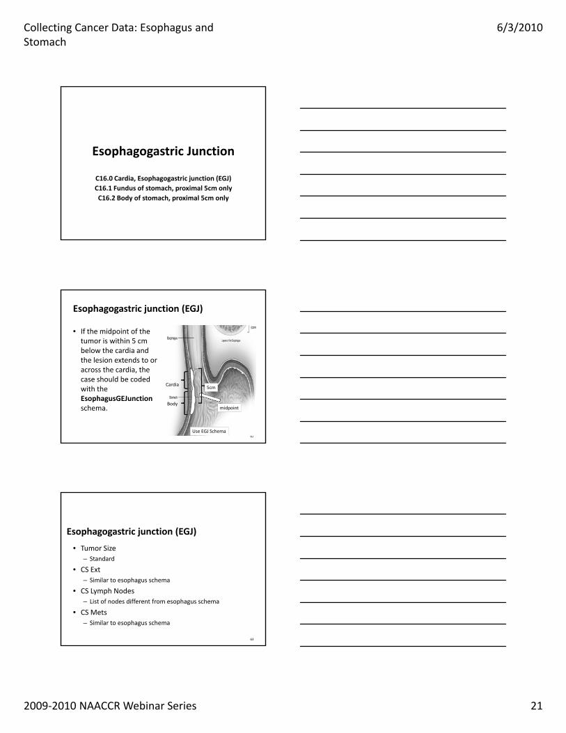
Esophagogastric Junction
C16.0 Cardia, Esophagogastric junction (EGJ) C16.1 Fundus of stomach, proximal 5cm only C16.2 Body of stomach, proximal 5cm only
Esophagogastric junction (EGJ)
• If the midpoint of the tumor is within 5 cm below the cardia and the lesion extends to or across the cardia, the case should be coded with the EsophagusGEJunction schema.
62
Cardia
Body
5cm
midpoint
Use EGJ Schema
Esophagogastric junction (EGJ)
• Tumor Size– Standard
• CS Ext• CS Ext– Similar to esophagus schema
• CS Lymph Nodes– List of nodes different from esophagus schema
• CS Mets– Similar to esophagus schema
63
Collecting Cancer Data: Esophagus and Stomach
6/3/2010
2009‐2010 NAACCR Webinar Series 22
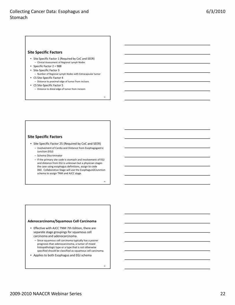
Site Specific Factors• Site Specific Factor 1 (Required by CoC and SEER)
– Clinical Assessment of Regional Lymph Nodes
• Specific Factor 2 = 988• Site‐Specific Factor 3
– Number of Regional Lymph Nodes with Extracapsular tumor
• CS Site‐Specific Factor 4– Distance to proximal edge of tumor from incisors
• CS Site‐Specific Factor 5– Distance to distal edge of tumor from incisors
64
Site Specific Factors
• Site Specific Factor 25 (Required by CoC and SEER)– Involvement of Cardia and Distance from Esophagogastric Junction (EGJ)( )
– Schema Discriminator
– If the primary site code is stomach and involvement of EGJ and distance from EGJ is unknown but a physician stages the case using esophagus definitions, assign to code 060. Collaborative Stage will use the EsophagusGEJunction schema to assign TNM and AJCC stage.
65
Adenocarcinoma/Squamous Cell Carcinoma
• Effective with AJCC TNM 7th Edition, there are separate stage groupings for squamous cell carcinoma and adenocarcinoma.carcinoma and adenocarcinoma. – Since squamous cell carcinoma typically has a poorer prognosis than adenocarcinoma, a tumor of mixed histopathologic type or a type that is not otherwise specified should be classified as squamous cell carcinoma.
• Applies to both Esophagus and EGJ schema
66
Collecting Cancer Data: Esophagus and Stomach
6/3/2010
2009‐2010 NAACCR Webinar Series 23
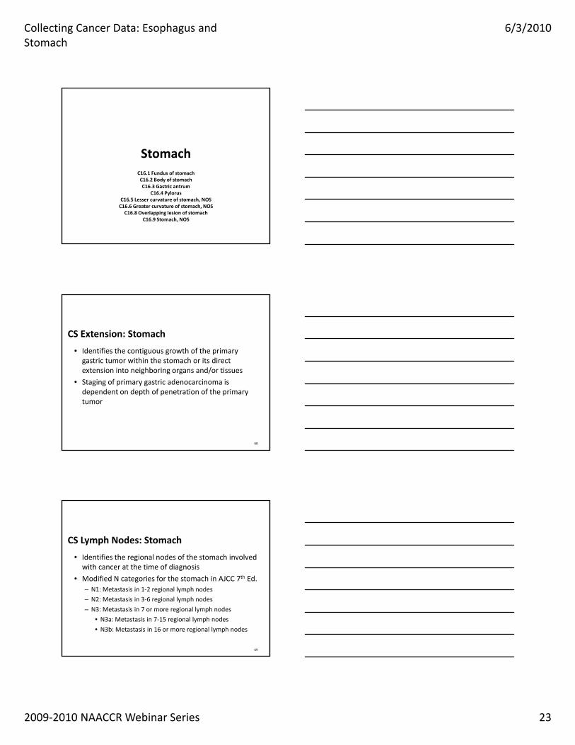
StomachC16.1 Fundus of stomach C16.2 Body of stomach C16.3 Gastric antrum
C16.4 Pylorus C16.5 Lesser curvature of stomach, NOS C16.6 Greater curvature of stomach, NOS C16.8 Overlapping lesion of stomach
C16.9 Stomach, NOS
CS Extension: Stomach
• Identifies the contiguous growth of the primary gastric tumor within the stomach or its direct extension into neighboring organs and/or tissuesextension into neighboring organs and/or tissues
• Staging of primary gastric adenocarcinoma is dependent on depth of penetration of the primary tumor
68
CS Lymph Nodes: Stomach
• Identifies the regional nodes of the stomach involved with cancer at the time of diagnosis
• Modified N categories for the stomach in AJCC 7th Ed• Modified N categories for the stomach in AJCC 7th Ed.– N1: Metastasis in 1‐2 regional lymph nodes
– N2: Metastasis in 3‐6 regional lymph nodes
– N3: Metastasis in 7 or more regional lymph nodes
• N3a: Metastasis in 7‐15 regional lymph nodes
• N3b: Metastasis in 16 or more regional lymph nodes
69
Collecting Cancer Data: Esophagus and Stomach
6/3/2010
2009‐2010 NAACCR Webinar Series 24
CS Lymph Nodes Eval: Stomach
• Used primarily to derive staging basis for N category
• Records how CS Lymph Nodes code was determined based on diagnostic methods used and their intentbased on diagnostic methods used and their intent
• Designates N category as clinical or pathologic based on intent– Staging basis is clinical when intent is workup
– Staging basis is pathologic when intent is treatment
70
CS Lymph Nodes Eval: Stomach
• Microscopic assessment of regional nodes that is part of workup to choose treatment plan is clinical stagingstaging– T category is clinical
• Microscopic assessment of regional nodes that is part of treatment is pathologic staging– T category is pathologic
71
CS Lymph Nodes Eval: Stomach
• Microscopic assessment of highest N category is always pathologic
• If lymph node dissection is not performed after• If lymph node dissection is not performed after neoadjuvant treatment, use code 0 or 1
• Only codes 5 and 6 are used if node assessment is performed after neoadjuvant therapy
72
Collecting Cancer Data: Esophagus and Stomach
6/3/2010
2009‐2010 NAACCR Webinar Series 25
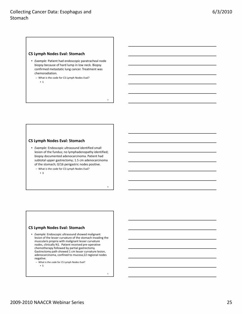
CS Lymph Nodes Eval: Stomach
• Example: Patient had endoscopic paratracheal node biopsy because of hard lump in low neck. Biopsy confirmed metastatic lung cancer. Treatment wasconfirmed metastatic lung cancer. Treatment was chemoradiation.– What is the code for CS Lymph Nodes Eval?
• 1
73
CS Lymph Nodes Eval: Stomach
• Example: Endoscopic ultrasound identified small lesion of the fundus; no lymphadenopathy identified; biopsy documented adenocarcinoma. Patient hadbiopsy documented adenocarcinoma. Patient had subtotal upper gastrectomy; 1.5 cm adenocarcinoma of the stomach; 0/16 perigastric nodes positive.– What is the code for CS Lymph Nodes Eval?
• 3
74
CS Lymph Nodes Eval: Stomach• Example: Endoscopic ultrasound showed malignant
lesion of the lesser curvature of the stomach invading the muscularis propria with malignant lesser curvature
d li i ll N1 P ti t i d tinodes, clinically N1. Patient received pre‐operative chemotherapy followed by partial gastrectomy. Gastrectomy path showed 1 cm lesser curvature lesion, adenocarcinoma, confined to mucosa;22 regional nodes negative.– What is the code for CS Lymph Nodes Eval?
• 5
75
Collecting Cancer Data: Esophagus and Stomach
6/3/2010
2009‐2010 NAACCR Webinar Series 26
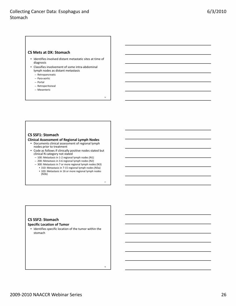
CS Mets at DX: Stomach
• Identifies involved distant metastatic sites at time of diagnosis
• Classifies involvement of some intra‐abdominal lymph nodes as distant metastasis– Retropancreatic– Para‐aortic– Portal– Retroperitoneal– Mesenteric
76
CS SSF1: StomachClinical Assessment of Regional Lymph Nodes
• Documents clinical assessment of regional lymph nodes prior to treatment
• Code as follows if clinically positive nodes stated but li i l N t t t t dclinical N category not stated – 100: Metastasis in 1‐2 regional lymph nodes (N1)– 200: Metastasis in 3‐6 regional lymph nodes (N2)– 300: Metastasis in 7 or more regional lymph nodes (N3)
• 310: Metastasis in 7‐15 regional lymph nodes (N3a)• 320: Metastasis in 16 or more regional lymph nodes (N3b)
77
CS SSF2: StomachSpecific Location of Tumor
• Identifies specific location of the tumor within the stomach
78
Collecting Cancer Data: Esophagus and Stomach
6/3/2010
2009‐2010 NAACCR Webinar Series 27
SSF13: StomachCarcinoembryonic Antigen (CEA)
• Code the interpretation of highest lab value of CEA prior to treatment
79
CS SSF14: StomachCarcinoembryonic Antigen (CEA) Lab Value
• Record in nanograms/milliliter highest CEA lab value prior to treatment
80
CS SSF15: StomachCA 19‐9 Lab Value
• Record in units/milliliter the highest CA 19‐9 lab value prior to treatment
81
Collecting Cancer Data: Esophagus and Stomach
6/3/2010
2009‐2010 NAACCR Webinar Series 28
CS SSF25: StomachInvolvement of Cardia & Distance from EGJ
• Fundus (C16.1) and body (C16.2) of stomach can be assigned to either EsophagusGEJunction or Stomach CSv2 schemaCSv2 schema
• SSF25 is schema discriminator field needed for CS algorithm to determine which schema to select when site is C16.1 or C16.2
82
Treatment
NCCN Guidelines http://www.nccn.org/professionals/phy
sician_gls/f_guidelines.asp
Esophagus
Collecting Cancer Data: Esophagus and Stomach
6/3/2010
2009‐2010 NAACCR Webinar Series 29
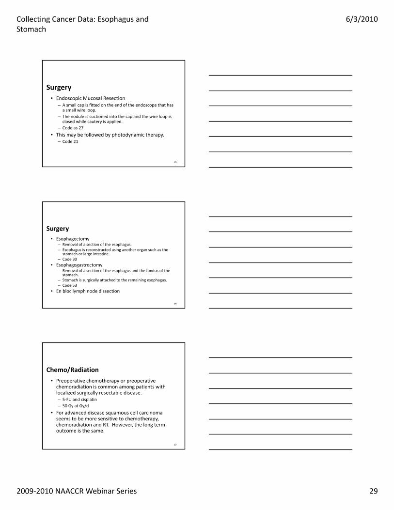
Surgery• Endoscopic Mucosal Resection
– A small cap is fitted on the end of the endoscope that has a small wire loop.
– The nodule is suctioned into the cap and the wire loop is closed while cautery is applied.
– Code as 27
• This may be followed by photodynamic therapy. – Code 21
85
Surgery• Esophagectomy
– Removal of a section of the esophagus.– Esophagus is reconstructed using another organ such as the
stomach or large intestine.stomach or large intestine.– Code 30
• Esophagogastrectomy– Removal of a section of the esophagus and the fundus of the
stomach.– Stomach is surgically attached to the remaining esophagus.– Code 53
• En bloc lymph node dissection
86
Chemo/Radiation
• Preoperative chemotherapy or preoperative chemoradiation is common among patients with localized surgically resectable disease.– 5‐FU and cisplatin– 50 Gy at Gy/d
• For advanced disease squamous cell carcinoma seems to be more sensitive to chemotherapy, chemoradiation and RT. However, the long term outcome is the same.
87
Collecting Cancer Data: Esophagus and Stomach
6/3/2010
2009‐2010 NAACCR Webinar Series 30
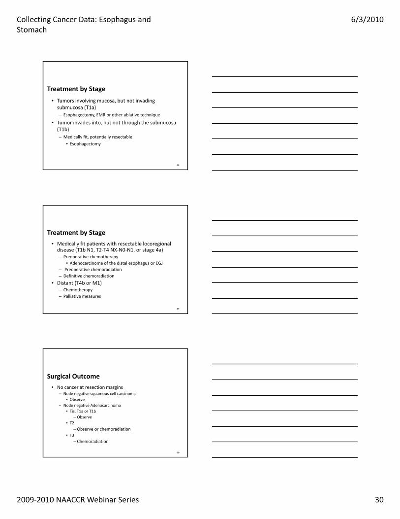
Treatment by Stage
• Tumors involving mucosa, but not invading submucosa (T1a)– Esophagectomy EMR or other ablative techniqueEsophagectomy, EMR or other ablative technique
• Tumor invades into, but not through the submucosa (T1b)– Medically fit, potentially resectable
• Esophagectomy
88
Treatment by Stage
• Medically fit patients with resectable locoregional disease (T1b N1, T2‐T4 NX‐N0‐N1, or stage 4a)– Preoperative chemotherapy
• Adenocarcinoma of the distal esophagus or EGJ– Preoperative chemoradiation– Definitive chemoradiation
• Distant (T4b or M1)– Chemotherapy– Palliative measures
89
Surgical Outcome• No cancer at resection margins
– Node negative squamous cell carcinoma• Observe
– Node negative Adenocarcinoma• Tis, T1a or T1b
– Observe• T2
– Observe or chemoradiation• T3
– Chemoradiation
90
Collecting Cancer Data: Esophagus and Stomach
6/3/2010
2009‐2010 NAACCR Webinar Series 31
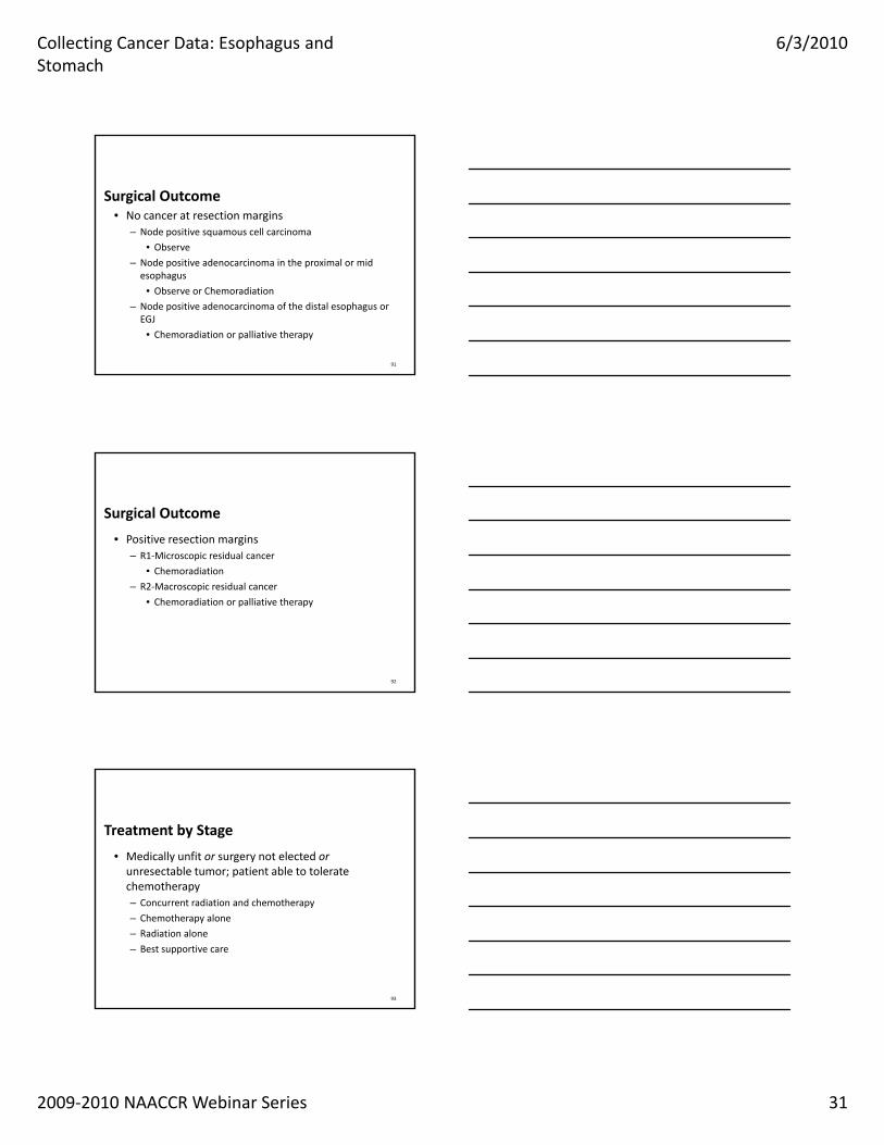
Surgical Outcome• No cancer at resection margins
– Node positive squamous cell carcinoma
• Observe
– Node positive adenocarcinoma in the proximal or mid esophagus
• Observe or Chemoradiation
– Node positive adenocarcinoma of the distal esophagus or EGJ
• Chemoradiation or palliative therapy
91
Surgical Outcome
• Positive resection margins– R1‐Microscopic residual cancer
• ChemoradiationChemoradiation
– R2‐Macroscopic residual cancer
• Chemoradiation or palliative therapy
92
Treatment by Stage
• Medically unfit or surgery not elected or unresectable tumor; patient able to tolerate chemotherapychemotherapy– Concurrent radiation and chemotherapy
– Chemotherapy alone
– Radiation alone
– Best supportive care
93
Collecting Cancer Data: Esophagus and Stomach
6/3/2010
2009‐2010 NAACCR Webinar Series 32
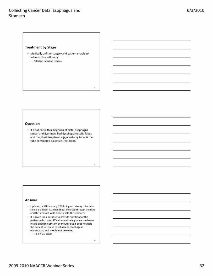
Treatment by Stage
• Medically unfit or surgery and patient unable to tolerate chemotherapy– Palliative radiation therapyPalliative radiation therapy
94
Question
• If a patient with a diagnosis of distal esophagus cancer and liver mets had dysphagia to solid foods and the physician placed a jejunostomy tube, is theand the physician placed a jejunostomy tube, is the tube considered palliative treatment?
95
Answer
• Updated in I&R January, 2010 ‐ A gastrostomy tube (also called a G‐tube) is a tube that’s inserted through the skin and the stomach wall, directly into the stomach.
• It is given for a purpose to provide nutrition for the patients who have difficulty swallowing or are unable to intake enough nutrition by mouth, but it does not help the patient to relieve dysphasia or esophageal obstruction, and should not be coded. – (I & R Team) 22886
96

Collecting Cancer Data: Esophagus and Stomach
6/3/2010
2009‐2010 NAACCR Webinar Series 33
Stomach
Treatment by Stage
• Tumors involving mucosa but not invading submucosa– Endoscopic mucosal resection (EMR) or surgery
• Locoregional tumors– Medically fit, potentially resectable
• Surgery• Preoperative chemotherapy followed by surgery• Preoperative chemoradiation followed by surgery• Postoperative treatment depends on surgical outcome
98
Treatment by Stage
• Locoregional tumors– Medically fit, unresectable
• Radiation therapy + concurrent 5‐FU radiosensitization• Chemotherapy
– Medically unfit• Radiation therapy + concurrent 5‐FU radiosensitization• Palliative care
• Distant metastasis– Palliative care
99
Collecting Cancer Data: Esophagus and Stomach
6/3/2010
2009‐2010 NAACCR Webinar Series 34
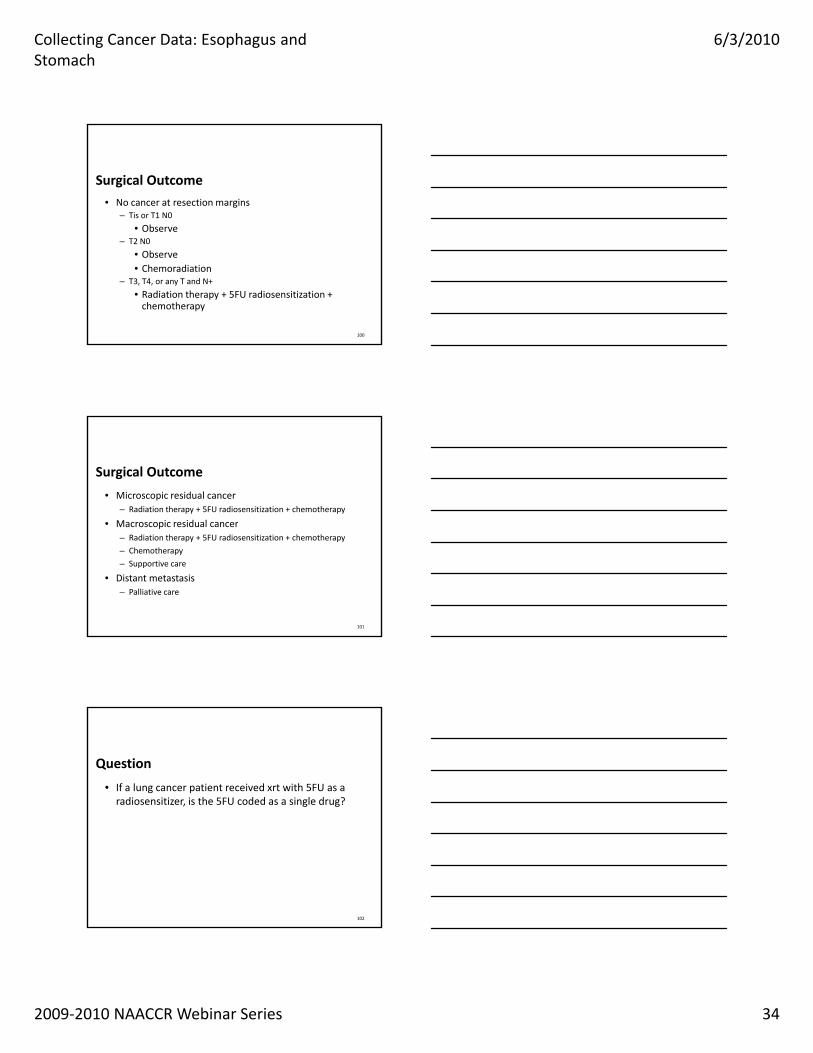
Surgical Outcome• No cancer at resection margins
– Tis or T1 N0
• Observe– T2 N0
• Observe• Chemoradiation
– T3, T4, or any T and N+
• Radiation therapy + 5FU radiosensitization + chemotherapy
100
Surgical Outcome
• Microscopic residual cancer– Radiation therapy + 5FU radiosensitization + chemotherapy
• Macroscopic residual cancerMacroscopic residual cancer– Radiation therapy + 5FU radiosensitization + chemotherapy
– Chemotherapy
– Supportive care
• Distant metastasis– Palliative care
101
Question
• If a lung cancer patient received xrt with 5FU as a radiosensitizer, is the 5FU coded as a single drug?
102
Collecting Cancer Data: Esophagus and Stomach
6/3/2010
2009‐2010 NAACCR Webinar Series 35
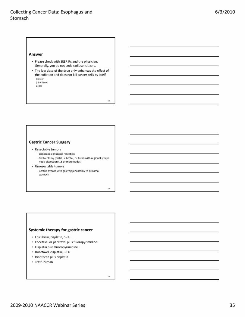
Answer
• Please check with SEER Rx and the physician. Generally, you do not code radiosensitizers.
• The low dose of the drug only enhances the effect of• The low dose of the drug only enhances the effect of the radiation and does not kill cancer cells by itself. Curator
(I & R Team)
24087
103
Gastric Cancer Surgery
• Resectable tumors– Endoscopic mucosal resection
– Gastrectomy (distal subtotal or total) with regional lymphGastrectomy (distal, subtotal, or total) with regional lymph node dissection (15 or more nodes)
• Unresectable tumors– Gastric bypass with gastrojejunostomy to proximal stomach
104
Systemic therapy for gastric cancer
• Epirubicin, cisplatin, 5‐FU
• Cocetaxel or paclitaxel plus fluoropyrimidine
Ci l ti l fl i idi• Cisplatin plus fluoropyrimidine
• Docetaxel, cisplatin, 5‐FU
• Irinotecan plus cisplatin
• Trastuzumab
105
Collecting Cancer Data: Esophagus and Stomach
6/3/2010
2009‐2010 NAACCR Webinar Series 36
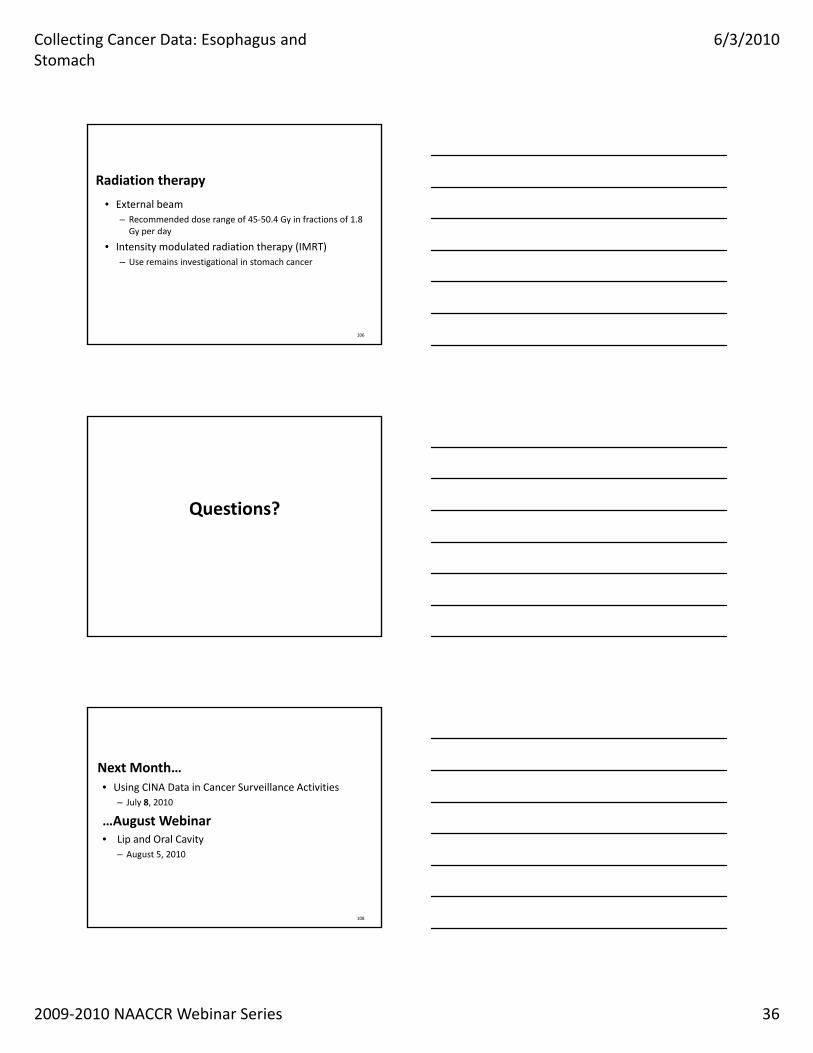
Radiation therapy
• External beam– Recommended dose range of 45‐50.4 Gy in fractions of 1.8 Gy per dayy p y
• Intensity modulated radiation therapy (IMRT)– Use remains investigational in stomach cancer
106
Questions?
Next Month…• Using CINA Data in Cancer Surveillance Activities
– July 8, 2010
A t W bi…August Webinar• Lip and Oral Cavity
– August 5, 2010
108