Collagen Membrane and Immune Response in Guided Bone ...
15
REVIEW ARTICLE Collagen Membrane and Immune Response in Guided Bone Regeneration: Recent Progress and Perspectives Chenyu Chu, DDS, 1, * Jia Deng, DDS, 1, * Xianchang Sun, MD, 2 Yili Qu, DDS, PhD, 1 and Yi Man, DDS, PhD 1,3 Collagen is one of the important components of collagen membranes as well as the extracellular matrix (ECM). Most previous studies have focused on combining collagen membranes with various cross-linking agents, grafting materials, and cytokines to enhance their mechanical properties and bioactivities. Moreover, collagen membranes are often designed to minimize foreign body reactions involving macrophages. However, macro- phages were recently found to play a pivotal role during bone regeneration based on their polarization into both proinflammatory and anti-inflammatory phenotypes. Because of the abilities to modulate macrophage polarization and mediate the balance of proinflammatory and anti-inflammatory microenvironments, immune-responsive collagen membranes may be an innovative strategy for promoting bone regeneration. Herein, following a brief review of collagen membranes and the background of macrophages, recent modulations and studies of immune- responsive collagen are described to express the potential of collagen interacting with macrophages and the necessity of further studies in the field of immune-responsive collagen membranes. Keywords: biomaterials, collagen, macrophage, regeneration, tissue engineering Introduction C ollagen is the main component of the extracellular matrix (ECM) and is involved in important cellular processes, including cell adhesion, proliferation, migration, and differentiation. 1–4 Moreover, collagen membranes have been widely utilized in guided bone regeneration (GBR), a technique often used for augmenting deficient alveolar ridge volume to ensure successful implant placement. 1–4 However, similar to other foreign materials, collagen can lead to inflammation, although it shows desirable bio- compatibility and low antigenicity. The material-dependent inflammatory reaction elicited by the introduction of bio- material is known as the foreign body reaction (FBR). 5–7 It is reported that the imbalance of FBR may lead to peri- implantitis and further promote bone resorption, resulting in the failure of implant surgeries. 8 Therefore, it is crucial to embrace the concept of immune-modulating biomaterials to achieve the balance of FBR. Macrophages play pivotal roles in the FBR, and their role in healing has been increasingly recognized in recent years because of their polarization into proinflammatory ( M1) and anti-inflammatory (M2) phenotypes. The ‘‘classical’’ M1 macrophages are activated by bacterial lipopolysaccharide (LPS) and interferon-g (IFN-g), which promote an inflam- matory response by secreting proinflammatory factors. 9 In contrast, ‘‘alternative’’ M2 macrophages are activated by cytokines such as interleukin-4 (IL-4), interleukin-10 (IL- 10), and interleukin-13 (IL-13), which are mainly respon- sible for immune regulation and tissue remodeling. 10–12 The body responds to biomaterials in several ways, depending on the degradability of the biomaterial. After implantation of nondegradable and slowly degradable bio- materials, macrophages cannot sufficiently spread and sub- sequently fuse into multinucleate giant cells that can be detected even years postimplantation. 6 In contrast, a study showed that M1 cells switch to enriched M2 cells by 7–14 days postimplantation of a scaffold composed of collagen-based materials. 13–15 M1 cells are thought to secrete proinflammatory factors that orchestrate the degradation of such degradable materials. However, delays or the prevention of macrophage- mediated degradation by cross-linking agents may inhibit the beneficial M2 response and result in decreased tissue 1 State Key Laboratory of Oral Diseases, West China Hospital of Stomatology, Sichuan University, Chengdu, China. 2 Yantai Zhenghai Bio-Tech, Laboratory of Shandong Province, Yantai, China. 3 Department of Oral Implantology, West China Hospital of Stomatology, Sichuan University, Chengdu, China. *Both these authors contributed equally to this work. TISSUE ENGINEERING: Part B Volume 00, Number 00, 2017 ª Mary Ann Liebert, Inc. DOI: 10.1089/ten.teb.2016.0463 1
Transcript of Collagen Membrane and Immune Response in Guided Bone ...

REVIEW ARTICLE
Collagen Membrane and Immune Responsein Guided Bone Regeneration:Recent Progress and Perspectives
Chenyu Chu, DDS,1,* Jia Deng, DDS,1,* Xianchang Sun, MD,2 Yili Qu, DDS, PhD,1 and Yi Man, DDS, PhD1,3
Collagen is one of the important components of collagen membranes as well as the extracellular matrix (ECM).Most previous studies have focused on combining collagen membranes with various cross-linking agents,grafting materials, and cytokines to enhance their mechanical properties and bioactivities. Moreover, collagenmembranes are often designed to minimize foreign body reactions involving macrophages. However, macro-phages were recently found to play a pivotal role during bone regeneration based on their polarization into bothproinflammatory and anti-inflammatory phenotypes. Because of the abilities to modulate macrophage polarizationand mediate the balance of proinflammatory and anti-inflammatory microenvironments, immune-responsivecollagen membranes may be an innovative strategy for promoting bone regeneration. Herein, following a briefreview of collagen membranes and the background of macrophages, recent modulations and studies of immune-responsive collagen are described to express the potential of collagen interacting with macrophages and thenecessity of further studies in the field of immune-responsive collagen membranes.
Keywords: biomaterials, collagen, macrophage, regeneration, tissue engineering
Introduction
Collagen is the main component of the extracellularmatrix (ECM) and is involved in important cellular
processes, including cell adhesion, proliferation, migration,and differentiation.1–4 Moreover, collagen membranes havebeen widely utilized in guided bone regeneration (GBR), atechnique often used for augmenting deficient alveolar ridgevolume to ensure successful implant placement.1–4
However, similar to other foreign materials, collagencan lead to inflammation, although it shows desirable bio-compatibility and low antigenicity. The material-dependentinflammatory reaction elicited by the introduction of bio-material is known as the foreign body reaction (FBR).5–7 Itis reported that the imbalance of FBR may lead to peri-implantitis and further promote bone resorption, resulting inthe failure of implant surgeries.8 Therefore, it is crucial toembrace the concept of immune-modulating biomaterials toachieve the balance of FBR.
Macrophages play pivotal roles in the FBR, and their rolein healing has been increasingly recognized in recent yearsbecause of their polarization into proinflammatory (M1) and
anti-inflammatory (M2) phenotypes. The ‘‘classical’’ M1macrophages are activated by bacterial lipopolysaccharide(LPS) and interferon-g (IFN-g), which promote an inflam-matory response by secreting proinflammatory factors.9 Incontrast, ‘‘alternative’’ M2 macrophages are activated bycytokines such as interleukin-4 (IL-4), interleukin-10 (IL-10), and interleukin-13 (IL-13), which are mainly respon-sible for immune regulation and tissue remodeling.10–12
The body responds to biomaterials in several ways,depending on the degradability of the biomaterial. Afterimplantation of nondegradable and slowly degradable bio-materials, macrophages cannot sufficiently spread and sub-sequently fuse into multinucleate giant cells that can bedetected even years postimplantation.6 In contrast, a studyshowed that M1 cells switch to enriched M2 cells by 7–14 dayspostimplantation of a scaffold composed of collagen-basedmaterials.13–15 M1 cells are thought to secrete proinflammatoryfactors that orchestrate the degradation of such degradablematerials.
However, delays or the prevention of macrophage-mediated degradation by cross-linking agents may inhibit thebeneficial M2 response and result in decreased tissue
1State Key Laboratory of Oral Diseases, West China Hospital of Stomatology, Sichuan University, Chengdu, China.2Yantai Zhenghai Bio-Tech, Laboratory of Shandong Province, Yantai, China.3Department of Oral Implantology, West China Hospital of Stomatology, Sichuan University, Chengdu, China.*Both these authors contributed equally to this work.
TISSUE ENGINEERING: Part BVolume 00, Number 00, 2017ª Mary Ann Liebert, Inc.DOI: 10.1089/ten.teb.2016.0463
1
formation,13 which may occur through the interaction be-tween immune cells and their degradation products.16
In addition, immune cells play a pivotal role in promotingnew bone formation around bone-implanted devices and areassociated with inflammatory fibrous tissue encapsulation. Itis important to focus on the integration of immune responseto the materials and the effects of immune environmenton bone cells during biomaterial-mediated osteogenesis.17
Therefore, with advancements in the field of osteoimmu-nology, a better understanding of the immune cell responseto various bone biomaterials is necessary, and collagen ma-terials based on the modulation of macrophage polarizationcan be fabricated to promote bone regeneration ultimately.
Herein, this review aims to (1) characterize and reviewprevious studies involving collagen membranes and theirpivotal roles in GBR, (2) provide background knowledge onmacrophages and their effects on bone regeneration, (3)review the current literature of collagen membranes inter-acting with macrophages, and (4) prospect the potential ofthe immune-mediated collagen materials in GBR.
Application of Collagen Membranes in GBR
In GBR applications, a barrier membrane is typicallyutilized to prevent epithelial cell and connective tissue mi-gration and create a space for osteoblast growth, which isbeneficial for bone and tissue healing.18–20 The nonabsorb-able expanded polytetrafluoroethylene (e-PTFE) membranesmay lead to exposure of membranes and the inconvenienceof a second surgery.21–23 Improvements have been madewith the use of controlled biodegradable collagen mem-branes, and advantages of collagen membranes include theirnaturally low antigenicity and high biocompatibility.
However, pure collagen lacks mechanical properties andwill be resorbed in a relatively degradation rate. Previousstudies have mainly focused on promoting bone regenerationby enhancing the mechanical properties of collagen mem-branes, combining collagen with other materials or cyto-kines, or reducing the inflammatory response, which will bereviewed in this section.
Improvement of mechanical properties
Collagen membranes must maintain their structure for asufficient period of time to prevent epithelial migration andkeep space for bone ingrowth during early wound healing.24
Cross-linking agent can effectively prolong the degradationtime of collagen membranes. Glutaraldehyde is the mostwidely used chemical cross-linking agent and is reported toimprove mechanical properties of various implantable col-lagen devices.25,26 However, it may also induce adverseeffects such as inflammatory response, calcification, andcytotoxicity.26–28
However, a prolonged degradation time may not alwaysresult in greater bone regeneration. Bresaola et al.29 evalu-ated the effects of low- and rapid-resorption-rate bioab-sorbable collagen membranes using a maxillary sinusaugmentation procedure and observed similar healing pat-terns and bone histomorphometry results among groups. Inaddition, the exposure of barrier membranes, which is theprimary factor contributing to GBR failure, is often ob-served for nonabsorbable e-PTFE membranes and highlycross-linked collagen membranes.23 Early exposure may
lead to the invasion of bacteria, resulting in early degrada-tion of the biomaterials.30
To overcome the limitations of cytotoxic residues ofchemical cross-linking agents, other cross-linking agentshave been developed. 1-Ethyl-3-(3-dimethylaminopropyl)carbodiimide hydrochloride (EDC) shows great potential,particularly for tissue-engineered scaffolds composed spe-cifically of native polymers (e.g., collagen), because it is azero-length cross-linking agent (the agent itself is not in-corporated into the macromolecule) and has not been shownto cause any cytotoxic reactions. Barnes et al.31 used 20 mMEDC alone in an ethanol solvent, which showed significantlygreater cross-linking and mechanical properties compared toglutaraldehyde.
In a recent study by Park and colleagues, an EDC cross-linked type-I collagen membrane was found to be biocom-patible, showed adequate tissue integration and resorptionkinetics, and promoted bone regeneration when used withbone grafts, indicating the potential of EDC cross-linkedcollagen membranes in GBR.
In addition, hexamethylene diisocyanate is a commonlyused cross-linker and is less toxic than glutaraldehyde.32
The potential of hexamethylene diisocyanate cross-linkedacellular porcine dermal collagen mesh was reported foronlay parastomal hernia repair, showing good recovery withno complications.
In conclusion, most cross-linking agents are used toenhance the mechanical properties of collagen membranes.However, few cross-linking agents show low cytotoxicityand possess the ability to recruit proregenerative macro-phages to promote healing. Therefore, additional studies ofcross-linking are needed.
Combination of collagen membraneswith grafting materials
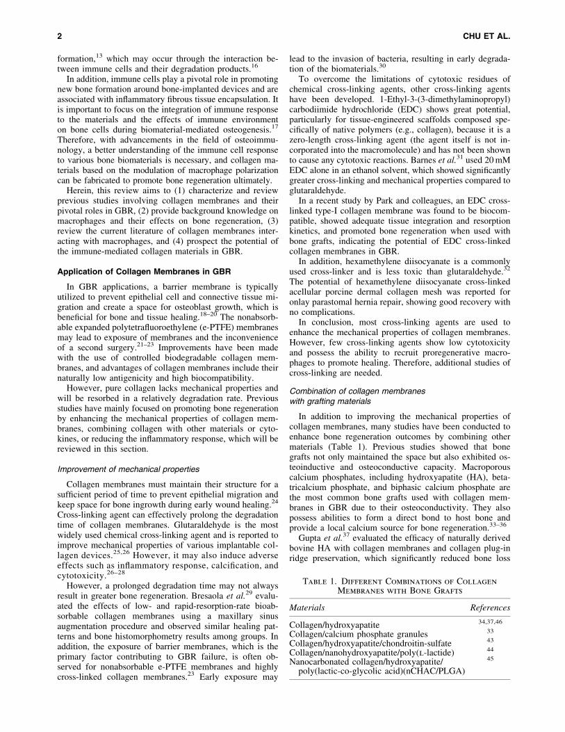
In addition to improving the mechanical properties ofcollagen membranes, many studies have been conducted toenhance bone regeneration outcomes by combining othermaterials (Table 1). Previous studies showed that bonegrafts not only maintained the space but also exhibited os-teoinductive and osteoconductive capacity. Macroporouscalcium phosphates, including hydroxyapatite (HA), beta-tricalcium phosphate, and biphasic calcium phosphate arethe most common bone grafts used with collagen mem-branes in GBR due to their osteoconductivity. They alsopossess abilities to form a direct bond to host bone andprovide a local calcium source for bone regeneration.33–36
Gupta et al.37 evaluated the efficacy of naturally derivedbovine HA with collagen membranes and collagen plug-inridge preservation, which significantly reduced bone loss
Table 1. Different Combinations of Collagen
Membranes with Bone Grafts
Materials References
Collagen/hydroxyapatite 34,37,46
Collagen/calcium phosphate granules 33
Collagen/hydroxyapatite/chondroitin-sulfate 43
Collagen/nanohydroxyapatite/poly(l-lactide) 44
Nanocarbonated collagen/hydroxyapatite/poly(lactic-co-glycolic acid)(nCHAC/PLGA)
45
2 CHU ET AL.
compared to naturally healing extraction sites. In addition,chondroitin sulfate is an important structural component ofcartilage, and also provides its resistance to compression.38
The benefits of this HA/collagen/chondroitin-sulfate spacerhave been observed in several animal and human studies,39–42
demonstrating its potential in bone regeneration. Benqueet al.43 used collagen membranes with an HA/collagen/chondroitin-sulfate spacer and observed significant reduc-tion in pocket depth (the depth of the gingival sulcus nearthe point at which the gingival tissue contacts the tooth) andgain in attachment level (the distance between junctionalepithelium and enamel-cementum junction).
In recent years, the biomimetic mineralized collagen ma-terials, including nanoapatite/collagen composites, attractedmuch attention. Liao and Cui44 reported a biomimetic bonescaffold composite, nanohydroxyapatite/collagen/poly(l-lactide),to promote bone regeneration. Moreover, Liao et al.45 fabri-cated a nanocarbonated hydroxyapatite/collagen/poly (lactic-co-glycolic acid) (nCHAC/PLGA) porous membrane, whichshowed enough high mechanical strength and high biocom-patibility and osteoconductivity. While the combination ofcollagen and bone grafts has been extensively applied in oralreconstruction, the technology seems not to be limited. Theuse of a structural porous hydroxyapatite (HAp) graft with acollagen membrane is also beneficial for guide segmentallong-bone defects of the axial skeleton.46
In summary, additional bone grafting materials for spacecreation, osteoinduction, and osteoconduction may improvebone formation outcomes. However, macrophage homeo-stasis is difficult to achieve when using a combination ofbiomaterials, including the collagen membranes and bonegrafting materials applied in bone regeneration. To preventinflammation caused by various biomaterials, some studiesfocus on the combination of collagen membranes and cyto-kines to improve outcome of bone regeneration, which will bediscussed in ‘‘Addition of growth factors and immune-relatedcytokines to collagen membranes’’ section.
Addition of growth factors and immune-relatedcytokines to collagen membranes
Many attempts have been made to promote bone regen-eration by combining growth factors and immune-relatedcytokines with collagen.47 Li et al.48 prepared a double-layered collagen membrane incorporated with basic fibroblastgrowth factor (bFGF)-loaded chitosan–heparin nanoparticles.bFGF maintained its bioactivity after release from the mem-branes, which may have potential applications in tissue re-pair for significantly improving cell proliferation when thecells are seeded onto different layers of the membranein vitro. Bone morphogenetic proteins (BMPs) can triggerangiogenesis and the migration and proliferation of mes-enchymal cells, and their differentiation into osteoblasts.49
In rat calvarial defects, Jo et al. investigated the effect ofsequential delivery of BMP-2 and BMP-7 on bone regener-ation with a HA/tricalcium phosphate bone graft coveredwith a collagen membrane, which significantly induced newbone formation.50 In addition, growth/differentiation factor5 is essential for the development and formation of bone,joints, tendons, and ligaments in the axial and appendicu-lar skeleton.51,52 Yamano et al. demonstrated that growth/differentiation factor 5 released from collagen membranes
significantly increased cell proliferation osteogenic geneexpression without showing cytotoxicity in MC3T3-E1 cells,and significantly enhanced and accelerated bone regenera-tion in a mandibular defect model.53
In addition, some studies applied the immune-relatedcytokines on collagen materials. Minardi et al.54 fabricated acollagen scaffold releasing IL-4, which revealed an over-expression of anti-inflammatory associated genes (Il-10,Mrc1, and Arg1) at the first 48 h, resulting in acceleration ofresolution of the inflammatory phase. Moreover, IFN-g andIL-4 were loaded on a collagen scaffold to harness the an-giogenesis, which was done by Spiller et al.55
Overall, collagen membranes associated with cytokinesare beneficial for bone regeneration and can be used as res-ervoirs to achieve spatiotemporal control of cytokine deliv-ery. However, the amount as well as the release of cytokinesand drugs is difficult to control, and the achievement ofspatiotemporal delivery is challenging. This was recentlyexemplified in a study by Chu et al.,56 who showed that ahigher concentration of epigallocatechin-3-gallate (EGCG),a component extracted from green tea with various biolog-ical effects, did not improve cell viability and even had aninhibitory effect, although a lower concentration of EGCGsignificantly improved the mechanical properties of collagenmembranes and cell viability compared to pure collagen.
However, after applying polyethylene glycol (PEG)modification on the surface by Chu et al., collagen mem-branes cross-linked by a higher concentration of EGCG alsopossessed an improved cell viability with lower level ofsecretion of tumor necrosis factor alpha (TNF-a).57 In ad-dition, another study by Mah et al.58 suggested that theosteogenic effect of EGCG depended largely on the con-centration and may switch from promotion to inhibition atincreasing concentrations. Therefore, the amounts of cyto-kines and drugs loaded onto collagen membranes for boneregeneration require further analysis and are difficult tocontrol, risking the outcomes of healing. Nevertheless, af-ter the implantation of collagen materials, it is crucial toachieve the balance of biomaterial-caused FBR.
Regulation of collagen-based FBR
Collagen can also elicit a material-dependent inflamma-tory reaction. Moreover, the inappropriate FBR will leadto peri-implantitis and boss loss, resulting in failure of im-plant surgeries.8 Therefore, many efforts have been made toreduce the FBR caused by the introduction of collagenmembranes. Wang et al.59 fabricated an anti-inflammatorycell-free collagen/resveratrol scaffold with improved mechan-ical strength and excellent cytocompatibility. After implanta-tion into osteochondral defects in rabbits, inflammatory-relatedgenes were downregulated and bone- and cartilage-relatedgenes were upregulated compared to untreated defects. Inaddition, a recent study by Chu et al.56 presented a novelcollagen membrane cross-linked by EGCG and successfullyenhanced the mechanical properties and regulated levelsof inflammatory cytokines secreted by preosteoblast cells.
However, the rapid physiological resolution of inflam-mation is beneficial for bone healing60,61 and an initial M1population is required to clear the wound site. Therefore, thedesign of implantable collagen membranes for bone re-generation should attempt to modulate the inflammatory
COLLAGEN MEMBRANE IN GUIDED BONE REGENERATION 3

response, while also avoiding the presence of chronic inflam-mation,62 but not blindly repressing the inflammatory reaction.
Applications as pseudo-periosteumin bone regeneration
Because numerous studies have focused on scaffolds asalternatives for bone defects,63–67 the periosteum has notbeen widely examined. However, the periosteum can supplysufficient blood and nutrition for bone tissue. Therefore,GBR based on functions of periosteum have gained in-creasing focus these years.68 The periosteum not only pro-vides mechanical support to tendons but also plays a keyrole in bone regeneration,69–73 and is involved in cell ar-rangement, collagen fiber alignment, direction of bone de-velopment, and osteoblast formation, which are necessaryfor remodeling injured bones.74–76 In addition, studies havealso indicated that the periosteum increases the rate andquantity of bone formation and improves vascular invasionin large segmental defects.77
Collagen is the main component of the fibrous layer of theperiosteum; moreover, it has been reported that collagenfiber orientation in the periosteum is preferentially associ-ated and aligned in the direction of long bone growth.71
Therefore, collagen membranes may serve as pseudo-periosteums. Shi et al.78 used a stem-cell- and endothelial-cell-laden collagen membrane as a pseudo-periosteum tocover a three-dimensional porous scaffold and observed re-markable osteogenesis compared to that in periosteum-freescaffolds. In addition, collagen membranes can support andpromote the proliferation of human periosteal cells.79
However, the requirement for the hierarchical topo-graphic surface of the periosteum, which is important forbone elongation, may not be satisfied by collagen mem-branes, as they cannot effectively anchor bone scaffoldsas nanoscale materials.80 In addition, osteal macrophages(OsteoMacs) were found to be distributed within both theendosteum and the periosteum,81 which are closely asso-ciated with bone modeling and will be discussed later.Moreover, it was reported that macrophages are responsiblefor both the initiation and the progression of early endo-chondral ossification by periosteal callus formation.82 How-ever, few studies have examined the mechanism of action ofthe pseudo-periosteum on the immune system, including theinteraction with OsteoMacs. Further studies of applicationsof collagen membranes as pseudo-periosteums are needed.
Collagen membranes have been widely used in GBR, andstudies have mainly focused on its direct effects on osteo-blasts and bone by prolonging the degradation time, in-corporating with grafting materials, adding cytokines, anddownregulating the inflammatory response, or serving as thepseudo-periosteum. However, the importance of immunecells and their reactions with materials that facilitate newbone formation have not been widely examined in the fieldof bone regeneration and collagen-based materials, whichwill be later discussed in this article.
Macrophages: Key Roles in GBR
Immune cells play a pivotal role in the process of healingafter implantation of collagen materials, which involves acharacteristic sequence of events, including hemostasis, in-
flammation, proliferation, and remodeling.83 After the he-mostasis phase, molecular factors secreted by immune cellsare involved in inflammatory reactions. In addition, mac-rophages mediate cellular proliferation, angiogenesis, newECM deposition, and granulation tissue formation in theproliferation cascade by secreting cytokines, including epi-dermal growth factor (EGF), bFGF, transforming growthfactor alpha and beta (TGF-a, TGF-b), and vascular endo-thelial growth factor (VEGF).83–85
Moreover, the outcome of the subsequent remodelingphase may involve macrophage participation and polariza-tion during the proliferation stage. The remodeling phaseinvolves matrix metalloprotease (MMP) and tissue inhibitorof metalloproteinase-mediated degradation and remodelingof the newly deposited collagen, which also involves mac-rophages.83,86 Macrophages are thought to mainly reside inthe peripheral blood and tissue and play different roles de-pending on their functional diversity, which will be discussedin ‘‘Blood-resident macrophages’’ and ‘‘Tissue-resident mac-rophages’’ section.
Blood-resident macrophages
Macrophages derived from circulating monocytes circu-late in the peripheral blood after leaving the bone marrowand become activated after migration into inflamed tissue(Fig. 1). Related to their functional diversity, they polarizeinto the proinflammatory M1 and anti-inflammatory M2state (Table 2 for details and the following studies),10–12,87–93
mirroring the T helper cell 1/T helper cell 2 (Th1/Th2)described for T helper cells.94
The ‘‘classically activated’’ M1 phenotype, induced bychemoattractants such as IFN-g and LPS,9 is capable ofproducing proinflammatory cytokines (such as IFN-g andinterleukin-2 [IL-2] in response to intracellular pathogens bypromoting Th1 differentiation of lymphocytes). These cellsalso kill intracellular pathogens by iron restriction, pre-venting the growth of bacterial infections.10 In addition, anincreased proinflammatory response of M1 macrophagesmay harm nearby cells by producing toxic reactive oxygenintermediates.88 In addition, a severe FBR as well as fibrousencapsulation escalated by M1 macrophages may result inthe failure of biomaterial integration.16
However, participation of M1 macrophages is essential forpromoting healing by the implantation of degradable biomate-rials such as collagen. As discussed in the Introduction, a delayor prevention in macrophage-mediated degradation by cross-linking agents may inhibit the beneficial M2 response anddecrease tissue formation,13 which may occur through inter-actions between host cells and their degradation products.16
The ‘‘alternatively activated’’ M2 phenotype of macro-phages, induced by signals such as IL-4 and IL-13, releaseanti-inflammatory cytokines such as IL-10,11 which con-tribute to tissue remodeling10 in contrast to M1 macro-phages. M2 macrophages are further divided into differentsubsets (i.e., M2a, M2b, and M2c) based on their func-tional diversity.89 Briefly, M2a is associated with Th2 anti-inflammatory responses. The M2b phenotype performsimmune regulatory functions (both proinflammatory andanti-inflammatory function).90 In contrast, the M2c phe-notype plays a major role in matrix deposition, tissue re-modeling, and prohealing.
4 CHU ET AL.

FIG. 1. The roles of macrophages on bone regeneration. Monocytes leaving from bone marrow circulate in blood anddifferentiate into M1 and M2 macrophages. M1 phenotype is induced by IFN-g and LPS, and may have characteristic of M2cells when it becomes endotoxin tolerance. Meanwhile, M2 macrophages is induced by signals such as IL-4 and IL-13, andcan be induced to be M1-like cells when exposed to LPS or IFN-g. In addition, tissue-resident macrophages (OsteoMacs)also play roles in bone regeneration. After arriving at the bone defects, M2 macrophages and OsteoMacs promote boneregeneration by secreting proregenerative cytokines, including IL-10, VEGF, and BMP-2, while M1 phenotype functions bysecreting proinflammatory cytokines such as IL-1b and TNF-a. BMP, bone morphogenetic protein; IFN-g, interferon-gamma; IL, interleukin; LPS, lipopolysaccharide; TNF-a, tumor necrosis factor alpha; VEGF, vascular endothelial growthfactor. Color images available online at www.liebertpub.com/teb
Table 2. Subsets of Macrophages
Type Inducers SecretionsExpressedmarkers
Signal pathways(transcription
factors) Functions References
M1 LPS, IFN-g IL-1b, 6, 12, 15, 18,23, IL-10low, TNF-a,CCL-15, 20, CXCL-9,10, 11, 13, RNI, ROI
MHCII, CD86,TLR, IFN-gR
MyD88, TRIF,Jak/NF-kB(p65 and p50),AP-1, IRF3,STAT1
(1) Promotion ofTh1 response
10,11,88
(2) Proinflammatory(3) Clearance of
intracellularpathogensclearance
(4) Tissue damage
M2a IL-4, IL-13,IL-33
IL-10, 1ra, IL-12low,CCL-13, 14, 17, 18,22, 23, 24, 26, IGF-1,PDGF, TGF-b, YM1,FIZZ1, arginase,polyamines
MR, GR, SR,CD163, CD23,IL-1 decoyreceptor,Lyve-1, ST2,IL-4Ra, FR
Jak/STAT6,IRF4,PPAR-g, p50
(1) Anti-inflammatory 12,90
(2) Clearanceof parasites
(3) Promotion ofallergic responses
M2b Immunecomplexes,LPS, IL-1b,
IL-10, IL-1blow,IL-12low, IL-6low,TNF-alow, CCL-1,20, CXCL-1, 2, 3
IL-1R, IL-10R,TLR4, FcgR
PI(3)K, Syk,SHP-1, SHIP
(1) Immunoregulation 87,92
(2) Interaction withB cells
M2c IL-10, GC,TGF-b
CCL-16, 18, CXCL13,TGF-b, BMP-2,matrix (versican,PTX3, a anti-trypsin)
CD163, CD206,IL10R, PD-1
Jak/STAT3 (1) Matrix deposition 11,89,91,93
(2) Tissue remodeling
BMP, bone morphogenetic protein; CCL, chemokine ligand; CD, cluster of differentiation; CXCL-a, chemokine ligands; FR, folatereceptor; GF, insulin-like growth factor; GR, galactose receptor; IFN-gR, IFN-g receptor; IFN-g, interferon-gamma; IL, interleukin; IRF,IFN regulatory factor; Jak, Janus kinase; LPS, lipopolysaccharide; MHC, major histocompatibility complex; MR, mannose receptor; PDGF,platelet-derived growth factor; PTX3, pentraxin 3; RNI, reactive nitrogen intermediate; ROI, reactive oxygen intermediate; SR, scavengingreceptor; STAT, signal transducers and activators of transcription; TGF, transforming growth factor; Th1, T helper cell 1; TLR, toll-likereceptor; TNF-a, tumor necrosis factor alpha.
5
Although M2 macrophages have been identified as pro-healing cells that promote ECM production by secretingIL-4 and TGF-b, overstimulation of ECM deposition maylead to unfavorable fibrosis, as both IL-4 and TGF-b arestrong inducers of fibrosis without tight regulation.95 Inaddition, monocyte chemotactic protein 1/chemokine ligand2 (MCP-1/CCL-2) has been identified as a profibrotic me-diator implicated in the stimulation of M2 polarization.96,97
However, inhibition of M2 macrophages and these moleculesmay terminally lead to impaired tissue healing.98 Therefore,identifying a method to stimulate sufficient functional ECMdeposition without collagen encapsulation and fibrosis re-mains a primary challenge in bone regeneration.
In summary, the presence of macrophage polarization inresponse to various microenvironments may be advanta-geous for constructive remodeling, while minimizing un-desirable inflammatory reactions.
Tissue-resident macrophages
In contrast to the original theory that macrophages de-rived from circulating monocyte precursors are recruited tosites of inflammation, more recent research has confirmedthat some tissue-resident macrophages subdivide and pro-liferate to sustain their local populations during injuryevents.99,100 Previously, monocyte subsets in human bloodwere categorized into two main subpopulations, CD14+
monocytes and CD14lowCD16+ monocytes.101 Mouse stud-ies have suggested that CD14+ monocytes in humans ter-minally represent classical monocytes in the blood stream.In contrast, resident macrophages102,103 may be differenti-ated by CD14lowCD16+ monocytes. OsteoMac is a CD68+
cell type of macrophage in bony tissues and is derived fromresident macrophages such as macrophages in other tissues,serving as a special canopy structure overlying mature os-teoblasts (Fig. 1).99,104,105
The general role of OsteoMacs has been described asimmune surveillance in the bone microenvironment. Previousstudies have shown that OsteoMacs can detect bacterialproducts106,107 and are highly involved in antigen presenta-tion.108,109 Further evidence suggests that these cells respondto LPS.81 Collectively, OsteoMacs may also function asphagocytes with some characteristics of the M1 macrophagediscussed in ‘‘Tissue-resident macrophages’’ section.
However, Chang et al. observed a 23-fold reduction inmineral deposition after removing macrophages from pri-mary osteoblast cultures.81 Currently, OsteoMacs are knownto produce a wide variety of either proinflammatory or anti-inflammatory cytokines. OsteoMacs have been shown tosecrete proinflammatory factors, including TNF-a,110,111 IL-6,112
IL-1b,110,113 and IFN-g114,115 under various conditions. How-ever, further studies showed that OsteoMacs produce anumber of bioactive cytokines, including TGF-b,116 osteo-pontin,117 1,25-dihydroxy-vitamin D3,118 and BMP-2,119
which are classical characteristics of the M2 macrophage andserve as inducers of ECM deposition and new bone formation.These observations demonstrate that the specific functions ofOsteoMacs are difficult to clearly define because of the po-tential cross-lineage plasticity and cross talk between cells.
OsteoMacs were found to encapsulate functionally ma-ture osteoblasts, described as ‘‘forming a canopy-like cellstructure,’’ suggesting that they are heavily involved in the
bone modeling process.81,120 Moreover, in a macrophageFas-induced apoptosis (Mafia) transgenic mouse model,inducing macrophage depletion, the complete loss of ma-ture osteoblasts and bone modeling at the bone inter-face was observed when the OsteoMac canopy architecturewas disrupted, demonstrating the functional importance ofOsteoMacs.121 Moreover, OsteoMacs are involved in boneregeneration by secreting VEGF and BMP-2.119
In contrast to bone modeling, the process of bone remodelinginvolves a careful and coordinated balance between bone-resorbing osteoclasts and bone-forming osteoblasts.122,123
TGF-b and ephrin B2 are possible coupling factors betweenosteoclasts and osteoblasts.122,124–126 Previous studies showedthat OsteoMacs are capable of producing TGF-b116 andephrin B2127,128 as discussed in ‘‘Tissue-resident macro-phages’’ section, and OsteoMacs may also be required for boneremodeling. Moreover, based on their close proximity to bonesurfaces as a canopy structure and well-known ability to detectdying cells, OsteoMacs may be candidates for detecting andresponding to bone damage, which is critical for osteoclastrecruitment that initiates the bone remodeling phase.129
Regulation of macrophage polarization
Macrophages polarization into proinflammatory (M1)and anti-inflammatory (M2) phenotypes based on func-tional diversity has been increasingly recognized theseyears. Many studies have focused on the mechanisms ofmacrophage polarization. The global regulatory landscapecontrolled by the E26 transformation-specific (ETS) familytranscription factor PU.1 represents the general transcriptionfactors regulated by external stimuli to modulate macro-phage function.130
PU.1 is constantly expressed at high levels to induce andmaintain macrophage differentiation.131–133 PU.1 binding issufficient to create small open regions of accessible DNAthat can be bound by other transcription factors that prob-ably contribute to the differentiation of macrophages, suchas IFN regulatory factor 8 (IRF8),134 or for functionalspecialization, such as IRF4 and IRF5 (which are involvedin M2 and M1 polarization, respectively) and CCAAT/enhancer-binding protein-b (C/EBPb).135–137
Some key transcription factors translate signals in themicroenvironment into a polarized macrophage phenotype.The signal transducers and activators of transcription(STAT) family play an important role in M1 and M2 mac-rophage polarization. M1 characteristics (major histocom-patibility class II molecules, nitric oxide synthase 2, andIL-12)99 are mainly promoted by IFN-g-mediated Januskinase–signal transducer and activator of transcription (JAK–STAT) signaling.138 In addition, polarization of M1 macro-phages is activated by LPS, which depends on the autocrineproduction of IFN-b through TRIF-dependent signalingfrom Toll-like receptor 4 (TLR4) to IRF3.139
M2 factors, including arginase 1, macrophage mannosereceptor 1 (also known as Cd206), resistin-like a (alsoknown as Fizz1), and chitinase 3-like 3 (also known asYm1),90 are regulated by IL-4Ra signals through a JAK–STAT6 pathway.140 Importantly, IL-4 triggers the activationof phosphoinositide 3-kinase,141 which is also crucial for inM2 macrophage polarization. Adipose tissue macrophagestypically have an anti-inflammatory M2-like phenotype.
6 CHU ET AL.
Peroxisome proliferator-activated receptor-gamma (PPAR-g) is a master regulator of lipid metabolism in M2 polari-zation,142 which can also be induced by IL-4 and IL13.143
The reciprocal regulation of M1 and M2 genes by thesame transcription factors is common in macrophage po-larization, as is exemplified by the mutual antagonism ofSTAT1 and STAT6. cAMP responsive element bindingprotein (CREB) may be a crucial transcription factor in mac-rophage polarization by promoting M2 polarization, whilerepressing M1 activation. CREB-induced C/EBPb is respon-sible for arginase 1 expression in response to TLR ligands,which promote M2 macrophages. In addition, CREB plays ananti-inflammatory role in response to LPS, which repressesM1 activation by mediating p38 mitogen-activated proteinkinase and mitogen- and stress-activated kinases 1 and 2.144,145
IFN regulatory factors also direct the differentiation ofM1 and M2 macrophages. IL-4-induced IRF4 was shown tospecifically regulate M2 macrophage polarization in re-sponse to fungi and helminthes involving JMJD3, whichis also controlled by the expression of IRF4.138,146,147 How-ever, overexpression of IRF5 is associated with granulocyte–macrophage colony-stimulating factor-induced humanmacrophages (an M1-like phenotype), which also showedthat the IFN regulatory factor is one of the reciprocal reg-ulations of M1 and M2 genes.
Moreover, macrophages possess dynamic and plasticphenotypes in response to local microenvironmental sig-nals,148–151 which depend on the type, concentration, andduration of the polarizing signals. It is found that a mixtureof macrophage phenotypes is shown at any time amongmacrophage population. There are even macrophages in astate that express markers of both M1 and M2 phenotypes.16
This may due to the changes in stimuli. Experiments haveshown that secretion of LPS-stimulated macrophages maychange from proinflammatory cytokines to anti-inflammatoryfactors such as IL-10, when the macrophages become TLRtolerant.151–153 Similarly, M2 cells can be readily induced tohave characteristic of M1 cells when exposure to LPS orIFN-g. However, the mechanisms of macrophage phenotypeplasticity are not well understood.
Modulation of Collagen Membranes Basedon Response to Immune Cells
As described above, the introduction of collagen membranesmay cause the FBR involving macrophages, which may impairbiomaterial integration. A previous study contributed to re-ducing the material-based inflammatory response.56,59 How-ever, with advancements in the understanding of macrophageresponses to implanted biomaterials, immune-mediated tis-sue regeneration elicited by biomaterials such as collagenis emerging as an innovative regenerative strategy.154
The integration among immune cells, bone cells,and collagen membranes
Generally, after the implantation of collagen membranes,a layer of proteins from the surrounding vasculature adsorbsonto the surface, leading to infiltration and adherence ofcells such as platelets, monocytes, and macrophages. Mac-rophages then recruit tissue repair cells (e.g., fibroblastsand mesenchymal stem cells) to the inflammation site byreleasing cytokines and chemokines. These sequences con-
stitute the natural innate immune response following bio-material implantation.155
The close relationship that exists between the immunesystems and collagen membranes is a double-edged sword.To be specific, a favorable FBR creates an osteogenic mi-croenvironment, while an inappropriate FBR may lead to thechronic inflammation and the formation of a fibrous capsulearound the implant. The fibrous capsule prevents direct inter-action between surrounding environment and the biomaterialsso that the materials can be maintained in the host body forsufficient time.156 However, the fibrous encapsulation alsoseparates the biomaterials with bone marrow. Undesirably,bone cells cannot attach to the surface of materials to form newbone, and the defect will be filled by fibrous tissue instead.
It is reported that the process of bone biomaterial-mediated de novo bone formation process can be dividedinto three phases: early, bone formation, and remodelingphase. The early stage is dominated by the inflammatoryphase, when the majority of macrophages would be the in-flammatory M1 phenotype. It is crucial to have a timelyswitch from M1 to M2 macrophage phenotype, which willlead to osteogenic cytokine release and new bone formation.However, a prolonged M1 polarization phase leads to anincrease in polarization of M2 macrophages that havefibrosis-enhancing cytokine release pattern, resulting in fi-brous encapsulation and the failure of bone biomaterials.157
Therefore, the osteoimmunomodulation (OIM) is impor-tant during biomaterial-mediated osteogenesis. The OIMnot only considers the immune response to the materials butalso focuses on the effects of the immune environment onthe behavior of bone cells after biomaterial implantation.17
The immune and bone cells are closely related by sharinga number of cytokines, receptors, signaling molecules, andtranscription factors,158,159 and immune cells, includingOsteoMacs, play a key role in bone homeostasis. For in-stance, immunomodulatory cytokines such as IFNs par-ticipate in the regulation of RANKL (an osteoclastogeniccytokine) signaling and inflammatory bone loss.
In addition, immunoglobulin-like receptors are critically in-volved in bone homeostasis.160 Altogether, better understand-ing and treatment of bone augmentation by immune-mediatedbiomaterials are greatly emerging. As the new generation ofbone biomaterials, they should possess abilities to modulate thelocal immune environment that favors osteogenesis and os-seointegration,17 including mediation of macrophages recruit-ment and polarization, which will be discussed in ‘‘Regulationof immune cells by biomaterials, including collagen’’ section.
Regulation of immune cells by biomaterials,including collagen
The behaviors of immune cells can be regulated by thebiomaterial surface properties, particle size, porosity, andthe released ions from the biomaterials. Previous studiesdeveloped strategies for reducing protein adsorption, initialcell adhesion, and foreign body giant cell fusion aroundbiomaterials,161 such as collagen, to reduce the FBR bychanging the surface chemistry of the materials.
Accordingly, biomaterial surface properties, includingwettability and surface charge, are strong regulators ofmacrophages in tissue engineering.162 Studies have dem-onstrated that the release of IL-1b, TNF-a, IL-6, IL-8, and
COLLAGEN MEMBRANE IN GUIDED BONE REGENERATION 7
macrophage inflammatory protein-1b preferred hydrophilicsurfaces, reducing the adhesion of macrophages comparedto hydrophobic surfaces.162–164 These may be determined bythe interaction of absorbed protein with immune cells andsubsequent behaviors of immune cells.165
Most proteins are hydrophilic on their outside and thustend to bind on the hydrophobic surfaces.166 In addition, it isalso generally accepted that positively charged surfaces aremore feasible to promote inflammatory response than neg-atively charged and neutral surfaces.167 Therefore, changeson surface properties of collagen membranes may be ben-eficial for the modulation of macrophages.
In addition, porosity and pore size of bone materials aretwo important factors to determine the ingrowth tissue types(inflammatory granuloma tissue, vascular tissue, and bonetissue).168 It is more beneficial for the ingrowth of bonetissue when the scaffolds possess higher porosity of 80–88%and macroporosity (pore size >50 mm).168 Moreover, po-rosity and pore size are involved in macrophage polariza-tion, which has been studied in the collagen-based materials.
Koyal et al.169 investigated the effect of fiber and poresize of a collagen-based scaffold on the polarization ofmouse bone marrow-derived macrophages toward regener-ative (M2) or inflammatory (M1) phenotypes. As fiber di-ameter, pore size, and porosity increased, expression of theM2 marker arginase 1 increased and expression of the M1marker inducible nitric oxide synthase decreased. Secretionof the angiogenic cytokines VEGF, TGF-b1, and bFGF wasalso higher in larger fibers and larger pore-size scaffolds.
Moreover, their results showed that pore size is morecrucial for macrophage polarization than fiber diameter.This may be because macrophages are more prone to infil-trate in materials with larger pores. In addition, it is reportedthat maximum vascularization and minimal fibrotic re-sponse, with an increased number of M2 phenotype mac-rophages, are observed in the cardiac implantation ofhydrogels with pore diameters of 30–40 mm.170
Meanwhile, ions released from the biomaterials duringdegradation can alter the local biological environment, re-sulting in modulation of immune response. Calcium (Ca) isfound to enhance inflammation through the Wnt5A/Ca2+
signaling pathway.171 Silicon (Si) participates in the earlymineralization phase of bone regeneration around activecalcification sites.172 Mg2+ ions can downregulate secretionof inflammatory cytokine by inhibiting the TLR pathway,173
which is the pathway macrophages used to recognize for-eign bodies during the process of rejection.7 TLR3 can onlysignal by the adaptor protein toll-like receptor adaptormolecule (Ticam), which is also known as TIR domaincontaining adapter inducing IFN-b. However, TLR4 canproceed by both pathways (TRIF and MyD88 pathways).Both MyD88-dependent and TRIF-dependent pathways ex-press inflammatory cytokines by recruiting NF-kB.174
However, the related irons released from collagen mem-branes have not been studied. Further studies can investigatethe effects of Ca2+, Si, and Mg2+ released from collagenmembranes on regulation of immune cells.
Collagen based on modulation of macrophages in GBR
Several studies have shown that collagen can recruitmacrophages. It was reported that the molecular events of
the collagen membrane in the GBR are associated withthe upregulation of cell recruitment and bone remodelinggenes in early and late periods and accumulation ofFGF-2 and BMP-2 proteins and immunoreactive cellswere found around the implanted membranes.175
Moreover, Sicari et al.176 used an acellular biologicscaffold (the main component of the scaffold is collagen) toaid in volumetric muscle loss. They found that acellularscaffold implantation was associated with the mobilizationand accumulation of perivascular stem cells and de novoformation of skeletal muscle cells at the injury site, resultingin functional improvement.
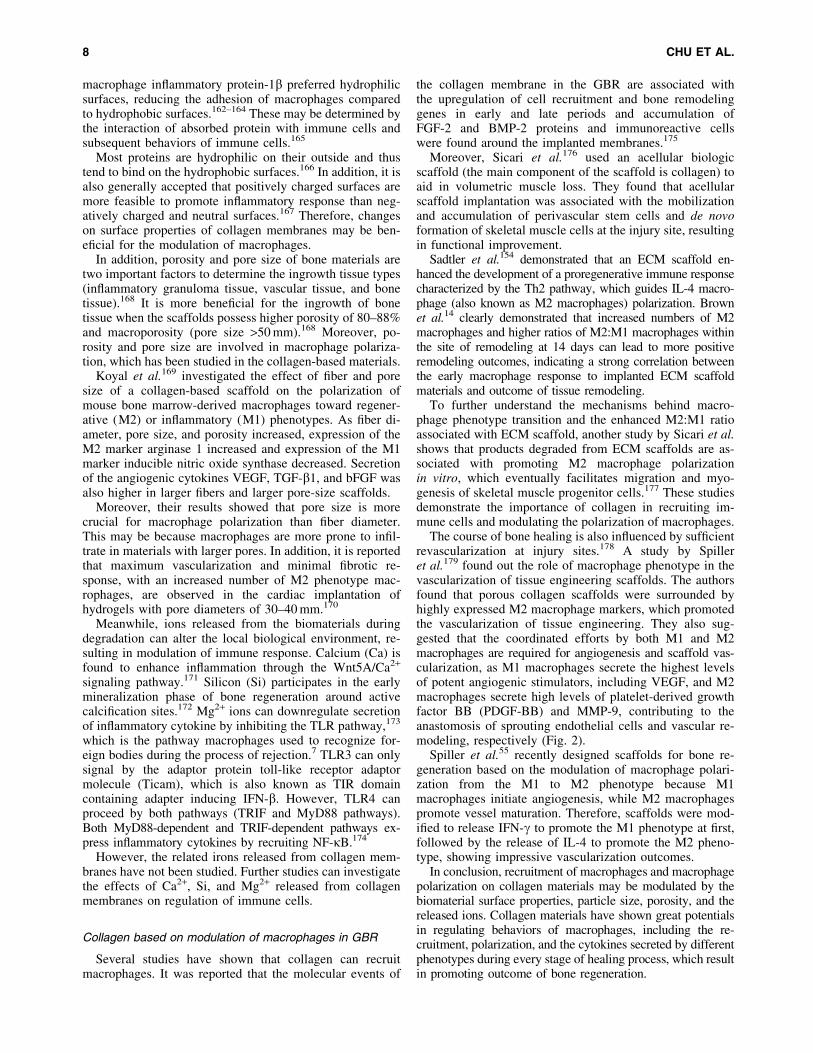
Sadtler et al.154 demonstrated that an ECM scaffold en-hanced the development of a proregenerative immune responsecharacterized by the Th2 pathway, which guides IL-4 macro-phage (also known as M2 macrophages) polarization. Brownet al.14 clearly demonstrated that increased numbers of M2macrophages and higher ratios of M2:M1 macrophages withinthe site of remodeling at 14 days can lead to more positiveremodeling outcomes, indicating a strong correlation betweenthe early macrophage response to implanted ECM scaffoldmaterials and outcome of tissue remodeling.
To further understand the mechanisms behind macro-phage phenotype transition and the enhanced M2:M1 ratioassociated with ECM scaffold, another study by Sicari et al.shows that products degraded from ECM scaffolds are as-sociated with promoting M2 macrophage polarizationin vitro, which eventually facilitates migration and myo-genesis of skeletal muscle progenitor cells.177 These studiesdemonstrate the importance of collagen in recruiting im-mune cells and modulating the polarization of macrophages.
The course of bone healing is also influenced by sufficientrevascularization at injury sites.178 A study by Spilleret al.179 found out the role of macrophage phenotype in thevascularization of tissue engineering scaffolds. The authorsfound that porous collagen scaffolds were surrounded byhighly expressed M2 macrophage markers, which promotedthe vascularization of tissue engineering. They also sug-gested that the coordinated efforts by both M1 and M2macrophages are required for angiogenesis and scaffold vas-cularization, as M1 macrophages secrete the highest levelsof potent angiogenic stimulators, including VEGF, and M2macrophages secrete high levels of platelet-derived growthfactor BB (PDGF-BB) and MMP-9, contributing to theanastomosis of sprouting endothelial cells and vascular re-modeling, respectively (Fig. 2).
Spiller et al.55 recently designed scaffolds for bone re-generation based on the modulation of macrophage polari-zation from the M1 to M2 phenotype because M1macrophages initiate angiogenesis, while M2 macrophagespromote vessel maturation. Therefore, scaffolds were mod-ified to release IFN-g to promote the M1 phenotype at first,followed by the release of IL-4 to promote the M2 pheno-type, showing impressive vascularization outcomes.
In conclusion, recruitment of macrophages and macrophagepolarization on collagen materials may be modulated by thebiomaterial surface properties, particle size, porosity, and thereleased ions. Collagen materials have shown great potentialsin regulating behaviors of macrophages, including the re-cruitment, polarization, and the cytokines secreted by differentphenotypes during every stage of healing process, which resultin promoting outcome of bone regeneration.
8 CHU ET AL.

Potentials of Collagen Membranes Basedon Macrophage Polarization
Using collagen materials to manipulate the adaptive im-mune system may promote both systemic and local pro-regenerative immune responses and ultimately stimulatetissue repair.154 Modulation of collagen membranes basedon the response to immune cells is still faced with obstacles,including the reactions of macrophage phenotypes in re-sponse to different microenvironments and the interactionsamong various biomaterials, macrophages, and bone cells.
Putten et al.180 suggested that many macrophages in theFBR induced by hexamethylene diisocyanate-cross-linkeddermal sheep collagen do not always fit into the classicalM1 or M2 dichotomy, as some macrophages exhibited anM2 phenotype, while the M1 phenotype was not detected.This may be because macrophages present different phe-notypes in response to different microenvironments of theFBR. Macrophage phenotypes are associated with the out-come of bone regeneration.
However, it is reported that the time of M1 macrophagespresent during the first stage of healing process may beinvolved with polarization of M2 macrophages that haveosteogenesis cytokine release pattern or fibrosis cytokinerelease pattern.157 This phenomenon may explain why un-expected results still exist even though the phenotypes aredefined. Therefore, further studies are needed to determinewhether macrophage phenotypes can be used to predict re-modeling following implantation of collagen membranes.
Moreover, the influence of various biomaterials is un-known. In dental implant treatment, collagen membranes areusually in contact with two materials, including a titanium
dental implant and bone grafting material within the tiny 1-mm region, when collagen membranes are introduced topromote bone regeneration. It is possible that, while thesetypes of biomaterials favor macrophage homeostasis andtissue integration, the others may cause a severe inflam-matory reaction.
Therefore, deeper understandings of the interaction be-tween collagen membranes and macrophage, and the cor-relation among various biomaterials are urgently needed,considering its great potential in the field. Moreover, furtherstudies can investigate the effects of Ca2+, Si, and Mg2+
released from collagen membranes on regulation of immunecells because the studies of the related irons released fromcollagen membranes are few.
Conclusion
Modulation of collagen membranes in bone regenerationwas used to focus on enhancing mechanical properties andloading grafting materials or cytokines. However, the resultswere not satisfied. The recent surge in the application of col-lage membranes has advanced to modulating inflammatoryreactions based on the improved understanding of macrophagepolarization. By modifications of surface properties, particlesize, porosity, and the released ions, an ideal immune-mediatedcollagen membrane should promote anti-inflammatory M2macrophages and secretion of proregenerative cytokines, withthe abilities of preventing migration of epithelium and main-taining space for bone ingrowth.
Further research should aim to increase the understandingof blood- and tissue-resident macrophage polarization and
FIG. 2. The schematic diagram of the new generation of GBR membranes. The new generation of GBR membranes isbased on regulation of foreign body reaction. By surface modification, the new GBR membranes may possess ability topromote recruitment and adhesion of M2 macrophages (proregenerative phenotype), which further increases secretion ofproregenerative cytokines such as VEGF, PDGF, and BMP-2 for revascularization and bone growth. Meanwhile, theyshould also maintain the functions as barriers to prevent the migration of fibroblasts and maintain space for osteoblastadhesion, proliferation, and differentiation. GBR, guided bone regeneration; PDGF, platelet-derived growth factor. Colorimages available online at www.liebertpub.com/teb
COLLAGEN MEMBRANE IN GUIDED BONE REGENERATION 9
the inflammatory reactions caused by various biomaterialsbecause the interactions among materials may lead tocomplicated FBR that make the outcome unpredictable.Improvements in collagen materials based on immunemodulation will have great potentials and ultimately lead tobetter outcomes in applications of GBR.
Acknowledgments
This work was supported by grants from the NationalNatural Science Foundation of China (No. 81671023) andthe National Key Research and Development Program ofChina (No. 2016YFA0201703/2016YFA0201700). In addi-tion, special appreciation was sent to Dr. Zetao Chen for hishelpful contribution and advice to this work.
Disclosure Statement
No competing financial interests exist.
References
1. Wang, H.L., and Boyapati, L. ‘‘PASS’’ principles forpredictable bone regeneration. Implant Dent 15, 8, 2006.
2. Tonelli, P., Duvina, M., Barbato, L., Biondi, E., Nuti, N.,Brancato, L., and Rose, G.D. Bone regeneration in den-tistry. Clin Cases Miner Bone Metab 8, 24, 2011.
3. Kostopoulos, L., and Karring, T. Augmentation of the ratmandible using guided tissue regeneration. Clin OralImplants Res 5, 75, 1994.
4. Dahlin, C., Sennerby, L., Lekholm, U., Linde, A., andNyman, S. Generation of new bone around titanium im-plants using a membrane technique: an experimental studyin rabbits. Int J Oral Maxillofac Implants 4, 19, 1989.
5. Luttikhuizen, D.T., Harmsen, M.C., and Van Luyn, M.J.Cellular and molecular dynamics in the foreign body re-action. Tissue Eng 12, 1955, 2006.
6. Anderson, J.M., Rodriguez, A., and Chang, D.T. Foreignbody reaction to biomaterials. Semin Immunol 20, 86,2008.
7. Franz, S., Rammelt, S., Scharnweber, D., and Simon, J.C.Immune responses to implants—a review of the implica-tions for the design of immunomodulatory biomaterials.Biomaterials 32, 6692, 2011.
8. Trindade, R., Albrektsson, T., Tengvall, P., and Wenner-berg, A. Foreign body reaction to biomaterials: on mech-anisms for buildup and breakdown of osseointegration. ClinImplant Dent Relat Res 18, 192, 2014.
9. Mosser, D.M. The many faces of macrophage activation. JLeukoc Biol 73, 209, 2003.
10. Recalcati, S., Locati, M., Marini, A., Santambrogio, P.,Zaninotto, F., De Pizzol, M., Zammataro, L., Girelli, D.,and Cairo, G. Differential regulation of iron homeostasisduring human macrophage polarized activation. Eur JImmunol 40, 824, 2010.
11. Koh, T.J., and DiPietro, L.A. Inflammation and woundhealing: the role of the macrophage. Expert Rev Mol Med13, e23, 2011.
12. Martinez, F.O., Sica, A., Mantovani, A., and Locati, M.Macrophage activation and polarization. Front Biosci 13,453, 2008.
13. Badylak, S.F., Valentin, J.E., Ravindra, A.K., Mccabe,G.P., and Stewartakers, A.M. Macrophage phenotype as adeterminant of biologic scaffold remodeling. Tissue EngPart A 14, 1835, 2008.
14. Brown, B.N., Londono, R., Tottey, S., Li, Z., Kukla, K.A.,Wolf, M.T., Daly, K.A., Reing, J.E., and Badylak, S.F.Macrophage phenotype as a predictor of constructive re-modeling following the implantation of biologically de-rived surgical mesh materials. Acta Biomater 8, 978,2011.
15. Brown, B.N., Valentin, J.E., Stewartakers, A.M., McCabe,G.P., and Badylak, S.F. Macrophage phenotype and re-modeling outcomes in response to biologic scaffolds withand without a cellular component. Biomaterials 30, 1482,2009.
16. Brown, B.N., Ratner, B.D., Goodman, S.B., Amar, S., andBadylak, S.F. Macrophage polarization: an opportunityfor improved outcomes in biomaterials and regenerativemedicine. Biomaterials 33, 3792, 2012.
17. Chen, Z., Klein, T., Murray, R.Z., Crawford, R., Chang, J.,Wu, C., and Xiao, Y. Osteoimmunomodulation for thedevelopment of advanced bone biomaterials. Mater Today19, 304, 2016.
18. American Academy of Periodontology. Glossary ofPeriodontal Terms, 4th Edition, Chicago, IL: AmericanAcademy of Periodontology, 2001.
19. Garrett, S. Periodontal regeneration around natural teeth.Ann Periodontol 1, 621, 1996.
20. Fritz, M.E. Implant therapy II. Ann Periodontol 1, 796,1996.
21. Pitaru, S., Tal, H., Soldinger, M., and Noff, M. Collagenmembranes prevent apical migration of epithelium and sup-port new connective tissue attachment during periodontalwound healing in dogs. J Periodontal Res 24, 247, 1989.
22. Black, B.S., Gher, M.E., Sandifer, J.B., Fucini, S.E., andRichardson, A.C. Comparative study of collagen and ex-panded polytetrafluroethylene membranes in the treatmentof human class II furcation defects. J Periodontol 65, 598,1994.
23. Crigger, M., Bogle, G.C., Garrett, S., and Gantes, B.G.Repair following treatment of circumferential periodontaldefects in dogs with collagen and expanded polytetra-fluoroethylene barrier membranes. J Periodontol 67, 403,1996.
24. Bunyaratavej, P., and Wang, H.L. Collagen membranes: areview. J Periodontol 72, 215, 2001.
25. McPherson, J.M., Sawamura, S., and Armstrong, R. Anexamination of the biologic response to injectable, glu-taraldehyde cross-linked collagen implants. J BiomedMater Res A 20, 93, 1986.
26. Simmons, D.M., and Kearney, J.N. Evaluation of collagencross-linking techniques for the stabilization of tissuematrices. Biotechnol Appl Biochem 17 (Pt 1), 23, 1993.
27. Rault, I., Frei, V., Herbage, D., Abdul-Malak, N., andHuc, A. Evaluation of different chemical methods forcros-linking collagen gel, films and sponges. J Mater SciMater Med 7, 215, 1996.
28. Speer, D.P., Chvapil, M., Eskelson, C.D., and Ulreich, J.Biological effects of residual glutaraldehyde in glutaraldehyde-tanned collagen biomaterials. J Biomed Mater Res 14,753, 1980.
29. Bresaola, M.D., Matsumoto, M.A., Zahoui, A., Biguetti,C.C., and Nary-Filho, H. Influence of rapid- and slow-rateresorption collagen membrane in maxillary sinus aug-mentation. Clin Oral Implants Res 28, 320, 2016.
30. Paul, B.F., Mellonig, J.T., and Gray, J.L. Use of a collagenbarrier to enhance healing in human periodontal furcationdefects. Int J Periodontics Restorative Dent 12, 123, 1992.
10 CHU ET AL.
31. Barnes, C.P., Pemble, C.W., Brand, D.D., Simpson, D.G.,and Bowlin, G.L. Cross-linking electrospun type II col-lagen tissue engineering scaffolds with carbodiimide inethanol. Tissue Eng 13, 1593, 2007.
32. Grieshaber, S.E., Farran, A.J.E., Lin-Gibson, S., Kiick,K.L., and Jia, X. Synthesis and characterization of elastin-mimetic hybrid polymers with multiblock, alternatingmolecular architecture and elastomeric properties. Mac-romolecules 42, 2532, 2009.
33. Jegoux, F., Goyenvalle, E., Cognet, R., Malard, O.,Moreau, F., Daculsi, G., and Aguado, E. Mandibularsegmental defect regenerated with macroporous biphasiccalcium phosphate, collagen membrane, and bone marrowgraft in dogs. Arch Otolaryngol Head Neck Surg 136, 971,2010.
34. Benque, E., Zahedi, S., Brocard, D., Marin, P., Brunel, G.,and Elharar, F. Tomodensitometric and histologic evalu-ation of the combined use of a collagen membrane and ahydroxyapatite spacer for guided bone regeneration: aclinical report. Int J Oral Maxillofac Implants 14, 258,1999.
35. Jung, R.E., Lecloux, G., Rompen, E., Ramel, C.F., Buser,D., and Hammerle, C.H. A feasibility study evaluating anin situ formed synthetic biodegradable membrane forguided bone regeneration in dogs. Clin Oral Implants Res20, 151, 2009.
36. Guda, T., Appleford, M., Oh, S., and Ong, J.L. A cellularperspective to bioceramic scaffolds for bone tissue engi-neering: the state of the art. Curr Top Med Chem 8, 290,2008.
37. Gupta, D., Gundannavar, G., Chinni, D.D., and Alampalli,R.V. Ridge preservation done immediately following ex-traction using bovine bone graft, collagen plug and col-lagen membrane. Int J Oral Implantol Clin Res 3, 8, 2012.
38. Leeb, B.F., Schweitzer, H., Montag, K., and Smolen, J.S.A metaanalysis of chondroitin sulfate in the treatment ofosteoarthritis. J Rheumatol 27, 205, 2000.
39. Ogilvie, A., Frank, R.M., Benque, E.P., Gineste, M.,Heughebaert, M., and Hemmerle, J. The biocompatibilityof hydroxyapatite implanted in the human periodontium. JPeriodontal Res 22, 270, 1987.
40. Bouvier, M., Couble, M.L., Hartmann, D.J., Gauthier, J.P.,and Magloire, H. Ultrastructural and immunocytochemicalstudy of bone-derived cells cultured in three-dimensionalmatrices: influence of chondroitin-4 sulfate on mineraliza-tion. Differentiation 45, 128, 1990.
41. Bouvier, M., Joffre, A., and Magloire, H. In vitro miner-alization of a three-dimensional collagen matrix by humandental pulp cells in the presence of chondroitin sulphate.Arch Oral Biol 35, 301, 1990.
42. Santarelli, G., Parodi, R., and Carusi, G. The use of aslowly resorbable collagen barrier in the regeneration ofbone in deep wide defects: a case report. Int J PeriodonticsRestorative Dent 16, 68, 1996.
43. Benque, E., Zahedi, S., Brocard, D., Oscaby, F., Justumus,P., and Brunel, G. Combined collagen membrane andhydroxyapatite/collagen chondroitin-sulfate spacer place-ment in the treatment of 2-wall intrabony defects inchronic adult and rapidly progressive periodontitis pa-tients. J Clin Periodontol 24, 550, 1997.
44. Liao, S.S., and Cui, F.Z. In vitro and in vivo degradationof mineralized collagen-based composite scaffold: nano-hydroxyapatite/collagen/poly(L-lactide). Tissue Eng 10,73, 2004.
45. Liao, S., Wang, W., Uo, M., Ohkawa, S., Akasaka, T.,Tamura, K., Cui, F., and Watari, F. A three-layered nano-carbonated hydroxyapatite/collagen/PLGA composite mem-brane for guided tissue regeneration. Biomaterials 26,7564, 2005.
46. Guda, T., Walker, J.A., Singleton, B., Hernandez, J.W.,Son, J., Kim, S., Oh, D.S., Appleford, M.R., Ong, J.L., andWenke, J.C. Guided bone regeneration in long-bone de-fects with a structural hydroxyapatite graft and collagenmembrane. Tissue Eng Part A 19, 1879, 2013.
47. Chu, C., Deng, J., Liu, L., Cao, Y., Wei, X., Li, J., andMan, Y. Nanoparticles combined with growth factors: re-cent progress and applications. RSC Adv 6, 90856, 2016.
48. Li, X., Wang, J., Su, G., Zhou, Z., Shi, J., Liu, L., Guan,M., and Zhang, Q. Spatiotemporal control over growthfactor delivery from collagen-based membrane. J BiomedMater Res A 100A, 396, 2012.
49. Kim, J.W., Choi, K.H., Yun, J.H., Jung, U.W., Kim, C.S.,Choi, S.H., and Cho, K.S. Bone formation of block andparticulated biphasic calcium phosphate lyophilized withEscherichia coli-derived recombinant human bone mor-phogenetic protein 2 in rat calvarial defects. Oral SurgOral Med Oral Pathol Oral Radiol Endod 112, 298,2011.
50. Jo, J.Y., Jeong, S.I., Shin, Y.M., Kang, S.S., Kim, S.E.,Jeong, C.M., and Huh, J.B. Sequential delivery of BMP-2and BMP-7 for bone regeneration using a heparinizedcollagen membrane. Int J Oral Maxillofac Surg 44, 921,2015.
51. Faiyaz-Ul-Haque, M., Ahmad, W., Wahab, A., Haque, S.,Azim, A.C., Zaidi, S.H.E., Teebi, A.S., Ahmad, M., Cohn,D.H., and Siddique, T. Frameshift mutation in the cartilage-derived morphogenetic protein 1 (CDMP1) gene and severeacromesomelic chondrodysplasia resembling Grebe-typechondrodysplasia. Am J Med Genet 111, 31, 2002.
52. Settle, S.H., Rountree, R.B., Sinha, A., Thacker, A.,Higgins, K., and Kingsley, D.M. Multiple joint and skel-etal patterning defects caused by single and double mu-tations in the mouse Gdf6 and Gdf5 genes. Dev Biol 254,116, 2003.
53. Yamano, S., Haku, K., Yamanaka, T., Dai, J., Takayama,T., Shohara, R., Tachi, K., Ishioka, M., Hanatani, S., andKarunagaran, S. The effect of a bioactive collagen mem-brane releasing PDGF or GDF-5 on bone regeneration.Biomaterials 35, 2446, 2014.
54. Minardi, S., Corradetti, B., Taraballi, F., Byun, J.H.,Cabrera, F., Liu, X., Ferrari, M., Weiner, B.K., and Tas-ciotti, E. IL-4 Release from a biomimetic scaffold for thetemporally controlled modulation of macrophage re-sponse. Ann Biomed Eng 44, 2008, 2016.
55. Spiller, K.L., Nassiri, S., Witherel, C.E., Anfang, R.R.,Ng, J., Nakazawa, K.R., Yu, T., and Vunjaknovakovic, G.Sequential delivery of immunomodulatory cytokines tofacilitate the M1-to-M2 transition of macrophages andenhance vascularization of bone scaffolds. Biomaterials37, 194, 2015.
56. Chu, C., Deng, J., Xiang, L., Wu, Y., Wei, X., Qu, Y., andMan, Y. Evaluation of epigallocatechin-3-gallate (EGCG)cross-linked collagen membranes and concerns on osteo-blasts. Mater Sci Eng C 67, 386, 2016.
57. Chu, C., Deng, J., Hou, Y., Xiang, L., Wu, Y., Qu, Y., andMan, Y. Application of PEG and EGCG modifiedcollagen-base membrane to promote osteoblasts prolifer-ation. Mater Sci Eng C 76, 31, 2017.
COLLAGEN MEMBRANE IN GUIDED BONE REGENERATION 11

58. Mah, Y.-J., Song, J.S., Kim, S.-O., Lee, J.-H., Jeon, M.,Jung, U.-W., Moon, S.J., Kim, J.-H., and Choi, H.-J. Theeffect of epigallocatechin-3-gallate (EGCG) on humanalveolar bone cells both in vitro and in vivo. Arch OralBiol 59, 539, 2014.
59. Wang, W., Sun, L., Zhang, P., Song, J., and Liu, W. Ananti-inflammatory cell-free collagen/resveratrol scaffoldfor repairing osteochondral defects in rabbits. Acta Bio-mater 10, 4983, 2014.
60. Thomas, M.V., and Puleo, D.A. Infection, inflammation,and bone regeneration: a paradoxical relationship. J DentRes 90, 1052, 2011.
61. Graves, D.T., Oates, T., and Garlet, G.P. Review of os-teoimmunology and the host response in endodontic andperiodontal lesions. J Oral Microbiol 3, 5304, 2011.
62. Williams, D.F. On the mechanisms of biocompatibility.Biomaterials 29, 2941, 2008.
63. Li, J., Lin, Z., Zheng, Q., Guo, X., Lan, S., Liu, S., andYang, S. Repair of rabbit radial bone defects using truebone ceramics combined with BMP-2-related peptide andtype I collagen. Mater Sci Eng C 30, 1272, 2010.
64. Lozano, D., Trejo, C.G., Gomez-Barrena, E., Manzano,M., Doadrio, J.C., Salinas, A.J., Vallet-Regı, M., Garcıa-Honduvilla, N., Esbrit, P., and Bujan, J. Osteostatin-loaded onto mesoporous ceramics improves the earlyphase of bone regeneration in a rabbit osteopenia model.Acta Biomater 8, 2317, 2012.
65. Sprio, S., Guicciardi, S., Dapporto, M., Melandri, C., andTampieri, A. Synthesis and mechanical behavior of b-tricalcium phosphate/titania composites addressed to re-generation of long bone segments. J Mech Behav BiomedMater 17, 1, 2013.
66. Mino-Farina, N., Munoz-Guzon, F.M., Lopez-Pena, M.,and Gonzalez-Cantalapiedra, A. Application of differentcalcium phosphate ceramics for bone regeneration. Bone48, S168, 2011.
67. Elghannam, A. Bone reconstruction: from bioceramicsto tissue engineering. Expert Rev Med Devices 2, 87,2005.
68. Lin, Z., Fateh, A., Salem, D.M., and Intini, G. Periosteum:biology and applications in craniofacial bone regenera-tion. J Dent Res 93, 109, 2014.
69. Petrochenko, P., and Narayan, R.J. Novel approaches tobone grafting: porosity, bone morphogenetic proteins,stem cells, and the periosteum. J Long Term Eff MedImplants 20, 303, 2010.
70. Uddstromer, L. The osteogenic capacity of tubular andmembranous bone periosteum. A qualitative and quanti-tative experimental study in growing rabbits. Scand J PlastReconstr Surg 12, 195, 1978.
71. Foolen, J., van Donkelaar, C., Nowlan, N., Murphy, P.,Huiskes, R., and Ito, K. Collagen orientation in peri-osteum and perichondrium is aligned with preferen-tial directions of tissue growth. J Orthop Res 26, 1263,2008.
72. Tonna, E.A., and Pillsbury, N. Changes in the osteoblasticand mitochondrial population of aging periosteum. Nature183, 337, 1959.
73. Fan, W., Crawford, R., and Xiao, Y. Structural and cel-lular differences between metaphyseal and diaphysealperiosteum in different aged rats. Bone 42, 81, 2008.
74. Augustin, G., Antabak, A., and Davila, S. The periosteum.Part 1: anatomy, histology and molecular biology. Injury38, 1115, 2007.
75. Tonna, E.A. Enzyme changes in the ageing periosteum.Nature 181, 486, 1958.
76. Caballero, M., Reed, C.R., Madan, G., and van Aalst, J.A.Osteoinduction in umbilical cord- and palate periosteum-derived mesenchymal stem cells. Ann Plast Surg 64, 605,2010.
77. Song, H.R., Puri, A., Lee, J.H., Park, H.B., Ra, D.K., Kim,G.S., and Yeon, S.C. Spontaneous bone regeneration insurgically induced bone defects in young rabbits. J PediatrOrthop B 11, 343, 2002.
78. Shi, X., Chen, S., Zhao, Y., Lai, C., and Wu, H. Enhancedosteogenesis by a biomimic pseudo-periosteum-involvedtissue engineering strategy. Adv Healthc Mater 2, 1229,2013.
79. Warnke, P.H., Douglas, T., Sivananthan, S., Wiltfang, J.,Springer, I.N.G., and Becker, S.T. Tissue engineering ofperiosteal cell membranes in vitro. Clin Oral Implants Res20, 761, 2009.
80. Shi, X., Fujie, T., Saito, A., Takeoka, S., Hou, Y., Shu, Y.,Chen, M., Wu, H., and Khademhosseini, A. Periosteum-mimetic structures made from freestanding microgroovednanosheets. Adv Mater 26, 3290, 2014.
81. Chang, M.K., Raggatt, L.J., Alexander, K.A., Kuliwaba,J.S., Fazzalari, N.L., Schroder, K., Maylin, E.R., Ripoll,V.M., Hume, D.A., and Pettit, A.R. Osteal tissue macro-phages are intercalated throughout human and mouse bonelining tissues and regulate osteoblast function in vitro andin vivo. J Immunol 181, 1232, 2008.
82. Raggatt, L.J., Wullschleger, M.E., Alexander, K.A., Wu,A.C.K., Millard, S.M., Kaur, S., Maugham, M.L., Gre-gory, L.S., Steck, R., and Pettit, A.R. Fracture healing viaperiosteal callus formation requires macrophages for bothinitiation and progression of early endochondral ossifica-tion. Am J Pathol 184, 3192, 2014.
83. Diegelmann, R.F., and Evans, M.C. Wound healing: anoverview of acute, fibrotic and delayed healing. FrontBiosci 9, 283, 2004.
84. Werner, S., and Grose, R. Regulation of wound healing bygrowth factors and cytokines. Physiol Rev 83, 835, 2003.
85. Knighton, D.R., Hunt, T.K., Scheuenstuhl, H., Halliday,B.J., Werb, Z., and Banda, M.J. Oxygen tension regulatesthe expression of angiogenesis factor by macrophages.Science 221, 1283, 1983.
86. Gill, S.E., and Parks, W.C. Metalloproteinases and theirinhibitors: regulators of wound healing. Int J BiochemCell Biol 40, 1334, 2008.
87. Sica, A., and Mantovani, A. Macrophage plasticity andpolarization: in vivo veritas. J Clin Invest 122, 787, 2012.
88. Mantovani, A., Sica, A., Sozzani, S., Allavena, P., Vecchi,A., and Locati, M. The chemokine system in diverseforms of macrophage activation and polarization. TrendsImmunol 25, 677, 2004.
89. Gordon, S., and Martinez, F.O. Alternative activation ofmacrophages: mechanism and functions. Immunity 32,593, 2010.
90. Martinez, F.O., Helming, L., and Gordon, S. Alternativeactivation of macrophages: an immunologic functionalperspective. Annu Rev Immunol 27, 451, 2009.
91. Mosser, D.M., and Edwards, J.P. Exploring the fullspectrum of macrophage activation. Nat Rev Immunol 8,958, 2008.
92. Biswas, S.K., and Mantovani, A. Macrophage plasticityand interaction with lymphocyte subsets: cancer as aparadigm. Nat Immunol 11, 889, 2010.
12 CHU ET AL.
93. Takebe, J., Champagne, C.M., Offenbacher, S., Ishibashi,K., and Cooper, L.F. Titanium surface topography alterscell shape and modulates bone morphogenetic protein 2expression in the J774A.1 macrophage cell line. J BiomedMater Res A 64, 207, 2003.
94. Mills, C.D., Kincaid, K., Alt, J., Heilman, M.J., and Hill,A.M. M-1/M-2 macrophages and the Th1/Th2 paradigm. JImmunol 164, 6166, 2000.
95. Loon, S.V., Smits, A., Driesen-Mol, A., Baaijens, F., andBouten, C. The immune response in in situ tissue engi-neering of aortic heart valves. J Pathol 214, 199, 2013.
96. Wynn, T.A. Cellular and molecular mechanisms of fi-brosis. J Pathol 214, 199, 2008.
97. Sun, L., Louie, M.C., Vannella, K.M., Wilke, C.A., Le-vine, A.M., Moore, B.B., and Shanley, T.P. New conceptsof IL-10-induced lung fibrosis: fibrocyte recruitment andM2 activation in a CCL2/CCR2 axis. Am J Physiol LungCell Mol Physiol 300, L341, 2011.
98. Low, Q.E., Drugea, I.A., Duffner, L.A., Quinn, D.G.,Cook, D.N., Rollins, B.J., Kovacs, E.J., and Dipietro, L.A.Wound healing in MIP-1alpha(-/-) and MCP-1(-/-) mice.Am J Pathol 159, 457, 2001.
99. Gordon, S., and Taylor, P.R. Monocyte and macrophageheterogeneity. Nat Rev Immunol 5, 953, 2005.
100. Yona, S., Kim, K.W., Wolf, Y., Mildner, A., Varol, D.,Breker, M., Strauss-Ayali, D., Viukov, S., Guilliams, M.,Misharin, A., Hume, D.A., Perlman, H., Malissen, B.,Zelzer, E., and Jung, S. Fate mapping reveals origins anddynamics of monocytes and tissue macrophages underhomeostasis. Immunity 38, 79, 2013.
101. Passlick, B., Flieger, D., and Ziegler-Heitbrock, H.W.Identification and characterization of a novel monocytesubpopulation in human peripheral blood. Blood 74, 2527,1989.
102. Auffray, C., Fogg, D., Garfa, M., Elain, G., Joinlambert,O., Kayal, S., Sarnacki, S., Cumano, A., Lauvau, G., andGeissmann, F. Monitoring of blood vessels and tissues bya population of monocytes with patrolling behavior. Sci-ence 317, 666, 2007.
103. Carlin, L.M., Stamatiades, E.G., Auffray, C., Hanna, R.N.,Glover, L., Vizcay-Barrena, G., Hedrick, C.C., Cook,H.T., Diebold, S., and Geissmann, F. Nr4a1-dependentLy6Clow monocytes monitor endothelial cells and or-chestrate their disposal. Cell 153, 362, 2013.
104. Hume, D.A. The mononuclear phagocyte system. CurrOpin Immunol 18, 49, 2006.
105. Taylor, P.R., Martinez-Pomares, L., Stacey, M., Lin, H.H.,Brown, G.D., and Gordon, S. Macrophage receptors andimmune recognition. Annu Rev Immunol 23, 901, 2005.
106. Kikuchi, T., Matsuguchi, T., Tsuboi, N., Mitani, A., Ta-naka, S., Matsuoka, M., Yamamoto, G., Hishikawa, T.,Noguchi, T., and Yoshikai, Y. Gene expression of osteo-clast differentiation factor is induced by lipopolysaccha-ride in mouse osteoblasts via toll-like receptors. J Immunol166, 3574, 2001.
107. Maruyama, K., Sano, G.-I., and Matsuo, K. Murine oste-oblasts respond to LPS and IFN-g similarly to macro-phages. J Bone Miner Metab 24, 454, 2006.
108. Ruiz, C., Perez, E., Vallecillo-Capilla, M.F., and Reyes-Botella, C. Phagocytosis and allogeneic T cell stimulationby cultured human osteoblast-like cells. Cell PhysiolBiochem 13, 309, 2003.
109. Reyes-Botella, C., Montes, M.J., Vallecillo-Capilla, M.F.,Olivares, E.G., and Ruiz, C. Expression of molecules in-
volved in antigen presentation and T cell activation (HLA-DR, CD80, CD86, CD44 and CD54) by cultured humanosteoblasts. J Periodontol 71, 614, 2000.
110. Stacey, K.J., Sweet, M.J., and Hume, D.A. Macrophagesingest and are activated by bacterial DNA. J Immunol157, 2116, 1996.
111. Kobayashi, K., Takahashi, N., Jimi, E., Udagawa, N.,Takami, M., Kotake, S., Nakagawa, N., Kinosaki, M.,Yamaguchi, K., Shima, N., Yasuda, H., Morinaga, T.,Higashio, K., Martin, T.J., and Suda, T. Tumor necrosisfactor alpha stimulates osteoclast differentiation by amechanism independent of the ODF/RANKL-RANK in-teraction. J Exp Med 191, 275, 2000.
112. Itoh, K., Udagawa, N., Kobayashi, K., Suda, K., Li, X.,Takami, M., Okahashi, N., Nishihara, T., and Takahashi,N. Lipopolysaccharide promotes the survival of osteo-clasts via Toll-like receptor 4, but cytokine productionof osteoclasts in response to lipopolysaccharide is dif-ferent from that of macrophages. J Immunol 170, 3688,2003.
113. Jimi, E., Nakamura, I., Duong, L.T., Ikebe, T., Takahashi,N., Rodan, G.A., and Suda, T. Interleukin 1 inducesmultinucleation and bone-resorbing activity of osteoclastsin the absence of osteoblasts/stromal cells. Exp Cell Res247, 84, 1999.
114. van Holten, J., Smeets, T.J., Blankert, P., and Tak, P.P.Expression of interferon beta in synovial tissue from pa-tients with rheumatoid arthritis: comparison with patientswith osteoarthritis and reactive arthritis. Ann Rheum Dis64, 1780, 2005.
115. Takayanagi, H., Kim, S., Matsuo, K., Suzuki, H., Suzuki,T., Sato, K., Yokochi, T., Oda, H., Nakamura, K., Ida, N.,Wagner, E.F., and Taniguchi, T. RANKL maintains bonehomeostasis through c-Fos-dependent induction of interferon-beta. Nature 416, 744, 2002.
116. Assoian, R.K., Fleurdelys, B.E., Stevenson, H.C., Miller,P.J., Madtes, D.K., Raines, E.W., Ross, R., and Sporn,M.B. Expression and secretion of type beta transforminggrowth factor by activated human macrophages. Proc NatlAcad Sci U S A 84, 6020, 1987.
117. Takahashi, F., Takahashi, K., Shimizu, K., Cui, R., Tada,N., Takahashi, H., Soma, S., Yoshioka, M., and Fukuchi,Y. Osteopontin is strongly expressed by alveolar macro-phages in the lungs of acute respiratory distress syndrome.Lung 182, 173, 2004.
118. Kreutz, M., Andreesen, R., Krause, S.W., Szabo, A., Ritz,E., and Reichel, H. 1,25-Dihydroxyvitamin D3 productionand vitamin D3 receptor expression are developmentallyregulated during differentiation of human monocytes intomacrophages. Blood 82, 1300, 1993.
119. Champagne, C.M., Takebe, J., Offenbacher, S., andCooper, L.F. Macrophage cell lines produce osteoinduc-tive signals that include bone morphogenetic protein-2.Bone 30, 26, 2002.
120. Parfitt, A.M. The bone remodeling compartment: a cir-culatory function for bone lining cells. J Bone Miner Res16, 1583, 2001.
121. Burnett, S.H., Kershen, E.J., Zhang, J., Zeng, L., Straley,S.C., Kaplan, A.M., and Cohen, D.A. Conditional mac-rophage ablation in transgenic mice expressing a Fas-based suicide gene. J Leukoc Biol 75, 612, 2004.
122. Martin, T.J., and Sims, N.A. Osteoclast-derived activity inthe coupling of bone formation to resorption. Trends MolMed 11, 76, 2005.
COLLAGEN MEMBRANE IN GUIDED BONE REGENERATION 13

123. Hattner, R., Epker, B.N., and Frost, H.M. Suggested se-quential mode of control of changes in cell behaviour inadult bone remodelling. Nature 206, 489, 1965.
124. Mundy, G.R., and Elefteriou, F. Boning up on Ephrinsignaling. Cell 126, 441, 2006.
125. Sandberg, M., Vuorio, T., Hirvonen, H., Alitalo, K., andVuorio, E. Enhanced expression of TGF-beta and c-fosmRNAs in the growth plates of developing human longbones. Development 102, 461, 1988.
126. Zhao, C., Irie, N., Takada, Y., Shimoda, K., Miyamoto, T.,Nishiwaki, T., Suda, T., and Matsuo, K. BidirectionalephrinB2-EphB4 signaling controls bone homeostasis. CellMetab 4, 111, 2006.
127. Yu, G., Luo, H., Wu, Y., and Wu, J. Mouse EphrinB3augments T-cell signaling and responses to T-cell receptorligation. J Biol Chem 278, 47209, 2003.
128. Yu, G., Mao, J., Wu, Y., Luo, H., and Wu, J. Ephrin-B1 iscritical in T-cell development. J Biol Chem 281, 10222,2006.
129. Jilka, R.L., Weinstein, R.S., Parfitt, A.M., and Manolagas,S.C. Perspective: quantifying osteoblast and osteocyteapoptosis: challenges and rewards. J Bone Miner Res 22,1492, 2007.
130. De Santa, F., Narang, V., Yap, Z.H., Tusi, B.K., Burgold,T., Austenaa, L., Bucci, G., Caganova, M., Notarbartolo,S., Casola, S., Testa, G., Sung, W.K., Wei, C.L., and Natoli,G. Jmjd3 contributes to the control of gene expression inLPS-activated macrophages. EMBO J 28, 3341, 2009.
131. Olson, M.C., Scott, E.W., Hack, A.A., Su, G.H., Tenen,D.G., Singh, H., and Simon, M.C. PU.1 is not essential forearly myeloid gene expression but is required for terminalmyeloid differentiation. Immunity 3, 703, 1995.
132. Nerlov, C., and Graf, T. PU.1 induces myeloid lineagecommitment in multipotent hematopoietic progenitors.Genes Dev 12, 2403, 1998.
133. DeKoter, R.P., and Singh, H. Regulation of B lymphocyteand macrophage development by graded expression ofPU.1. Science 288, 1439, 2000.
134. Holtschke, T., Lohler, J., Kanno, Y., Fehr, T., Giese, N.,Rosenbauer, F., Lou, J., Knobeloch, K.P., Gabriele, L.,Waring, J.F., Bachmann, M.F., Zinkernagel, R.M., Morse,H.C., 3rd, Ozato, K., and Horak, I. Immunodeficiency andchronic myelogenous leukemia-like syndrome in micewith a targeted mutation of the ICSBP gene. Cell 87, 307,1996.
135. Krausgruber, T., Blazek, K., Smallie, T., Alzabin, S.,Lockstone, H., Sahgal, N., Hussell, T., Feldmann, M., andUdalova, I.A. IRF5 promotes inflammatory macrophagepolarization and TH1-TH17 responses. Nat Immunol 12,231, 2011.
136. Satoh, T., Takeuchi, O., Vandenbon, A., Yasuda, K., Ta-naka, Y., Kumagai, Y., Miyake, T., Matsushita, K., Oka-zaki, T., Saitoh, T., Honma, K., Matsuyama, T., Yui, K.,Tsujimura, T., Standley, D.M., Nakanishi, K., Nakai, K.,and Akira, S. The Jmjd3-Irf4 axis regulates M2 macro-phage polarization and host responses against helminthinfection. Nat Immunol 11, 936, 2010.
137. Ruffell, D., Mourkioti, F., Gambardella, A., Kirstetter, P.,Lopez, R.G., Rosenthal, N., and Nerlov, C. A CREB-C/EBPbeta cascade induces M2 macrophage-specific geneexpression and promotes muscle injury repair. Proc NatlAcad Sci U S A 106, 17475, 2009.
138. Darnell, J.E., Jr., Kerr, I.M., and Stark, G.R. Jak-STATpathways and transcriptional activation in response to
IFNs and other extracellular signaling proteins. Science264, 1415, 1994.
139. Toshchakov, V., Jones, B.W., Perera, P.Y., Thomas, K.,Cody, M.J., Zhang, S., Williams, B.R., Major, J., Ha-milton, T.A., Fenton, M.J., and Vogel, S.N. TLR4, but notTLR2, mediates IFN-beta-induced STAT1alpha/beta-dependent gene expression in macrophages. Nat Immunol3, 392, 2002.
140. Takeda, K., Tanaka, T., Shi, W., Matsumoto, M., Minami,M., Kashiwamura, S., Nakanishi, K., Yoshida, N., Kishi-moto, T., and Akira, S. Essential role of Stat6 in IL-4signalling. Nature 380, 627, 1996.
141. Fruman, D.A., Snapper, S.B., Yballe, C.M., Davidson, L.,Yu, J.Y., Alt, F.W., and Cantley, L.C. Impaired B celldevelopment and proliferation in absence of phosphoino-sitide 3-kinase p85alpha. Science 283, 393, 1999.
142. Ricote, M., Li, A.C., Willson, T.M., Kelly, C.J., and Glass,C.K. The peroxisome proliferator-activated receptor-gammais a negative regulator of macrophage activation. Nature391, 79, 1998.
143. Huang, J.T., Welch, J.S., Ricote, M., Binder, C.J.,Willson, T.M., Kelly, C., Witztum, J.L., Funk, C.D.,Conrad, D., and Glass, C.K. Interleukin-4-dependentproduction of PPAR-gamma ligands in macrophages by12/15-lipoxygenase. Nature 400, 378, 1999.
144. Kim, C., Wilcox-Adelman, S., Sano, Y., Tang, W.J.,Collier, R.J., and Park, J.M. Antiinflammatory cAMPsignaling and cell migration genes co-opted by the anthraxbacillus. Proc Natl Acad Sci U S A 105, 6150, 2008.
145. Marigo, I., Bosio, E., Solito, S., Mesa, C., Fernandez, A.,Dolcetti, L., Ugel, S., Sonda, N., Bicciato, S., Falisi, E.,Calabrese, F., Basso, G., Zanovello, P., Cozzi, E., Man-druzzato, S., and Bronte, V. Tumor-induced tolerance andimmune suppression depend on the C/EBPbeta transcrip-tion factor. Immunity 32, 790, 2010.
146. De Santa, F., Totaro, M.G., Prosperini, E., Notarbartolo,S., Testa, G., and Natoli, G. The histone H3 lysine-27demethylase Jmjd3 links inflammation to inhibition ofpolycomb-mediated gene silencing. Cell 130, 1083, 2007.
147. El Chartouni, C., Schwarzfischer, L., and Rehli, M.Interleukin-4 induced interferon regulatory factor (Irf) 4participates in the regulation of alternative macrophagepriming. Immunobiology 215, 821, 2010.
148. Sunderkotter, C., Nikolic, T., Dillon, M.J., Rooijen, N.V.,Stehling, M., Drevets, D.A., and Leenen, P.J.M. Subpopula-tions of mouse blood monocytes differ in maturation stageand inflammatory response. J Immunol 172, 4410, 2004.
149. Stout, R.D., Watkins, S.K., and Suttles, J. Functionalplasticity of macrophages: in situ reprogramming of tumor-associated macrophages. J Leukoc Biol 86, 1105, 2009.
150. Porcheray, F., Viaud, S., Rimaniol, A.C., Leone, C., Sa-mah, B., Dereuddre-Bosquet, N., Dormont, D., and Gras,G. Macrophage activation switching: an asset for the reso-lution of inflammation. Clin Exp Immunol 142, 481, 2005.
151. Stout, R.D., Jiang, C., Matta, B., Tietzel, I., Watkins, S.K.,and Suttles, J. Macrophages sequentially change theirfunctional phenotype in response to changes in microen-vironmental influences. J Immunol 175, 342, 2005.
152. Biswas, S.K., and Lopez-Collazo, E. Endotoxin tolerance:new mechanisms, molecules and clinical significance.Trends Immunol 30, 475, 2009.
153. Foster, S.L., Hargreaves, D.C., and Medzhitov, R. Gene-specific control of inflammation by TLR-induced chro-matin modifications. Nature 447, 972, 2007.
14 CHU ET AL.
154. Sadtler, K., Estrellas, K., Allen, B.W., Wolf, M.T., Fan,H., Tam, A.J., Patel, C.H., Luber, B.S., Wang, H., andWagner, K.R. Developing a pro-regenerative biomaterialscaffold microenvironment requires T helper 2 cells.Science 352, 313, 2016.
155. Sridharan, R., Cameron, A.R., Kelly, D.J., Kearney, C.J.,and O’Brien, F.J. Biomaterial based modulation of mac-rophage polarization: a review and suggested designprinciples. Mater Today 18, 313, 2015.
156. Ma, M., Liu, W.F., Hill, P.S., Bratlie, K.M., Siegwart,D.J., Chin, J., Park, M., Guerreiro, J., and Anderson, D.G.Development of cationic polymer coatings to regulateforeign-body responses. Adv Mater 23, H189, 2011.
157. Chen, Z., Yuen, J., Crawford, R., Chang, J., Wu, C., andXiao, Y. The effect of osteoimmunomodulation on theosteogenic effects of cobalt incorporated b-tricalciumphosphate. Biomaterials 61, 126, 2015.
158. Takayanagi, H. Osteoimmunology: shared mechanismsand crosstalk between the immune and bone systems. NatRev Immunol 7, 292, 2007.
159. Arron, J.R., and Choi, Y. Osteoimmunology: bone versusimmune system. Nature 408, 535, 2000.
160. Takayanagi, H. Inflammatory bone destruction and os-teoimmunology. J Periodontal Res 40, 287, 2005.
161. Chen, S., Jones, J.A., Xu, Y., Low, H.-Y., Anderson, J.M.,and Leong, K.W. Characterization of topographical ef-fects on macrophage behavior in a foreign body responsemodel. Biomaterials 31, 3479, 2010.
162. Chang, D.T., Jones, J.A., Meyerson, H., Colton, E., Kwon,I.K., Matsuda, T., and Anderson, J.M. Lymphocyte/mac-rophage interactions: biomaterial surface-dependent cy-tokine, chemokine, and matrix protein production. JBiomed Mater Res A 87, 676, 2008.
163. Jones, J.A., Chang, D.T., Meyerson, H., Colton, E., Kwon,I.K., Matsuda, T., and Anderson, J.M. Proteomic analy-sis and quantification of cytokines and chemokinesfrom biomaterial surface-adherent macrophages andforeign body giant cells. J Biomed Mater Res A 83, 585,2007.
164. Chang, D.T., Colton, E., and Anderson, J.M. Paracrineand juxtacrine lymphocyte enhancement of adherentmacrophage and foreign body giant cell activation. JBiomed Mater Res A 89, 490, 2009.
165. Wilson, C.J., Clegg, R.E., Leavesley, D.I., and Pearcy,M.J. Mediation of biomaterial-cell interactions by ad-sorbed proteins: a review. Tissue Eng 11, 1, 2005.
166. Xu, L.-C., and Siedlecki, C.A. Effects of surface wetta-bility and contact time on protein adhesion to biomaterialsurfaces. Biomaterials 28, 3273, 2007.
167. Dobrovolskaia, M.A., and McNeil, S.E. Immunologicalproperties of engineered nanomaterials. Nat Nanotechnol2, 469, 2007.
168. Karageorgiou, V., and Kaplan, D. Porosity of 3D bioma-terial scaffolds and osteogenesis. Biomaterials 26, 5474,2005.
169. Koyal, G., Pullen, N.A., Oskeritzian, C.A., Ryan, J.J., andBowlin, G.L. Macrophage functional polarization (M1/M2) in response to varying fiber and pore dimensions ofelectrospun scaffolds. Biomaterials 34, 4439, 2013.
170. Madden, L.R., Mortisen, D.J., Sussman, E.M., Dupras,S.K., Fugate, J.A., Cuy, J.L., Hauch, K.D., Laflamme,M.A., Murry, C.E., and Ratner, B.D. Proangiogenicscaffolds as functional templates for cardiac tissue engi-neering. Proc Natl Acad Sci U S A 107, 15211, 2010.
171. De, A. Wnt/Ca2+ signaling pathway: a brief overview.Acta Biochim Biophys Sin (Shanghai) 43, 745, 2011.
172. Carlisle, E.M. Silicon: a possible factor in bone calcifi-cation. Science 167, 279, 1970.
173. Sugimoto, J., Romani, A.M., Valentin-Torres, A.M., Lu-ciano, A.A., Ramirez Kitchen, C.M., Funderburg, N.,Mesiano, S., and Bernstein, H.B. Magnesium decreasesinflammatory cytokine production: a novel innate immu-nomodulatory mechanism. J Immunol 188, 6338, 2012.
174. Kawai, T., and Akira, S. The role of pattern-recognitionreceptors in innate immunity: update on Toll-like recep-tors. Nat Immunol 11, 373, 2010.
175. Turri, A., Elgali, I., Vazirisani, F., Johansson, A., Ema-nuelsson, L., Dahlin, C., Thomsen, P., and Omar, O.Guided bone regeneration is promoted by the molecularevents in the membrane compartment. Biomaterials 84,167, 2016.
176. Sicari, B.M., Rubin, J.P., Dearth, C.L., Wolf, M.T.,Ambrosio, F., Boninger, M., Turner, N.J., Weber, D.J.,Simpson, T.W., and Wyse, A. An acellular biologic scaf-fold promotes skeletal muscle formation in mice and hu-mans with volumetric muscle loss. Sci Transl Med 6,234ra58, 2014.
177. Sicari, B.M., Dziki, J.L., Siu, B.F., Medberry, C.J.,Dearth, C.L., and Badylak, S.F. The promotion of a con-structive macrophage phenotype by solubilized extracel-lular matrix. Biomaterials 35, 8605, 2014.
178. Claes, L., Recknagel, S., and Ignatius, A. Fracture healingunder healthy and inflammatory conditions. Nat RevRheumatol 8, 133, 2012.
179. Spiller, K.L., Anfang, R.R., Spiller, K.J., Ng, J., Naka-zawa, K.R., Daulton, J.W., and Vunjak-Novakovic, G.The role of macrophage phenotype in vascularization oftissue engineering scaffolds. Biomaterials 35, 4477, 2014.
180. Putten, S.M.V., Ploeger, D.T.A., Popa, E.R., and Bank,R.A. Macrophage phenotypes in the collagen-induced for-eign body reaction in rats. Acta Biomater 9, 6502, 2013.
Address correspondence to:Yi Man, DDS, PhD
Department of Oral ImplantologyState Key Laboratory of Oral Diseases
West China Hospital of StomatologySichuan University
14, 3rd section, Renmin South RoadChengdu 610041
China
E-mail: [email protected]
Yili Qu, DDS, PhDState Key Laboratory of Oral Diseases
West China Hospital of StomatologySichuan University
14, 3rd section, Renmin South RoadChengdu 610041
China
E-mail: [email protected]
Received: November 1, 2016Accepted: March 31, 2017
Online Publication Date: May 5, 2017
COLLAGEN MEMBRANE IN GUIDED BONE REGENERATION 15