collagen in patients with osteoarthritis. Evidence for ... · Evidence for altered synthesis of...
12
Evidence for altered synthesis of type II collagen in patients with osteoarthritis. F Nelson, … , P Dieppe, A Robin Poole J Clin Invest. 1998; 102(12):2115-2125. https://doi.org/10.1172/JCI4853. There is evidence to suggest that the synthesis of type II collagen is increased in osteoarthritis (OA). Using an immunoassay, we show that the content of the C-propeptide of type II procollagen (CPII), released extracellularly from the newly synthesized molecule, is directly related to the synthesis of this molecule in healthy and osteoarthritic articular cartilages. In OA cartilage, CPII content is often markedly elevated (mean 7.6-fold), particularly in the mid and deep zones, reaching 29.6% of the content in newborn. Synthesis is also directly related to total collagen II content in OA, suggesting its importance in maintaining collagen content and cartilage structure. The release of CPII from cartilage is correlated directly with cartilage content. However, the increase in CPII in OA cartilage is not reflected in serum, where a significant reduction is observed. Together these studies provide evidence for alterations in procollagen II synthesis in vivo in patients with OA. Research Article Find the latest version: http://jci.me/4853-pdf
Transcript of collagen in patients with osteoarthritis. Evidence for ... · Evidence for altered synthesis of...
Evidence for altered synthesis of type IIcollagen in patients with osteoarthritis.
F Nelson, … , P Dieppe, A Robin Poole
J Clin Invest. 1998;102(12):2115-2125. https://doi.org/10.1172/JCI4853.
There is evidence to suggest that the synthesis of type II collagen is increased inosteoarthritis (OA). Using an immunoassay, we show that the content of the C-propeptide oftype II procollagen (CPII), released extracellularly from the newly synthesized molecule, isdirectly related to the synthesis of this molecule in healthy and osteoarthritic articularcartilages. In OA cartilage, CPII content is often markedly elevated (mean 7.6-fold),particularly in the mid and deep zones, reaching 29.6% of the content in newborn. Synthesisis also directly related to total collagen II content in OA, suggesting its importance inmaintaining collagen content and cartilage structure. The release of CPII from cartilage iscorrelated directly with cartilage content. However, the increase in CPII in OA cartilage isnot reflected in serum, where a significant reduction is observed. Together these studiesprovide evidence for alterations in procollagen II synthesis in vivo in patients with OA.
Research Article
Find the latest version:
http://jci.me/4853-pdf
Collagen II Synthesis in Arthritis
2115
J. Clin. Invest.© The American Society for Clinical Investigation, Inc.0021-9738/98/12/2115/11 $2.00Volume 102, Number 12, December 1998, 2115–2125http://www.jci.org
Evidence for Altered Synthesis of Type II Collagen in Patients with Osteoarthritis
Fred Nelson, Leif Dahlberg, Sheila Laverty,*
‡
Agnes Reiner,* Isabelle Pidoux,* Mirela Ionescu,* Graeme L. Fraser,*
Emerson Brooks,
§
Michael Tanzer,
§
Lawrence C. Rosenberg,
i
Paul Dieppe,
¶
and A. Robin Poole*
*Joint Diseases Laboratory, Shriners Hospitals for Children, Division of Surgical Research, Department of Surgery, McGill University,
Montreal, Quebec, H3G 1A6, Canada;
‡
Faculté de Médecine Vétérinaire, Université de Montréal, Ste-Hyacinthe, Quebec, J2S 7C6, Canada;
§
Division of Orthopaedics, Department of Surgery, Montreal General Hospital, McGill University, Montreal, Quebec, H36 1A6, Canada;
i
Orthopaedic Research Laboratories, Montefiore Hospital and Medical Center, Bronx, New York 10467;
¶
Department of Rheumatology, University of Bristol Medical School, Bristol Royal Infirmary, Bristol BS2 8HW, United Kingdom
Abstract
There is evidence to suggest that the synthesis of type II col-lagen is increased in osteoarthritis (OA). Using an immu-noassay, we show that the content of the C-propeptide oftype II procollagen (CPII), released extracellularly from thenewly synthesized molecule, is directly related to the syn-thesis of this molecule in healthy and osteoarthritic articu-lar cartilages. In OA cartilage, CPII content is often mark-edly elevated (mean 7.6-fold), particularly in the mid anddeep zones, reaching 29.6% of the content in newborn. Syn-thesis is also directly related to total collagen II content inOA, suggesting its importance in maintaining collagen con-tent and cartilage structure. The release of CPII from carti-lage is correlated directly with cartilage content. However,the increase in CPII in OA cartilage is not reflected in se-rum, where a significant reduction is observed. Togetherthese studies provide evidence for alterations in procollagenII synthesis in vivo in patients with OA. (
J. Clin. Invest.
1998. 102:2115–2125
.
) Key words: cartilage
•
osteoarthritis
•
collagen
•
synthesis
•
immunoassay
Introduction
Type II collagen is the main component of cartilage, compris-ing 15–25% of the wet weight, approximately half the dry
weight (1), and representing 90–95% of the total collagen con-tent of this tissue (2, 3). It forms fibrils that endow cartilagewith tensile strength (4, 5).
The fibrillar collagens (types I, II, and III) are synthesizedas procollagen
a
chains (6). The procollagen molecule is se-creted from the cell into the extracellular matrix where itforms fibrils. This is accompanied by the removal of the C- andN-propeptides, by specific C- and N-proteinases, as collagenfibrils are formed (7).
Three identical 35-kD chains, covalently bonded by inter-chain disulfide bonds, comprise the C-propeptide of type IIprocollagen (CPII)
1
procollagen, within 15 min of labeling (8);it can be detected immunohistochemically in the extracellularmatrix of cartilage (9) in association with collagen fibrils (10).It represents a potential marker of type II procollagen synthe-sis in vivo as well as in vitro. An immunoassay for the C-pro-peptide of type II procollagen has been developed (11) andused to study its content in growth plate cartilages (12) and inthe circulation of patients with rheumatoid arthritis where itscontent is increased (13). Newly synthesized type II procol-lagen and the removal of the CPII (by C-proteinase activity)can also be studied by immunobinding of radiolabeled mole-cules and SDS-PAGE/autoradiography (8) and by immuno-blotting (14). In view of the fact that the majority (90–95%) ofcartilage collagen (by mass) is type II collagen, and hydroxy-proline is predominantly concentrated in this molecule (repre-senting 9.9% of the total amino acid composition of the mole-cule [15]), incorporation of
3
H
-proline into
3
H
-hydroxyprolinecan be used to study type II procollagen synthesis by using an-ion exchange chromatography to separate radiolabeled hy-droxyproline from radiolabeled proline fractions (16–18). Al-though this method is accurate, it is very cumbersome and canusually only be used to analyze synthesis in vitro, which, be-cause of environmental differences, may not accurately reflectsynthesis in vivo. We sought to establish whether the contentof CPII could be used as an index of the synthesis of this mole-cule. A good correlation would mean that its measurement byimmunoassay, even in frozen cartilage, could provide a conve-nient analysis of procollagen II synthesis in vivo (not previ-ously possible), as well as in vitro. This correlation would likelydepend on a relatively short half-life for CPII.
The survival of the extracellular matrix of cartilage de-pends on a balance between synthesis and degradation of keymatrix molecules such as type II collagen. In osteoarthritis(OA), where degeneration of articular cartilage is an underly-
P. Dieppe and A.R. Poole are equal senior authors.A preliminary report of part of this study was presented as an ab-
stract at the Trans. 40th Annual Meeting of the Orthopaedic Re-search Society in 1994.
Address correspondence to Dr. A.R. Poole, Joint Diseases Labo-ratory, Shriners Hospitals for Children, Division of Surgical Re-search, Department of Surgery, McGill University, 1529 Cedar Ave-nue, Montreal, Quebec, H36 1A6, Canada. Phone: 514-849-6208;FAX: 514-849-9684; E-mail: [email protected] F. Nelson’scurrent address is Orthopaedics Medical Group of San Diego, 7910Frost St., Suite 202, San Diego, CA 92123. J.L. Dahlberg’s current ad-dress is Department of Orthopaedics, University Hospital MAS,Malmö, Sweden. E. Brooks’ current address is Queen ElizabethHospital, Riverside Dr., Charlottetown, P.E.I., C1A 8T5 Canada. P.Dieppe’s current address is MRC Health Services Research Collabo-ration, University of Bristol, Bristol, BS8 2PR, United Kingdom.
Received for publication 7 August 1998 and accepted in revisedform 22 October 1998.
1.
Abbreviations used in this paper:
CHAPS, 3-(3-[(cholamidopro-pyl)dimethylammonio]-1-propanesulfonate); CPII, C-propeptide oftype II procollagen; IGF-1, insulin-like growth factor; OA, osteoar-thritis; SF, synovial fluid.

2116
Nelson et al.
ing feature of the condition, there is limited evidence for in-creased synthesis of collagen in early degeneration (19, 20) aswell as of proteoglycan (21, 22). Analyses of mRNA, by in situhybridization, have also pointed to an activation of type II pro-collagen expression in this human disease (23, 24). The de-generation of articular cartilage in OA is accompanied byincreased damage to type II collagen (3, 25) that involves en-hanced collagenase activity (26). This starts at and close to thearticular surface and extends progressively, with increasing de-generation, into the middle and deep zones (3, 27). The largeproteoglycan aggrecan is also lost wherever type II collagendamage is most pronounced (27). These changes lead to a netloss of type II collagen (3) and of the proteoglycan aggrecan(21, 22).
In this article, we show that by measuring CPII, we can nowmeasure and study type II procollagen synthesis in human car-tilages in vitro and in vivo. These observations confirm and ex-tend evidence for the increased synthesis in OA articular carti-lage and demonstrate the potential for repair of this moleculein articular cartilage, in OA. Studies of circulating CPII in se-rum indicate that this increase in type II procollagen synthesisin OA cartilage is not reflected systemically.
Methods
Subjects
After obtaining local ethics committee approval for the study, we col-lected serum samples from 93 ambulatory subjects attending a blooddonor center, none of whom had any evidence of arthritis or anyother inflammatory disease. This group comprised 52 women and 41men (Table I). Serum and synovial fluid (SF) samples were also col-lected without anticoagulant, from patients with knee arthritis requir-ing withdrawal of SF for diagnostic or therapeutic reasons. The serumsamples were collected at the same time that the joint was aspirated.Patients were excluded if they had had a steroid injection into the in-dex knee joint within 3 mo of the study; other therapy was not takeninto account. There were 56 patients with active RA and synovitis ofthe knees (36 women and 20 men [Table I]) and 59 patients with OAof the knee and an effusion (43 women and 16 men [Table I]). All pa-tients had been referred to the Department of Rheumatology at theBristol Royal Infirmary; RA was diagnosed by American College ofRheumatology criteria (28) and OA on the basis of use-related painin the index joint, combined with radiological features of OA, includ-ing definite joint space narrowing.
SFs were centrifuged at 200
g
for 10 min, and the supernatantswere frozen at
2
70
8
C before analysis. Analyses of SF and recordingof other clinical parameters were as described (29).
Cartilages
Human fetal knee articular/epiphyseal cartilages were obtained fromnine therapeutic abortions (at 16–18 wk) and two stillbirths. Humanarticular cartilages, macroscopically normal in appearance, fromadult femoral condyles were obtained at autopsy within 16 h of death(41–81 yr). These persons had not received chemotherapy and did notinclude those who suffered from diabetes. The same sites where carti-lage remained in patients at arthroplasty for OA of the knee (50–80yr) were also studied. Osteophytic cartilages were not examined.
All adult human cartilages were histologically graded by themethod of Mankin et al. for degenerative changes (19). For furtherdetails see Rizkalla et al. (22).
Fetal bovine articular/epiphyseal cartilages were isolated from163–180-d gestation fetuses from a local abattoir. The ages were de-termined from a veterinary formula based upon measurements of tib-ial length (30).
Materials
GIBCO BRL (Burlington, Ontario, Canada) Dulbecco’s modifiedEagle’s medium (DMEM) was from Life Technologies (Grand Is-land, NY). Tritiated proline and
L
-[
35
S]-methionine were from Amer-sham (Oakville, Ontario, Canada). Human recombinant insulin (por-cine pancreas) and pepstatin A was from Sigma (St. Louis, MO).Insulin-like growth factor-1 (IGF-1, 2 mg/ml in water) was a gift fromDr. A. Skottner (Pharmacia-Upjohn, Stockholm, Sweden). ProteinA–Sepharose CL-4B was purchased from Pharmacia-Upjohn (Tor-onto, Ontario, Canada). Resin AG 50W 8
3
200–400 m was suppliedby Bio-Rad (Mississauga, Ontario, Canada).
Cartilage preparation, culture, and biolabeling of collagen
Bovine studies.
Fetal bovine femoral condylar articular/epiphysealcartilage was used at a stage when there is no significant epiphysealcenter of ossification. 6-mm diameter cartilage plugs were cut to adepth of 1 cm with a stainless steel punch and maintained in Eagle’sDMEM (see below) containing 50 mM Hepes buffer. Starting fromthe articular surface, 1-mm thick discs were isolated using a razorblade in conjunction with a special plastic cutting jig. In view of tissueheterogeneity, each of eight discs was divided into 16 wedges of equalsize, and two pieces of each disc were apportioned to each of eightseparate wells of 24-well Falcon 3047 plates (Becton Dickinson, Lin-coln Park, NJ). Thus, each of the wells received a total of 16 pieces ofcartilage in 1 ml culture medium. Larger well numbers were createdin the same manner. The total wet tissue weight per well, at the timeof harvest, was recorded after removal of excess fluid. It was between40 and 60 mg (mean 50 mg). Quadruplicate wells were studied foreach experimental point.
In all explant experiments, the freshly isolated cartilage waswashed for 10 min in 2.5 mg/ml amphotericin B (fungizone) followedby amphotericin B plus 10 times the normal concentration of penicil-lin and streptomycin (see below), before being washed with standardserum-free culture medium. The culture medium was Eagle’s DMEMwith penicillin (100 U/ml), streptomycin (100
m
g/ml), 50 mM Hepesbuffer, pH 7.4, bovine serum albumin (100
m
g/ml), and fresh ascorbicacid (50
m
g/ml). Media were changed every 2 d and frozen beforeassay.
To establish steady state tissue turnover, explants in all 24 wellswere maintained in the standard serum-free culture medium for 10 d.Tritiated proline was then added for a further 2 d with 0, 12.5, 25, 50,100, and 200 ng IGF-1/ml.
In studies of the relationship of CPII content in bovine cartilageto the release of CPII into culture media, conditions were as de-scribed except for the following exceptions. Cartilages were firstmaintained in standard serum-free culture medium for 6 d. This wasfollowed by the addition of a mixture of insulin (I), selenium (S), andtransferrin (T) (10:10:0.01) at 5, 89, 300, 625, 2,500, 5,000, and 10,000ng/ml (ITS supplement). Tissues and media were then harvested after48 h (when media were changed) and at 96 h. In these studies, weused Costar ultra-low cluster plates (Corning Costar Corporation,
Table I. Details of Human Subjects Who Were Examined for the Concentration of CPII in Their Body Fluids
Age (yr) Controls OA RA
20–30 — — 3 (2 F, 1 M)30–40 2 (1 F, 1 M) — 2 (1 F, 1 M)40–50 24 (13 F, 11 M) 7 (4 F, 3 M) 3 (1 F, 2 M)50–60 21 (12 F, 9 M) 16 (12 F, 4 M) 17 (10 F, 7 M)60–70 22 (12 F, 10 M) 15 (10 F, 5 M) 22 (14 F, 8 M)70–80 10 (6 F, 4 M) 17 (13 F, 4 M) 8 (7 F, 1 M)80–90 14 (8 F, 6 M) 4 (4 F) 1 (1 F)Total 93 (52 F, 41 M) 59 (43 F, 16 M) 56 (36 F, 20 M)
F, female; M, male.

Collagen II Synthesis in Arthritis
2117
Cambridge, MA), which had been previously coated with 2 ml ofDMEM containing 0.1 mg/ml BSA for 20 h at 37
8
C. The combinationof these plates and conditions prevented absorption of CPII onto theplastic surface of the well, which otherwise hindered detection in cul-ture medium.
Human cartilage studies
Human adult femoral condylar OA cartilages from joint replacementwere dissected and maintained in Eagle’s DMEM medium as de-scribed above. They were cut into small pieces (
z
1 mm
3
) and ran-domly mixed and divided into weighed samples of
z
50 mg (wetweight) per 1 ml medium per well (24-well plate) as above for cultureor for direct analysis. After culture in DMEM, which in this case alsocontained additional insulin (1
m
g/ml), transferrin (1
m
g/ml), and so-dium selenite (1 ng/ml) and BSA (1 mg/ml) with a medium change at2 and 4 d, 25
m
Ci/ml of
3
H
-proline was added on day 4 for a further 2 d.Cultures were then harvested and frozen as above.
Half-life of CPII
To measure the half-life of CPII in bovine fetal cartilages, 24 explantcultures prepared as above were maintained in DMEM without IGF-1for 10 d. On day 10, all explants were cultured in methionine-freeDMEM with 25 ng/ml IGF-1 for 3 h and then cultured with 50
m
Ci/ml
35
S-methionine for 2 h at 37
8
C. The explants were rinsed in normal cul-ture media for 15 min. Four wells were harvested (time zero), and theremaining wells were pulse-chased by culturing in methionine-suffi-cient DMEM for 6, 12, 18, 24, and 36 h. As above, quadruplicate cul-tures were studied at each time point. These studies were repeatedwith human normal (
n
5
1) and OA (
n
5
2) femoral articular carti-lages.
35
S-methionine–biolabeled CPII was isolated from cartilage ex-tracts with affinity-purified rabbit antibody to CPII (see below), asdescribed previously (8). 10% SDS-PAGE was used to analyze theantibody-bound antigens. Autoradiograms of SDS-PAGE gels wereanalyzed by densitometry. Results were expressed as relative areas ofthe CPII band equated to mg. wet weight of cartilage.
Extraction of CPII
To extract CPII and any extractable newly synthesized collagen incultured cartilages, the cartilages from each well were extracted for48 h at 4
8
C with 1 ml 4 M guanidinium chloride (GuCl) containing theproteinase inhibitors 1 mM EDTA, 1 mM iodoacetamide, 1 mM phe-nylmethyl sulfonyl fluoride, and 5
m
g/ml pepstatin A, in 50 mM so-dium acetate, pH 5.8, and 1% 3-(3-[(cholamidopropyl)dimethylam-monio]-1-propanesulfonate) (CHAPS). Aliquots (400 ml) were thenexhaustively dialyzed against 50 mM sodium acetate, pH 6.3, using amicrodialysis unit (Bethesda Research Laboratory, Gaithersburg,MD). The volume of the dialysate was measured by weighing so thatchanges in volume could be accounted for in calculating results.
Uncultured human cartilages were extracted in essentially thesame way but after frozen sectioning of the cartilage (see ref. 22).
Tritiated hydroxyproline determination
This was based on the method used by Tyler and Benton (18). Thetissue residue (rinsed with water), dialyzed aliquots of the 4 M GuClextracts and lyophilized culture media were hydrolyzed for 18 h at110
8
C in sealed glass tubes containing 1 ml 6 N HCl. Hydrolyzateswere then vacuum-dried, and each was reconstituted in 1 ml 0.26 N ci-trate-phosphate buffer, pH 3.1. Tritiated hydroxyproline was isolatedfrom an aliquot of the sample on an AG 50W 8
3
200–400 m cationexchange resin column (1.5
3
7.5 cm) with 0.26 M citrate-phosphate,pH 3.1. 36 1-ml fractions were collected at a flow rate of 0.5 ml/min.Columns were standardized using
3
H
-proline and with hydroxypro-line, which was determined colorimetrically (31). Fractions were di-luted in scintillation fluid and analyzed for tritium with a PackardCA1900 scintillation counter (Meriden, CT). Approximately 85–90%of the
3
H
-hydroxyproline was recovered by chromatography, small
radiolabeled fragments of collagen having been lost during dialysis ofGuCl extracts and media.
Purification of CPII
Bovine fetal CPII was purified, and its concentration determinedspectrophotometrically as previously described (32). Recombinanthuman type II procollagen was a generous gift from Dr. A. Fertalaand D. Prockop (Allegheny University, Philadelphia). Where indi-cated it was digested with purified bacterial collagenase (AdvanceBiofactures Corp., Lynbrook, NY).
Antibodies to CPII
These studies involved the use of monospecific rabbit antisera to theC-propeptide of bovine type II procollagen. This was prepared as de-scribed (9, 32). In brief, rabbits were immunized with purified bovineC-propeptide by repeated immunization, starting with Freund’s com-plete adjuvant. The properties of these sera were extremely similar.Very similar results were obtained in all the analyses described here.
RIA assay for CPII
Assays on cartilage were performed as described previously. Usuallypurified bovine CPII was used. In some cases, human recombinantprocollagen or CPII generated from the latter by bacterial collage-nase digestion was studied (11). The GuCl dialysate (75
m
l) was di-luted in 25 ml of 1% CHAPS, 2 M NaCl, 0.4 M EDTA in 0.4 M potas-sium acetate, pH 7.4, before assay. For assays of body fluids, themethod we described earlier was used (13). Immunoassay revealedthat bovine collagen was 10 times more reactive than human CPII: in-hibition curves exhibited excellent parallelity (data not shown). Acorrection factor of 10
3
was applied to convert data obtained withbovine CPII to human CPII equivalents for purposes of approxima-tion where results are discussed in terms of total collagen synthesis inhuman cartilages.
Affinity purification of antibody R3233 for use in immunoprecipitation of CPII
1.5 g amino-hexyl Sepharose (Pharmacia-Upjohn) was washed fourtimes in 0.1 M carbonate, pH 9.0, by briefly centrifuging at
z
500
g.
Itwas combined with 8 mg CPII and mixed gently for 24 h at 4
8
C. Thecolumn was packed and washed with PBS, followed by 25 ml each of5 mM Tris, pH 7.5, 100 mM Tris, pH 6.7, and 5 mM Tris, pH 7.5. Af-ter rinsing in PBS, 3 ml of rabbit antiserum was added with 2 ml PBS.The column was rocked overnight at 4
8
C. It was washed with PBS un-til the optical density of the elute fell to
,
0.02 OD. The bound anti-bodies were eluted with 0.1 M glycine HCl, pH 3.0. The pH was im-mediately adjusted to pH 7.0 by the addition of 2.5 M Tris; pH8.0.E
280
was used to detect the fractions containing immunoglobulin.These were pooled and concentrated by ultrafiltration with a YM 30membrane (30-kD cutoff; Millipore, Bedford, MA) to a final volumeof 2 ml. They were stored at 4
8
C.
Distribution with depth of CPII in human articular cartilages
In a separate study, normal and osteoarthritic human articular carti-lages were obtained from autopsy and surgical specimens, respec-tively. Full-depth blocks (0.5 cm
2
) were frozen sectioned to produce20-
m
m thick sections cut parallel to the articular surface from the lat-ter to the deep zone. Groups of five serial sections (100
m
m depth)were pooled (20–30 mg) and extracted with 1 ml 4 M GuCl for 24 h,at 4
8
C and dialyzed, and CPII was determined by RIA as described(12). The number of 100-
m
m thick levels ranged from 10–25 accord-ing to the thickness of the specimens.
Immunoassay of type II collagen content in OA cartilages
Cartilage was extracted with
a
-chymotrypsin and further solubilizedwith proteinase K. Total type II collagen content (COL2-3/4mepitope) was measured using these digests by immunoassay as de-scribed (3).
2118
Nelson et al.
Immunoassay of aggrecan epitopes in body fluids
These were performed for the 846 epitope of aggrecan, a putativemarker of synthesis (13) and for a keratan sulfate epitope using a Fabassay as described previously (29).
DNA content of cartilages
This was determined fluorimetrically (33) on proteinase K digestsprepared as described (3).
Immunohistochemistry of CPII
CPII was localized using a rabbit polyclonal antiserum to CPII andpepsin-generated F(ab
9
)
2
in the immunoperoxidase method as de-scribed (25). Controls were prepared by preabsorbing the F(ab
9
)
2
with 100
m
g/ml of CPII for 1 h at 37
8
C.
Statistics
Where indicated, box plots were prepared to show analyses where theupper and lower margins of the boxes represent 75 and 25% percen-tiles, respectively. The upper and lower bars represent 90 and 10%limits, respectively, and the outliers are shown as symbols. Mediansare indicated within the boxes. Medians
6
standard deviations are alsoshown. Spearman rank correlations were performed and analyzed forsignificance. Mann-Whitney analyses were used to determine signifi-cant differences between groups.
Results
Correlation of
3
H-hydroxyproline synthesis with CPII content in cultured articular epiphyseal cartilages
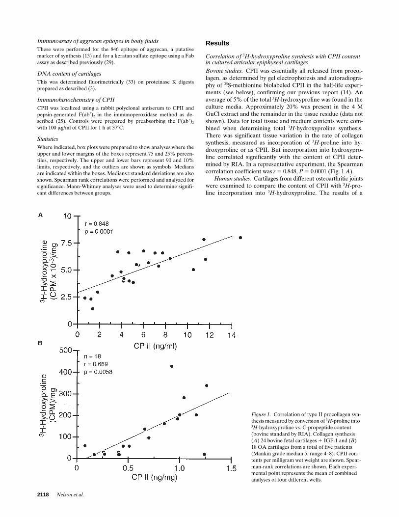
Bovine studies.
CPII was essentially all released from procol-lagen, as determined by gel electrophoresis and autoradiogra-phy of
35
S-methionine biolabeled CPII in the half-life experi-ments (see below), confirming our previous report (14). Anaverage of 5% of the total
3
H
-hydroxyproline was found in theculture media. Approximately 20% was present in the 4 MGuCl extract and the remainder in the tissue residue (data notshown). Data for total tissue and medium contents were com-bined when determining total
3
H
-hydroxyproline synthesis.There was significant tissue variation in the rate of collagensynthesis, measured as incorporation of
3
H
-proline into hy-droxyproline or as CPII. But incorporation into hydroxypro-line correlated significantly with the content of CPII deter-mined by RIA. In a representative experiment, the Spearmancorrelation coefficient was
r
5
0.848,
P
5
0.0001 (Fig. 1
A
).
Human studies.
Cartilages from different osteoarthritic jointswere examined to compare the content of CPII with
3
H
-pro-line incorporation into
3
H
-hydroxyproline. The results of a
Figure 1. Correlation of type II procollagen syn-thesis measured by conversion of 3H-proline into 3H-hydroxyproline vs. C-propeptide content (bovine standard by RIA). Collagen synthesis (A) 24 bovine fetal cartilages 1 IGF-1 and (B) 18 OA cartilages from a total of five patients (Mankin grade median 5, range 4–8). CPII con-tents per milligram wet weight are shown. Spear-man-rank correlations are shown. Each experi-mental point represents the mean of combined analyses of four different wells.

Collagen II Synthesis in Arthritis
2119
representative experiment on five OA cartilages are shown inFig. 1
B.
As in the case of the bovine studies, there was a signif-icant correlation between CPII content and
3
H
-hydroxyprolinecontent (
r
5
0.69;
P
5
0.0058).
Half-life of CPII in bovine and human cartilages
The analysis of
35
S-methionine–labeled CPII revealed that theradiolabel was predominantly in free CPII removed from pro-collagen. The data for bovine cartilage is shown in Fig. 2. Simi-lar results were obtained for human cartilages (data notshown). A major band and a secondary minor, faster movingband were observed. The latter represents a natural degrada-tion product lacking 10 residues at the amino terminus (34).Similar results were obtained with the all the antisera (data notshown). There was a progressive reduction of labeled CPIIwith time (Fig. 2). Densitometric analyses of the main CPIIband were made and corrected for total wet weight of carti-lage. These revealed that the half-life of fetal bovine CPII was14.5 h. This was similar to other data obtained from a prelimi-nary experiment (
z
16 h). Studies of cartilages from patientswith OA revealed half-lives of similar duration, namely 16 hcompared with 14 h and 19 h in other experiments (data notshown).
Relationship of CPII content in cartilage to release into culture medium in bovine studies
Experiments were performed to determine whether CPII re-leased from bovine cartilage reflected the content of CPII withinthe tissue. Cartilages were cultured in different concentrations ofITS supplement to stimulate variable collagen synthesis.
Analyses of cartilages and culture media, after 48 or 96 h ofculture, revealed a significant correlation between tissue andmedia contents where r 5 0.769, P 5 , 0.0001 (Fig. 3).
Total contents of CPII in normal and osteoarthritichuman cartilages
Having shown that the content of CPII accurately reflectedrates of procollagen II synthesis and that it had a relatively
short half-life, we examined the content of CPII in differenthuman articular cartilages. These were maximal in fetal carti-lage, being reduced almost threefold at birth and reaching verylow levels (mean 3.9% of newborn) in normal adult cartilage(Table II). But in human OA cartilage, CPII was significantlyelevated (mean 3.4361.81 ng/mg) compared with the nor-mal adult (mean 0.4560.27 ng/mg; Table II). This elevationbrought the mean content from 3.9% of that at birth in thenormal adult to 29.6% of the neonatal value in OA cartilage.The elevation was maintained in the more degenerate carti-lages, determined by Mankin grade (Table II). There was,however, no correlation in OA cartilages with Mankin grade,as determined by Spearman rank analyses (r 5 0.208; P 520.4893). The difference in mean CPII content in osteoar-
Figure 2. Half-life of C-propeptide in bovine fetal cartilage. SDS-PAGE autoradiography of immunobound C-propeptide with time af-ter labeling with 35S-methionine. Each experimental point represents the combined analysis of four different wells at the time (h) indicated. Molecular weight markers are shown.
Figure 3. Correlation of CPII content in cartilage with release into culture medium of bovine cul-tures (CPII by RIA using bovine standard). Each experimental point represents the mean6SD of combined analyses of four different wells.

2120 Nelson et al.
thritic compared with human fetal cartilages (z 9.9-fold) wasof the same order of difference observed between fetal bovinecartilages and human OA cartilages in the radiolabeling stud-ies (Fig. 1).
In a separate study, 50 OA cartilages were analyzed forcontents of CPII and total type II collagen. Spearman rankanalyses revealed a correlation value of r 5 0.3608 (P 5 0.01).Thus synthesis appears to be a determining factor in total typeII collagen content.
Regional variation in CPII content in human articular cartilages
In ageing and OA, changes in articular cartilage can be very lo-calized and regionally quite different. Hence, it was importantto determine whether CPII content varied within a femoralcondyle in a joint, be it normal or OA. Three pairs of normalcartilage and five pairs of OA cartilages were examined. Spec-imens were usually z 0.5–1.0 cm distance from their neighbor-ing samples. In both sets of joints, there was often significantvariation in content (Table II).
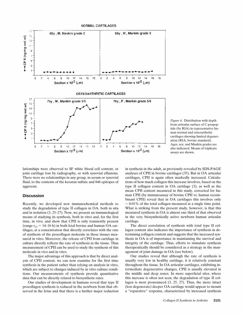
Variation with depth of CPII in human articular cartilage
Studies of the distribution with depth of CPII by RIA, re-vealed that in normal cartilages the small amount that waspresent was distributed fairly evenly throughout the cartilage.Examples are shown in Fig. 4. In contrast, in OA cartilage of
low-moderate Mankin grades, with an intact articular surface,it was usually more concentrated in the mid and deep layersthan at the articular surface. Representative examples areshown in Fig. 4.
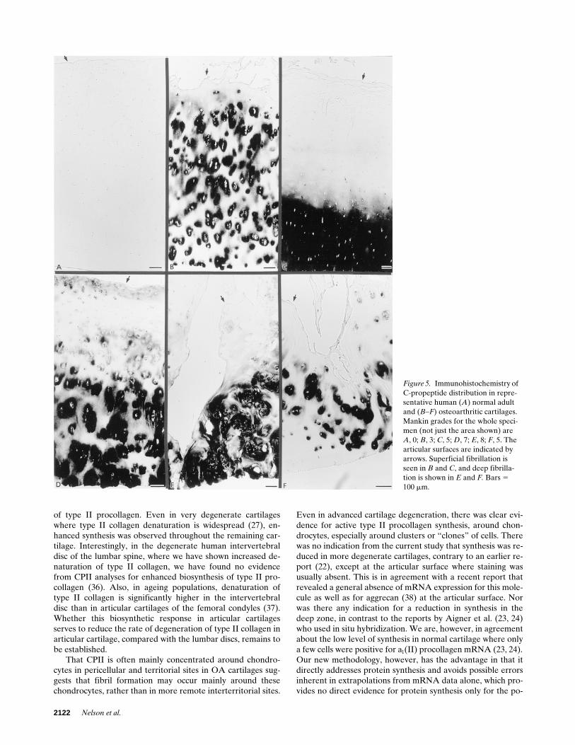
Immunohistochemical localization of CPII produced simi-lar results. With this method for immunostaining of CPII, thismolecule was not detectable in normal cartilages of ages 28(n 5 2), 41, 68, and 69 yr, of which an example is shown (Fig. 5A). In contrast, OA specimens (n 5 16, age range 52–82 yr,mean 68 yr) always stained strongly, usually in pericellular andterritorial sites (Fig. 5, B and D–F). Specificity of antibodybinding was confirmed by the absence of staining in sectionstreated with nonimmune rabbit F(ab9)2. Moreover, stainingwas absent, or considerably reduced, when antibody F(ab9)2
preparations were preabsorbed with purified CPII (data notshown). In 75% of OA specimens, staining was restricted tothe mid and deep zones (Fig. 5, C and D). It was usually absentfrom the natural or new articular surface as we show here.Sometimes, there was staining around chondrocytes through-out most of the cartilage except at the articular surface (Fig. 5B). In very fibrillated degenerate specimens, intense stainingwas observed around many chondrocytes, especially wherethere were clusters of chondrocytes (Fig. 5, D–F).
Concentrations of CPII in body fluids
Serum. To determine whether these increases in CPII contentwere accompanied by increases in serum as we had previouslyobserved in patients with RA (13), we examined CPII contentin sera.
Comparisons of sera from patients with OA with thosefrom patients with RA and nonarthritic controls revealed that,whereas CPII was significantly elevated (P 5 0.0001) in RA,confirming our earlier report for a much smaller group of pa-tients (13), patients with OA exhibited a significant reduction(P 5 0.0001) in circulating CPII compared with controls (Fig.6). Moreover, the distribution of concentrations was surpris-ingly narrow and even tighter than that observed in controls.Patients with RA exhibited much more variability in content.There were no correlations in any patient group of CPII tokeratan sulfate or 846 epitopes of aggrecan.
SF
In contrast, SF contents of CPII were similar in OA and RAand not significantly different (P 5 0.4722; Fig. 7). Analyses ofthe ratio of CPII contents in SF to serum of individual patientsrevealed that CPII was significantly elevated (median 2.05-fold) in SF in OA (P 5 0.0001) but not in RA (Fig. 8), butlevels of CPII in SF were not correlated with those in sera ineither group. Whereas 89.5% of patients with OA showed asignificant elevation over serum (median 5 2.05), only 64.8%showed an elevation in synovial fluids in RA (median 5 1.08).
When levels of CPII in SF were compared with the corre-sponding CPII contents (mean 1.3562.17) of single cartilagesamples from the same joint at arthroplasty for OA (n 5 37),we found no evidence of a significant correlation (data notshown).
Correlations between CPII and clinical measurements in controls, OA, and RA
In OA, there was an inverse correlation between serum CPIIcontent and disease duration (r 5 20.403, P 5 0.0157, n 5 37).A relationship of serum CPII to joint pain, which was not quitesignificant, was also noted (r 5 0.327, P 5 0.06, n 5 34). No re-
Table II. Concentrations of the C-Propeptide of Type II Procollagen in Human Articular Cartilages Determined by RIA (Bovine Standard)
Group/patient Age/sample Mankin grade C-Propeptide (ng/mg wet wt)
Fetal (n = 9) 16–18 wk — 33.8612.6Newborn (n = 2) 0 yr — 11.661.5Normal Years
N1 47a 0 0.38*47b 0 0.43*
N2 73a 3 0.37*73b 1 1.03*
N3 79a 2 0.41*79b 3 0.71*
N4 81 1 0.28*N5 79 1 0.28*N6 81 2 0.13*
OsteoarthriticOA1 74a 2 0.89‡
74b 2 0.67‡
OA2 70a 2 5.79‡
70b 7 5.98‡
OA3 77a 9 5.70‡
77b 7 1.71‡
OA4 73a 10 3.57‡
73b 8 3.42‡
OA5 59a 13 2.56‡
59b 10 4.26‡
OA6 74 5 3.64‡
OA7 74 6 2.96‡
a and b represent two samples from the same condyle. *Mean6SD0.4560.27; ‡mean6SD 3.4361.81 significantly different from normaladult (P , 0.0003) by Mann-Whitney analysis.
Collagen II Synthesis in Arthritis 2121
lationships were observed to SF white blood cell content, orjoint cartilage loss by radiography, or with synovial effusions.There were no relationships in any group, in serum or synovialfluid, to the contents of the keratan sulfate and 846 epitopes ofaggrecan.
Discussion
Recently, we developed new immunochemical methods tostudy the degradation of type II collagen in OA, both in situand in isolation (3, 25–27). Now, we present an immunologicalmeans of studying its synthesis, both in vitro and, for the firsttime, in vivo, and show that CPII is only transiently present(range t1/2 5 14–16 h) in both fetal bovine and human OA car-tilages, at a concentration that directly correlates with the rateof synthesis of the procollagen molecule in these tissues mea-sured in vitro. Moreover, the release of CPII from cartilage inculture directly reflects the rate of synthesis in the tissue. Thusmeasurement of CPII can be used to study the synthesis of thismolecule in vivo and in vitro.
The major advantage of this approach is that by direct anal-ysis of CPII content, we can now examine for the first timesynthesis in the patient rather than relying on analysis in vitro,which are subject to changes induced by in vitro culture condi-tions. Our measurements of synthesis provide quantitativedata that can be directly related to biosynthetic rates.
Our studies of development in humans reveal that type IIprocollagen synthesis is reduced in the newborn from that ob-served in the fetus and that there is a further major reduction
in synthesis in the adult, as previously revealed by SDS-PAGEanalyses of CPII in bovine cartilages (35). But in OA articularcartilages, CPII is again often markedly increased. Calcula-tions of how much collagen this increase involves, based on thetype II collagen content in OA cartilage (3), as well as themean CPII content measured in this study, corrected for hu-man CPII (by immunoassay of bovine CPII vs. human recom-binant CPII) reveal that in OA cartilages this involves onlyz 0.01% of the total collagen measured at a single time point.What is striking from the present study, however, is that thismeasured synthesis in OA is almost one third of that observedin the very biosynthetically active newborn human articularcartilage.
The direct correlation of synthesis with total type II col-lagen content also indicates the importance of synthesis in de-termining collagen content and suggests that the increased syn-thesis in OA is of importance in maintaining the survival andintegrity of the cartilage. Thus, efforts to stimulate synthesistherapeutically should be considered as a strategy in the man-agement of joint damage in OA (see below).
Our studies reveal that although the rate of synthesis isusually very low in healthy cartilage, it is relatively constantthroughout the tissue. In OA articular cartilages, exhibiting in-termediate degenerative changes, CPII is usually elevated inthe middle and deep zones. In more superficial sites, wherethis increase is often not seen, the degradation of type II col-lagen is most pronounced (3, 25, 27). Thus, the more intact(less degenerate) deeper OA cartilage would appear to mounta “reparative” response, characterized by increased synthesis
Figure 4. Distribution with depth from articular surface of C-propep-tide (by RIA) in representative hu-man normal and osteoarthritic cartilages showing limited degener-ation (RIA, bovine standard). Ages, sex, and Mankin grades are also indicated. Means of triplicate assays are shown.
2122 Nelson et al.
of type II procollagen. Even in very degenerate cartilageswhere type II collagen denaturation is widespread (27), en-hanced synthesis was observed throughout the remaining car-tilage. Interestingly, in the degenerate human intervertebraldisc of the lumbar spine, where we have shown increased de-naturation of type II collagen, we have found no evidencefrom CPII analyses for enhanced biosynthesis of type II pro-collagen (36). Also, in ageing populations, denaturation oftype II collagen is significantly higher in the intervertebraldisc than in articular cartilages of the femoral condyles (37).Whether this biosynthetic response in articular cartilagesserves to reduce the rate of degeneration of type II collagen inarticular cartilage, compared with the lumbar discs, remains tobe established.
That CPII is often mainly concentrated around chondro-cytes in pericellular and territorial sites in OA cartilages sug-gests that fibril formation may occur mainly around thesechondrocytes, rather than in more remote interterritorial sites.
Even in advanced cartilage degeneration, there was clear evi-dence for active type II procollagen synthesis, around chon-drocytes, especially around clusters or “clones” of cells. Therewas no indication from the current study that synthesis was re-duced in more degenerate cartilages, contrary to an earlier re-port (22), except at the articular surface where staining wasusually absent. This is in agreement with a recent report thatrevealed a general absence of mRNA expression for this mole-cule as well as for aggrecan (38) at the articular surface. Norwas there any indication for a reduction in synthesis in thedeep zone, in contrast to the reports by Aigner et al. (23, 24)who used in situ hybridization. We are, however, in agreementabout the low level of synthesis in normal cartilage where onlya few cells were positive for a1(II) procollagen mRNA (23, 24).Our new methodology, however, has the advantage in that itdirectly addresses protein synthesis and avoids possible errorsinherent in extrapolations from mRNA data alone, which pro-vides no direct evidence for protein synthesis only for the po-
Figure 5. Immunohistochemistry of C-propeptide distribution in repre-sentative human (A) normal adult and (B–F) osteoarthritic cartilages. Mankin grades for the whole speci-men (not just the area shown) are A, 0; B, 3; C, 5; D, 7; E, 8; F, 5. The articular surfaces are indicated by arrows. Superficial fibrillation is seen in B and C, and deep fibrilla-tion is shown in E and F. Bars 5 100 mm.

Collagen II Synthesis in Arthritis 2123
tential for synthesis. Clearly, both sets of information comple-ment each other and will, in the future, provide the mostvaluable insights into our understanding of synthesis at thelevel of individual cells.
Research in animals has also provided evidence in supportof increased procollagen II synthesis in degenerative cartilage.In experimental OA in rabbits, there is a marked increase intotal procollagen II synthesis (39). In dogs with natural degen-erative disease, there is increased procollagen synthesis earlyin the disease and a decrease as it progresses (40). Interest-ingly, this increase was also shown to be accompanied by an in-crease in procollagen II (41). Moreover, experimental OA indogs is accompanied by a much more pronounced increase intype II procollagen mRNA for type II collagen than for pro-teoglycan aggrecan (42, 43).
The identification of these increases in type II procollagensynthesis in human and animal articular cartilages, in natural
and experimental OA, is especially important because theydemonstrate the potential for replacement of damaged col-lagen fibrils in the extracellular matrix. Since in less damagedcartilage biosynthesis is often enhanced in deeper layers wherecollagen damage is often less pronounced (3, 27), it may bepossible, with an appropriate therapeutic approach, to fosterthis biosynthetic process as well as that involving the pro-teoglycan aggrecan. Thus, the therapeutic stimulation or en-hancement of repair of articular cartilage may be better aimedat preserving and building on this natural repair process. Theuse of inhibitors designed to arrest collagen degradation mayhelp encourage repair mechanisms.
The observed increase in CPII in diseased articular carti-lages in OA was not, however, reflected by an increase in theserum CPII content in OA although the release of CPII fromcartilage in culture reflects CPII content in the tissue. This is incontrast to patients with RA where an increase was observed,
Figure 7. CPII contents (RIA bo-vine standard) of SF of subjects in OA and RA subjects. The medians are significantly different (P 5 0.0001).
Figure 6. CPII contents (RIA bovine standard) of sera of subjects in con-trol, OA, and RA groups. Significant differences are indicated (each is P 5 0.0001).

2124 Nelson et al.
as in our previous studies (13) but for which cartilage data waslacking. However, in children CPII is elevated during growthand drops as growth ceases (44). Although a significant in-crease in CPII in SF over serum was observed in OA com-pared to RA patients, the elevation over serum levels wassmall compared with other disease-related epitopes such as the846 epitope of aggrecan (29). Although an increase in CPII hasbeen reported in OA joint fluid over normal levels (45), therewas no evidence in our studies of any overall increase in serumin OA, as observed in RA here and in previous studies (13), orof any correlations between articular cartilage content andjoint fluid and serum content of CPII in OA. Thus, the circu-lating CPII in serum likely reflects whole body cartilage me-tabolism in OA where few joints are ordinarily involved com-pared with RA. If this is the case, the decrease in serum CPIIin OA may represent reduced synthesis of this molecule withinother cartilages. Whether this is a consequence of the diseaseor causally related to its development remains to be seen.
Acknowledgments
This study was funded by the Shriners Hospitals for Children, theMedical Research Council of Canada, and the National Institutes ofHealth (to A.R.P.), the Arthritis and Rheumatism Council (to P.D.),by the National Institutes of Health (to L.R.), the Fonds de la Re-cherche en Santé du Quebec (to R.C.B.) and by the Swedish Societyof Medicine, Wenner-Gren Center Foundation, Swedish Society forResearch in Sport Medicine and the Swedish Society of Medicine (toL.D.).
References
1. Poole, A.R. 1993. Cartilage in health and disease. In Arthritis and AlliedConditions. A Textbook in Rheumatology, 12th ed. D. McCarthy and W.Koopman, editors. Lea and Febiger, Philadelphia. 279–333.
2. Mayne, R. 1989. Cartilage collagens. What is their function, and are theyinvolved in articular disease. Arthritis Rheum. 32:241–246.
3. Hollander, A., T. Heathfield, C. Webber, Y. Iwata, R. Bourne, C. Rora-beck, and A.R. Poole. 1994. Increased damage to type II collagen in osteoar-thritic articular cartilage detected by a new immunoassay. J. Clin. Invest. 93:1722–1732.
4. Kempson, G.E., H. Muir, C. Pollard, and M. Tuke. 1973. The tensileproperties of the cartilage of human femoral condyles related to the content ofcollagen and glycosaminoglycans. Biochim. Biophys. Acta. 297:456–472.
5. Mow, V.C., L.A. Setton, D.S. Ratcliffe, D.S. Howell, and J.A. Buck-walter. 1990. Structure-function relationships of articular cartilage and the ef-fects of joint instability and trauma on cartilage function. In Cartilage Changesin Osteoarthritis. K.D. Brandt, editor. Indiana University School of Medicine,Ciba-Geigy. 22–42.
6. Prockop, D.J., K.I. Kivirikko, L. Tuderman, and N.A. Guzman. 1979.The biosynthesis of collagen and its disorders. N. Eng. J. Med. 301:13–23.
7. Peltonen, L., R. Halila, and L. Ryhänen. 1985. Enzymes converting pro-collagens to collagens. J. Cell. Biochem. 28:15–21.
8. Kujawa, M.J., M. Weitzhandler, A.R. Poole, L. Rosenberg, and A.I. Cap-lan. 1989. Association of the C-propeptide of type II collagen with mineraliza-tion of embryonic chick long bone and sternal development. Connect. TissueRes. 23:179–199.
9. Poole, A.R., I. Pidoux, A. Reiner, H. Choi, and L.C. Rosenberg. 1984.Association of an extracellular protein (chondrocalcin) with the calcification ofcartilage in enchondral bone formation. J. Cell Biol. 98:54–65.
10. Lee, E.R., and A.R. Poole. 1996. Ultrastructural localization of the C-pro-peptide released from type II procollagen in fetal bovine growth plate cartilage.J. Histochem. Cytochem. 44:433–443.
11. Hinek, A., A. Reiner, and A.R. Poole. 1987. The calcification of carti-lage matrix in chondrocyte culture: studies of the C-propeptide of type II col-lagen (chondrocalcin). J. Cell Biol. 104:1435–1441.
12. Alini, M., Y. Matsui, G.R. Dodge, and A.R. Poole. 1992. The extracellu-lar matrix of cartilage in the growth plate before and during calcification in thegrowth plate before and during calcification: changes in the composition anddegradation of type II collagen. Calcif. Tissue Int. 50:327–335.
13. Mänsson, B., D. Carey, M. Alini, M. Ionescu, L.C. Rosenberg, A.R.Poole, and D. Heinegård. 1995. Cartilage and bone metabolism in rheumatoidarthritis. Differences between rapid and slow progression of disease identifiedby serum markers of cartilage metabolism. J. Clin. Invest. 95:1071–1077.
14. Lee, E.R., Y. Matsui, and A.R. Poole. 1990. Immunochemical and im-munocytochemical studies of the C-propeptide of type II procollagen in chon-drocytes of the growth plate. J. Histochem. Cytochem. 38:659–693.
15. Nimni, M.E. 1983. Collagen: structure, function, and metabolism in nor-mal and fibrotic tissues. Semin. Arthritis Rheum. 13:1–86.
16. Spanheimer, R.G., and B. Peterkofsky. 1985. A specific decrease in col-lagen synthesis in acutely fasted, vitamin C-supplemented, guinea pigs. J. Biol.Chem. 260:3955–3962.
17. Spanheimer, R.G., T.A. Bird, and B. Peterkofsky. 1986. Regulation ofcollagen synthesis and mRNA levels in articular cartilage of scorbutic guineapigs. Arch. Biochem. Biophys. 246:33–41.
Figure 8. Ratio of CPII contents (RIA, bovine standard) in SF compared with sera measured in the same patient in those with OA and RA. The ratios are significantly different (P 5 0.0001).

Collagen II Synthesis in Arthritis 2125
18. Tyler, J.A., and H.P. Benton. 1988. Synthesis of type II collagen is de-creased in cartilage cultured with interleukin 1 while the rate of intracellulardegradation remains unchanged. Coll. Relat. Res. 8:393–405.
19. Mankin, H.J., H. Dorfman, L. Lippiello, and A. Zarins. 1971. Biochemi-cal and metabolic abnormalities in articular cartilage from osteo-arthritic hu-man hips. II. Correlation of morphology with biochemical and metabolic data.J. Bone Joint Surg. Am. 53:523–537.
20. Lippiello, L., D. Hall, and H.J. Mankin. 1977. Collagen synthesis in nor-mal and osteoarthritic human cartilage. J. Clin. Invest. 59:593–600.
21. Thompson, R.C., and T.R. Oegema. 1979. Metabolic activity of articularcartilage in osteoarthritis. J. Bone Joint Surg. Am. 61:407–416.
22. Rizkalla, G., A. Reiner, E. Bogoch, and A.R. Poole. 1992. Studies of thearticular cartilage proteoglycan aggrecan in health and osteoarthritis. Evidencefor molecular heterogeneity and extensive molecular changes in disease. J. Clin.Invest. 90:2268–2277.
23. Aigner, T., H. Stoss, G. Weseloh, G. Zeiler, and K. von der Mark. 1992.Activation of collagen type II expression in osteoarthritic and rheumatoid carti-lage. Virchows Arch. B Cell Pathol. 62:337–345.
24. Aigner, T., W. Bertling, H. Stöss, G. Weseloh, and K. von der Mark.1993. Independent expression of fibril-forming collagens I, II and III in chon-drocytes of human osteoarthritic cartilage. J. Clin. Invest. 91:829–837.
25. Dodge, G.R., and A.R. Poole. 1989. Immunohistochemical detectionand immunochemical analysis of type II collagen degradation in human normal,rheumatoid, and osteoarthritic cartilages and in explants of bovine articularcartilage cultured with interleukin 1. J. Clin. Invest. 83:647–661.
26. Billinghurst, R.C., L. Dahlberg, M. Ionescu, A. Reiner, R. Bourne, C.Rorabeck, P. Mitchell, J. Hambor, O. Diekmann, H. Tschesche, et al. 1997. En-chanced cleavage of type II collagen by collagenases in osteoarthritic articularcartilage. J. Clin. Invest. 99:1534–1545.
27. Hollander, A.P., I. Pidoux, A. Reiner, C. Rorabeck, R. Bourne, andA.R. Poole. 1995. Damage to type II collagen in ageing and osteoarthritis: startsat the articular surface, originates around chondrocytes, and extends into thecartilage with progressive degeneration. J. Clin. Invest. 96:2859–2869.
28. Arnett, F.C., S.M. Edworthy, D.A. Block, D.J. McShane, J.F. Fries, N.S.Cooper, L.A. Healey, S.R. Kaplan, M.H. Liang, H.S. Luthra, et al. 1988. TheAmerican Rheumatism Association 1987 revised criteria for the classificationof rheumatoid arthritis. Arthritis Rheum. 31:315–324.
29. Poole, A.R., M. Ionescu, A. Swan, and P.A. Dieppe. 1994. Changes incartilage metabolism in arthritis are reflected by altered serum and synovialfluid levels of the cartilage proteoglycan aggrecan: implications for pathogene-sis. J. Clin. Invest. 94:25–33.
30. Pal, S., L.H. Tang, H. Choi, E. Haberman, L.C. Rosenberg, P.J. Rough-ley, and A.R. Poole. 1981. Structural changes during development in bovine fe-tal epiphyseal cartilage. Coll. Relat. Res. 1:151–176.
31. Burleigh, M.C., A.J. Barrett, and G.S. Lazarus. 1974. Cathepsin B1. Alysosomal enzyme that degrades native collagen. Biochem. J. 137:387–398.
32. Choi, H.U., L.H. Tang, T.L. Johnson, S. Pal, L.C. Rosenberg, A. Reiner,and A.R. Poole. 1983. Isolation and characterization of a 35,000 molecularweight subunit fetal cartilage matrix protein. J. Biol. Chem. 258:655–661.
33. Labarca, C., and K. Paigen. 1980. A simple, rapid and sensitive DNA as-say procedure. Anal. Biochem. 102:344–352.
34. Van Der Rest, M., L.C. Rosenberg, B.R. Olsen, and A.R. Poole. 1986.Chondrocalcin is identical with the C-propeptide of type II procollagen. Bio-chem. J. 237:923–925.
35. Niyibizi, C., J.J. Wu, and D.R. Eyre. 1987. The carboxypropeptide tri-mer of type II collagen is a prominent component of immature cartilages andintervertebral-disc tissues. Biochim. Biophys. Acta. 916:493–499.
36. Antoniou, J., F. Nelson, T. Steffen, N. Winterbottom, A.P. Hollander,A.R. Poole, M. Aebi, and M. Alini. 1996. The human lumbar intervertebraldisc: evidence for changes in the biosynthesis and denaturation of the extracel-lular matrix with growth, maturation, ageing, and degeneration. J. Clin. Invest.98:996–1003.
37. Hollander, A.P., T.F. Heathfield, J.J. Liu, I. Pidoux, P.J. Roughley, J.S.Mort, and A.R. Poole. 1996. Enhanced denaturation of the a1(II) chains oftype-II collagen in normal adult human intervertebral discs compared with fem-oral articular cartilage. J. Orthop. Res. 14:61–66.
38. Aigner, T.Y., S.I. Vornehm, G. Zeiler, J. Dudhia, K. Von der Mark, andM.T. Bayliss. 1997. Suppression of cartilage matrix gene expression in upperzone chondrocytes of osteoarthritic cartilage. Arthritis Rheum. 40:562–569.
39. Eyre, D.R., C.A. McDevitt, M.E.J. Billingham, and H. Muir. 1980. Bio-synthesis of collagen and other matrix proteins by articular cartilage in experi-mental osteoarthrosis. Biochem. J. 188:823–837.
40. Burton-Wurster, N., C.S. Hui-Chou, H.A. Greisen, and G. Lust. 1982.Reduced deposition of collagen in the degenerated articular cartilage of dogswith degenerative joint disease. Biochem. Biophys. Acta. 718:74–84.
41. Miller, D.R., and G. Lust. 1979. Accumulation of procollagen in the de-generative articular cartilage of dogs with osteoarthritis. Biochim. Biophys.Acta. 583:218–231.
42. Matyas, J.R., M.E. Adams, D. Huang, and L.J. Sandell. 1995. Discoordi-nate gene expression of aggrecan and type II collagen in experimental osteoar-thritis. Arthritis Rheum. 38:420–425.
43. Matyas, J.R., M.E. Adams, D. Huang, and L.J. Sandell. 1997. Major roleof collagen IIB in the elevation of total type II procollagen messenger RNA inthe hypertrophic phase of experimental osteoarthritis. Arthritis Rheum. 40:1046–1049.
44. Carey, D.E., M. Alini, M. Ionescu, J.S. Hyams, J.C. Rowe, L.C. Rosen-berg, and A.R. Poole. 1997. Serum content of the C-propeptide of the cartilagemolecule type II collagen in children. Clin. Exp. Rheum. 15:325–328.
45. Lohmander, S.Y., Y. Yoshihara, H. Roos, T. Kokayashi, H. Yamada,and M. Shinmei. 1996. Procollagen II c-propeptide in joint fluid: changes in con-centrations with age, time after joint injury and osteoarthritis. J. Rheumatol. 23:1765–1769.