COLD ADAPTATIONS STUDY OF GLYCOSYL HYDROLASE...
42
COLD ADAPTATIONS STUDY OF GLYCOSYL HYDROLASE ENZYMES VIA COMPUTATIONAL METHODS SEPIDEH PARVIZPOUR A thesis submitted in fulfilment of the requirements for the award of the degree of Doctor of Philosophy (Bioscience) Faculty of Biosciences and Medical Engineering Universiti Teknologi Malaysia SEPTEMBER 2015
Transcript of COLD ADAPTATIONS STUDY OF GLYCOSYL HYDROLASE...

COLD ADAPTATIONS STUDY OF GLYCOSYL HYDROLASE ENZYMES
VIA COMPUTATIONAL METHODS
SEPIDEH PARVIZPOUR
A thesis submitted in fulfilment of the
requirements for the award of the degree of
Doctor of Philosophy (Bioscience)
Faculty of Biosciences and Medical Engineering
Universiti Teknologi Malaysia
SEPTEMBER 2015

iii
“Dedicated to my beloved husband, my son, my parents and parents-in-law”

iv
ACKNOLEDGEMENTS
I would like to express my true and sincere thanks and gratitude to my
supervisor, Assoc. Professor Dr. Mohd Shahir Shamsir for his patience, guidance,
encouragement, invaluable comments, and supports that made this research possible
and completed early.
I would like to thank all members of the Bioinformatics lab (BIRG) for their
continues support in many aspects of this research.
The completion of this work would never have been possible without
persistent and inflexible support of my husband Dr. Jafar Razmara. He placed before
his unavoidable opportunities to prove his managerial, organizational and adaptive
qualities. I have no words to exactly define his level of patience and sacrifice that he
exercised to create and maintain highly demanded balance in my family. During the
whole process he did everything to provide us a happy family. I would like to
appreciate him and my lovely son Amir Hossein for all the moral support. They are
the reason why I worked hard.
On top of everything, my dream to achieve the highest educational degree
would never have got materialized without wishes, prayers, moral and material
support of my parents and parents-in-law. I owe a debt of gratitude to them that
always have been there to encourage and support me.
v
ABSTRACT
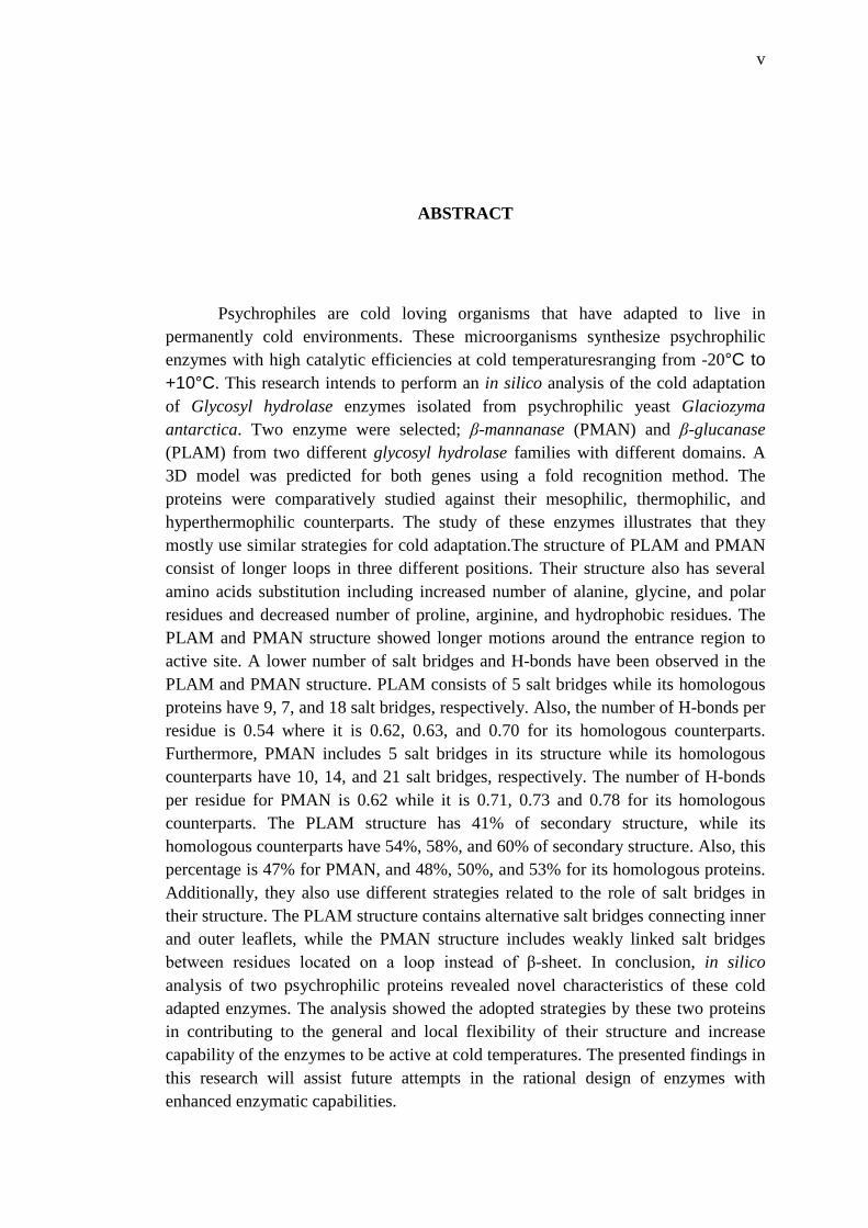
Psychrophiles are cold loving organisms that have adapted to live in permanently cold environments. These microorganisms synthesize psychrophilic enzymes with high catalytic efficiencies at cold temperaturesranging from -20°C to +10°C. This research intends to perform an in silico analysis of the cold adaptation of Glycosyl hydrolase enzymes isolated from psychrophilic yeast Glaciozyma antarctica. Two enzyme were selected; β-mannanase (PMAN) and β-glucanase (PLAM) from two different glycosyl hydrolase families with different domains. A 3D model was predicted for both genes using a fold recognition method. The proteins were comparatively studied against their mesophilic, thermophilic, and hyperthermophilic counterparts. The study of these enzymes illustrates that they mostly use similar strategies for cold adaptation.The structure of PLAM and PMAN consist of longer loops in three different positions. Their structure also has several amino acids substitution including increased number of alanine, glycine, and polar residues and decreased number of proline, arginine, and hydrophobic residues. The PLAM and PMAN structure showed longer motions around the entrance region to active site. A lower number of salt bridges and H-bonds have been observed in the PLAM and PMAN structure. PLAM consists of 5 salt bridges while its homologous proteins have 9, 7, and 18 salt bridges, respectively. Also, the number of H-bonds per residue is 0.54 where it is 0.62, 0.63, and 0.70 for its homologous counterparts. Furthermore, PMAN includes 5 salt bridges in its structure while its homologous counterparts have 10, 14, and 21 salt bridges, respectively. The number of H-bonds per residue for PMAN is 0.62 while it is 0.71, 0.73 and 0.78 for its homologous counterparts. The PLAM structure has 41% of secondary structure, while its homologous counterparts have 54%, 58%, and 60% of secondary structure. Also, this percentage is 47% for PMAN, and 48%, 50%, and 53% for its homologous proteins. Additionally, they also use different strategies related to the role of salt bridges in their structure. The PLAM structure contains alternative salt bridges connecting inner and outer leaflets, while the PMAN structure includes weakly linked salt bridges between residues located on a loop instead of β-sheet. In conclusion, in silico analysis of two psychrophilic proteins revealed novel characteristics of these cold adapted enzymes. The analysis showed the adopted strategies by these two proteins in contributing to the general and local flexibility of their structure and increase capability of the enzymes to be active at cold temperatures. The presented findings in this research will assist future attempts in the rational design of enzymes with enhanced enzymatic capabilities.

vi
ABSTRAK
Organisma psikrofilik adalah organisma yang telah menyesuaikan diri untuk hidup dalam persekitaran yang sejuk kekal. Mikroorganisma-mikroorganisma ini mensintesis enzim psikrofilik dengan tujuan untuk mengekalkan kecekapan pemangkin pada suhu sejuk antara -20 ° C hingga + 10 ° C. Kajian ini bertujuan untuk melakukan analisis komputeran adaptasi suhu sejuk enzim glikosil hidrolase yang telah diasingkan daripada yis psikrofilik Glaciozyma antarctica. Dua enzim yang dipencil telah dipilih; β-mannanase (PMAN) dan β-glukanase (PLAM) daripada dua keluarga enzim glikosil hidrolase yang berbeza. Model 3D telah diramalkan untuk kedua-dua gen menggunakan kaedah "pengecaman lipatan". Protein dikaji secara perbandingan terhadap enzim mesofilik, termofilik, dan hipertermofilik. Kajian enzim ini menggambarkan bahawa kebanyakan enzim menggunakan strategi yang sama untuk mengadaptasi kepada keadaan sejuk. Struktur PLAM dan PMAN terdiri daripada gelungan-gelungan pada tiga kedudukan yang berbeza. Struktur mereka juga mempunyai beberapa perubahan asid amino seperti jumlah peningkatan alanina, glisin, jujuk amino polar dan beberapa prolin, arginina, dan jujuk amino hidrofobik yang dikurangkan jumlahnya. Struktur PLAM dan struktur PMAN menunjukkan pergerakkan lebih dinamik di sekitar kawasan pintu masuk ke tapak aktif enzim. Beberapa ciri yang menunjukkan penurunan dalam struktur PLAM dan PMAN adalah jambatan garam dan rangkaian H. PLAM mempunyai 5 jambatan garam manakala homolog mempunyai 9, 7, dan 18 jambatan garam. Bilangan rangkai H untuk setiap jujuk asid amino adalah 0.54 berbanding dengan 0.62, 0.63, dan 0.70 untuk protein homolog. Tambahan pula, PMAN mempunyai hanya 5 jambatan garam dalam struktur berbanding dengan protein homolog yang mempunyai 10, 14, dan 21 jambatan garam. Bilangan rangkai H untuk setiap jujuk asid amino untuk PMAN adalah 0.62 berbanding dengan 0.71, 0.73 dan 0.78 protein homolognya. Struktur PLAM mempunyai 41% daripada struktur sekunder, manakala rakan-rakan homolog yang mempunyai 54%, 58%, dan 60% daripada struktur sekunder. Peratusan ini adalah 47% untuk PMAN, dan 48%, 50%, dan 53% berbanding protein homolog. Selain itu, mereka juga menggunakan strategi yang berbeza untuk menggunapakai jambatan garam dalam struktur mereka. Struktur PLAM mengandungi jambatan garam alternatif menyambung bahagian dalaman dan luaran, manakala struktur PMAN mempunyai jambatan garam yang lemah untuk mengaitkan di antara asid amino yang dua terletak pada gelungan antara lembaran β. Kesimpulannya, analisis komputeran dua protein psikrofilik menunjukkan beberapa ciri-ciri unik yang membolehkan enzim ini berfungsi di dalam suhu sejuk kekal. Analisa ini menunjukkan strategi yang diguna pakai oleh kedua-dua protein dalam menyumbang kepada fleksibiliti secara umum dan khusus terhadap keupayaan struktur dan pengekalan keupayaan mereka menjadi enzim aktif pada suhu yang sejuk. Hasil kajian ini akan membantu dalam memperolehi enzim dengan aktiviti keupayaan tinggi melalui rekabentuk rasional enzim.

vii
TABLE OF CONTENTS
CHAPTER TITLE PAGE
TITLE PAGE i
DECLARTION ii
DEDICATION iii
ACKNOWLEDGMENTS iv
ABSTRACT v
ABSTRAK vi
TABLE OF CONTENTS i
LIST OF TABLES xi
LIST OF FIGURES xiii
LIST OF ABBREVIATIONS xvii
1 INTRODUCTION 1
1.1 Overview 1
1.2 Challenges in characterization of psychrophiles 2
1.3 Problem Statement 3
1.4 Research Goal and Objectives 4
1.5 Scope of the Study 5
1.6 Thesis organization 5
2 LITERATURE REVIEW 7
2.1 Introduction 7
2.2 Extremophiles 7
2.3 Psychrophiles 9
2.4 Psychrophilic enzymes 9

viii
2.5 The application of psychrophilic enzymes 11
2.6 Glycosyl hydrolase 14
2.6.1 Glycoside hydrolase classification 15
2.6.2 Glycosyl hydrolase mechanisms 17
2.6.3 Glycosyl hydrolase applications 18
2.7 Psychrophilic yeast, Glaciozyma Antarctica PI12 19
2.8 β-glucans 20
2.8.1 Enzymatic hydrolysing of glucan 21
2.9 β-glucanase 21
2.9.1 β-glucanase family classification and structural
determination 22
2.9.2 Applications of β-glucanase 23
2.10 Mannans 24
2.10.1 Enzymatic hydrolysis of mannan 26
2.11 β-mannanase 27
2.11.1 β-mannanase occurrence and regulation 28
2.11.2 β-mannanase family classification and structural
determination 29
2.11.3 Applications of β-mannanases 29
2.12 Protein structure prediction 33
2.12.1 Comparative modeling 36
2.12.2 Protein fold recognition 37
2.12.3 New fold prediction 38
2.13 Structure refinement 40
2.14 Structure validation 40
2.15 Molecular Dynamic (MD) simulation 42
2.16 Protein function prediction 42
2.16.1 Surface accessibility prediction 44
2.16.2 Identification of catalytic residues by amino acid
substitution 44
2.16.3 Docking studies 45
3 RESEARCH METHODOLOGY 47
3.1 Introduction 47

ix
3.2 Operational Framework of the research 47
3.3 Materials and Methods 49
3.3.1 Sequence-based analysis 49
3.3.2 Structure-based analysis 51
3.3.3 Cold adaptation study 54
4 STRUCTURE PREDICTION AND COLD ADAPTATION
ANALYSIS OF A NOVEL LAMINARINASE 57
4.1 Sequence retrieval 57
4.2 PLAM primary sequence characteristics 57
4.3 PLAM Signal peptide prediction 60
4.4 PLAM sequence analysis 61
4.5 PLAM phylogenetic study 62
4.6 3D-Model prediction 63
4.7 Structure refinement 66
4.8 Evaluation of the model 67
4.9 Analysis of the model 70
4.10 Protein-Substrate interaction 72
4.11 Tunnel prediction 78
4.12 Cold adaptation study 79
4.12.1 Comparative primary sequence analysis 79
4.12.2 Molecular Dynamic (MD) simulation 85
4.12.3 Generalized order parameter analysis 87
4.12.4 Principal component analysis (PCA) 88
4.12.5 Comparative analysis of structural parameters 90
4.12.6 Study of the protein unfolding 98
5 STRUCTURE PREDICTION AND COLD ADAPTATION
ANALYSIS OF THE NOVEL MANNANASE 101
5.1 Sequence retrieval 101
5.2 PMAN primary sequence characteristics 101
5.3 PMAN Signal peptide prediction 104
5.4 PMAN sequence analysis 105

x
5.5 PMAN phylogenetic study 106
5.6 3D-Model prediction 107
5.7 Structure refinement 110
5.8 Evaluation of the model 111
5.9 Analysis of the model 114
5.10 Protein-Substrate interaction 119
5.11 Tunnel prediction 124
5.12 Cold adaptation study 126
5.12.1 Comparative primary sequence analysis 126
5.12.2 Molecular Dynamic (MD) simulation 132
5.12.3 Generalized order parameter analysis 134
5.12.4 Principal component analysis (PCA) 135
5.12.5 Comparative analysis of structural parameters 137
5.12.6 Study of the protein unfolding 145
6 CONCLUSION 148
6.1 Research conclusion 148
6.2 Future works 151
REFERENCES 152

xi
LIST OF TABLES
TABLE NO TITLE PAGE
2.1 A summary of psychrophilic enzymes applications 12
2.2 The established clans for glycoside hydrolases 16
4.1 Summary of amino acids composition in PLAM using
protparam 60
4.2 Summary of amino acids characterized groups in PLAM using
Colorseq 60
4.3 Top three proposed templates by different threading tools 65
4.4 Evaluation of the best models created by the MODELLER
program for the alignments produced by different servers 65
4.5 A summary of model evaluations using different tools 67
4.6 The binding residues of different laminarinase substrates 74
4.7 Predicted tunnels into the active sites of PLAM prepared by
Mole software 78
4.8 Cavity information for the predicted tunnels of PLAM in Table
4.7 79
4.9 Comparison of amino acid substitution between PLAM and
mesophilic, thermophilic, and hyperthermophilic laminarinases 83
4.10 Amino acid content of PLAM for thermostability-related
residues compared to averages of family GH-16 members 85
4.11 Structural parameters from MD simulation of the homologous
Laminarinases at 12°C and 90°C 92
4.12 Data computed from MD simulation of PLAM at 4°C, 12°C and
18°C 93
4.13 Secondary structure percentages of laminarinases at 12°C 97

xii
4.14 Secondary structure percentages of laminarinases at 90°C 97
5.1 Summary of amino acids composition in PMAN 104
5.2 Summary of amino acids characterized groups percentage in
PMAN 104
5.3 Top three proposed templates by different threading tools 108
5.4 evaluation of the best models created by MODELLER program
for the alignments produced by different servers 109
5.5 Model evaluation summary using different tools 111
5.6 The binding residues of different mannanase substrates 121
5.7 Predicted tunnels into the active sites of PMAN prepared by
Mole software 125
5.8 Cavity information for the predicted tunnels for PMAN in Table
5.7 126
5.9 Comparison of amino acid substitution between PMAN and
mesophilic and thermophilic mannanases 130
5.10 Structural parameters from MD simulation of the homologous
Mannanases at 12°C and 80°C 141
5.11 Secondary structure percentages of mannanases at 12°C 142
5.12 Secondary structure percentages of mannanases at 90°C 142
xiii
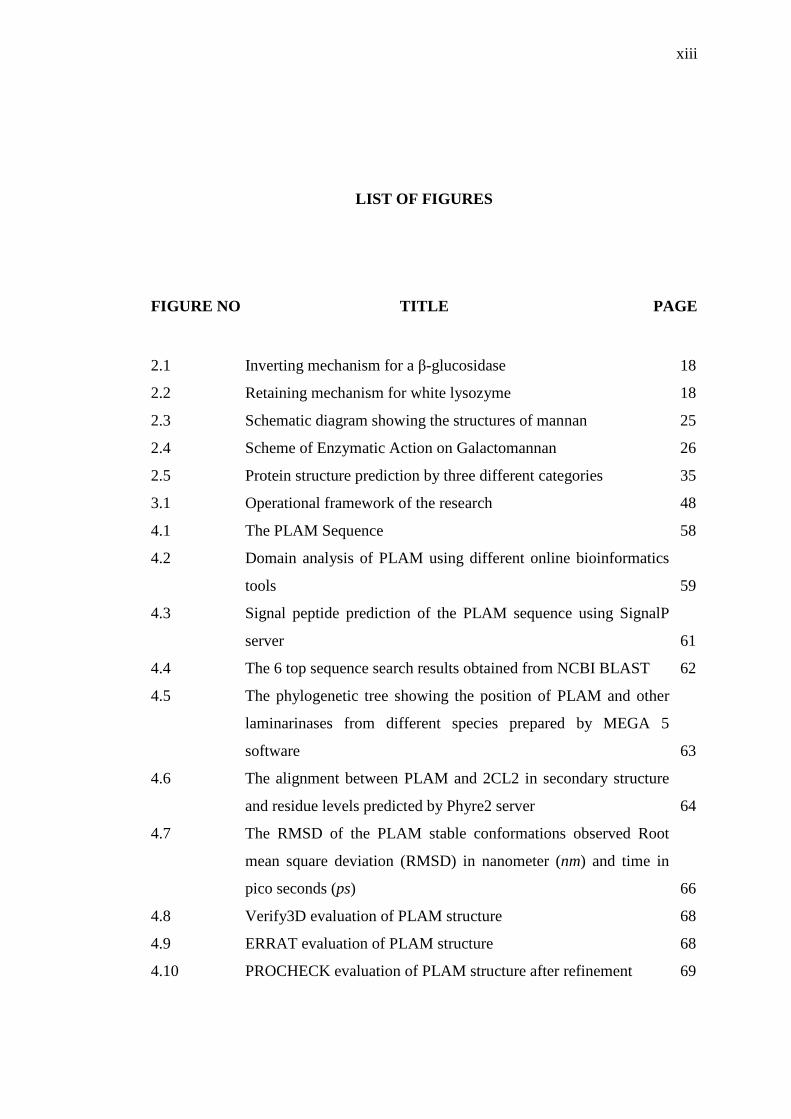
LIST OF FIGURES
FIGURE NO TITLE PAGE
2.1 Inverting mechanism for a β-glucosidase 18
2.2 Retaining mechanism for white lysozyme 18
2.3 Schematic diagram showing the structures of mannan 25
2.4 Scheme of Enzymatic Action on Galactomannan 26
2.5 Protein structure prediction by three different categories 35
3.1 Operational framework of the research 48
4.1 The PLAM Sequence 58
4.2 Domain analysis of PLAM using different online bioinformatics
tools 59
4.3 Signal peptide prediction of the PLAM sequence using SignalP
server 61
4.4 The 6 top sequence search results obtained from NCBI BLAST 62
4.5 The phylogenetic tree showing the position of PLAM and other
laminarinases from different species prepared by MEGA 5
software 63
4.6 The alignment between PLAM and 2CL2 in secondary structure
and residue levels predicted by Phyre2 server 64
4.7 The RMSD of the PLAM stable conformations observed Root
mean square deviation (RMSD) in nanometer (nm) and time in
pico seconds (ps) 66
4.8 Verify3D evaluation of PLAM structure 68
4.9 ERRAT evaluation of PLAM structure 68
4.10 PROCHECK evaluation of PLAM structure after refinement 69

xiv
4.11 3D-model of PLAM representing the secondary structure
elements in cartoon form 71
4.12 Superimposition of PLAM and its template, 2CL2 71
4.13 Distance between two catalytic residuesGlu117 and Glu122of
PLAMprepared by Pymol 74
4.14 Water molecules during the PLAM-Laminarin complex
simulation around catalytic active sites 75
4.15 Water molecules during PLAM-Lichenin complex simulation
around catalytic active sites 76
4.16 Water molecules during PLAM-Barley complex simulation
around catalytic active sites 77
4.17 The predicted tunnel for PLAM and its cavity 79
4.18 Multiple sequence alignment of laminarinases 81
4.19 Structural alignment of laminarinases 82
4.20 Dynamic changes of PLAM during MD simulation compared
with its homologous proteins based on RMSD at 12°C and 90°C 86
4.21 N-H generalized order parameter S2 at 12°C. The order
parameters are shown as a function of residue numbers 87
4.22 Dominant motions simulation for PLAM, 2CL2, 2HYK and 3ILN
proteins at 12°Cusing Principal Component Analysis (PCA) 89
4.23 Projection of simulation frames in the 3D-subspace formed by the
first eigenvectors for 2CL2, 2HYK, 3ILN, and PLAM proteins at
12°C 90
4.24 Topological features of the salt bridges of simulated laminarinase 94
4.25 Topological positions of salt bridges of different laminarinases 96
4.26 Secondary structure percentages of different laminarinases at
12°C 97
4.27 Secondary structure percentages of different laminarinases at
12°C 97
4.28 Secondary structure plot of PLAM during MD simulation for
three longer loop locations 98
4.29 DSSP plot of the PLAM and 3ILN structures at 285K,363K,
400K, and 500K 100
5.1 The PMAN Sequence 102

xv
5.2 Domain analysis of PMAN using different online bioinformatics
tools 103
5.3 Signal peptide prediction of the PMAN sequence using SignalP
server 105
5.4 The 6 top sequence search results obtained from NCBI BLAST 105
5.5 The phylogenetic tree showing the position of PMAN and other
mannanases from different species 106
5.6 The alignment between PMAN and 1RH9 in secondary structure
and residue levels predicted by HHpred server 109
5.7 The RMSD of the PMAN stable conformations observed Root
mean square deviation (RMSD) in nanometer (nm) and time in
pico seconds (ps) 110
5.8 Verify3D evaluation of PMAN structure 112
5.9 ERRAT evaluation of PMAN structure 112
5.10 PROCHECK evaluation of PMAN structure 113
5.11 3D-model of PMAN representing the secondary structure
elements 114
5.12 Superimposition of PMAN and its template, 1RH9 in a cartoon
representation 115
5.13 The 3D-structure of 1RH9 and PMAN including an α/β-barrel and
some other additional α-helices and β-strands 116
5.14 Multiple sequence alignment of mannanases 117
5.15 The alignment between PMAN and 2C0H in secondary structure
and residue levels predicted by HHpred server 119
5.16 Distance between two catalytic residues Glu184 and Glu273 of
PMAN prepared by pymol 120
5.17 Water molecules during the PMAN-Mannobios complex
simulation around catalytic active sites 122
5.18 Water molecules during PMAN-Mannotrios complex simulation
around catalytic active sites 123
5.19 Water molecules during PMAN-Mannopentaose complex
simulation around catalytic active sites 124
5.20 Top predicted channels into the active sites of different
mannanases 126

xvi
5.21 Multiple sequence alignments of mannanases 128
5.22 Multiple structural alignment of mannanases 129
5.23 Dynamic changes of PMAN during MD simulation compared
with its homologous proteins based on RMSD at 12°C and 80°C 133
5.24 N-H generalized order parameter S2 at 12°C. The order
parameters are shown as a function of residue numbers 134
5.25 Dominant motions simulation for PMAN, 1RH9, 4QP0 and 3PZ9
proteins at 12°C using Principal Component Analysis (PCA) 136
5.26 Projection of simulation frames in the 3D-subspace formed by the
first eigenvectors for 1RH9, 3PZ9, 4QPO, and PMAN proteins 137
5.27 Secondary structure plot of PMAN during MD simulation for
three longer loop locations 140
5.28 Secondary structure percentages of mannanases at 12°C 142
5.29 Secondary structure percentages of mannanases at 80°C 142
5.30 Topological features of the salt bridges of simulated mannanases 144
5.31 Topological positions of salt bridges of different mannanases 145
5.32 DSSP plot of the PMAN and 3PZ9 structures at 285K, 353K,
400K, and 500K 146

xvii
LIST OF ABBREVIATIONS
3D - Three-Dimensional
AFP - Anti-Freeze Protein
DNA - Deoxy-ribo Nucleic Acid
GANDB - Glaicozyma Antarctica Genome DataBase
GH - glycoside hydrolase
HB - hydrogen bonds
MD - Molecular Dynamic
PDB - Protein Data Bank
PCA - Principal component analysis
PLAM - Psychrophilic Laminarinase
PMAN - Psychrophilic Mannanase
RMSD - Root Mean Square Deviation
SASA - Solvent accessible surface area
SB - Salt bridge
TM-score - Template Modeling score

1
CHAPTER 1
1 INTRODUCTION
1.1 Overview
The wide spectrum of different environments presented by earth's biosphere
require a variety of adaptive strategies to be live by organisms. Temperature is the
key factor that affects the biochemical adaptation of living organisms to their
environment. Organisms inhabiting extreme temperatures have been of particular
interest because the isolated proteins from these organisms can remain stable and
function at these environments. These proteins are often desirable for industrial
processes and engineering proteins from organisms living at moderate temperatures.
They also provide a unique opportunity for researchers to study relationships
between their structural characteristics and biological functions.
The majority (>80%) of the Earth’s biosphere is permanently exposed to
temperatures below 5 °C (Margesin and Miteva, 2011). Psychrophiles are cold loving
microorganisms that have adapted to live in permanently cold environments that are
close to the freezing point of water. These microorganisms synthesize psychrophilic
enzymes with high catalytic efficiencies at cold temperatures. This adaptation
requires an adjustment in various cellular components, including the membrane,
protein synthesis machinery, energy-generating systems, and other physicochemical
characteristics. Enzymes from psychrophiles are supposed to be structurally more
flexible than their mesophilic and thermophilic counterparts. This structural
flexibility improves the ability of the protein to undergo conformational changes
during catalysis and creates an enhanced catalytic efficiency at low temperature with

2
an inherent decrease in the chemical reaction rates. This establishes the proper
plasticity around the active site that is important for the thermolability of enzymes to
obtain high catalytic efficiencies at low temperatures (Margesin and Miteva, 2011).
These specific characteristics of psychrophilic enzymes provide potential industrial
applications in biotechnology and related fields.
Psychrophiles can be found in a large range of microorganisms including
Bacteria, Archaea, and Eukarya. They are mostly represented by bacteria (Gounot,
1991; Russell et al., 1998), archaea (Siddiqui and Cavicchioli, 2006), algae (Morgan-
Kiss et al., 2006), yeast (Buzzini et al., 2012), plants and animals (Margesin et al.,
2007; Doucet et al., 2009), whereas the biggest psychrophiles are the polar fish
thriving beneath the icepack (Eastman, 1993; Prisco et al., 1998; Giordano et al.,
2012). Accordingly, among extremophiles, psychrophiles are the most widely found
microorganisms in terms of diversity, biomass, and distribution.
The cold-adapted enzymes have a high biotechnological value due to their
high thermolability at raised temperatures, their activity in organic solvents, and their
high kcat at low temperatures (Roman et al., 2012). The enzymes are more productive
than their mesophilic or thermophilic counterparts at low temperature, and thereby,
the production processes can be economically done by efficiently saving energy.
Therefore, psychrophilic enzymes are widely used in industrial applications such as
household molecular biology, detergents, and baking.
1.2 Challenges in characterization of psychrophiles
Psychrophiles synthesize cold-loving enzymes permanently at near-zero
temperatures to preserve their cell cycle. The activity of psychrophilic enzymes is
mostly optimized at the expense of substrate affinity decreasing the free energy
barrier of the transition state. Additionally, the moderate reduction of the catalytic
activity at cold environment is ensured by a weak temperature dependence of these
enzymes (Struvay and Feller, 2012). Furthermore, activity of enzymes at cold
temperature is optimized by destabilization of the whole molecule or the structures

3
carrying the active site. As a result, the number and strength of all types of weak
interactions are decreased, and therefore, dynamics of active site residues in the cold
temperature are improved (Struvay and Feller, 2012).
Recently, significant progresses have been obtained to illustrate cold
adaptation of enzymes to extreme temperatures. However, there are several questions
remain to be answered regarding the structural and functional properties of these
psychrophilic macromolecules. The existing challenges include folding reactions at
low temperature, kinetic parameters of cold-active enzymes, global and local
flexibility of cold-adapted enzymes, macromolecular dynamics, and extreme
environmental temperature. Biologists are highly interested to refine their knowledge
of the strategies adopted by psychrophilic proteins to be active at cold environment
using different related sciences including biochemistry, biophysics, microbiology,
and enzymology.
1.3 Problem Statement
Nowadays, the need of enzymes with the capacity to perform their catalysis at
low temperature is rapidly increasing. This could be due to their potential
environmental application and also their usefulness in industrial processes.
Psychrophiles have several remarkable biotechnological potential, which attract
researchers to utilize them in several biotechnological applications. Understanding
the molecular characteristics and behaviors of these enzymes has an enormous
importance to efficiently develop their application in different industries.
Glaciozyma antarctica is a pyschrophilic yeast living at cold, marine, and
Antarctic regions. The optimum growth temperature of G. antarctica strain PI12 is
12°C (D’Amico et al., 2003) where it can grow up to 18°C. Turchetti et al. (Turchetti
et al., 2011) proposed a new classification of the yeast from L. antarcticum to G.
Antarctica in 2011. Several cold-active proteins have been isolated from this yeast
including chitinase (Ramli et al., 2012), α-amylase (Ramli et al., 2013).

4
Cold-adapted and heat labile mannanases have been reported from several
psychrophilic bacteria, fungi and plants. However, there is no report on a cold-
adapted mannanase from psychrophilic or psychrotolerant yeast. Interest in the
potential application of β-mannanases has increased in several industrial processes
because of their important role in the bioconversion of lignocelluloses, one of the
most abundant reusable resources in nature.
Additionally, laminarinase is another cold-loving enzyme that is widely
spread throughout bacteria, archaea and eukaryotes. To our knowledge, laminarinase
has not been reported from yeast until now. The enzyme plays essential roles in the
degradation of microbial saccharides by hydrolysing the β-1,3-linkages of glucans
and, therefore, is crucial for nutrient uptake and energy production in these
microorganisms. The enzyme has received increased attention due to its potential use
in several biotechnological applications, including industrial processes, food
industries, and bioremediation.
The genome and proteome study revealed structural characteristics of
organisms and facilitates to simulate, predict and infer functional properties of genes.
The following main question has to be answered in this study:
(i) How different glycosyl hydrolase family enzymes have been adapted to
cold temperature?
1.4 Research Goal and Objectives
The main goal of this research is to study psychrophilic adaptation of
Glycosyl hydrolase enzymes from the psychrophilic G. antarctica pI12 yeast. To
achieve this goal, following objectives have to be met:
(i) To model the structure of two novel Glycosyl hydrolase family enzymes
from the psychrophilic yeast G. antarctica pI12 by comparative
modeling.

5
(ii) To study and analyze interactions between the chosen Glycosyl hydrolase
catalytic enzymes and the substrates.
(iii) To study cold adaptation of the chosen Glycosyl hydrolase family
enzymes from the psychrophilic yeast G. antarctica pI12 based on
primary sequence, structure analysis, and molecular dynamics simulation.
(iv) To investigate and establish novel strategies used by psychrophilic
Glycosyl hydrolase family enzymes from the psychrophilic yeast G.
antarctica pI12 to adapt with cold environment.
1.5 Scope of the Study
The research involves psychrophilic adaptation study of glycosyl hydrolase
enzymes using in silico approch. In this work, two genes were selected belonging to
G. antarctica including β-mannanase and β-glucanase, and subjected to comparative
modeling.
In order to effectively identify the cold adaptation mechanisms of glycosyl
hydrolase enzymes, all analysis were performed comparatively using mesophilic,
thermophilic, and hyperthermophilic counterparts of the selected genes. The 3D
structure of the genes was further subjected to docking and molecular dynamics
simulations and different structural and functional characteristics were studied via
MD simulations.
1.6 Thesis organization
The thesis is organized in the following chapters:
6
Chapter 1 describes the research outline. It presents background of the study
and problem statement. In sequel, the research goal and objectives are explained and
scope of the research is discussed.
Chapter 2 includes a review on literatures related to the study. As preliminary,
basic concepts of the related subjects are described, and then, the chapter moves to
description of related studies on research area. Finally, the recent trends of the
research are explained.
Chapter 3 presents the research methodology of this research including the
operational framework of the research to reach the main objectives. Furthermore, the
required methods and materials in this research are described.
Chapter 4 shows the results of the structure prediction and cold adaptation
study of a novel laminarinase (3.2.1.6, endo 1,3(4) β-glucanase). It includes the
results obtained from the conducted experiments and discussions related to the
defined objectives.
Chapter 5 shows the structure prediction results and cold adaptation analysis
of a novel mannanase (3.2.1.78, endo 1,4 β-mannanase). The results of conducted
experiments and discussions related to the defined objectives are included in this
chapter.
Chapter 6 concludes the thesis by a general discussion on the research results.
Furthermore, the chapter finally suggests the challenging and emerging trends for the
future studies.

152
REFERENCES
Ademark, P. et al., 1998. Softwood hemicellulose-degrading enzymes from Aspergillus
niger: purification and properties of a β-mannanase. Journal of Biotechnology,
63(3), pp.199–210.
Aehle, W., 2007. Enzymes in Industry, Weinheim, Germany: Wiley-VCH Verlag GmbH
& Co.
Ahmed, R., Jain, S.K. & Shukla, P.K., 2013. In-silico characterization of β-(1, 3)-
endoglucanase (ENGL1) from Aspergillus fumigatus by homology modeling
and docking studies. Bioinformation, 9(16), pp.802–807.
Akita, M. et al., 2004. Crystallization and preliminary X-ray study of alkaline man-
nanase from an alkaliphilic Bacillus isolate. Acta Crystallographica Section D
Biological Crystallography, 60, pp.1490–1492.
Alias, N. et al., 2014. Molecular Cloning and Optimization for High Level Expression
of Cold-Adapted Serine Protease from Antarctic Yeast Glaciozyma antarctica
PI12. Enzyme Research, 2014.
Alimenti et al., 2009. Molecular cold-adaptation: comparative analysis of two
homologous families of psychrophilic and mesophilic signal proteins of the
protozoan ciliate, Euplotes. IUBMB Life, 61(8), pp.838–845.
AlQuraishia, A.A. & Shokir, E.M., 2009. Viscosity and Density Correlations for
Hydrocarbon Gases and Pure and Impure Gas Mixtures. Petroleum Science and
Technology, 27(15), pp.1674–1689.
Altschul, S.F. et al., 1990. Basic Local Alignment Search Tool. Journal of molecular
biology, 215, pp.403–410.
Altschul, S.F. et al., 1997. Gapped BLAST and PSI-BLAST: a new generation of
protein database search programs. Nucleic Acid Research, 25(17), pp.3389–
3402.

153
Alvarado, V., Nonogaki, H. & Bradford, K.J., 2000. Expression of endo-b-mannanase
and SNF-related protein kinase genes in true potato seeds in relation to
dormancy, gibberellin and abscisic acid. In J. D. Viemont & J. Crabbé, eds.
Dormancy in Plants. CAB International, Wallingford, U.K., pp. 347–364.
Alvarez, M. et al., 1998. Triose phosphate isomerase (TIM) of the psychrophilic
Bacterium Vibrio marinus. Journal of Biological Chemistry, 273, pp.2199–2206.
Barrett, C.P., Hall, B.A. & Noble, M.E., 2004. Dynamite: a simple way to gain insight
into protein motions. Acta Crystallographica Section D Biological
Crystallography, 60, pp.2280–2287.
Bendtsen, J.D. et al., 2004. Improved prediction of signal peptides: SignalP 3.0. Journal
of molecular biology, 340, pp.783–795.
Benkert, P., Biasini, M. & Schwede, T., 2011. Toward the estimation of the absolute
quality of individual protein structure models. Bioinformatics, 27(3), pp.343–
350.
Bonneau, R. & Baker, D., 2001. Ab initio protein structure prediction: progress and
prospects. Annual review of biophysics and biomolecular structure, 30(1),
pp.173–189.
Bordoli, L. et al., 2008. Protein structure homology modeling using SWISS-MODEL
workspace. Nature Protocols, 4(1), pp.1–13.
Bourgault, R. et al., 2001. Endo-beta-mannanase activity in tomato and other ripening
fruits. HorScience, 36, pp.72–75.
Bourgault, R. et al., 2005. Three-dimensional structure of (1,4)- β -D-mannan
mannanohydrolase from tomato fruit. Protein Science, 14, pp.1233–1241.
Bourgault, R. & Bewley, J.D., 2002. Variation in Its C-Terminal Amino Acids
Determines Whether Endo-β-Mannanase Is Active or Inactive in Ripening
Tomato Fruits of Different Cultivars. Plant Phisiology, 130(3), pp.1254–1262.
Boyce, A. & Walsh, G., 2007. Production, purification and application-relevant
characterisation of an endo-1,3(4)-β-glucanase from Rhizomucor miehei.
Applied Microbiology and Biotechnology, 76, pp.835–841.
Bronsted, J.N., 1928. Acid and Basic Catalysis. Chemical Reviews, 5(3), pp.231–338.
154
Brooijmans, N., 2009. Docking methods, ligand design, and validating data sets in the
structural genomic era. Structural Bioinformatics, pp.635–663.
Brylinski, M. & Skolnick, J., 2008. A threading-based method (FINDSITE) for ligand-
binding site prediction and functional annotation. In Proceedings of the National
Academy of Sciences. pp. 129–134.
Buzzini, P. et al., 2012. Psychrophilic yeasts from worldwide glacial habitats: Diversity,
adaptation strategies and biotechnological potential. FEMS Microbiology
Ecology, 82, pp.217–241.
Capek, P. et al., 2000. Galactoglucomannan from the secondary cell wall of Picea
abies.L.Krast. Carbohydrate Research, 329, pp.635–645.
Cavicchioli, R. et al., 2011. Biotechnological uses of enzymes from psychrophiles.
Microbial biotechnology, 4(4), pp.449–60.
Cavicchioli, R. et al., 2002. Low-temperature extremophiles and their applications.
Current Opinion in Biotechnology, 13, pp.253–261.
Chaikumpollerta, O., Methacanonb, P. & Suchiva, K., 2004. Structural elucidation of
hemicelluloses from Vetiver grass. Carbohydrate Polymers, 57(2), pp.191–196.
Chaitanya, M. et al., 2010. Exploring the molecular basis for selective binding of
Mycobacterium tuberculosis Asp kinase toward its natural substrates and
feedback inhibitors: a docking and molecular dynamics study. Journal of
Molecular Modeling, 16(8), pp.1357–1367.
Chandra, M. et al., 2011. Isolation, Purification and Characterization of a Thermostable
β-Mannanase from Paenibacillus sp. DZ3. Journal of the Korean Society for
Applied Biological Chemistry, 54(3), pp.325–331.
Chanzy, H., Dube, M. & Marchessault, R.H., 2004. Single crystals and oriented
crystallization of ivory nut mannan. Biopoly, 18, pp.887–898.
Chauhan, P.S. et al., 2012a. Mannanases: microbial sources, production, properties and
potential biotechnological applications. Applied Microbiology and
Biotechnology, 93(5), pp.1817–1830.
Chauhan, P.S. et al., 2012b. Mannanases: microbial sources, production, properties and
potential biotechnological applications. Applied Microbiology and
Biotechnology, 93(5), pp.1817–1830.

155
Cherry, J.R. & Fidantsef, A.L., 2003. Directed evolution of industrial enzymes: an
updCherry, Joel R Fidantsef, Ana Late. Current Opinion in Biotechnology,
14(4), pp.438–443.
Chitale, M., Hawkins, T. & Kihara, D., 2008. Automated prediction of protein function
from sequence. Prediction of protein strucutre, functions, and interactions. ,
pp.63–86.
Chivian, D. et al., 2003. Automated prediction of CASP-5 structures using the Robetta
server. Proteins: Structure, Function, and Bioinformatics, 53(S6), pp.524–533.
Chovancova, E. et al., 2012. CAVER 3.0: A Tool for the Analysis of Transport
Pathways in Dynamic Protein Structures. PLoS Computational Biology, 8.
Clarke, J.H. et al., 2000. A comparison of enzyme-aided bleaching of softwood paper
pulp using combinations of xylanase, mannanase and alpha-galactosidase.
Applied microbiology and biotechnology, 53(6), pp.661–667.
Collins, T et al., 2006. Use of glycoside hydrolase family 8 xylanases in baking. Journal
of Cereal Science, 43, pp.79–84.
Colovos, C. & Yeates, T.O., 1993. Verification of protein structures: patterns of
nonbonded atomic interactions. Protein Science, 2(9), pp.1511–1519.
Corpet, F., 1988. Multiple sequence alignment with hierarchical clustering. Nucleic
acids research, 16(22), pp.10881–10890.
Cuff, J.A. & Barton, G.J., 2000. Application of multiple sequence alignment profiles to
improve protein secondary structure prediction. Proteins: Structure, Function
and Bioinformatics, 40(3), pp.502–511.
D’Amico, S. et al., 2006. Psychrophilic microorganisms: challenges for life. EMBO
reports, 7(4), pp.385–9.
D’Amico, S., Gerday, C. & Feller, G., 2003. Temperature adaptation of proteins:
Engineering mesophilic-like activity and stability in a cold-adapted alpha-
amylase. Journal of molecular biology, 332, pp.981–988.
Dake, M.S., Jadhav, J.P. & Patil, N.B., 2004. Induction and properties of (1→3)-β-D-
glucanase from. , 3(January), pp.58–64.

156
Damiano, V.B. et al., 2003. Application of crude xylanase from Basillus licheniformis
77-2 to the bleaching of eucalyptus kraft pulp. World Journal of Microbiology
and Biotechnology, 19, pp.139–144.
Daud, M.J. & Jarvis, M.C., 1992. Mannan of palm kernel. Phytochemistry, 31(2),
pp.463–464.
Davail, S. et al., 1994. Cold adaptation of proteins. Purification, characterization, and
sequence of the heatlabile subtilisin from the antarctic psychrophile Bacillus
TA41. Journal of Biological Chemistry, 269(26), pp.17448–17453.
Davies, G. et al., 1998. Snapshots along an enzymatic reaction coordinate: Analysis of a
retaining beta-Glycoside hydrolase. Biochemistry, 37(34).
Davies & Henrissat, B., 1995. Structures and mechanisms of glycosyl hydrolases.
Structure (London, England : 1993), 3(9), pp.853–9.
Dhingra, P. & Jayaram, B., 2013. A homology/ab initio hybrid algorithm for sampling
near native protein conformations. Journal of Computational Chemistry, 32(22),
pp.1925–1936.
Do, T. et al., 2013. Molecular characterization of a glycosyl hydrolase family 10
xylanase from Aspergillus niger. Protein Expression and purification, 92(2),
pp.196–202.
Dominik, S., Stalbrand, Henrik & Antony, R.W., 1999. Mannan-Degrading Enzymes
from Cellulomonas fimi. Applied Environmental Microbiology, 65(6), pp.2598–
2605.
Doucet, D., Walker, V.K. & Qin, W., 2009. The bugs that came in from the cold:
molecular adaptations to low temperatures in insects. Cellular and Molecular
Life Sciences, 66(8), pp.1404–1418.
Drew, K., Chivian, D. & Bonneau, R., 2009. De novo protein structure prediction:
methods and application. In J. Gu & P. E. Bourne, eds. Structural
Bioinformatics. John Wiley and Sons, Inc.
Eastman, J.T., 1993. Antarctic Fish Biology: Evolution in a Unique Environment.
Academic Press, San Diego, Calif, USA.

157
Eide, M.H., Homleid, J.P. & Mattsson, B., 2003. Life cycle assessment (LCA) of
cleaning-in-place processes in dairies. Lebensm Wiss Technology, 36, pp.303–
314.
Eisenberg, D., Lüthy, R. & Bowie, J.U., 1997. VERIFY3D: Assessment of protein
models with three-dimensional profiles. Methods in Enzymology, 277, pp.396–
404.
Emanuelsson, O. et al., 2007. Locating proteins in the cell using TargetP, SignalP, and
related tools. Nature Protocols, 2, pp.953–971.
Eswar, N. et al., 2007. Comparative protein structure modeling using MODELLER.
Current Protocols in Bioinformatics, 2(1), pp.1–30.
Feller, G et al., 1992. Purification, characterization, and nucleotide sequence of the
thermolabile alpha-amylase from the antarctic psychrotroph Alteromonas
haloplanctis A23. Journal of Biological Chemistry, 267, pp.5217–5221.
Fibriansah, G. et al., 2007. The 1.3 A˚ crystal structure of a novel endo-β-1,3-glucanase
of glycoside hydrolase family 16 from alkaliphilic Nocardiopsis sp. strain F96.
Proteins: Structure, Function, Bioinformatics, (April), pp.683–690.
Finn, R.D. et al., 2010. The Pfam protein families database. Nucleic Acid Research,
38(1), pp.D211–D222.
Fong, N.J. et al., 2001. Carotenoid accumulation in the psychrotrophic bacterium
Arthrobacter agilis in response to thermal and salt stress. Applied Microbiology
and Biotechnology, 56, pp.750–756.
Fornes, O. et al., 2009. ModLink+: improving fold recognition by using protein–protein
interactions. Bioinformatics, 25(12), pp.1506–1512.
Franco, P.F., Ferreira, H.M. & Filho, E.X., 2004. Production and characterization of
hemicellulase activities from Trichoderma harzianum strain T4. Biotechnology
and Applied Biochemistry, 40, pp.255–259.
Friesner, R.A. et al., 2006. Extra precision glide: docking and scoring incorporating a
model of hydrophobic enclosure for protein-ligand complexes. Journal of
Medicinal Chemistry, 49(21), pp.6177– 6196.
Galkin, A. et al., 1999. Cold adapted alanine dehydrogenases from two Antarctic
bacterial strains: gene cloning, protein characterization, and comparison with

158
mesophilic and thermophilic counterparts. Applied and environmental
microbiology, 65(9), pp.4014–4020.
Garsoux, G. et al., 2004. Kinetic and structural optimization to catalysis at low
temperatures in a psychrophilic cellulase from the Antarctic bacterium
Pseudoalteromonas haloplanktis. The Biochemical journal, 384(Pt 2), pp.247–
53.
Gasteiger, E. et al., 2005. Protein identification and analysis tools on the ExPASy
Server. In J. M. Walker, ed. The Proteomics Protocols Handbook. Humana
Press, pp. 571– 607.
Geer, L.Y. et al., 2002. CDART: protein homology by domain architecture. Genome
Research, 12(10), pp.1619– 1623.
Geralt, M. et al., 2013. Thermodynamic Stability of Psychrophilic and Mesophilic
Pheromones of the Protozoan Ciliate Euplotes. Biology, 2(1), pp.142–150.
Gilbert, J.A., Davies, P.L. & Laybourn-Parry, J., 2005. A hyperactive, Ca2+ -dependent
antifreeze protein in an Antarctic bacterium. FEMS microbiology letters, 245,
pp.67–72.
Giordano, D. et al., 2012. Molecular adaptations in Antarctic fish and marine
microorganisms. Marine Genomics, 6, pp.1–6.
Goncalves, A.M. et al., 2012. Endo-b-D-1,4-mannanase from Chrysonilia sitophila
displays a novel loop arrangement for substrate selectivity. Acta
Crystallographica Section D Biological Crystallography, 68, pp.1468–1478.
Gounot, A.M., 1991. Bacterial life at low temperature: physiological aspects and
biotechnological implications. Journal of Applied Bacteriology, 71(5), pp.386–
397.
Guex, N. & Peitsch, M., 1997. SWISS-MODEL and the Swiss-Pdb viewer: an
environment for comparative protein modeling. Electrophoresis, 18(15),
pp.2714–2723.
Guo, Q. et al., 2010. A food-grade industrial arming yeast expressing beta-1,3-1,4-
glucanase with enhanced thermal stability. Journal of Zhejiang University.
Science. B, 11(1), pp.41–51.

159
Gupta, R. & Brunak, S., 2002. Prediction of glycosylation across the human proteome
and the correlation to protein function. In Pacific Symposium on Biocomputing.
pp. 310–322.
Hagglund, P., 2002. Mannan-hydrolysis by hemicellulases,
Halperin, I. et al., 2002. Principles of docking: an overview of search algorithms and a
guide to scoring functions. Proteins: Structure, Function, and Bioinformatics,
47(4), pp.409–443.
Herning, T. et al., 1992. Role of proline residues in human lysozyme stability: a
scanning calorimetric study combined with x-ray structure analysis of proline
mutants. Biochemistry, 31(31), pp.7077–7085.
Hildebrand, A. et al., 2009. Fast and accurate automatic structure prediction with
HHpred. Proteins: Structure, Function, and Bioinformatics, 77(S9), pp.128–132.
Hilge, M. et al., 1998. High-resolution native and complex structures of thermostable
beta-mannanase from Thermomonospora fusca - substrate specificity in glycosyl
hydrolase family 5. Structure (London, England : 1993), 6(11), pp.1433–44.
Hongpattarakere, T., 2002. Hyperthermostable cellulolytic and hemicellulolytic
enzymes and their biotechnological applications. Journal of Science and
Technology, 24(3), pp.481–491.
Huang, J.W. et al., 2014. Improving the specific activity of β-mannanase from
Aspergillus niger BK01 by structure-based rational design. Biochimica et
Biophysica Acta, 1844(3), pp.:663–669.
Ilari, A. et al., 2009. Crystal structure of a family 16 endoglucanase from the
hyperthermophile Pyrococcus furiosus--structural basis of substrate recognition.
The FEBS journal, 276(4), pp.1048–58.
Jackson, M.E. et al., 2004. A dose-response study with the feed enzyme beta-mannanase
in broilers provided with corn-soybean meal based diets in the absence of
antibiotic growth promoters. Poultry Science, 83, pp.1992– 1996.
Jarvis, R. & Patrick, E., 1973. Clustering using a similarity Measure based on shared
near neighbors. IEEE Transact on Computers, 11, pp.1025–1034.

160
Jeon, J. et al., 2009. Characterization and its potential application of two esterases
derived from the Arctic sediment metagenome. Marine Biotechnology, 11,
pp.307–316.
Jia, Z. & Davies, P.L., 2002. Antifreeze proteins: an unusual receptor–ligand
interaction. Trends in Biochemical Science, 27, pp.101–106.
John, R.P. et al., 2011. Micro and macroalgal biomass: a renewable source for
bioethanol. Bioresource technology, 102(1), pp.186–193.
Jones, D.T., 1999. An efficient and reliable protein fold recognition method for genomic
sequences. Journal of molecular biology, 287(4), pp.797–815.
Karplus, M. & McCammon, J.A., 2002. Molecular dynamics simulations of
biomolecules. nature Structural Biology, 9, pp.646–652.
Kelley & Sternberg, M.J.E., 2009. Protein structure prediction on the web: a case study
using the Phyre server. Nature Protocols, 4(3), pp.363–371.
Kihara, D., Chen, H. & Yang, Y.D., 2009. Quality assessment of protein structure
models. Current Protein and Peptide Science, 10(3), pp.216–228.
Kim et al., 1999. Structural basis for cold adaptation. Journal of Biological Chemistry,
274(17), pp.11761–11767.
Kingsley, L.J. & Lill, M. a., 2015. Substrate tunnels in enzymes: Structure-function
relationships and computational methodology. Proteins: Structure, Function,
and Bioinformatics, (November 2014), p.n/a–n/a.
Kolinski, A., 2004. Protein modeling and structure prediction with a reduced
representation. Acta Biomedica Polonica-English Edition, 51, pp.349–372.
Konig, J. et al., 2002. Determination of xylanase, β-1,3-glucanase and cellulase activity.
Analytical and Bioanalytical Chemistry, 374, pp.80–87.
Koshland, D.E., 1953. Stereochemistry and the mechanism of enzymatic reactions.
Biological Reviews, 28, pp.416–436.
Kosiński, J. et al., 2008. Template based prediction of three-dimensional protein
structures: fold recognition and comparative modeling. Prediction of Protein
Structures, Functions, and Interactions, pp.87–116.
Krah, M. et al., 1998. The laminarinase from thermophilic eubacterium Rhodothermus
marinus--conformation, stability, and identification of active site carboxylic

161
residues by site-directed mutagenesis. European journal of biochemistry / FEBS,
257(1), pp.101–11.
Kuhad, R.C., Singh, A. & Eriksson, K.E., 1997. Microorganisms and enzymes involved
in the degradation of plant fiber cell walls. Advances in Biochemical
Engineering/Biotechnology, 57, pp.45–125.
Kulcinskaja, E. et al., 2013. Expression and characterization of a Bifidobacterium
adolescentis beta-mannanase carrying mannan-binding and cell association
motifs. Applied and environmental microbiology, 79(1), pp.133–40.
Kumar & Nussinov, 2004. Different roles of electrostatics in heat and in cold:
adaptation by citrate synthase. ChemBioChem, 5(3), pp.280–290.
Kumar & Nussinov, 1999. Salt bridge stability in monomeric proteins. Journal of
molecular biology, 293(5), pp.1241–1255.
Kumar, Sandeep & Nussinov, Ruth, 2002. Close-range electrostatic interactions in
proteins. ChemBioChem, 3(7), pp.604–617.
De la Cruz, J. et al., 1995. A novel endo-??-1,3-glucanase, BGN13.1, involved in the
mycoparasitism of Trichoderma harzianum. Journal of Bacteriology, 177(23),
pp.6937–6945.
Laroche, C. & Michaud, P., 2007. New Developments and Prospective Applications for
β ( 1 , 3 ) Glucans. , pp.59–73.
Larsson et al., 2006. Three-dimensional crystal structure and enzymic characterization
of b-mannanase Man5A from blue mussel Mytilus edulis. Journal of molecular
biology, 357, pp.1500–1510.
Laskowski, R.A. et al., 1993. PROCHECK: a program to check the stereochemical
quality of protein structures. Journal of Applied Crystallography, 26(2), pp.283–
291.
Laurie, T.A. & Jackson, 2005. Q-SiteFinder: an energy-based method for the prediction
of protein-ligand binding sites. Bioinformatics, 21, pp.1908–1916.
Li, H. & Chen, V., 2010. Membrane Fouling and Cleaning in Food and Bioprocessing.
In A Practical Guide to Membrane Technology and Applications in Food and
Bioprocessing. pp. 213–254.

162
Lorito, M. et al., 1994. Purification, characterization, and synergistic activity of a glucan
1,3-β-glu- cosidase and an N-acetyl-β-glucosaminidase from Trichoderma
harzianum. Phytopathology, 84, pp.398–405.
Lu, H. et al., 2013. A family 5 β-mannanase from the thermophilic fungus Thielavia
arenaria XZ7 with typical thermophilic enzyme features. Applied microbiology
and biotechnology, 97(18), pp.8121–8.
Lundqvista, J. et al., 2002. Isolation and characterization of galactoglucomannan from
spruce. Carbohydrate Polymers, 48, pp.29–39.
Luthy, R., Bowie, J.U. & Eisenberg, D., 1992. Assessment of protein models with three-
dimensional profiles. Nature, 356(6364), pp.83–85.
Mancuso Nichols, C.A., Guezennec, J. & Bowman, J.P., 2005. Bacterial
exopolysaccharides from extreme marine environments with special
consideration of the southern ocean, sea ice, and deep-sea hydrothermal vents: a
review. Marine Biotechnology, 7, pp.253–271.
Margesin, R, Neuner, G. & Storey, K.B., 2007. Cold-loving microbes , plants , and
animals — fundamental and applied aspects. , pp.77–99.
Margesin, Rosa & Miteva, V., 2011. Diversity and ecology of psychrophilic
microorganisms. Research in Microbiology, 162(3), pp.346–361.
Marshall, C.J., 1997. Cold-adapted enzymes. Trends in biotechnology, 15(9), pp.359–
64.
McCarter, J.D. & Withers, S.G., 1994. Mechanisms of enzymatic glycoside hydrolysis.
Current opinion in structural biology, 4, pp.885–892.
McCleary, B.V., 1988. β-D-Mannanase. In Methods Enzymol, Wood WA and Kellog ST.
Academic Press, California, USA, pp. 596–610.
McCleary, B.V. & Matheson, N.K., 1986. Enzymic analysis of polysaccharide structure.
Adv. Carbohydr. Chem. Biochem., pp.147–276.
McIntosh, M., Stone, B. & Stanisich, V., 2005. Curdlan and other bacterial (1->3)-beta-
d-glucans. Applied Microbiology and Biotechnology, 68(2), pp.163–173.
Metpally, R.P.R. & Reddy, B.V.B., 2009. Comparative proteome analysis of
psychrophilic versus mesophilic bacterial species: Insights into the molecular
basis of cold adaptation of proteins. BMC genomics, 10, p.11.

163
Montiel, M. et al., 2002. Evaluation of an endo-β-mannanase produced by Streptomyces
ipomoea CECT 3341 for the biobleaching of pine kraft pulps. Applied
Microbiology and Biotechnology, 58, pp.67–72.
Morgan-Kiss, R.M. et al., 2006. Adaptation and acclimation of photosynthetic
microorganisms to permanently cold environments. Microbiology and molecular
biology reviews : MMBR, 70(1), pp.222–252.
Morris, G.M. et al., 1998. Automated docking using a Lamarckian genetic algorithm
and an empirical binding free energy function. Journal of Computational
Chemistry, 19(14), pp.1639–1662.
Naganagouda, K., Salimath, P.V. & Mulimani, V.H., 2009. Purification and
Characterization of Endo-β-1 , 4 Mannanase from Aspergillus niger gr for
Application in Food Processing Industry. Journal of Microbiology and
Biotechnology, 19(1), pp.1184–1190.
Nonogaki, Hiroyuki, Gee, O.H. & Bradford, Kent J., 2000. A Germination-Specific
Endo-β-Mannanase Gene Is Expressed in the Micropylar Endosperm Cap of
Tomato Seeds. Plant Phisiology, 123(4), pp.1235–1246.
Nunes, F.M. et al., 2006. Characterization of galactomannan derivatives in roasted
coffee beverages. Journal of Agricultural and Food Chemistry, 54, pp.3428–
3439.
Odetallah, N.H., Parks, C.W. & Ferket, P.R., 2002. Effect of wheat enzyme preparation
on the performance characteristics of tom turkeys fed wheat-based rations.
Poultry Science, 81, pp.987–994.
Palackal, N. et al., 2007. A multifunctional hybrid glycosyl hydrolase discovered in an
uncultured microbial consortium from ruminant gut. Applied Microbiology and
Biotechnology, 74, pp.113–124.
Pang, Z. et al., 2004. Purification and characterization of an endo-1 , 3-β-glucanase
from Arthrobacter sp . , 4(2), pp.57–66.
Papaleo, E. et al., 2006. Flexibility and enzymatic cold-adaptation: a comparative
molecular dynamics investigation of the elastase family. Biochimica et
biophysica acta, 1764(8), pp.1397–406.

164
Papaleo, E. et al., 2008. Protein flexibility in psychrophilic and mesophilic trypsins.
Evidence of evolutionary conservation of protein dynamics in trypsin-like
serine-proteases. FEBS letters, 582(6), pp.1008–18.
Paredes, D.I. et al., 2011. Comparative void-volume analysis of psychrophilic and
mesophilic enzymes : Structural bioinformatics of psychrophilic enzymes
reveals sources of core flexibility. BMC Structural Biology, 11(1), p.42.
Parker, K.N. et al., 2001. Galactomannanases Man2 and Man5 from Thermotoga
species: growth physiology on galactomannans, gene sequence analysis, and
biochemical properties of recombinant enzymes. Biotechnology and
bioengineering, 75(3), pp.322–333.
Peng, J. & Xu, J., 2010. Low-homology protein threading. Bioinformatics, 26(12),
pp.i294–i300.
Petkowicz, C.L.D. et al., 2001. Linear mannan in the endosperm of Schizolobium
amazonicum. Carbohydrate Polymers, 44, pp.107–112.
Petsko, G.A. & Ringe, D., 2004. Protein Structure and Function, New Science Press
Ltd.
Pettersen, E. et al., 2004. UCSF Chimera-a visualization system for exploratory research
and analysis. Journal of Computational Chemistry, 25(13), pp.1605–1612.
Pettey, L.A. et al., 2002. Effect of beta-mannanase adding to corn-soybean meal diets
on growth performance, carcass traits, and nutrient digestibility of weanling and
growing-finishing pigs. Journal of Animal Sciences, 80, pp.1012–1019.
Phadtare, S., 2004. Recent developments in bacterial cold-shock response. Current
Issues in Molecular Biology, 6, pp.125–136.
Pitson, S.M., Seviour, R.J. & McDougall, B.M., 1993. Noncellulolytic fungal β-
glucanases: their physiology and regulation. Enzyme and Microbial Technology,
15, pp.178–192.
Poele, S.T. & Van der Graaf, J., 2005. Enzymatic cleaning in ultrafiltration of
wastewater treatment plant effluent. Desalination, 179, pp.73–81.
Polizeli, M.L. et al., 2005. Xylanases fron fungi: Properties and industrial applications.
Applied Microbiology and Biotechnology, 67, pp.577–591.

165
Prisco, G. di, Pisano, E. & Clarke, A., 1998. Fishes of AntarcticA: A Biological
Overview. Springer, Milano, Italy.
Puls, J., 1997. Chemistry and biochemistry of hemicelluloses-relationship between
hemicellulose structure and enzymes required for hydrolysis. In Macromol
Symposium. pp. 183–196.
Quevillon, E. et al., 2005. InterProScan: protein domains identifier. Nucleic Acid
Research, 33(2), pp.W116–W120.
Ramachandran, G.N. & Sasisekharan, V., 1968. Conformation of polypeptides and
proteins. Advances in Protein Chemistry, 23, pp.283–437.
Ramli, A.N.M. et al., 2013. Sequence and structural investigation of a novel
psychrophilic α-amylase from Glaciozyma antarctica PI12 for cold-adaptation
analysis. Journal of molecular modeling, 19(8), pp.3369–83.
Ramli, A.N.M. et al., 2012. Structural prediction of a novel chitinase from the
psychrophilic Glaciozyma antarctica PI12 and an analysis of its structural
properties and function. Journal of computer-aided molecular design, 26(8),
pp.947–61.
Rarey, M., Kramer, B. & Lengauer, T., 1999. The particle concept: placing discrete
water molecules during protein‐ligand docking predictions. Proteins: Structure,
Function, and Bioinformatics, 34(1), pp.17–28.
Redhu, S. & Jindal, A., 2013. Molecular modeling: a new scaffold for drug design.
International Journal of Pharmacy and Pharmaceutical Sciences, 5(1), pp.5–8.
Roman, D. et al., 2012. Historical biogeography of the North American glacier ice
worm, Mesenchytraeus solifugus (Annelida: Oligochaeta: Enchytraeidae).
Molecular Phylogenetics and Evolution, 63, pp.577–584.
Rost, B., 2009. Prediction of protein structure in 1D—secondary structure, membrane
regions, and solvent accessibility. Structural Bioinformatics, pp.679– 714.
Rost, B. & O’Donoghue, S., 1997. Sisyphus and prediction of protein structure.
Computer Applications in the Biosciences: CABIOS, 13(4), pp.345–356.
Roy, A., Kucukural, A. & Zhang, Y., 2010. I-TASSER: a unified platform for
automated protein structure and function prediction. Nature Protocols, 5(4),
pp.725–738.

166
Russell, R.J. et al., 1998. Structural adaptations of the cold-active citrate synthase from
an Antarctic bacterium. Structure (London, England : 1993), 6(3), pp.351–61.
Sabini, E. et al., 2000. The three-dimensional structure of a Trichoderma reesei β-
mannanase from glycoside hydrolase family 5. Acta Crystallographica Section
D Biological Crystallography, 56(1), pp.3–13.
Sachslehner, A. et al., 2000. Hydrolysis of isolated coffee mannan and coffee extract by
mannanases of Sclerotium rolfsii. Journal of Biotechnology, 80, pp.127–134.
Saha, B.C., 2003. Hemicellulose bioconversion. Journal of Industrial Microbiology &
Biotechnology, 30(5), pp.279–91.
Sánchez, R. & Sali, A., 1997. Evaluation of comparative protein structure modeling by
MODELLER-3. Proteins, 1, pp.50–58.
Sanner, M.F., 1999. Python: a programming language for software integration and
development. Journal of Molecular Graphics and Modeling, 17(1), pp.57–61.
Santos, C.R. et al., 2010. Cloning, expression, purification, crystallization and
preliminary X-ray diffraction studies of the catalytic domain of a
hyperthermostable endo-1,4-b-D-mannanase from Thermo- toga petrophila
RKU-1. Acta crystallographica. Section F, Structural biology and crystallization
communications, 66, pp.1078–1081.
Saunders et al., 2003. Mechanisms of Thermal Adaptation Revealed From the Genomes
of the Antarctic Archaea Methanogenium frigidum and Methanococcoides
burtonii. Genome Research, 13(7), pp.1241–1255.
Schaeffer, H.J., Leykan, J. & Walton, J.D., 1994. Cloning and targeted gene disruption
of EXG1, encoding exo-β-1,3-glucanase, in the phytopatho- genic fungus
Cochliobolus carbonum. Applied and Environmental Microbiology, 60, pp.594–
598.
Schoonman, M.J.L., Knegtel, R.M.A. & Grootenhuis, P.D.J., 1998. Practical evaluation
of comparative modelling and threading methods. Computers & Chemistry,
22(5), pp.369–375.
Sehnal, D. et al., 2013. MOLE 2.0: advanced approach for analysis of
biomacromolecular channels. Journal of Cheminformatics, 5(39).

167
Shenoy, S.R. & Jayaram, B., 2010. Proteins: sequence to structure and function –
current status. . 11,. Current Protein and Peptide Science, 11, pp.498–514.
Shi, J., Blundell, T.L. & Mizuguchi, K., 2001. FUGUE: sequence-structure homology
recognition using environment-specific substitution tables and structure-
dependent gap penalties. Journal of Molecular Biology, 310(1), pp.243–257.
Shobha, M.S. et al., 2005. Modification of guar galactomannan with the aid of
Aspergillus niger pectinase. Carbohydrate Polymers, 62, pp.267–273.
Siddiqui, K. S. & Cavicchioli, R., 2006. Cold-adapted enzymes. Annual review of
biochemistry, 75, pp.403–33.
Sinnott, M.L., 1990. Catalytic mechanisms of enzymic glycosyl transfer. chemical
reviews, 90, pp.1171–1202.
Söding, J., Biegert, A. & Lupas, A.N., 2005. The HHpred interactive server for protein
homology detection and structure prediction. Nucleic Acid Research, 33(2),
pp.W244–W248.
Van der Spoel, D. et al., 2005. GROMACS: fast, flexible, and free. Journal of
Computational Chemistry, 26(16), pp.1701–1718.
Stefanidi, E. & Vorgias, C., 2008. Molecular analysis of the gene encoding a new
chitinase from the marine psychrophilic bacterium oritella marina and
biochemical characterization of the recombinant enzyme. Extremophiles, 12,
pp.541–552.
Struvay, C. & Feller, Georges, 2012. Optimization to low temperature activity in
psychrophilic enzymes. International journal of molecular sciences, 13(9),
pp.11643–65.
Sundu, B. & Dingle, J.G., 2003. Use of enzymes to improve the nutritional value of
palm kernel meal and copra meal. In Queensland Poultry Science Symposium.
pp. 1–15.
Sunna, A. et al., 2000. A Gene Encoding a Novel Multidomain β-1,4-Mannanase from
Caldibacillus cellulovorans and Action of the Recombinant Enzyme on Kraft
Pulp.
Suraini, A.A. et al., 2008. Microbial degradation of chitin materials by Trichoderma
virens UKM. Journal of Biological Science, 8, pp.52–59.
168
Suurnakki, A. et al., 1997. Hemicellulases in the bleaching of chemical pulps. Advances
in Biochemical Engineering/Biotechnology, 57, pp.261–287.
Tailford, L.E. et al., 2009. Understanding how diverse beta-mannanases recognize
heterogeneous substrates. Biochemistry, 48(29), pp.7009–18.
Tamura, K. et al., 2011. MEGA5. Molecular evolutionary genetics analysis using
maximum likelihood, evolutionary distance, and maximum parsimony methods.
Molecular Biology Evolution, 28, pp.2731–2739.
Teng, D. et al., 2006. Cloning of β-1,3-1,4-glucanase gene from Bacillus licheniformis
EGW039 (CGMCC 0635) and its expression in Escherichia coli BL21 (DE3).
Applied Microbiology and Biotechnology, 72, pp.705–712.
Tenkanen, M., 1998. Action of Trichoderma reesei and Aspergillus oryzae esterases in
the deacetylation of hemicelluloses. Biotechnology and applied biochemistry,
27, pp.19–24.
Torrance, J.W. & Thornton, J.M., 2009. Structure-based prediction of enzymes and their
active sites. In J. M. Bujnicki, ed. Prediction of Protein Structures, Functions,
and Interactions. John Wiley & Sons, Ltd., pp. 187–209.
Tress, M. et al., 2009. Integrating prediction of structure, function, and interactions. In J.
M. Bujnicki, ed. Prediction of Protein Structures, Functions, and Interactions.
John Wiley & Sons, Ltd, pp. 259–279.
Tronelli, D. et al., 2007. Structural adaptation to low temperatures—analysis of the
subunit interface of oligomeric psychrophilic enzymes. FEBS Journal, 274(17),
pp.4595– 4608.
Turchetti, B., Hall, S.R.T. & Connell, L.B., 2011. Psychrophilic yeasts from Antarctica
and European glaciers: description of Glaciozyma gen. nov., Glaciozyma
martinii sp. nov. and Glaciozyma watsonii sp. nov,. Extremophiles, 15(5),
pp.573–586.
Tutino, M.L. et al., 2009. Cold-adapted esterases and lipases: from fundamentals to
application. Protein and peptide letters, 16, pp.1172–1180.
Valentini, F., Diamantia, A. & Palleschi, G., 2010. New bio-cleaning strategies on
porous building materials affected by biodeterioration event. Applied Surface
Science, 256(22), pp.6550–6563.

169
Vasur, J. et al., 2010. Synthesis of cyclic beta-glucan using laminarinase 16A
glycosynthase mutant from the basidiomycete Phanerochaete chrysosporium.
Journal of the American Chemical Society, 132(5), pp.1724–30.
Vasur, J. et al., 2009. X-ray crystal structures of Phanerochaete chrysosporium
Laminarinase 16A in complex with products from lichenin and laminarin
hydrolysis. The FEBS journal, 276(14), pp.3858–69.
Vasur, J. et al., 2006. X-ray crystallographic native sulfur SAD structure determination
of laminarinase Lam16A from Phanerochaete chrysosporium. Acta
crystallographica. Section D, Biological crystallography, 62(Pt 11), pp.1422–9.
Venselaar, H., Krieger, E. & Vriend, G., 2009. Homology modeling. Structural
Bioinformatics, pp.715–732.
Verdonk, M.L. et al., 2005. Modeling water molecules in protein-ligand docking using
GOLD. Journal of Medicinal Chemistry, 48(20), pp.6504–6515.
Vester, J.K., Glaring, M.A. & Stougaard, P., 2014. Discovery of novel enzymes with
industrial potential from a cold and alkaline environment by a combination of
functional metagenomics and and culturing. Microbial cell factories, 13(72).
Vyas, V.K. et al., 2012. Homology Modeling a Fast Tool for Drug Discovery: Current
Perspectives. Indian Journal of Pharmaceutical Sciences, 74(1), pp.1–17.
Wallner, B. & Elofsson, A., 2009. Quality assessment of protein models. In J. M.
Bujnicki, ed. Prediction of Protein Structures, Functions, and Interactions. John
Wiley and Sons, Ltd, pp. 143–157.
Wallon, G. et al., 1997. Sequence and homology model of 3-isopropylmalate
dehydrogenase from the psychrotrophic bacterium Vibrio sp. I5 suggest reasons
for thermal instability. Protein engineering, 10(6), pp.665–672.
Wang, M. et al., 2013. Purification, characterization, and production of β-mannanase
from Bacillus subtilis TJ-102 and its application in gluco-
mannooligosaccharides preparation. European Food Research and Technology.
Ward, N.E. & Fodge, D.W., 1996. Ingredients to counter anti-nutritional factors:
Soybean-based feeds need enzymetoo. Feed Manage, 47(10), pp.13–18.
Wei-Chun, L. et al., 2012. Engineering of dual-functional hybrid glucanases. PEDS,
Protein Engineering, Design & Selection, 25(11), pp.771–780.

170
Wiederstein, M. & Sippl, M.J., 2007. ProSA-web: interactive web service for the
recognition of errors in three-dimensional structures of proteins. Nucleic Acid
Research, 35(2), pp.W407–W410.
Williams, R.J. et al., 2014. Combined inhibitor free-energy landscape and structural
analysis reports on the mannosidase conformational coordinate. Angewandte
Chemie (International ed. in English), 53(4), pp.1087–91.
Wu, G. et al., 2005. Effects of β-mannanase in corn-soy diets on commercial leghorns in
second-cycle hens. Poultry Science, 84, pp.894– 897.
Wu, S. & Zhang, Y., 2008. MUSTER: improving protein sequence profile–profile
alignments by using multiple sources of structure information. Proteins:
Structure, Function, and Bioinformatics, 72(2), pp.547–556.
Xu, B. et al., 2002. Endo-β-1,4-Mannanases from blue mussel, Mytilus edulis:
purification, characterization, and mode of action. Journal of Biotechnology,
92(3), pp.267–277.
Yang et al., 2009. Principal component analysis of native ensembles of biomolecular
structures (PCA_NEST): insights into functional dynamics. Bioinformatics, 25,
pp.606–614.
Yang, Y. et al., 2011. Improving protein fold recognition and template-based modeling
by employing probabilistic-based matching between predicted one-dimensional
structural properties of query and corresponding native properties of templates.
Bioinformatics, 27(15), pp.2076–2082.
Zahller, M., Daggett, D. & Duncan, N., 2010. Life Cycle Assessment of Detergent for
Brewery Clean in Place Applications. In The American Center for Life Cycle
Assessment Conference.
Zakaria, M. et al., 1998. Optimization for beta-mannanase production of a psychrophilic
bacterium.pdf. Bioscience, Biotechnology, and Biochemistry, 62(4), p.Mia Md.
Zakaria et al.
Zhang, Y. & Skolnick, J., 2005. TM-align: a protein structure alignment algorithm
based on the TM-score. Nucleic Acid Research, 33(7), pp.2302–2309.
Zhao, Y. et al., 2008. Crystallization and preliminary X-ray study of alkaline beta-
mannanase from the alkaliphilic Bacillus sp. N16-5. Acta crystallographica.

171
Section F, Structural biology and crystallization communications, 64(Pt 10),
pp.957–9.
Zhao, Y. et al., 2011. Structural analysis of alkaline β-mannanase from alkaliphilic
Bacillus sp. N16-5: implications for adaptation to alkaline conditions. PloS one,
6(1), p.e14608.
Zuber, H., 1988. Temperature adaptation of lactate dehydrogenase Structural, functional
and genetic aspects. Biophysical Chemistry, 29(1-2), pp.171–179.
Zverlov, V. V et al., 1997. Highly thermostable endo-I , 3 = p = glucanase (
laminarinase ) LamA from Thermotoga neapolitana : nucleotide sequence of the
gene and characterization of the recombinant gene product. , pp.1701–1708.