Coiling and Maturation of a High-Performance Fibre in ...
13
Chapman University Chapman University Digital Commons Biology, Chemistry, and Environmental Sciences Faculty Articles and Research Biology, Chemistry, and Environmental Sciences 4-4-2014 Coiling and Maturation of a High-Performance Fibre in Hagfish Slime Gland read Cells Timothy Winegard Follow this and additional works at: hp://digitalcommons.chapman.edu/sees_articles is Article is brought to you for free and open access by the Biology, Chemistry, and Environmental Sciences at Chapman University Digital Commons. It has been accepted for inclusion in Biology, Chemistry, and Environmental Sciences Faculty Articles and Research by an authorized administrator of Chapman University Digital Commons. For more information, please contact [email protected]. Recommended Citation Winegard TM, Herr JE, Mena C, Lee B, Dinov I, Bird D, Bernards M, Hobel S, Van Valkenburgh B, Toga A & Fudge DS (2014) Coiling and maturation of a high performance fibre in hagfish slime gland thread cells, Nature Communications 5: 3534. DOI: 10.1038/ ncomms4534
Transcript of Coiling and Maturation of a High-Performance Fibre in ...

Chapman UniversityChapman University Digital CommonsBiology, Chemistry, and Environmental SciencesFaculty Articles and Research Biology, Chemistry, and Environmental Sciences
4-4-2014
Coiling and Maturation of a High-PerformanceFibre in Hagfish Slime Gland Thread CellsTimothy Winegard
Follow this and additional works at: http://digitalcommons.chapman.edu/sees_articles
This Article is brought to you for free and open access by the Biology, Chemistry, and Environmental Sciences at Chapman University DigitalCommons. It has been accepted for inclusion in Biology, Chemistry, and Environmental Sciences Faculty Articles and Research by an authorizedadministrator of Chapman University Digital Commons. For more information, please contact [email protected].
Recommended CitationWinegard TM, Herr JE, Mena C, Lee B, Dinov I, Bird D, Bernards M, Hobel S, Van Valkenburgh B, Toga A & Fudge DS (2014)Coiling and maturation of a high performance fibre in hagfish slime gland thread cells, Nature Communications 5: 3534. DOI: 10.1038/ncomms4534

Coiling and Maturation of a High-Performance Fibre in Hagfish SlimeGland Thread Cells
CommentsThis is a pre-copy-editing, author-produced PDF of an article accepted for publication in NatureCommunications, volume 5, in 2014 following peer review. The definitive publisher-authenticated version isavailable online at DOI: 10.1038/ncomms4534
Creative Commons License
This work is licensed under a Creative Commons Attribution 4.0 License.
CopyrightMacmillan Publishers Limited
This article is available at Chapman University Digital Commons: http://digitalcommons.chapman.edu/sees_articles/146

Coiling and maturation of a high performance fibre in hagfish slime gland thread cells
Timothy Winegard1, Julia Herr1, Carlos Mena2,3, Betty Lee2,3, Ivo Dinov2,3,5, Deborah Bird4, Mark Bernards1, Sam Hobel2,3, Blaire Van Valkenburgh4, Arthur Toga2,3, and Douglas Fudge1
1Department of Integrative Biology, University of Guelph, Guelph, Ontario, Canada
2Laboratory of Neuro Imaging (LONI), Institute for Neuroimaging and Informatics (INI), Keck School of Medicine, University of Southern California (USC), Los Angeles, CA
3Department of Neurology, Laboratory of Neuro Imaging, University of California Los Angeles, California, USA
4Department of Ecology and Evolutionary Biology, University of California Los Angeles, California, USA
5University of Michigan, UMSN 4311, Ann Arbor, Michigan, USA
Summary
The defensive slime of hagfishes contains thousands of intermediate filament protein threads1 that
are manufactured within specialized gland thread cells2–4. The outstanding material properties of
these threads, which rival spider dragline silks, make them an ideal model for biomimetic efforts
to produce sustainable protein materials5. The gland thread cell is remarkable because of the
strength of the thread it produces, but also because of the thread’s impressive length (~150 mm)1,
its exquisite packaging within the cytoplasm3,4, and its ability to deploy rapidly in seawater
without tangling6. The thread bundle (or “skein”) is organized into staggered loops that spiral
around the long axis of the cell. In mature cells, these highly organized loops fill most of the cell
volume. Although the exact site of thread assembly is unknown, the thinnest regions of the thread
are found adjacent to the nucleus7,8. Intermediate filaments and microtubules are known to
interact during early stages of thread assembly, but the mature thread is electron dense with no
discernable ultrastructure7,8. Until now, we have lacked information about gland thread cell
development, including high power images of very young cells that could provide insight into how
the thread is coiled and how it matures. Here we show (1) how changes in nuclear morphology,
size, and position can explain the three-dimensional pattern of thread coiling in gland thread cells,
Correspondence and requests for materials should be addressed to [email protected].
Author contributionsTW designed and carried out experiments and wrote the manuscript, Julia Herr prepared drawings for figures, Carlos Mena carried out the 3D modeling and created figures, Betty Lee created figures, Ivo Dinov and Sam Sobel carried out 3D analysis, image processing, and created figures, Deborah Bird and Blaire Van Valkenburgh assisted with 3D masking of FIB-SEM data and figure preparation, Arthur Toga provided software and hardware for 3D modeling, and DF designed experiments and wrote the manuscript. Funding was provided by NSERC Discovery and Accelerator grants to DF, and an NSERC CGS-M scholarship to TW.
Reprints and permissions information is available at www.nature.com/reprints.
The authors declare no competing financial interests.
NIH Public AccessAuthor ManuscriptNat Commun. Author manuscript; available in PMC 2015 April 04.
Published in final edited form as:Nat Commun. ; 5: 3534. doi:10.1038/ncomms4534.
NIH
-PA
Author M
anuscriptN
IH-P
A A
uthor Manuscript
NIH
-PA
Author M
anuscript
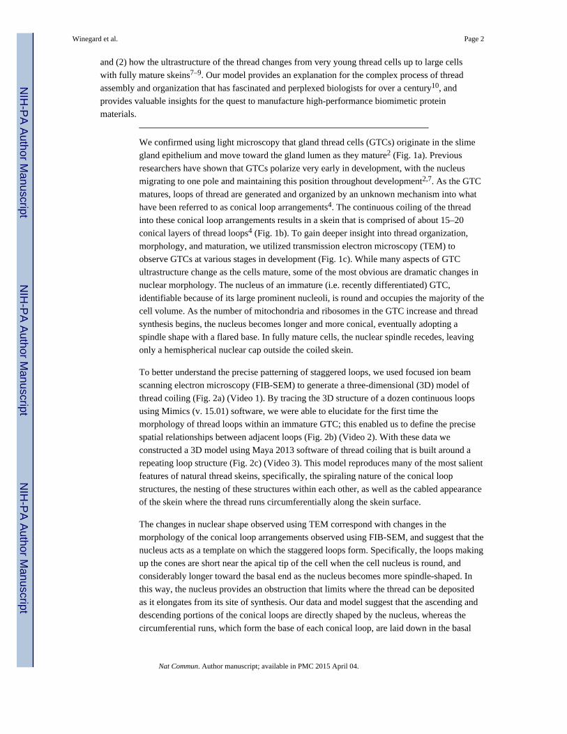
and (2) how the ultrastructure of the thread changes from very young thread cells up to large cells
with fully mature skeins7–9. Our model provides an explanation for the complex process of thread
assembly and organization that has fascinated and perplexed biologists for over a century10, and
provides valuable insights for the quest to manufacture high-performance biomimetic protein
materials.
We confirmed using light microscopy that gland thread cells (GTCs) originate in the slime
gland epithelium and move toward the gland lumen as they mature2 (Fig. 1a). Previous
researchers have shown that GTCs polarize very early in development, with the nucleus
migrating to one pole and maintaining this position throughout development2,7. As the GTC
matures, loops of thread are generated and organized by an unknown mechanism into what
have been referred to as conical loop arrangements4. The continuous coiling of the thread
into these conical loop arrangements results in a skein that is comprised of about 15–20
conical layers of thread loops4 (Fig. 1b). To gain deeper insight into thread organization,
morphology, and maturation, we utilized transmission electron microscopy (TEM) to
observe GTCs at various stages in development (Fig. 1c). While many aspects of GTC
ultrastructure change as the cells mature, some of the most obvious are dramatic changes in
nuclear morphology. The nucleus of an immature (i.e. recently differentiated) GTC,
identifiable because of its large prominent nucleoli, is round and occupies the majority of the
cell volume. As the number of mitochondria and ribosomes in the GTC increase and thread
synthesis begins, the nucleus becomes longer and more conical, eventually adopting a
spindle shape with a flared base. In fully mature cells, the nuclear spindle recedes, leaving
only a hemispherical nuclear cap outside the coiled skein.
To better understand the precise patterning of staggered loops, we used focused ion beam
scanning electron microscopy (FIB-SEM) to generate a three-dimensional (3D) model of
thread coiling (Fig. 2a) (Video 1). By tracing the 3D structure of a dozen continuous loops
using Mimics (v. 15.01) software, we were able to elucidate for the first time the
morphology of thread loops within an immature GTC; this enabled us to define the precise
spatial relationships between adjacent loops (Fig. 2b) (Video 2). With these data we
constructed a 3D model using Maya 2013 software of thread coiling that is built around a
repeating loop structure (Fig. 2c) (Video 3). This model reproduces many of the most salient
features of natural thread skeins, specifically, the spiraling nature of the conical loop
structures, the nesting of these structures within each other, as well as the cabled appearance
of the skein where the thread runs circumferentially along the skein surface.
The changes in nuclear shape observed using TEM correspond with changes in the
morphology of the conical loop arrangements observed using FIB-SEM, and suggest that the
nucleus acts as a template on which the staggered loops form. Specifically, the loops making
up the cones are short near the apical tip of the cell when the cell nucleus is round, and
considerably longer toward the basal end as the nucleus becomes more spindle-shaped. In
this way, the nucleus provides an obstruction that limits where the thread can be deposited
as it elongates from its site of synthesis. Our data and model suggest that the ascending and
descending portions of the conical loops are directly shaped by the nucleus, whereas the
circumferential runs, which form the base of each conical loop, are laid down in the basal
Winegard et al. Page 2
Nat Commun. Author manuscript; available in PMC 2015 April 04.
NIH
-PA
Author M
anuscriptN
IH-P
A A
uthor Manuscript
NIH
-PA
Author M
anuscript

groove where the nucleus and plasma membrane converge (Fig. 1c). In nearly mature GTCs,
the nucleus has reduced to only a fraction of its former size, and its full retreat in a basal
direction leads to very short circumferential runs and loops consisting of mostly back and
forth axial runs, called “inner core loops” by Fernholm4 (Fig. 3). Although interactions
between the intermediate filaments (IF) known as nuclear lamins and the inner nuclear
membrane are well described11,12 less is known about how the outer nuclear membrane
interacts with cytoplasmic elements13,14. The patterning of slime thread coiling by the GTC
nucleus may provide a new model for exploring these interactions in detail. Our data also
raise questions about how such dramatic changes in nuclear size and shape are regulated.
Our TEM studies largely substantiate previous studies7–9 and we believe add to our
understanding of the ultrastructural changes that take place during thread maturation (Figs.
4a,b). Others have shown that immature slime threads contain both IF and microtubules
(MT), but it was not clear which of these elements appears first. Our results clearly
demonstrate that thread production starts with a small bundle of IF in a paranuclear region
rich in mitochondria and ribosomes. The thread increases in both length and width via the
addition of more IF and eventually the incorporation of MT. The youngest threads are only
2–3 IFs in diameter (about 30 nm) and more mature ones are about 30 IFs in diameter (about
400 nm). During these stages, the thread is wrapped with a distinctive ~12-nm diameter
filament that either spirals around the thread at a steep angle7 or exists as separate rings (Fig.
4c); we are not able to distinguish between these two possibilities from our data at this time.
The most dramatic change in thread ultrastructure corresponds with the compaction of 12-
nm diameter IF into a dense superstructure in which discrete IF are no longer visible and
MT become surrounded by an electron lucent space presumably freed up by IF compaction.
This shift in thread ultrastructure likely corresponds with the post-translational modification
of IF proteins within mature GTCs9,15; it also coincides with the appearance of a fluffy rind
around the thread periphery7,8, which may indicate growth of the thread via the direct
addition of IF proteins or subunits rather than 12-nm IF. In fully mature threads, MT are
absent7,8, the spaces they occupy are completely filled in, the fluffy rind is gone, and
adjacent threads conform to each other leaving only a narrow space (about 40 nm) between
them (Figs. 4a,b).
Our model explains many aspects of how a single cell can produce a continuous 150 mm
long protein thread that can unravel without tangling, yet several questions remain. Our data
suggest that elongation occurs at a single site near the apical tip of the nucleus, but the
mechanism of elongation remains elusive. The role of intra-thread MT is also unclear,
although they may be involved in the delivery of IF and/or IF subunits16–17 to the interior of
the growing thread. Indeed, TEM and FIB-SEM images both demonstrate MT within the
thread and penetrating the thread from the side (Figs. 4c,d). The 12-nm filament that wraps
around the thread (Fig. 4e) before the IF condense is also puzzling. Downing and
colleagues7 propose that the filament is involved in the addition of IF to the growing thread,
but how exactly this would work is difficult to imagine. We suggest that the wrapping
filaments may prevent the merging of adjacent loops, which would have obvious negative
effects on thread deployment and ultimately the function of the slime1. Two other cell types,
the hagfish epidermal thread cell and the lamprey skein cell, notably also produce thick
helical IF bundles18,19, in contrast to the branching networks of IF that are far more common
Winegard et al. Page 3
Nat Commun. Author manuscript; available in PMC 2015 April 04.
NIH
-PA
Author M
anuscriptN
IH-P
A A
uthor Manuscript
NIH
-PA
Author M
anuscript

in metazoan cells20. In hagfish GTCs, this ability to produce a single, unbranched IF bundle
has been taken to the extreme and may be one of the key adaptations that allowed hagfishes
to evolve their remarkable fibrous defensive slime. While our model explains many aspects
of slime thread morphology, it does not explain how the precise staggering of loops arises.
The fact that each successive loop is translocated in a clockwise direction relative to its
younger neighboring loop is suggestive of a wheel-like mechanism at work. Although
wheels are extremely rare in biology21, thread coiling may involve just such a mechanism.
Recent efforts to make fibres from solubilized hagfish slime thread proteins yielded threads
with promising material properties, but they were clearly inferior to native slime threads22.
The changes in thread ultrastructure documented here suggest that the outstanding
mechanics of native slime threads likely arise via careful control of protein structure from IF
subunits to mature IF to dense IF superstructure. Unraveling the biochemical and
biophysical mechanisms underlying these transitions may lead us to new methodologies for
the production of sustainable high-performance protein materials.
Supplementary Material
Refer to Web version on PubMed Central for supplementary material.
Acknowledgments
The authors would like to thank the members of the Fudge Lab including Oualid Haddad, Atsuko Negishi, and Nicole Pinto, Dianne Moyles from the University of Guelph Electron Microscopy Unit, Glynis de Silveira and Julia Huang from McMaster University’s Canadian Center for Electron Microscopy, Amy Rowat and Dan Toso from UCLA, Susan Lapos from the University of Guelph’s Animal Health Laboratories, as well as Bob Frank, Matt Cornish, and Mike Davies from the University of Guelph’s Hagen Aqualab.
References
1. Fudge D, Levy N, Chiu S, Gosline J. Composition, morphology and mechanics of hagfish slime. J Exp Biol. 2005; 208:4613–4625. [PubMed: 16326943]
2. Newby W. The slime glands and thread cells of hagfish, Polistrotrema stoutii. J Morp. 1946; 78:397–409.
3. Downing S, et al. Threads in the hagfish slime gland thread cells: Organization, biochemical features, and length. Science. 1981; 212:326–328. [PubMed: 17792088]
4. Fernholm B. Thread cells from the slime glands of hagfish (Myxinidae). Acta Zoologica. 1981; 62:137–145.
5. Fudge D, Hillis S, Levy N, Gosline J. Hagfish slime threads as a biomimetic model for high performance protein fibres. Bioinspir Biomim. 2010; 5:035002. [PubMed: 20729569]
6. Winegard T, Fudge D. Deployment of hagfish thread skeins requires the transmission of mixing forces via mucin strands. J Exp Biol. 2010; 213:1235–1240. [PubMed: 20348334]
7. Downing S, Spitzer R, Koch E, Salo W. The hagfish slime gland thread cell. I. A unique cellular system for the study of intermediate filaments and intermediate filament-microtubule interactions. J Cell Biol. 1984; 98:653–669. [PubMed: 6537952]
8. Terakado K, Ogawa M, Hashimoto Y. Ultrastructure of the thread cells in the slime gland of Japanese hagfishes, Paramyxine atami and Eptatretus burgeri. Cell Tissue Res. 1975; 159:311–323. [PubMed: 1149101]
9. Spitzer R, Koch E, Downing S. Maturation of hagfish gland thread cells – composition and characterization of intermediate filament polypeptides. Cell Motil Cytoskel. 1988; 11:31–45.
10. Blomfield B. The thread cells and epidermis of Myxine. Quart J Micro Sci. 1882; 22:355–361.
Winegard et al. Page 4
Nat Commun. Author manuscript; available in PMC 2015 April 04.
NIH
-PA
Author M
anuscriptN
IH-P
A A
uthor Manuscript
NIH
-PA
Author M
anuscript
11. Gundersen G, Worman H. Nuclear positioning. Cell. 2013; 152(6):1376–1389. [PubMed: 23498944]
12. Burke B, Stewart C. The nuclear lamins: flexibility in function. Nat Rev Mol Cell Biol. 2013; 14:13–24. [PubMed: 23212477]
13. Maniotis A, Chen C, Ingber D. Demonstration of mechanical connections between integrins, cytoskeletal filaments, and nucleoplasm that stabilize nuclear structure. PNAS. 1997; 94:849–854. [PubMed: 9023345]
14. Lombardi M, Jaalouk D, Shanahan C, Burke B, Roux K, Lammerding J. The interaction between nesprins and SUN proteins at the nuclear envelope is critical for force transmission between the nucleus and cytoskeleton. J Biol Chem. 2011; 286:26743–26753. [PubMed: 21652697]
15. Spitzer R, Downing S, Koch E, Salo W, Saidel J. Hagfish slime gland thread cells. II. Isolation and characterisation of intermediate filament components associated with the thread. J Cell Biol. 1984; 98:670–677. [PubMed: 6537953]
16. Helfand B, Loomis P, Yoon M, Goldman R. Rapid transport of neural intermediate filament protein. J Cell Sci. 2003; 116:2345–2359. [PubMed: 12711702]
17. Uchida A, Alami N, Brown A. Tight functional coupling of kinesin-1A and dynein motors in the bidirectional transport of neurofilaments. Mol Biol Cell. 2009; 20:4997–5006. [PubMed: 19812246]
18. Downing S, Novales R. The fine structure of lamprey epidermis III. Granular cells. J Ultrastruct Res. 1971; 35:304–313. [PubMed: 4104246]
19. Quay W. Integument and the environment: Glandular composition, function and evolution. Am Zool. 1972; 12:95–108.
20. Coulombe P, Omary M. ‘Hard’ and ‘soft’ principles defining the structure, function and regulation of keratin intermediate filaments. Curr Op Cell Biol. 2002; 14:110–122. [PubMed: 11792552]
21. LaBarbera M. Why the wheels won’t go. Amer Nat. 1983; 121:395–408.
22. Negishi A, Armstrong C, Kreplak L, Rheinstadter M, Lim L-T, Gillis T, Fudge D. The production of fibers and films from solubilized hagfish slime thread proteins. Biomacromolecules. 2012; 13:3475–3482. [PubMed: 23016557]
Winegard et al. Page 5
Nat Commun. Author manuscript; available in PMC 2015 April 04.
NIH
-PA
Author M
anuscriptN
IH-P
A A
uthor Manuscript
NIH
-PA
Author M
anuscript

Figure 1. Hagfish slime gland, gland thread cells, and thread skeinsa, H&E stained cross-section of slime gland showing: gland thread cells (GTC), gland
mucus cells (GMC), gland pore (p), striated muscle around gland (m), and skin (s). Insets
show an immature GTC near the gland epithelium, and a mature GTC within the gland
lumen. b, SEM of coiled thread from a mature GTC that has broken open, revealing the
organization of staggered loops (partially highlighted in red), which form layers of conical
loop arrangements that spiral around the skein (one layer highlighted in purple). c,
Developmental progression of M. glutinosa GTCs revealed with TEM illustrating dramatic
Winegard et al. Page 6
Nat Commun. Author manuscript; available in PMC 2015 April 04.
NIH
-PA
Author M
anuscriptN
IH-P
A A
uthor Manuscript
NIH
-PA
Author M
anuscript

changes in nuclear size and shape. Immature GTCs lacking a slime thread can be identified
by their prominent nuclei and nucleoli. As the slime thread increases in length and diameter,
the nucleus becomes more spindle like, eventually receding to the basal end of the cell in
mature cells. Scale bars are 5 μm.
Winegard et al. Page 7
Nat Commun. Author manuscript; available in PMC 2015 April 04.
NIH
-PA
Author M
anuscriptN
IH-P
A A
uthor Manuscript
NIH
-PA
Author M
anuscript

Figure 2. 3D reconstruction of thread coiling within an immature GTC from FIB-SEM dataa, Stacks of images from FIB-SEM were assembled into a 3D volume using Mimics
software. This image was created by combining axial and transverse sections of the rendered
volume with an SEM image of a separate complete thread skein. b, Segmentation of a dozen
continuous loops within a developing GTC revealed the precise 3D pattern of thread coiling
(single loop in green). Segmentation of the nucleus (light blue), nucleolus (dark blue), and
mitochondria (maroon) shows the relative position of these structures to the developing
thread. The loops shown are not the most recent ones laid down on the nucleus (these were
too small to follow), but they do reflect nuclear shape at the time of synthesis. Inset shows
the position of the rendered structures within the cell. c, Based on the 3D structure of a
single loop and its relationship to adjacent loops, our model (built in Maya 2013) reproduces
the spiraling nature of the conical loops, the nesting of these conical structures, as well as
the cabled appearance of the skein where the thread runs circumferentially along the skein
surface.
Winegard et al. Page 8
Nat Commun. Author manuscript; available in PMC 2015 April 04.
NIH
-PA
Author M
anuscriptN
IH-P
A A
uthor Manuscript
NIH
-PA
Author M
anuscript

Figure 3. Temporal and spatial models of thread assembly and coiling in GTCsGTC growth and maturation is characterized by dramatic changes in nuclear size and
morphology (left), which correspond with the shape of conical loop arrangements laid down
during successive stages of thread production (right). Regular staggered loops are laid down
in the space defined by previous loops and the apical surface of the nucleus, and their
morphology changes as the nucleus becomes more spindle-shaped and retreats toward the
basal end of the cell. The result of manufacturing the skein in the manner depicted is a
mature, ovoid skein that can be ejected through the gland pore and unravel to its full
extended length of ~150 mm without tangling.
Winegard et al. Page 9
Nat Commun. Author manuscript; available in PMC 2015 April 04.
NIH
-PA
Author M
anuscriptN
IH-P
A A
uthor Manuscript
NIH
-PA
Author M
anuscript

Figure 4. Developmental series of thread ultrastructurea, TEM sections of slime threads from very immature to fully mature GTCs. The thread
depicted in (1) consists of a bundle of about ten, 12-nm IF. Threads increase in girth by the
addition of more IF, and eventually by the addition of MT (2). In the next stage (3), 12-nm
IF become packed more tightly, which creates electron lucent halos (asterisk) around the
MT. Further IF compaction is accompanied by the appearance of a fluffy rind (arrowheads)
on the thread surface (4), which likely corresponds to the direct addition of IF subunits or
proteins to the thread rather than the bundling of mature IF. In the final stage, IF proteins
compact further, MT are lost, the spaces they occupied are filled in, and the fluffy rind
disappears. In fully mature GTCs, the thread takes up the vast majority of the cell volume
and adjacent threads are packed so tightly that they conform to each other (5). Scale bars are
all 200 nm. b, Models of thread development corresponding to each of the stages depicted in
Winegard et al. Page 10
Nat Commun. Author manuscript; available in PMC 2015 April 04.
NIH
-PA
Author M
anuscriptN
IH-P
A A
uthor Manuscript
NIH
-PA
Author M
anuscript
(a). The second panel illustrates the 12-nm diameter filament (arrows) that wraps around the
thread at this stage in development c, TEM of thread longitudinal section depicting MT
within a developing thread. d, Cross-section of a thread from stage (4) showing a MT
penetrating from the side (3). e, Young thread from stage (2) showing the wrapping
filament, which is evident as regularly spaced circular sections and corresponding lines that
cross the thread at an average angle of about 86°. Scale bars in c, d, and e are 500 nm.
Winegard et al. Page 11
Nat Commun. Author manuscript; available in PMC 2015 April 04.
NIH
-PA
Author M
anuscriptN
IH-P
A A
uthor Manuscript
NIH
-PA
Author M
anuscript