Cognitive recovery and restoration of cell proliferation ... · Abstract Alzheimer’s disease (AD)...
13
RESEARCH ARTICLE Cognitive recovery and restoration of cell proliferation in the dentate gyrus in the 5XFAD transgenic mice model of Alzheimer’s disease following 2-hydroxy-DHA treatment Maria A. Fiol-deRoque • Raquel Gutierrez-Lanza • Silvia Tere ´s • Manuel Torres • Pere Barcelo ´ • Rube ´n V. Rial • Alexei Verkhratsky • Pablo V. Escriba ´ • Xavier Busquets • Jose ´ J. Rodrı ´guez Received: 6 May 2013 / Accepted: 30 August 2013 Ó Springer Science+Business Media Dordrecht 2013 Abstract Alzheimer’s disease (AD) is the most common neurodegenerative disorder in the elderly. In the last years, abnormalities of lipid metabolism and in particular of docosahexaenoic acid (DHA) have been recently linked with the development of the disease. According to the recent studies showing how hydroxylation of fatty acids enhances their biological activity, here we show that chronic treatment with a hydroxylated derivative of DHA, the 2-hydroxy-DHA (2OHDHA) in the 5XFAD transgenic mice model of AD improves performance in the radial arm maze test and restores cell proliferation in the dentate gyrus, with no changes in the presence of beta amyloid (Ab) plaques. These results suggest that 2OHDHA induced restoration of cell proliferation can be regarded as a major component in memory recovery that is inde- pendent of Ab load thus, setting the starting point for the development of a new drug for the treatment of AD. Keywords Alzheimer’s disease Lipid metabolism 2OHDHA 5XFAD Alzheimer model Cognition Neurogenesis Introduction Alzheimer’s disease (AD) is the most common neurodegenerative disease and the main form of progressive dementia in the elderly (Schaeffer et al. M. A. Fiol-deRoque S. Tere ´s M. Torres P. V. Escriba ´(&) X. Busquets (&) Molecular Cell Biomedicine, Department of Biology, Institut Universitari d’Investigacions en Cie `ncies de la Salut, University of the Balearic Islands, Bldg. Guillem Colom, Crta. Valldemossa, Km 7,5, 07122 Palma, Balearic Islands, Spain e-mail: [email protected] X. Busquets e-mail: [email protected] R. Gutierrez-Lanza J. J. Rodrı ´guez (&) Department of Neuroscience, University of the Basque Country UPV/EHU and CIBERNED, Technological Park, Bldg. 205, Floor-1, Laida Bidea, 48170 Zamudio, Bizkaia, Spain e-mail: [email protected] P. Barcelo ´ R. V. Rial Laboratorio de Neurofisiologı ´a, Department of Biology, University of the Balearic Islands, Palma, Spain A. Verkhratsky Faculty of Life Sciences, The University of Manchester, Manchester, UK J. J. Rodrı ´guez IKERBASQUE, Basque Foundation for Science, Bilbao, Spain 123 Biogerontology DOI 10.1007/s10522-013-9461-4
Transcript of Cognitive recovery and restoration of cell proliferation ... · Abstract Alzheimer’s disease (AD)...
RESEARCH ARTICLE
Cognitive recovery and restoration of cell proliferationin the dentate gyrus in the 5XFAD transgenic mice modelof Alzheimer’s disease following 2-hydroxy-DHA treatment
Maria A. Fiol-deRoque • Raquel Gutierrez-Lanza • Silvia Teres • Manuel Torres •
Pere Barcelo • Ruben V. Rial • Alexei Verkhratsky • Pablo V. Escriba •
Xavier Busquets • Jose J. Rodrıguez
Received: 6 May 2013 / Accepted: 30 August 2013
� Springer Science+Business Media Dordrecht 2013
Abstract Alzheimer’s disease (AD) is the most
common neurodegenerative disorder in the elderly.
In the last years, abnormalities of lipid metabolism and
in particular of docosahexaenoic acid (DHA) have
been recently linked with the development of the
disease. According to the recent studies showing how
hydroxylation of fatty acids enhances their biological
activity, here we show that chronic treatment with a
hydroxylated derivative of DHA, the 2-hydroxy-DHA
(2OHDHA) in the 5XFAD transgenic mice model of
AD improves performance in the radial arm maze test
and restores cell proliferation in the dentate gyrus,
with no changes in the presence of beta amyloid (Ab)
plaques. These results suggest that 2OHDHA induced
restoration of cell proliferation can be regarded as a
major component in memory recovery that is inde-
pendent of Ab load thus, setting the starting point for
the development of a new drug for the treatment of
AD.
Keywords Alzheimer’s disease � Lipid
metabolism � 2OHDHA � 5XFAD Alzheimer
model � Cognition � Neurogenesis
Introduction
Alzheimer’s disease (AD) is the most common
neurodegenerative disease and the main form of
progressive dementia in the elderly (Schaeffer et al.
M. A. Fiol-deRoque � S. Teres � M. Torres �P. V. Escriba (&) � X. Busquets (&)
Molecular Cell Biomedicine, Department of Biology,
Institut Universitari d’Investigacions en Ciencies de la
Salut, University of the Balearic Islands, Bldg. Guillem
Colom, Crta. Valldemossa, Km 7,5, 07122 Palma,
Balearic Islands, Spain
e-mail: [email protected]
X. Busquets
e-mail: [email protected]
R. Gutierrez-Lanza � J. J. Rodrıguez (&)
Department of Neuroscience, University of the Basque
Country UPV/EHU and CIBERNED, Technological Park,
Bldg. 205, Floor-1, Laida Bidea, 48170 Zamudio, Bizkaia,
Spain
e-mail: [email protected]
P. Barcelo � R. V. Rial
Laboratorio de Neurofisiologıa, Department of Biology,
University of the Balearic Islands, Palma, Spain
A. Verkhratsky
Faculty of Life Sciences, The University of Manchester,
Manchester, UK
J. J. Rodrıguez
IKERBASQUE, Basque Foundation for Science, Bilbao,
Spain
123
Biogerontology
DOI 10.1007/s10522-013-9461-4
2011). The pathophysiology of AD is characterized by
loss of synapses and neurons accompanied with
formation of extracellular deposits of fibrillar b-
amyloid (Ab), these deposits known as senile plaques,
and by the intraneuronal accumulation of neurofibril-
lary tangles, which result from the abnormal hyper-
phosphorylation of tau protein (Zhang et al. 2007).
Cognitive deficits arise from the damage of brain
regions associated with learning and memory, such as
the hippocampus, one of the most vulnerable brain
regions to AD (Thompson et al. 2004). The subgran-
ular zone (SGZ) of the granular cell layer (GCL) of the
hippocampal dentate gyrus (DG) is one of the two
neurogenic niches in the mammalian adult brain,
along with the anterior part of the subventricular zone
(Abrous et al. 2005). The role of impaired neurogen-
esis in the progression of AD has recently gained
relevance not only in animal models but also in
humans (Rodrıguez and Verkhratsky 2011). In numer-
ous studies on AD animal models the decrease of
neurogenesis was described (Donovan et al. 2006;
Demars et al. 2010; Rodrıguez et al. 2008), although
the opposite data also exist (Jin et al. 2004b; Jin et al.
2004a; Lopez-Toledano and Shelanski 2007). Despite
these contradictions, the restoration or potentiation of
neurogenesis has been recently considered as a
potential therapeutic strategy that may reduce or delay
early cognitive symptoms of the disease (Rodrıguez
et al. 2011; Mu and Gage 2011).
Lipid metabolism is considered to contribute to the
development of AD (Hartmann et al. 2007), with
indications that alterations in cholesterol, triglycerides
or omega-3 (n-3) fatty acids (FA) are associated with
the cognitive decline observed in AD (Morley and
Banks 2010). Furthermore, it has been suggested that
Ab production is directly affected by the lipid
composition of the membrane, these being key settings
for either the amyloidogenic or the non amyloidogenic
pathway (Hartmann et al. 2007; Grimm et al. 2011).
The docosahexaenoic acid (DHA, 22:6 n-3) is the
most abundant n-3 polyunsaturated FA (PUFA) in the
brain (Hashimoto et al. 2011). DHA is involved in the
development and function of the central nervous
system, contributing to synaptogenesis, synaptic func-
tion, cognition and neuroprotection (Salem et al.
2001). The DHA is primarily obtained through the diet
and its deficiency is related to cognitive decline in
aged brains and to neurodegenerative diseases such as
AD (Uauy and Dangour 2006). Several animal studies
have demonstrated that oral intake of DHA or fish oil
reduces AD-like brain pathology (Lim et al. 2005;
Green et al. 2007). Treatment with DHA significantly
improves cognitive deficits, protects from loss of
synaptic proteins and lowers insoluble Ab in trans-
genic AD mouse models (Calon et al. 2004; Lim et al.
2005). Moreover, numerous epidemiological studies
indicate an inverse correlation between DHA intake
and AD incidence, associating high levels of DHA
with a reduced risk of impaired cognitive functions
(Kalmijn et al. 1997; Morris et al. 2003; Johnson and
Schaefer 2006). However, direct administration of
DHA has no clear effect on cognitive decline (Freund-
Levi et al. 2006; Quinn et al. 2010). Recently, some
studies have also related DHA to neurogenesis
suggesting that this n-3 PUFA could modulate the
generation of new functional neurons (Su 2010). DHA
is able to promote proliferation in cultured embryonic
neural stem cells and neurogenesis in the hippocampus
of adult and aged rats with an improvement in learning
and cognition (Kawakita et al. 2006; Dyall et al. 2010).
Transgenic mice for n-3 PUFA, which have a high
level of DHA, have shown increased hippocampal
neurogenesis and neurite outgrowth as well as
improvements in spatial learning (He et al. 2009).
These findings indicate a potential mechanism by
which DHA improves the neuronal function in hippo-
campus thus facilitating or promoting learning and
behavior by generating new hippocampal neurons (Su
2010). It has been suggested that the biological
activity of DHA could be modulated trough neuro-
protectin D1 (NPD1; 10,17S-docosatrien) (Lukiw and
Bazan 2008). NPD1 is a natural hydroxylated deriv-
ative of DHA that is able to modulate an intrinsic
neuroprotective, anti-inflammatory, and antiapoptotic
gene-expression program that promotes cell survival
and reverts Ab induced neuronal death (Lukiw et al.
2005). It has been reported previously that hydroxyl-
ation of FA enhances their biological activity. A
hydroxylated synthetic derivative of oleic acid, 2-hy-
droxyoleic acid (Minerval), has become a potent
antitumor compound that modifies the composition
and structure of lipid membranes (Llado et al. 2009,
2010; Barcelo-Coblijn et al. 2011; Teres et al. 2012).
According to these data, and the apparently
enhanced power of hydroxylated FA, we propose to
use synthetic PUFA 2-hydroxy-DHA (2OHDHA) as a
new tool for the treatment of AD-associated cognitive
deficits. 2OHDHA is a hydroxylated derivative of
Biogerontology
123
DHA that has been developed by hydroxylating the
alpha-carbon of the precursor lipid. Here we report the
effect of 2OHDHA on memory and on cell prolifer-
ation in the dentate gyrus in the transgenic AD model
mice.
Materials and methods
All the experiments were performed in agreement with
the Bioethical Committee of the University of Balearic
Islands (Permit Number): #XB023-2010. All efforts
were made to reduce the number of animals by
following the 3R’s.
Animals and treatments
We used male 5XFAD double transgenic mice (line
Tg6799, Jackson Laboratories�, USA). 5XFAD mice
co-express Familial AD (FAD) mutant forms of
human APP (the Swedish mutation: K670N, M671L;
the Florida mutation: I716V; the London mutation:
V717I) and presenilin-1 (M146L; L286V) transgenes
under transcriptional control of the neuron-specific
mouse Thy-1 promoter (Oakley et al. 2006). 5XFAD
animals (B6/SJL genetic background) were main-
tained by crossing heterozygous transgenic mice with
B6/SJL F1 breeders. All 5XFAD transgenic mice were
heterozygous with respect to both transgenes, and non-
transgenic (Non-Tg) wild-type littermate mice were
used as controls.
Non-Tg controls (n = 10) and 5XFAD (n = 11)
animals were treated with vehicle (5 % intraesophagic
ethanol solution, 15 ml/kg/day), whilst the experi-
mental group, 5XFAD?2OHDHA, (n = 12) was
treated with 2OHDHA (15 mg/kg/day dissolved in
5 % ethanol; Lipopharma Therapeutics, Spain). The
treatment was carried out according to the disease
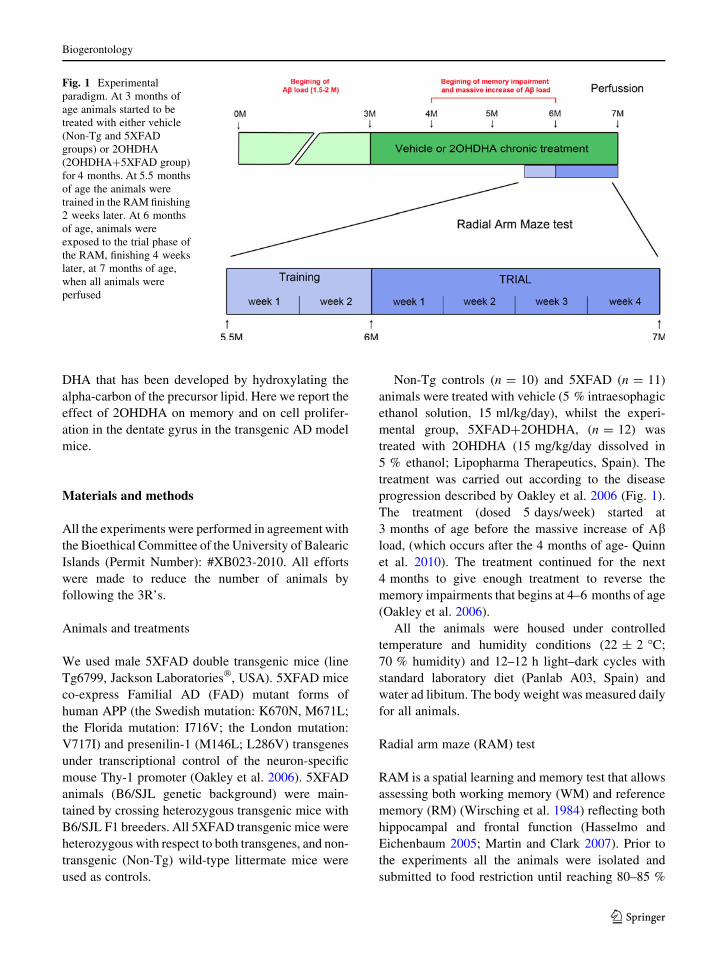
progression described by Oakley et al. 2006 (Fig. 1).
The treatment (dosed 5 days/week) started at
3 months of age before the massive increase of Abload, (which occurs after the 4 months of age- Quinn
et al. 2010). The treatment continued for the next
4 months to give enough treatment to reverse the
memory impairments that begins at 4–6 months of age
(Oakley et al. 2006).
All the animals were housed under controlled
temperature and humidity conditions (22 ± 2 �C;
70 % humidity) and 12–12 h light–dark cycles with
standard laboratory diet (Panlab A03, Spain) and
water ad libitum. The body weight was measured daily
for all animals.
Radial arm maze (RAM) test
RAM is a spatial learning and memory test that allows
assessing both working memory (WM) and reference
memory (RM) (Wirsching et al. 1984) reflecting both
hippocampal and frontal function (Hasselmo and
Eichenbaum 2005; Martin and Clark 2007). Prior to
the experiments all the animals were isolated and
submitted to food restriction until reaching 80–85 %
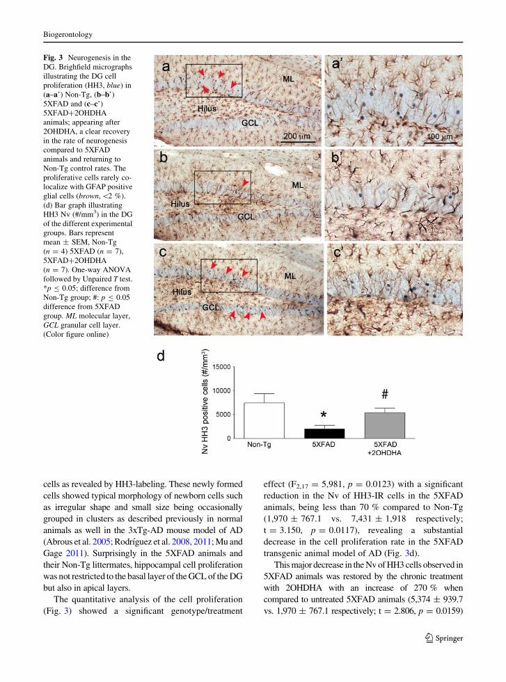
Fig. 1 Experimental
paradigm. At 3 months of
age animals started to be
treated with either vehicle
(Non-Tg and 5XFAD
groups) or 2OHDHA
(2OHDHA?5XFAD group)
for 4 months. At 5.5 months
of age the animals were
trained in the RAM finishing
2 weeks later. At 6 months
of age, animals were
exposed to the trial phase of
the RAM, finishing 4 weeks
later, at 7 months of age,
when all animals were
perfused
Biogerontology
123
of the normal body weight in ad libitum feeding and
were maintained in these conditions until the end of
the test. After food restriction and 2 weeks before
trials started, the animals were trained in the eight
RAM (LE766/8, Panlab SL, Barcelona, Spain). Each
mouse was placed in the centre of the maze and
allowed to look for the reward, a 45 mg food pellet
(Dustless Precision Pellets, Bio-Serv, US) located at
the end of every arm. Each session finished when the
animal either succeeded in finding the eight baited
arms or failed in complete all the arms after 10 min.
The movement of every animal was recorded with a
digital video tracking system (LE 8300 with software
SEDACOM v1.3, Panlab, SL, Spain). The training
was repeated 5 days/week, during 2 weeks. After the
training, when the mice reached 6 months of age, the
experimental paradigm started (Fig. 1). In all the
sessions just four arms were baited compared to the
training protocol. Each session finished when the
animals either succeeded in finding the four baited
arms or failed after 10 min. The performance was
assessed taking into account (1) the time to achieve the
test (2) the number of WM errors (WME: re-entrances
in a previous visited baited arm), (3) the number of
RM errors (RME: entrances in an unbaited arm) and
(4) the total number of errors (WME ? RME). The
test was repeated 5 days/week for 4 weeks. Once the
RAM test was finished, the animals were sacrificed
and the brains were used for the subsequent anatom-
ical studies.
Fixation and tissue processing
Non-Tg (n = 4), 5XFAD (n = 7) and 5XFAD?2OH-
DHA (n = 7) males were anaesthetized, after behav-
ioral analysis, with an intraperitoneal injection of
Ketamine/Xylacine (100/10 mg/Kg) at 7 months of
age and then transcardically perfused with 25 ml of
0.9 % saline solution followed by 75 ml of 4 %
paraformaldehyde in 0.1 M phosphate buffer (PB), pH
7.4. Brains were removed from the cranium and
bisected down the midline. The tissue was then post-
fixed in the same fixative for 24 h and kept in 0.1 M
PB, pH 7.4. Sagital sections of the hemibrains were cut
into 40–50 lm thickness using a vibrating microtome
(VT1000S, Leica, Milton Keynes, UK). Free floating
brain sections in 0.1 M PB pH 7.4, were collected and
stored in cryoprotectant solution (25 % sucrose and
3.5 % glycerol in 0.05 M PB at pH 7.4). Sagital
vibratome sections between levels L 1.20 mm to L
2.16 mm (hippocampus) were selected for immuno-
histochemistry (IHC) according to the mouse brain
atlas of Paxinos and Franklin (2004).
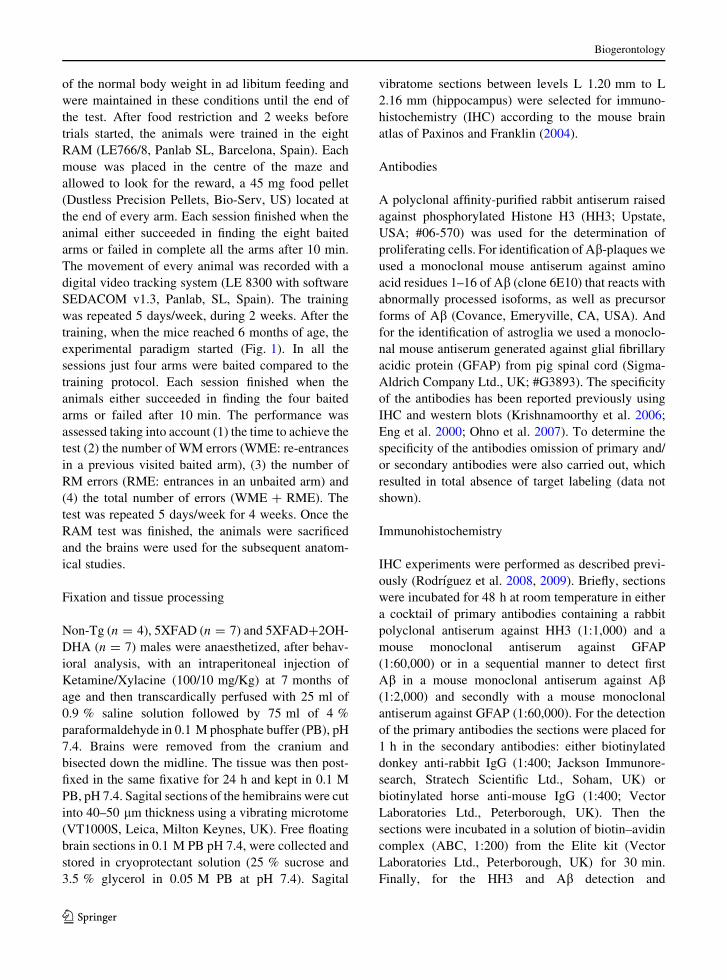
Antibodies
A polyclonal affinity-purified rabbit antiserum raised
against phosphorylated Histone H3 (HH3; Upstate,
USA; #06-570) was used for the determination of
proliferating cells. For identification of Ab-plaques we
used a monoclonal mouse antiserum against amino
acid residues 1–16 of Ab (clone 6E10) that reacts with
abnormally processed isoforms, as well as precursor
forms of Ab (Covance, Emeryville, CA, USA). And
for the identification of astroglia we used a monoclo-
nal mouse antiserum generated against glial fibrillary
acidic protein (GFAP) from pig spinal cord (Sigma-
Aldrich Company Ltd., UK; #G3893). The specificity
of the antibodies has been reported previously using
IHC and western blots (Krishnamoorthy et al. 2006;
Eng et al. 2000; Ohno et al. 2007). To determine the
specificity of the antibodies omission of primary and/
or secondary antibodies were also carried out, which
resulted in total absence of target labeling (data not
shown).
Immunohistochemistry
IHC experiments were performed as described previ-
ously (Rodrıguez et al. 2008, 2009). Briefly, sections
were incubated for 48 h at room temperature in either
a cocktail of primary antibodies containing a rabbit
polyclonal antiserum against HH3 (1:1,000) and a
mouse monoclonal antiserum against GFAP
(1:60,000) or in a sequential manner to detect first
Ab in a mouse monoclonal antiserum against Ab(1:2,000) and secondly with a mouse monoclonal
antiserum against GFAP (1:60,000). For the detection
of the primary antibodies the sections were placed for
1 h in the secondary antibodies: either biotinylated
donkey anti-rabbit IgG (1:400; Jackson Immunore-
search, Stratech Scientific Ltd., Soham, UK) or
biotinylated horse anti-mouse IgG (1:400; Vector
Laboratories Ltd., Peterborough, UK). Then the
sections were incubated in a solution of biotin–avidin
complex (ABC, 1:200) from the Elite kit (Vector
Laboratories Ltd., Peterborough, UK) for 30 min.
Finally, for the HH3 and Ab detection and
Biogerontology
123

visualization, the ABC was revealed by the incubation
in a solution prepared from the SGZ kits (Vector
Laboratories Ltd., Peterborough, UK) for 5 min; and
for the GFAP in a solution containing 0.022 % of 3,30
diaminobenzidine (Aldrich, Gillingham, UK) and
0.003 % H2O2 in 0.1 M Tris-buffered saline for
2.5 min.
HH3 and Ab plaques determination and analysis
We determined the numerical density (Nv) of HH3-
immunoreactive neurons (HH3-IR) as labeled cells per
volume unit (#/mm3) on 3–12 non-consecutive sagital
sections per animal. Subsequently, we determined the
Nv and size (mm2) of Ab plaques in the GCL of the
DG on 4 non-consecutive sections of the same animals
used for the proliferation analysis. Brain sections were
analyzed and photomicrographs were taken using a
Nikon Eclipse 80i microscope coupled with an 8001
MicroFiRE camera and all the area measurements
were done using computer-assisted imaging analysis
(ImageJ 1.43, NIH, USA). The entire HH3 positive
cell and Ab plaque counting as well as the IOD
measurement in the DG were conducted by a single
investigator (M.A.F-dR.) who was blind to the subject
number and group assignment. All slides were coded
prior by another lab member (R.G.) and codes were
not broken until all measurements were completed.
Inverse optical density (IOD) measurement
Using computer-assisted imaging analysis (Image J
1.43) we analyzed the density of intraneuronal Ab on
the same four sections used for the Ab plaques
counting. We measured the optical density (OD) of the
GCL of the DG as described previously (Noristani
et al. 2010). Briefly, to exclude any experimental
errors all images were taken at constant light intensity
and keeping the same optical filters, used to ensure the
specificity of the signal recorded by the camera.
The staining was observed throughout the thickness
of the section (40 lm) using light microscopy. The
OD was calculated from a relative scale of intensity
ranging from 0 to 255, with readout of 255 corre-
sponding to the area with very low Ab-immunoreac-
tive (Ab-IR) and 0 corresponding to the densest area of
labeling. The calibration density was kept constant for
the whole procedure to avoid experimental variances.
The sections background OD was determined from the
corpus callosum (CC) that was considered as blank
since Ab labeling in the CC is virtually absent, and CC
also presents a similar labeling to GFAP that DG.
To analyze the change in Ab-IR density against
constant control, the 255 was divided by control region
(CC) and the obtained factor was multiplied by the
region of interest (GCL) in every given section. IOD
was obtained by subtracting from the obtained back-
ground level (set at 255).
IOD ¼ 255� GCL255
CC
� �
Measurements of mean density were taken and
averaged, after background subtraction, from the GCL
of the ML of each slice. The results are shown as
inverse Ab-IR density (IOD/pixel).
Statistical analysis
One-way analysis of variance (ANOVA) followed by
either post hoc Bonferroni analysis or unpaired t test
comparisons were used to determine differences
between Non-Tg controls, 5XFAD or 5XFAD?2OH-
DHA animals (implemented by GraphPad Prism 4.0;
GraphPad Software, Inc., USA). All the data is
presented as Mean ± SEM.
Results
Effect of 2OHDHA on radial maze learning ability
On the fourth week of trial 5XFAD showed a
significant delay of 65 % in the time performance
compared to Non-Tg control animals (124.4 ± 12.38
vs. 75.69 ± 4.779 s respectively; p \ 0.01; Fig. 2a).
This delay was paralleled by a significant increase in
the total number of errors, close to 50 %, compared to
the Non-Tg control group (12.27 ± 1.048 vs.
8.200 ± 0.5993 respectively; p \ 0.05; Fig. 2b).
Chronic treatment with 2OHDHA showed no evi-
dent changes in the time performance and total number
of errors during the first 3 weeks of trial neither in Non-
Tg, nor in 5XFAD nor in 5XFAD?2OHDHA. How-
ever, on the fourth week of trial a clear performance
improvement in treated animals when compared to the
continuous alteration was observed in 5XFAD animals,
which was similar to Non-Tg control values; as showed
by the significant genotype/treatment effect in both the
Biogerontology
123

time performances (F2,32 = 4.056, p = 0.0276) and the
total number of errors (F2,32 = 5.771, p = 0.0076;
Fig. 2a, b).
Chronic 2OHDHA treatment showed an improve-
ment in the time performance since 5XFAD treated
animals complete the RAM test 15 % faster than non-
treated 5XFAD animals (105.4 ± 10.34 vs.
124.4 ± 12.38 s respectively) even if this difference
was not significant. Nevertheless, and more impor-
tantly, the total number of errors was massively
reduced. 5XFAD?2OHDHA animals showed a sig-
nificant decrease in total number of errors (Fig. 2b) by
more than 30 % compared to the 5XFAD animals
(8.287 ± 1.086 vs. 12.27 ± 1.048 respectively;
p \ 0.05) reaching similar levels to those seen in
Non-Tg control animals.
When separately considered WM and RM, presented
as WME and RME, the untreated 5XFAD animals
showed a significant impairment, which was completely
reverted after the chronic treatment with 2OHDHA.
However, there was a more relevant improvement in
WME than in RME, as revealed by the significant
genotype/treatment effect (F2,32 = 5.685, p = 0.0081;
F2,32 = 4.591, p = 0.0182 respectively).
5XFAD animals showed a significant increase in the
number of WME (Fig. 2c) being higher than 70 %
compared to Non-Tg control animals (3.955 ± 0.4770
vs. 2.320 ± 0.3200 respectively; p \ 0.05). The num-
ber of RME (Fig. 2d) was also higher, with an increase
of 40 % compared to Non-Tg controls (8.318 ± 0.6428
vs. 5.880 ± 0.3901 respectively; p \ 0.05).
When considering 5XFAD?2OHDHA animals,
they showed a significant decrease in both kinds of
errors, being of 45 % in WME (Fig. 2c), compared to
5XFAD animals (2.217 ± 0.4011 vs. 3.955 ± 0.4770
respectively; p \ 0.05), reaching normal Non-Tg
values. 5XFAD?2OHDHA animals also showed a
significant decrease in the number of RME (Fig. 2d),
around 30 % compared to 5XFAD animals
(6.071 ± 0.7387 vs. 8.318 ± 0.6428 respectively;
p \ 0.05) returning to the same level of errors seen
in Non-Tg control animals. These results suggest that
the memory recovery after the chronic treatment with
2OHDHA can modify both WM and RM, although
there is a more important recovery in WM, as revealed
by a further decrease of 15 %.
2OHDHA effects on cell proliferation
in the dentate gyrus
In both, Non-Tg controls and 5XFAD animals, there was
a consistent and detectable number of newly generated
Fig. 2 Learning ability on
the RAM. Bar graphs
showing the RAM
performance at the fourth
week of trial (5 trials/week)
at 7 months of age after
4 months of treatment
(5 days/week): Non-Tg
(n = 10), 5XFAD (n = 11)
and 5XFAD?2OHDHA
(n = 12). a Time
performance, b total number
of errors, c number of
working memory errors
(WME, re-entrance in
visited arms) and c number
of reference memory errors
(RME, entrance in un-baited
arms). Bars represent
mean ± SEM. One-way
ANOVA followed by
Bonferroni post hoc test.
*p B 0.05; **p B 0.01;
difference from Non-Tg
group; #: p B 0.05
difference from 5XFAD
group
Biogerontology
123
cells as revealed by HH3-labeling. These newly formed
cells showed typical morphology of newborn cells such
as irregular shape and small size being occasionally
grouped in clusters as described previously in normal
animals as well in the 3xTg-AD mouse model of AD
(Abrous et al. 2005; Rodrıguez et al. 2008, 2011; Mu and
Gage 2011). Surprisingly in the 5XFAD animals and
their Non-Tg littermates, hippocampal cell proliferation
was not restricted to the basal layer of the GCL of the DG
but also in apical layers.
The quantitative analysis of the cell proliferation
(Fig. 3) showed a significant genotype/treatment
effect (F2,17 = 5,981, p = 0.0123) with a significant
reduction in the Nv of HH3-IR cells in the 5XFAD
animals, being less than 70 % compared to Non-Tg
(1,970 ± 767.1 vs. 7,431 ± 1,918 respectively;
t = 3.150, p = 0.0117), revealing a substantial
decrease in the cell proliferation rate in the 5XFAD
transgenic animal model of AD (Fig. 3d).
This major decrease in the Nv of HH3 cells observed in
5XFAD animals was restored by the chronic treatment
with 2OHDHA with an increase of 270 % when
compared to untreated 5XFAD animals (5,374 ± 939.7
vs. 1,970 ± 767.1 respectively; t = 2.806, p = 0.0159)
Fig. 3 Neurogenesis in the
DG. Brighfield micrographs
illustrating the DG cell
proliferation (HH3, blue) in
(a–a’) Non-Tg, (b–b’)
5XFAD and (c–c’)
5XFAD?2OHDHA
animals; appearing after
2OHDHA, a clear recovery
in the rate of neurogenesis
compared to 5XFAD
animals and returning to
Non-Tg control rates. The
proliferative cells rarely co-
localize with GFAP positive
glial cells (brown,\2 %).
(d) Bar graph illustrating
HH3 Nv (#/mm3) in the DG
of the different experimental
groups. Bars represent
mean ± SEM, Non-Tg
(n = 4) 5XFAD (n = 7),
5XFAD?2OHDHA
(n = 7). One-way ANOVA
followed by Unpaired T test.
*p B 0.05; difference from
Non-Tg group; #: p B 0.05
difference from 5XFAD
group. ML molecular layer,
GCL granular cell layer.
(Color figure online)
Biogerontology
123

(Fig. 3d). HH3-IR cells in the 5XFAD?2OHDHA ani-
mals were reaching values closer to those seen in Non-Tg
controls. Indeed, no statistical significant differences were
found between Non-Tg and 5XFAD?2OHDHA mice
(7,431 ± 1,918 vs. 5,374 ± 939.7 respectively;
t = 1.092; p = 0.3031).
It is also remarkable that after the chronic treatment
with 2OHDHA all newly generated cells appeared in
both suprapyramidal and infrapyramidal blades of
GCL of the DG, whilst in Non-Tg animals the majority
of the newly generated cells were localized in the
suprapyramidal blade of the GCL (Fig. 3a–c). When
considering the phenotype of proliferating cells in the
GCL of the DG we found that HH3-IR cells were
rarely co-localized with GFAP. In fact, less than 2 %
of HH3-positive cells showed glial phenotype, sug-
gesting that the proliferation cells are of neural lineage
as previously reports in the 3xTg-AD model (Rodrı-
guez et al. 2008, 2009, 2011).
2OHDHA and Ab accumulation in the GCL
of the dentate gyrus
As expected, in Non-Tg control animals there was no
Ab accumulation in any of the hippocampal subfields
including the DG (Fig. 4a). In contrast, in 5XFAD a
massive presence of Ab was detected throughout the
whole hippocampus (Fig. 4b): including the subicu-
lum, CA1 and CA3 as well as the layers of the DG
including both upper and lower blade of de ML,
together with the hilus and the GCL The Ab load was
not changed after the chronic treatment with 2OH-
DHA (Fig. 4c) and the GFAP-IR distribution
remained the same in all the groups.
Since previous results showed that hippocampal HH3-
IR is located in the DG, we also assessed and analyzed the
Ab load in this hippocampal region. No significant
changes were observed in the DG of the hippocampus in
the 5XFAD?2OHDHA animals when compared to the
untreated 5XFAD animals neither in the Nv of Abplaques (3,560 ± 534.5 vs. 2,659 ± 303.3 respectively;
t = 1.361 p = 0.2157) nor in the area size of the plaques
(0.0007856 ± 7,462e-005 vs. 0.0008818 ± 9,136e-
005 respectively; t = 0.8250, p = 0.4366). In both
groups the total area of the DG occupied by Ab plaques
was similar comprising 9–11 % of the total area. The
analysis of the Ab deposit specifically in the GCL of the
DG did not reveal significant changes. When the
5XFAD?2OHDHA animals where compared to the
5XFAD no changes in the Nv of Ab plaques
(2,049 ± 317.7 vs. 1,453 ± 286.5 respectively;
t = 1.356, p = 0.2172) or in the area size of the plaques
(0.0009025 ± 8.820e-005 vs. 0.0008469 ± 6.194e-
005 respectively; t = 2.434, p = 0.0591) were detected.
Again, the total area of the GCL occupied by Ab plaques
Fig. 4 Ab load in the DG. Brightfield micrographs illustrating
Ab plaques accumulation (blue) and GFAP positive glial cells
(brown) in the DG of the hippocampus of a Non-Tg, b 5XFAD
and c 5XFAD?2OHDHA animals. In 5XFAD animals there
was a massive accumulation of Ab plaques which is not changed
by the treatment (see Results). DG dentate gyrus, GCL granule
cell layer, Mol.L molecular layer, S.Mol stratum lacunosum
molecular, S.Or stratum oriens, S.Pyr stratum pyramidale,
S.Rad stratum radiatum. (Color figure online)
Biogerontology
123
was similar in both groups, comprising a 6–7 % of the
total area. Similarly, there were no differences in the
intracellular Ab-IR in the GCL in 5XFAD?2OHDHA
animals compared to 5XFAD.
Discussion
The modification of naturally active molecules, such
as lipids, represents a new tool in the development of
medicaments (Escriba 2006). In the past we have
demonstrated that the 2-hydroxylation of a natural-
occurring FA as oleic acid produced a new molecule
with antitumoral activity (Llado et al. 2009) by
interfering with the sphingolipid synthesis pathway
by activating the sphingomyelin synthase (Barcelo-
Coblijn et al. 2011).
In the present study, we used 5XFAD transgenic
mice to investigate the effects of 2OHDHA chronic
treatment on memory performance and neurogenesis
in AD, as well as their relation to the Ab accumulation
in the GCL of the hippocampus. We found a signif-
icant memory impairment in the 5XFAD mouse model
in both WM and RM by the RAM test together with a
decrease in hippocampal cell proliferation at 7 months
of age, when there is a massive accumulation of Ab.
Chronic treatment with 2OHDHA resulted in recovery
of the spatial memory impairment, with a major
improvement in WM. This improvement in spatial
memory was concomitant with a significant recovery
in the rate of hippocampal cell proliferation.
The observed impairment in memory in the 5XFAD
mouse model of AD is in agreement with previous
behavioral studies in this and other AD transgenic
models, for example APP single transgenic, PS1xAPP
double transgenic and 3xTg-AD (Ashe 2001; Oddo
et al. 2003). In addition, we have described impair-
ments in both RM (reflecting hippocampal function)
and WM (reflecting both hippocampal and prefrontal
cortex functions). Previous studies had already
described a decline in various types of hippocampus-
dependent memory in the 5XFAD mouse model
including spatial memory in the Morris water maze
and temporal memory in the auditory trace fear
conditioning (Ohno et al. 2006) as well as contextual
fear memory (Ohno 2009). Importantly, these memory
impairments start to occur at 6 months of age in
accordance with the massive increase of Ab levels and
amyloid burden, which, in the 5XFAD model, appear
between 4 and 6 months of age (Oakley et al. 2006).
This reported memory impairment is obviously linked
with the onset of hippocampal synaptic dysfunction
such as reduced levels of baseline transmission and
deficient LTP in 5XFAD mice (Kimura and Ohno
2009).
Similarly to other findings (Haughey et al. 2002;
Wen et al. 2002; Dong et al. 2004), which demon-
strated impaired neurogenesis in transgenic mice
having mutant forms of APP and/or presenilin-1 and
our recent results in the 3xTg-AD mouse model of AD
(Rodrıguez et al. 2008, 2009, 2011); we have detected
a significant decrease of cell proliferation in the
5XFAD animals at 7 months of age. The impaired cell
proliferation observed in 5XFAD animals could be
due to a reduced level of neurotrophic factors, like the
brain-derived neurotrophic factor (BDNF), that starts
early in the course of human AD (Peng et al. 2005;
Devi and Ohno 2012) and observed in the 5XFAD
animals from 3 months of age (Devi and Ohno 2012).
Chronic treatment with 2OHDHA (or DHA) up-
regulate cell proliferation to normal values observed in
the healthy brain. Concievably, this increased hippo-
campal cell proliferation could enhance the hippo-
campal network promoting the long-term potentiation
(LTP, that is known to be affected in AD-Yamin 2009)
thus improving in the RM. However, we have showed
that the chronic treatment with 2OHDHA results in a
larger improvement of WM than RM. In this sense, the
recovery of the hippocampal cellular network can
have an indirect effect on the WM; due to the dense
projection from the hippocampus to the prefrontal
cortex with strong excitatory action (Vertes et al.
2007). However, not only the prefrontal cortex but
also the hippocampus subserve spatial WM in rodents
(Gordon 2011) indicating that the improvement seen
in the spatial WM could be a concomitant effect due to
the recovery of local and hippocampus-cortex con-
nection. Similar improvement in RM in young rats and
an improvement in both WM and RM in aged rats after
chronic treatments with DHA have been described
previously (Gamoh et al. 2001, 1999).
The 2OHDHA being a direct derivative of DHA
may act through the same pathways than DHA. In this
sense, it has been shown that the n-3 deficiency reduce
the levels of BDNF and signaling through the BDNF
receptor TrkB proportionally to brain DHA levels
(Bhatia et al. 2011) and that the n-3 PUFA supple-
mentation enhance BDNF expression (Katsuki et al.
Biogerontology
123
2009) correlating with an increase of neurogenesis in
mouse hippocampus (Blondeau et al. 2009). More-
over, it has been showed that BDNF promotes LTP
improvement (Pang and Lu 2004; Lu et al. 2008)
setting a possible connection between DHA, BDNF
and LTP. In addition, the 2OHDHA being a hydrox-
ylated derivative of DHA, as well as NPD1, may
possibly demonstrate a neuroprotective (Zhao et al.
2011) and antiapoptotic capacity (Calandria et al.
2012), enhancing the maintenance of the hippocam-
pus, not only as an inducer of cell proliferation, but
also protecting existing neurons.
Although there is some evidence indicating that
DHA directs the amyloidogenic processing of APP
towards the non-amyloidogenic pathway, with a
consequent reduction of Ab (Grimm et al. 2011), we
failed to detect changes in the Ab load neither in the
whole DG nor in the GCL of the hippocampus after the
chronic treatment with 2OHDHA. Besides not being
exactly the same molecule, this difference may be due
to different factors such as the dose and the treatment
length, as well as the animal model of AD used. In this
case, the 5XFAD is a hyperamyloidosis model that
rapidly accumulates high levels of Ab, in both plaques
and intracellularly (Oakley et al. 2006), which could
mask small changes in the total pool of Ab. Never-
theless, we have reported a recovery of memory and
enhanced cell proliferation in the 5XFAD?2OHDHA
animals despite of absence of changes in Ab,
suggesting that cell proliferation is not directly
affected by Ab plaques in the DG of the hippocampus.
Clinical perspectives
Although evidence from this and other studies suggest
that omega 3-fatty acids may protect from cognitive
decline, clinical trials carried out with DHA showed
no solid evidence on cognitive function benefits
(Quinn et al. 2010; Sydenham et al. 2012). The
cognitive recovery after treatment with 2OHDHA in
5XFAD mice and in human APP/Tau transgenic
Drosophila models is greater than the obtained with
DHA (manuscript in preparation). This opens the
possibility for positive results in future clinical studies
using 2OHDHA.
Acknowledgements This work was supported by grants from
the Spanish Government: TRACE [grant number PET2008/
0172-01 (to X.B.)]; INNPACTO [grant number IPT-010000-
2010–16 (to X.B.)]; Plan Nacional de I?D?I 2008–2011 and
ISCIII-Subdireccion General de Evaluacion y Fomento de la
investigacion co-financed by FEDER [grant number PI10/02738
(to J.J.R. and A.V.)]; and the Government of the Basque Country
grants [grant numbers AE-2010-1-28, AEGV10/16 and GV-
2011111020 (to J.J.R.)]; as well as by [BIO2010-21132 from the
MICINN (to P.E.)]. M.A.F-dR was supported by a fellowship
from the Govern de les Illes Balears (Conselleria d’Educacio,
Cultura i Universitats) operational program co-funded by the
European Social Fund. M.A.F-dR and M.T. were also recipients
of an IPT-Spanish government fellowship.
References
Abrous DN, Koehl M, Le Moal M (2005) Adult neurogenesis:
from precursors to network and physiology. Physiol Rev
85(2):523–569. doi:10.1152/physrev.00055.2003
Ashe KH (2001) Learning and memory in transgenic mice
modeling Alzheimer’s disease. Learn Mem 8(6):301–308.
doi:10.1101/lm.43701
Barcelo-Coblijn G, Martin ML, de Almeida RF, Noguera-Salva
MA, Marcilla-Etxenike A, Guardiola-Serrano F, Luth A,
Kleuser B, Halver JE, Escriba PV (2011) Sphingomyelin
and sphingomyelin synthase (SMS) in the malignant
transformation of glioma cells and in 2-hydroxyoleic acid
therapy. Proc Natl Acad Sci USA 108(49):19569–19574.
doi:10.1073/pnas.1115484108
Bhatia HS, Agrawal R, Sharma S, Huo YX, Ying Z, Gomez-
Pinilla F (2011) Omega-3 fatty acid deficiency during brain
maturation reduces neuronal and behavioral plasticity in
adulthood. PLoS ONE 6(12):e28451. doi:10.1371/journal.
pone.0028451
Blondeau N, Nguemeni C, Debruyne DN, Piens M, Wu X, Pan
H, Hu X, Gandin C, Lipsky RH, Plumier JC, Marini AM,
Heurteaux C (2009) Subchronic alpha-linolenic acid
treatment enhances brain plasticity and exerts an antide-
pressant effect: a versatile potential therapy for stroke.
Neuropsychopharmacology 34(12):2548–2559. doi:10.
1038/npp.2009.84
Calandria JM, Mukherjee PK, de Rivero Vaccari JC, Zhu M,
Petasis NA, Bazan NG (2012) Ataxin-1 poly-Q-induced
proteotoxic stress and apoptosis are attenuated in neural
cells by docosahexaenoic acid-derived neuroprotectin D1.
J Biol Chem. doi:10.1074/jbc.M111.287078
Calon F, Lim GP, Yang F, Morihara T, Teter B, Ubeda O,
Rostaing P, Triller A, Salem N, Ashe KH, Frautschy SA,
Cole GM (2004) Docosahexaenoic acid protects from
dendritic pathology in an Alzheimer’s disease mouse
model. Neuron 43(5):633–645. doi:10.1016/j.neuron.2004.
08.013
Demars M, Hu YS, Gadadhar A, Lazarov O (2010) Impaired
neurogenesis is an early event in the etiology of familial
Alzheimer’s disease in transgenic mice. J Neurosci Res
88(10):2103–2117. doi:10.1002/jnr.22387
Devi L, Ohno M (2012) 7,8-Dihydroxyflavone, a small-mole-
cule TrkB agonist, reverses memory deficits and BACE1
elevation in a mouse model of Alzheimer’s disease. Neu-
ropsychopharmacology 37(2):434–444. doi:10.1038/npp.
2011.191
Biogerontology
123
Dong H, Goico B, Martin M, Csernansky CA, Bertchume A,
Csernansky JG (2004) Modulation of hippocampal cell
proliferation, memory, and amyloid plaque deposition in
APPsw (Tg2576) mutant mice by isolation stress. Neuro-
science 127(3):601–609. doi:10.1016/j.neuroscience.2004.
05.040
Donovan MH, Yazdani U, Norris RD, Games D, German DC,
Eisch AJ (2006) Decreased adult hippocampal neurogen-
esis in the PDAPP mouse model of Alzheimer’s disease.
J Comp Neurol 495(1):70–83. doi:10.1002/cne.20840
Dyall SC, Michael GJ, Michael-Titus AT (2010) Omega-3 fatty
acids reverse age-related decreases in nuclear receptors and
increase neurogenesis in old rats. J Neurosci Res
88(10):2091–2102. doi:10.1002/jnr.22390
Eng LF, Ghirnikar RS, Lee YL (2000) Glial fibrillary acidic
protein: GFAP-thirty-one years (1969–2000). Neurochem
Res 25(9–10):1439–1451
Escriba PV (2006) Membrane-lipid therapy: a new approach in
molecular medicine. Trends Mol Med 12(1):34–43. doi:10.
1016/j.molmed.2005.11.004
Freund-Levi Y, Eriksdotter-Jonhagen M, Cederholm T, Basun
H, Faxen-Irving G, Garlind A, Vedin I, Vessby B, Wahl-
und LO, Palmblad J (2006) Omega-3 fatty acid treatment
in 174 patients with mild to moderate Alzheimer disease:
OmegAD study: a randomized double-blind trial. Arch
Neurol 63(10):1402–1408. doi:10.1001/archneur.63.10.
1402
Gamoh S, Hashimoto M, Sugioka K, Shahdat Hossain M, Hata
N, Misawa Y, Masumura S (1999) Chronic administration
of docosahexaenoic acid improves reference memory-
related learning ability in young rats. Neuroscience
93(1):237–241
Gamoh S, Hashimoto M, Hossain S, Masumura S (2001)
Chronic administration of docosahexaenoic acid improves
the performance of radial arm maze task in aged rats. Clin
Exp Pharmacol Physiol 28(4):266–270
Gordon JA (2011) Oscillations and hippocampal-prefrontal
synchrony. Curr Opin Neurobiol 21(3):486–491. doi:10.
1016/j.conb.2011.02.012
Green KN, Martinez-Coria H, Khashwji H, Hall EB, Yurko-
Mauro KA, Ellis L, LaFerla FM (2007) Dietary docosa-
hexaenoic acid and docosapentaenoic acid ameliorate
amyloid-beta and tau pathology via a mechanism involving
presenilin 1 levels. J Neurosci 27(16):4385–4395. doi:10.
1523/JNEUROSCI.0055-07.2007
Grimm MO, Kuchenbecker J, Grosgen S, Burg VK, Hunds-
dorfer B, Rothhaar TL, Friess P, de Wilde MC, Broersen
LM, Penke B, Peter M, Vıgh L, Grimm HS, Hartmann T
(2011) Docosahexaenoic acid reduces amyloid beta pro-
duction via multiple pleiotropic mechanisms. J Biol Chem
286(16):14028–14039. doi:10.1074/jbc.M110.182329
Hartmann T, Kuchenbecker J, Grimm MO (2007) Alzheimer’s
disease: the lipid connection. J Neurochem 103(Suppl
1):159–170. doi:10.1111/j.1471-4159.2007.04715.x
Hashimoto M, Tozawa R, Katakura M, Shahdat H, Haque AM,
Tanabe Y, Gamoh S, Shido O (2011) Protective effects of
prescription n-3 fatty acids against impairment of spatial
cognitive learning ability in amyloid b-infused rats. Food
Funct 2(7):386–394. doi:10.1039/c1fo00002k
Hasselmo ME, Eichenbaum H (2005) Hippocampal mecha-
nisms for the context-dependent retrieval of episodes.
Neural Netw 18(9):1172–1190. doi:10.1016/j.neunet.
2005.08.007
Haughey NJ, Nath A, Chan SL, Borchard AC, Rao MS, Mattson
MP (2002) Disruption of neurogenesis by amyloid beta-
peptide, and perturbed neural progenitor cell homeostasis,
in models of Alzheimer’s disease. J Neurochem
83(6):1509–1524
He C, Qu X, Cui L, Wang J, Kang JX (2009) Improved spatial
learning performance of fat-1 mice is associated with
enhanced neurogenesis and neuritogenesis by docosahexa-
enoic acid. Proc Natl Acad Sci USA 106(27):11370–11375.
doi:10.1073/pnas.0904835106
Jin K, Galvan V, Xie L, Mao XO, Gorostiza OF, Bredesen DE,
Greenberg DA (2004a) Enhanced neurogenesis in Alz-
heimer’s disease transgenic (PDGF-APPSw, Ind) mice.
Proc Natl Acad Sci USA 101(36):13363–13367. doi:10.
1073/pnas.0403678101
Jin K, Peel AL, Mao XO, Xie L, Cottrell BA, Henshall DC,
Greenberg DA (2004b) Increased hippocampal neurogen-
esis in Alzheimer’s disease. Proc Natl Acad Sci USA
101(1):343–347. doi:10.1073/pnas.2634794100
Johnson EJ, Schaefer EJ (2006) Potential role of dietary n-3 fatty
acids in the prevention of dementia and macular degener-
ation. Am J Clin Nutr 83(6 Suppl):1494S–1498S
Kalmijn S, Launer LJ, Ott A, Witteman JC, Hofman A, Breteler
MM (1997) Dietary fat intake and the risk of incident
dementia in the Rotterdam Study. Ann Neurol
42(5):776–782. doi:10.1002/ana.410420514
Katsuki H, Kurimoto E, Takemori S, Kurauchi Y, Hisatsune A,
Isohama Y, Izumi Y, Kume T, Shudo K, Akaike A (2009)
Retinoic acid receptor stimulation protects midbrain
dopaminergic neurons from inflammatory degeneration via
BDNF-mediated signaling. J Neurochem 110(2):707–718.
doi:10.1111/j.1471-4159.2009.06171.x
Kawakita E, Hashimoto M, Shido O (2006) Docosahexaenoic
acid promotes neurogenesis in vitro and in vivo. Neuro-
science 139(3):991–997. doi:10.1016/j.neuroscience.2006.
01.021
Kimura R, Ohno M (2009) Impairments in remote memory
stabilization precede hippocampal synaptic and cognitive
failures in 5XFAD Alzheimer mouse model. Neurobiol Dis
33(2):229–235. doi:10.1016/j.nbd.2008.10.006
Krishnamoorthy T, Chen X, Govin J, Cheung WL, Dorsey J,
Schindler K, Winter E, Allis CD, Guacci V, Khochbin S,
Fuller MT, Berger SL (2006) Phosphorylation of histone
H4 Ser1 regulates sporulation in yeast and is conserved in
fly and mouse spermatogenesis. Genes Dev 20(18):
2580–2592. doi:10.1101/gad.1457006
Lim GP, Calon F, Morihara T, Yang F, Teter B, Ubeda O, Salem N,
Frautschy SA, Cole GM (2005) A diet enriched with the
omega-3 fatty acid docosahexaenoic acid reduces amyloid
burden in an aged Alzheimer mouse model. J Neurosci
25(12):3032–3040. doi:10.1523/JNEUROSCI.4225-04.2005
Llado V, Gutierrez A, Martınez J, Casas J, Teres S, Higuera M,
Galmes A, Saus C, Besalduch J, Busquets X, Escriba PV
(2010) Minerval induces apoptosis in Jurkat and other
cancer cells. J Cell Mol Med 14(3):659–670. doi:10.1111/j.
1582-4934.2008.00625.x
Llado V, Teres S, Higuera M, Alvarez R, Noguera-Salva MA,
Halver JE, Escriba PV, Busquets X (2009) Pivotal role of
dihydrofolate reductase knockdown in the anticancer
Biogerontology
123

activity of 2-hydroxyoleic acid. Proc Natl Acad Sci USA
106(33):13754–13758. doi:10.1073/pnas.0907300106
Lopez-Toledano MA, Shelanski ML (2007) Increased neuro-
genesis in young transgenic mice overexpressing human
APP(Sw, Ind). J Alzheimers Dis 12(3):229–240
Lu Y, Christian K, Lu B (2008) BDNF: A key regulator for
protein synthesis-dependent LTP and long-term memory?
Neurobiol Learn Mem 89(3):312–323. doi:10.1016/j.nlm.
2007.08.018
Lukiw WJ, Bazan NG (2008) Docosahexaenoic acid and the
aging brain. J Nutr 138(12):2510–2514. doi:10.3945/jn.
108.096016
Lukiw WJ, Cui JG, Marcheselli VL, Bodker M, Botkjaer A,
Gotlinger K, Serhan CN, Bazan NG (2005) A role for
docosahexaenoic acid-derived neuroprotectin D1 in neural
cell survival and Alzheimer disease. J Clin Invest
115(10):2774–2783. doi:10.1172/JCI25420
Martin SJ, Clark RE (2007) The rodent hippocampus and spatial
memory: from synapses to systems. Cell Mol Life Sci
64(4):401–431. doi:10.1007/s00018-007-6336-3
Morley JE, Banks WA (2010) Lipids and cognition. J Alzhei-
mers Dis 20(3):737–747. doi:10.3233/JAD-2010-091576
Morris MC, Evans DA, Bienias JL, Tangney CC, Bennett DA,
Wilson RS, Aggarwal N, Schneider J (2003) Consumption
of fish and n-3 fatty acids and risk of incident Alzheimer
disease. Arch Neurol 60(7):940–946. doi:10.1001/archneur.
60.7.940
Mu Y, Gage FH (2011) Adult hippocampal neurogenesis and its
role in Alzheimer’s disease. Mol Neurodegener 6:85.
doi:10.1186/1750-1326-6-85
Noristani HN, Olabarria M, Verkhratsky A, Rodrıguez JJ (2010)
Serotonin fibre sprouting and increase in serotonin trans-
porter immunoreactivity in the CA1 area of hippocampus
in a triple transgenic mouse model of Alzheimer’s disease.
Eur J Neurosci 32(1):71–79. doi:10.1111/j.1460-9568.
2010.07274.x
Oakley H, Cole SL, Logan S, Maus E, Shao P, Craft J, Guillozet-
Bongaarts A, Ohno M, Disterhoft J, Van Eldik L, Berry R,
Vassar R (2006) Intraneuronal beta-amyloid aggregates,
neurodegeneration, and neuron loss in transgenic mice with
five familial Alzheimer’s disease mutations: potential fac-
tors in amyloid plaque formation. J Neurosci 26(40):
10129–10140. doi:10.1523/JNEUROSCI.1202-06.2006
Oddo S, Caccamo A, Shepherd JD, Murphy MP, Golde TE,
Kayed R, Metherate R, Mattson MP, Akbari Y, LaFerla FM
(2003) Triple-transgenic model of Alzheimer’s disease
with plaques and tangles: intracellular Abeta and synaptic
dysfunction. Neuron 39(3):409–421
Ohno M (2009) Failures to reconsolidate memory in a mouse
model of Alzheimer’s disease. Neurobiol Learn Mem
92(3):455–459. doi:10.1016/j.nlm.2009.05.001
Ohno M, Chang L, Tseng W, Oakley H, Citron M, Klein WL,
Vassar R, Disterhoft JF (2006) Temporal memory deficits
in Alzheimer’s mouse models: rescue by genetic deletion
of BACE1. Eur J Neurosci 23(1):251–260. doi:10.1111/j.
1460-9568.2005.04551.x
Ohno M, Cole SL, Yasvoina M, Zhao J, Citron M, Berry R,
Disterhoft JF, Vassar R (2007) BACE1 gene deletion
prevents neuron loss and memory deficits in 5XFAD APP/
PS1 transgenic mice. Neurobiol Dis 26(1):134–145.
doi:10.1016/j.nbd.2006.12.008
Pang PT, Lu B (2004) Regulation of late-phase LTP and long-
term memory in normal and aging hippocampus: role of
secreted proteins tPA and BDNF. Ageing Res Rev
3(4):407–430. doi:10.1016/j.arr.2004.07.002
Paxinos G, Franklin KBJ (2004) The mouse brain in stereotaxic
coordinates, 2nd edn. Academic, London
Peng S, Wuu J, Mufson EJ, Fahnestock M (2005) Precursor form
of brain-derived neurotrophic factor and mature brain-
derived neurotrophic factor are decreased in the pre-clini-
cal stages of Alzheimer’s disease. J Neurochem 93(6):
1412–1421. doi:10.1111/j.1471-4159.2005.03135.x
Quinn JF, Raman R, Thomas RG, Yurko-Mauro K, Nelson EB,
Van Dyck C, Galvin JE, Emond J, Jack CR, Weiner M,
Shinto L, Aisen PS (2010) Docosahexaenoic acid supple-
mentation and cognitive decline in Alzheimer disease: a
randomized trial. JAMA 304(17):1903–1911. doi:10.1001/
jama.2010.1510
Rodrıguez JJ, Verkhratsky A (2011) Neurogenesis in Alzhei-
mer’s disease. J Anat 219(1):78–89. doi:10.1111/j.1469-
7580.2011.01343.x
Rodrıguez JJ, Jones VC, Tabuchi M, Allan SM, Knight EM,
LaFerla FM, Oddo S, Verkhratsky A (2008) Impaired adult
neurogenesis in the dentate gyrus of a triple transgenic
mouse model of Alzheimer’s disease. PLoS ONE
3(8):e2935. doi:10.1371/journal.pone.0002935
Rodrıguez JJ, Jones VC, Verkhratsky A (2009) Impaired cell
proliferation in the subventricular zone in an Alzheimer’s
disease model. NeuroReport 20(10):907–912. doi:10.1097/
WNR.0b013e32832be77d
Rodrıguez JJ, Noristani HN, Olabarria M, Fletcher J, Somerville
TD, Yeh CY, Verkhratsky A (2011) Voluntary running and
environmental enrichment restores impaired hippocampal
neurogenesis in a triple transgenic mouse model of Alz-
heimer’s disease. Curr Alzheimer Res 8(7):707–717
Salem N, Litman B, Kim HY, Gawrisch K (2001) Mechanisms
of action of docosahexaenoic acid in the nervous system.
Lipids 36(9):945–959
Schaeffer EL, Figueiro M, Gattaz WF (2011) Insights into
Alzheimer disease pathogenesis from studies in trans-
genic animal models. Clinics (Sao Paulo) 66(Suppl 1):
45–54
Su HM (2010) Mechanisms of n-3 fatty acid-mediated devel-
opment and maintenance of learning memory performance.
J Nutr Biochem 21(5):364–373. doi:10.1016/j.jnutbio.
2009.11.003
Sydenham E, Dangour AD, Lim WS (2012) Omega 3 fatty acid
for the prevention of cognitive decline and dementia.
Cochrane Database Syst Rev 6:CD005379. doi:10.1002/
14651858.CD005379.pub3
Teres S, Llado V, Higuera M, Barcelo-Coblijn G, Martin ML,
Noguera-Salva MA, Marcilla-Etxenike A, Garcıa-Verdugo
JM, Soriano-Navarro M, Saus C, Gomez-Pinedo U, Bus-
quets X, Escriba PV (2012) 2-Hydroxyoleate, a nontoxic
membrane binding anticancer drug, induces glioma cell
differentiation and autophagy. Proc Natl Acad Sci USA.
doi:10.1073/pnas.1118349109
Thompson PM, Hayashi KM, De Zubicaray GI, Janke AL, Rose
SE, Semple J, Hong MS, Herman DH, Gravano D, Dodd-
rell DM, Toga AW (2004) Mapping hippocampal and
ventricular change in Alzheimer disease. Neuroimage
22(4):1754–1766. doi:10.1016/j.neuroimage.2004.03.040
Biogerontology
123
Uauy R, Dangour AD (2006) Nutrition in brain development
and aging: role of essential fatty acids. Nutr Rev 64(5 Pt
2):S24–S33 (discussion S72–91)
Vertes RP, Hoover WB, Szigeti-Buck K, Leranth C (2007)
Nucleus reuniens of the midline thalamus: link between the
medial prefrontal cortex and the hippocampus. Brain Res
Bull 71(6):601–609. doi:10.1016/j.brainresbull.2006.12.002
Wen PH, Shao X, Shao Z, Hof PR, Wisniewski T, Kelley K,
Friedrich VL, Ho L, Pasinetti GM, Shioi J, Robakis NK,
Elder GA (2002) Overexpression of wild type but not an
FAD mutant presenilin-1 promotes neurogenesis in the
hippocampus of adult mice. Neurobiol Dis 10(1):8–19.
doi:10.1006/nbdi.2002.0490
Wirsching BA, Beninger RJ, Jhamandas K, Boegman RJ, El-
Defrawy SR (1984) Differential effects of scopolamine on
working and reference memory of rats in the radial maze.
Pharmacol Biochem Behav 20(5):659–662
Yamin G (2009) NMDA receptor-dependent signaling path-
ways that underlie amyloid beta-protein disruption of LTP
in the hippocampus. J Neurosci Res 87(8):1729–1736.
doi:10.1002/jnr.21998
Zhang C, McNeil E, Dressler L, Siman R (2007) Long-lasting
impairment in hippocampal neurogenesis associated with
amyloid deposition in a knock-in mouse model of familial
Alzheimer’s disease. Exp Neurol 204(1):77–87. doi:10.
1016/j.expneurol.2006.09.018
Zhao Y, Calon F, Julien C, Winkler JW, Petasis NA, Lukiw WJ,
Bazan NG (2011) Docosahexaenoic acid-derived neuro-
protectin D1 induces neuronal survival via secretase- and
PPARgamma-mediated mechanisms in Alzheimer’s dis-
ease models. PLoS ONE 6(1):e15816. doi:10.1371/journal.
pone.0015816
Biogerontology
123