Coagulase and Deoxyribonuclease Activities Staphylococci Isolated...
9
AseaED MICRoBIOLOGY, Apr. 1972, p. 725-733 Copyright @ 1972 American Society for Microbiology Vol. 23, No. 4 Printed in U.S.A. Coagulase and Deoxyribonuclease Activities of Staphylococci Isolated From Clinical Sources' HARRY E. MORTON ANm JUDITH COHN William Pepper Laboratory, Microbiology Division, Department of Pathology, School of Medicine, University of Pennsylvania, Philadelphia, Pennsylvania 19104 Received for publication 2 August 1971 A total of 504 clinical isolates of the family Micrococcaceae were tested for coagulase, deoxyribonuclease, clumping factor, and phosphatase to determine whether there is a correlation between the results of these tests and the patho- genicity of staphylococci. In the tests for coagulase production, it was found that either human or rabbit plasma could be used with broth cultures, whereas rabbit but not human plasma was satisfactory when microorganisms removed from solid culture medium were used. Deoxyribonuclease production correlated better than the fermentation of mannitol with coagulase production. The use of methyl green, Toluidine Blue 0, or acridine orange offered no advantage over the use of HCl for detecting the production of deoxyribonuclease. Neither the presence of clumping factor nor the production of phosphatase correlated well with coagulase production. Strains of staphylococci that did not produce coagu- lase and deoxyribonuclease were isolated as frequently as, and from a greater variety of clinical sources than, strains which produced these substances. It is concluded that the production of coagulase and deoxyribonuclease are proper- ties of staphylococci which are not necessarily indicative of potential pathoge- nicity of the organisms for man. An investigation of the tests for coagulase, deoxyribonuclease (DNase), clumping factor, and phosphatase was undertaken to determine whether any of these in vitro tests might be useful for predicting potential pathogenicity of staphylococci for man. It was considered nec- essary to determine whether either human or rabbit plasma could be used in the slide test for clumping factor and in the tube test for coagulase. Cadness-Graves et al. (6) stated that with human plasma coagulase-negative strains do not give positive slide tests for clumping factor. Using human plasma and growth from solid medium Needham et al. (22) found approximately 95% correlation between the slide test for clumping factor and the tube test for coagulase, and observed that a few strains reversed their reactions when subcul- tured and subsequently retested. Decrease or loss of clumping factor was also observed by Steward and Kelly (32) after storage of cul- tures and frozen cell suspensions but not when the cultures were lyophilized. Knowledge of this change in properties is important in the ' Presented at the 71st Annual Meeting of the American Society for Microbiology, Minneapolis, Minn., 2-7 May 1971. maintenance of cultures for quality control. Manuals for clinical microbiology are not consistent or sufficiently specific in their rec- ommendations for performing the slide test for clumping factor or for performing the coagu- lase test. In the American Society for Micro- biology Manual of Clinical Microbiology, Ivler stated that the slide test for clumping factor is not recommended for routine diagnostic work and would not describe it in his chapter on staphylococci (14). However, in the same man- ual, Paik described the procedure for the slide test and recommended confirming all negative tests with a tube test (24). This gives the im- pression that a positive slide test for clumping factor indicates a positive tube test reaction for coagulase. In the American Public Health Association manual of diagnostic procedures, Blair recommended either human or rabbit plasma for the tube test for coagulase with growth from solid medium and in the slide test for clumping factor (3). Confirmation of nega- tive slide tests with tube tests was also recom- mended and it was pointed out that some skill is required in performing and reading the slide test. The DNase test was reported by Jarvis and Wynne to be helpful in the case of strains 725 on July 15, 2018 by guest http://aem.asm.org/ Downloaded from
Transcript of Coagulase and Deoxyribonuclease Activities Staphylococci Isolated...

AseaED MICRoBIOLOGY, Apr. 1972, p. 725-733Copyright @ 1972 American Society for Microbiology
Vol. 23, No. 4Printed in U.S.A.
Coagulase and Deoxyribonuclease Activities ofStaphylococci Isolated From Clinical Sources'
HARRY E. MORTON ANm JUDITH COHN
William Pepper Laboratory, Microbiology Division, Department of Pathology, School of Medicine,University of Pennsylvania, Philadelphia, Pennsylvania 19104
Received for publication 2 August 1971
A total of 504 clinical isolates of the family Micrococcaceae were tested forcoagulase, deoxyribonuclease, clumping factor, and phosphatase to determinewhether there is a correlation between the results of these tests and the patho-genicity of staphylococci. In the tests for coagulase production, it was foundthat either human or rabbit plasma could be used with broth cultures, whereasrabbit but not human plasma was satisfactory when microorganisms removedfrom solid culture medium were used. Deoxyribonuclease production correlatedbetter than the fermentation of mannitol with coagulase production. The use ofmethyl green, Toluidine Blue 0, or acridine orange offered no advantage over
the use of HCl for detecting the production of deoxyribonuclease. Neither thepresence of clumping factor nor the production of phosphatase correlated wellwith coagulase production. Strains of staphylococci that did not produce coagu-
lase and deoxyribonuclease were isolated as frequently as, and from a greatervariety of clinical sources than, strains which produced these substances. It isconcluded that the production of coagulase and deoxyribonuclease are proper-ties of staphylococci which are not necessarily indicative of potential pathoge-nicity of the organisms for man.
An investigation of the tests for coagulase,deoxyribonuclease (DNase), clumping factor,and phosphatase was undertaken to determinewhether any of these in vitro tests might beuseful for predicting potential pathogenicity ofstaphylococci for man. It was considered nec-essary to determine whether either human orrabbit plasma could be used in the slide testfor clumping factor and in the tube test forcoagulase. Cadness-Graves et al. (6) statedthat with human plasma coagulase-negativestrains do not give positive slide tests forclumping factor. Using human plasma andgrowth from solid medium Needham et al. (22)found approximately 95% correlation betweenthe slide test for clumping factor and the tubetest for coagulase, and observed that a fewstrains reversed their reactions when subcul-tured and subsequently retested. Decrease orloss of clumping factor was also observed bySteward and Kelly (32) after storage of cul-tures and frozen cell suspensions but not whenthe cultures were lyophilized. Knowledge ofthis change in properties is important in the
' Presented at the 71st Annual Meeting of the AmericanSociety for Microbiology, Minneapolis, Minn., 2-7 May1971.
maintenance of cultures for quality control.Manuals for clinical microbiology are not
consistent or sufficiently specific in their rec-ommendations for performing the slide test forclumping factor or for performing the coagu-lase test. In the American Society for Micro-biology Manual of Clinical Microbiology, Ivlerstated that the slide test for clumping factor isnot recommended for routine diagnostic workand would not describe it in his chapter onstaphylococci (14). However, in the same man-ual, Paik described the procedure for the slidetest and recommended confirming all negativetests with a tube test (24). This gives the im-pression that a positive slide test for clumpingfactor indicates a positive tube test reactionfor coagulase. In the American Public HealthAssociation manual of diagnostic procedures,Blair recommended either human or rabbitplasma for the tube test for coagulase withgrowth from solid medium and in the slide testfor clumping factor (3). Confirmation of nega-tive slide tests with tube tests was also recom-mended and it was pointed out that some skillis required in performing and reading the slidetest. The DNase test was reported by Jarvisand Wynne to be helpful in the case of strains
725
on July 15, 2018 by guesthttp://aem
.asm.org/
Dow
nloaded from
726 MORTON
that give doubtful reactions in the coagulasetest (15).Although many authors have referred to the
production of coagulase and DNase as indica-tive of the pathogenicity of staphylococci forman, an examination of the strains from clin-ical material does not appear to have beenmade recently.
MATERIALS AND METHODSCultures. Strains of staphylococci were isolated
from clinical specimens submitted to the laboratory.To include numerous coagulase-negative cultures inthe study, some strains of gram-positive cocci wereisolated from blood-agar plates that had been ex-posed to room air during the routine monitoring ofair in the operating rooms. The cultures were main-tained on heart infusion agar (HIA) slants, andusually not more than two or three transfers weremade before performance of the various tests.Plasmas. Human plasma was removed from those
blood specimens which had been collected in Vacu-tainers (lavender-colored stoppers) containing EDTA(either the sodium or potassium salt of ethylenedi-aminetetraacetic acid) for hematological studies.Usually 1 to 2 ml of plasma could be removed fromeach tube. Although the blood was not collectedaseptically, the plasma was handled aseptically. Theplasma from a number of tubes was removed to yielda pool of at least 150 ml. This was centrifuged at2,000 rev/min for 15 min to remove the blood cells,and then was dispensed in 10 to 12-ml amounts intotubes with'screw-cap tops and stored at -10 C untilused. Just prior to use, a sufficient quantity of theplasma was thawed and the contents of each tubewere mixed thoroughly.
Rabbit plasma was Difco Coagulase PlasmaEDTA obtained in the desiccated form. After beingreconstituted according to the manufacturer's direc-tions, it was either used promptly or stored at -10C.Media. DNase test agar (DTA), from either Difco
or BBL, was prepared according to the manufactur-er's directions and dispensed into sterile plasticpetri dishes (90 by 15 mm).DTA-MG medium was prepared by adding 1 ml
of 0.5% methyl green solution to each 100 ml of DTAmedium prior to autoclaving at 121 C for 15 min.After cooling to 50 C, the medium was dispensedinto sterile plastic petri dishes. A stock solution of0.5 g of methyl green (Allied Chemical NA 0420) wasprepared in 100 ml of deionized water and repeat-edly extracted with equal volumes of chloroform in aseparatory funnel until the chloroform became color-less, as recommended by Smith et al. (31). The stocksolution of methyl green was stored at 4 C.DTA-TBO medium was prepared by adding 10
mg of Toluidine Blue 0 (Allied Chemical NA 0641)to each 100 ml of DTA medium prior to autoclavingat 121 C for 15 min (29). After cooling to 50 C, themedium was dispensed into sterile petri dishes.DTA-AO medium was similar to that described
by Omenn and Friedman (23), and was prepared by
A,ND COHN APPL. MICROBIOL.
adding 1 ml of a stock solution of 0.4% acridine or-ange (basic orange 14; Matheson, Coleman and Bell,AX 305) to each 100 ml of DTA medium before auto-claving. After cooling to 50 C, the medium was dis-pensed into sterile petri dishes.
Phenolphthalein diphosphate agar (PDP medium)was prepared by adding 2 ml of a 0.5% stock solutionof phenolphthalein diphosphate sodium salt and 3ml of sterile defibrinated sheep blood to each 100 mlof HIA which had been melted and cooled to 50 C;the PDP medium was dispensed into sterile plasticpetri dishes. The stock solution was prepared by dis-solving 0.5 g of phenolphthalein diphosphate sodiumsalt in 100 ml of deionized water which had beensterilized by filtering through a 0.22-,um membranefilter (Millipore Corp.); the stock solution was storedat - 10 C (2).HIA and heart infusion broth (HIB) were obtained
from Difco. Both media were stated by the manufac-turer to have a final pH of 7.4.The media for fermentation tests were prepared
by adding 1% glucose or 1% mannitol to purple brothbase (Difco). These media were dispensed in 6-mlamounts into 16 by 150 mm tubes containing 10 by35 mm inverted tubes, which had been fitted withMorton closures (Scientific Products no. T1390-16)and sterilized by autoclaving at 121 C for 15 min.
Tests. Tube coagulase tests with broth cultureswere performed by adding 0.5 ml of overnight HIBculture to 0.5 ml of human or rabbit plasma (dilutedto 1 part rabbit plasma and 4 parts HIB) and incu-bating the tubes in a water bath at 37 C. The tubeswere examined after 1 to 2, 4, and 6 hr. Any coagula-tion of the plasma was recorded as a positive reac-tion. Tubes in which no coagulation occurred wereincubated overnight, and a final reading was madeafter 24 hr. If coagulation occurred, it took placeusually within 1 to 2 or 4 hr and was in the form of afirm or nearly firm clot. Rarely did a positive reac-tion occur during overnight incubation. The reactingmixtures were contained in sterile 13 by 100 mmtubes with stainless-steel closures (Scientific Prod-ucts no. T1390-13).Tube coagulase tests with growth from solid me-
dium were performed by suspending some growthfrom an overnight HIA slant in 0.5-ml amounts ofhuman and undiluted rabbit plasma with a platinumloop. The tubes were similar to those used in thecoagulase test with broth cultures. Readings weremade after 1 and 3 hr and after overnight incubationin a water bath at 37 C. Any coagulation of theplasma was recorded as a positive reaction.
Slide tests for the clumping factor were performedby mixing some growth from an HIA slant with adrop of 0.85% sodium chloride solution on a micro-scope slide. If the suspension remained uniform, adrop of human or rabbit plasma was added and rap-idly mixed by gently rotating the slide. Clumping ofthe organisms within 10 sec was recorded as a posi-tive reaction.
For determining the ability of the organisms toferment glucose and mannitol, one tube of glucosebroth and one tube of mannitol broth were inocu-lated with the growth from a HIA slant, placed in a
on July 15, 2018 by guesthttp://aem
.asm.org/
Dow
nloaded from

ENZYME ACTIVITIES OF STAPHYLOCOCCI
GasPak (BBL) anaerobic jar, and incubated at 36 Covernight. The production of acid was evidenced bythe change in color of the bromocresol purple indi-cator from purple to yellow.The test for phosphatase was made by inoculating
plates of PDP medium with up to eight cultures withthe growth from HIA slants. Inoculations were madeby making a streak about 2 cm long with an inocu-lating needle. After overnight incubation at 36 C, theopened plates were held over an open ammoniabottle. The growth containing phosphatase-pro-ducing organisms turned a bright-pink color,whereas no color change took place in the absence ofphosphatase.The test for DNase was made by inoculating
plates of DTA with up to eight cultures with thegrowth from HIA slants by making streaks about 2cm long with an inoculating needle. After overnightincubation at 36 C, the plates were flooded with 1 NHCl (16). The production of DNase by the growingcultures was evidenced by a clear zone remainingaround the growth; in the absence of DNase, themedium turned opaque.
In many cases, parallel tests for the production ofDNase were made with DTA-MG, DTA-TBO, andDTA-AO media. Plates of DTA-MG and DTA-TBOwere inoculated and incubated similarly to the DTAplates. On DTA-MG, the production of DNase bythe growing cultures produced a clear zone aroundthe growth; the medium in the absence of DNaseremained green. On DTA-TBO, the production ofDNase was indicated by a rose-pink zone around thegrowth; the medium in the absence of DNase re-mained blue. Plates of DTA-AO were inoculatedwith up to 8 cultures with the growth from tubes ofHIB by making short streaks with an inoculatingloop. After overnight incubation at 36 C, the openedplates were examined in a darkened room with ultra-violet light by use of a Mineralite, model SL2537, 9w, 110 v, short-wave ultraviolet filter (Ultra-VioletProducts, Inc., South Pasadena, Calif.). A zone ofnonfluorescence surrounded the growing cultureswhich produced DNase, whereas the medium flu-oresced brightly in areas where DNase had not beenproduced.
RESULTSIn Table 1 are summarized the results ob-
tained by testing 224 strains of staphylococcigrown in broth for coagulase production by useof human and rabbit plasmas in the tube test.Also included are the results obtained by usingthe growth of the organisms from solid me-dium for detecting the presence of clumpingfactor in the slide test and the production ofcoagulase in the tube tests with the two plas-mas, the production of DNase on DTA, andthe ability to utilize glucose and mannitol an-aerobically.
All 224 strains of staphylococci producedcoagulase as determined by the tube test withbroth cultures and at least one of the plasmas;
223 of the 224 strains coagulated rabbitplasma, and 219 of the 224 strains coagulatedhuman plasma. When growth from solid me-dium was used in the tube test, 221 of the 224strains of staphylococci coagulated rabbitplasma but only 36 of the 224 strains coagu-lated human plasma. When some of the growthfrom the same cultures on solid medium usedin the tube tests was used for the slide testswith the same lots of plasma, only 152 of the224 strains clumped in rabbit plasma and 184strains clumped in human plasma. It is evi-dent that rabbit plasma is better than humanplasma for performing the tube coagulase testwith growth from either solid or liquid media.If human plasma has to be used for the coagu-lase test, only broth cultures should be used.The presence of clumping factor as detected inthe slide test was not a good indicator of coag-ulase production, as only 68% of the 224 cul-tures reacted with rabbit plasma and 82% re-acted with human plasma.
Only 2 (0.89%) of the 224 coagulase-positivestrains of staphylococci failed to give a posi-tive test for DNase. These two strains alsofailed to ferment mannitol, failed to give apositive coagulase test with rabbit plasma inthe tube test with growth from solid medium,and did not possess clumping factor. Thebroth cultures coagulated human and rabbitplasma in the tube test (line 15, Table 1).These strains were isolated from air.The broth cultures of 69 strains of micro-
cocci failed to coagulate human and rabbitplasmas in the tube tests, and it is this prop-erty which characterizes this group of culturessummarized in Table 2. The reason for se-lecting as the most reliable results those ob-tained in the coagulase test in which equalamounts of broth culture and plasma werecontained in a tube, as proposed by Cowan in1938 (7), was that it was shown by Williamsand Harper (35) to be the most satisfactorytechnique. Only one strain fermented glucoseand mannitol (line 12, Table 2); this strainfailed to produce DNase and coagulase by anyof the methods of testing, and it did not pos-sess clumping factor. Only three strains coagu-lated rabbit plasma in the tube test withgrowth from solid medium, but the numbers offalse-positive reactions were much greater withhuman plasma and in the slide tests forclumping factor. Five of the 69 cultures pro-duced DNase.
Only 200 of the 224 coagulase-positive cul-tures listed in Table 1 and 19 of the 96 coagu-lase-negative cultures listed in Table 2 pro-duced phosphatase. Additional tests for phos-
727VOL. 23, 1972
on July 15, 2018 by guesthttp://aem
.asm.org/
Dow
nloaded from

MORTON AND COHN APPL. MICROBIOL.
TABLE 1. Clumping factor, coagulase, and DNase tests with staphylococcia
Clumping factor Coagulase tests AnaerobicNo. of slide test . . fermentationLine cultu SoldmediumBroth medium DNase
H R H R H R G M
1 16 + + + + + + + + +2 127 + + - + + + + + +3 9 + - + + + + + + +4 7 - - + + + + + + +5 29 - - - + + + + + +6 18 + - - + + + + + +7 7 + + - + + + + - +8 1 + - + + + + + - +9 1 + - - + + + + - +10 1 + - - + - + + - +11 1 + + - + - + + + +12 1 + - + + - + + + +13 1 + - - + + - + + +14 1 - - - + + + + - +15 2 + + + +
16 1 - - - + - + + + +17 1 + + - - - + + + +
Total reacting 224 184 152 36 221 219 223 224 211 222
strainsIa H = human plasma; R = rabbit plasma; G = glucose; M = mannitol; + = clumping in the slide tests
for clumping factor, coagulation in the tube tests for coagulation production, only acid produced in the fer-mentation tests, and production of DNase in the DNase test; - = no reaction.
TABLE 2. Clumping factor, coagulase, and DNase tests with Micrococcaceaea
Clumping factor Coagulase tests AnaerobicLine No. of slide test Solid medium Broth medium fermentation DNasecultures
H R H R H R G M
2 22 +
3 5 + +
4 5 + +
5 2 + +
6 1 + + + - - - - - -7 3 + + +
8 1 + +
9 1 +-
10 3r 6 +1 1 3 411 1 +
12 1 + +-
13 7 + + --
14 4 + +
15 1 + + +
16 1 + + + - - - + - -
Total reacting 69 17 10 13 3 0 0 47 1 5strains
a See footnote a of Table 1.
728
on July 15, 2018 by guesthttp://aem
.asm.org/
Dow
nloaded from
ENZYME ACTIVITIES OF STAPHYLOCOCCI
phatase production were discontinued sincethe correlation between it and coagulase pro-duction was poor.
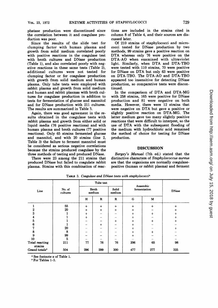
Since the results of the slide test forclumping factor with human plasma andgrowth from solid medium correlated poorlywith positive reactions in the coagulase testwith broth cultures and DNase production(Table 1), and also correlated poorly with neg-ative reactions in these same tests (Table 2),additional cultures were not tested forclumping factor or for coagulase productionwith growth from solid medium and humanplasma. Only tube tests were employed withrabbit plasma and growth from solid mediumand human and rabbit plasmas with broth cul-tures for coagulase production in addition totests for fermentation of glucose and mannitoland for DNase production with 211 cultures.The results are summarized in Table 3.
Again, there was good agreement in the re-
sults obtained in the coagulase tests withrabbit plasma and growth from either solid or
liquid media (76 positive reactions) and withhuman plasma and broth cultures (77 positivereactions). Only 65 strains fermented glucoseand mannitol, and with 20 strains (line 2,Table 3) the failure to ferment mannitol mustbe considered as serious negative correlationsbecause the strains produced coagulase by thethree methods of testing and produced DNase.There were 23 among the 211 strains that
produced DNase but failed to coagulate rabbitplasma. Strains with this combination of reac-
tions are included in the strains cited incolumn 8 of Table 4, and their sources are dis-cussed later.Of 210 strains of staphylococci and micro-
cocci tested for DNase production by twomethods, 99 strains gave a positive reaction on
DTA whereas only 76 were positive on theDTA-AO when examined with ultravioletlight. Similarly, when DTA and DTA-TBOwere tested with 110 strains, 70 were positivefor DNase on DTA but only 63 were positiveon DTA-TBO. The DTA-AO and DTA-TBOappeared too insensitive for detecting DNaseproduction, so comparative tests were discon-tinued.
In the comparison of DTA and DTA-MGwith 258 strains, 165 were positive for DNaseproduction and 81 were negative on bothmedia. However, there were 12 strains thatwere negative on DTA but gave a positive or
slightly positive reaction on DTA-MG. Thelatter medium gave too many slightly positivereactions that were difficult to interpret, so theuse of DTA with the subsequent flooding ofthe medium with hydrochloric acid remainedthe method of choice for testing for DNaseproduction.
DISCUSSIONBergey's Manual (7th ed.) stated that the
distinctive characters of Staphylococcus aureus
are that the organisms are normally coagulase-positive (human or rabbit plasma) and ferment
TABLE 3. Coagulase and DNase tests with staphylococcia
Tube testAnaerobic
Line No. of Broth Solid fermentation DNaecultures medium medium as
H R R G M
1 55 + + + + + +2 20 + + + + -+3 1 + + + +4 1+ - - + -+5 1- - + + + +6 + -+--
7 20 --+ -+
8 8 _ _ _ + + _9 99 _ _ _ + _ _10 5
Total reacting 211 77 76 76 206 65 98strains
Grand totals" 504 296 299 300 477 277 325
a See footnote a of Table 1.b For Tables 1-3.
VOL. 23, 1972 729
on July 15, 2018 by guesthttp://aem
.asm.org/
Dow
nloaded from

TABLE 4. Summary of the reactions of 504 strains of Micrococcaceae in the tests for coagulase, DNase, andfermentation of glucose and mannitol
Columns and combinations of reactions"Test
1 2 3 4 5 6 7 8 9 10 11 12
Coagulase5......... + - + + - + - + +DNasec. + - ++ + + - + +Glucosed.+ - + + + + ++ +Mannitold ............... + - - - - _ + + +Total strains (504) ......... 267 26 31 5 138 1 026 1 0 0 0
a Symbols: + = positive reaction; - = no reaction on the substrate.b Coagulation of rabbit plasma in the tube test with growth from either solid or liquid medium.c Production of DNase on DTA when tested with 1 N HCI.d Production of acid during anaerobic incubation for 24 hr.
both glucose and mannitol under anaerobicconditions. Evans (10, 11) stressed the impor-tance of anaerobic incubation of fermentationtests with mannitol and reported that each of19 coagulase-positive (by rabbit plasma)strains of staphylococci fermented mannitolbut 4% of 66 coagulase-negative strains alsofermented mannitol. In a more extensivestudy, Mossel (21) reported that 2.1% of 389coagulase-positive strains failed to fermentmannitol and 4.8% of 188 coagulase-negativestrains fermented mannitol (anaerobically).Kimler (19, 20) found that about 1% of hisstrains of staphylococci were coagulase-posi-tive and mannitol-negative and about 8% werecoagulase-negative and mannitol-positive. Hestated that based on 112 coagulase-positiveand 49 coagulase-negative strains there wasbetter agreement with mannitol fermentationand other reactions in the case of the coagu-lase-positive strains than in the case of coagu-lase-negative strains. Baird-Parker (1) ob-served a rather high frequency of coagulase-negative strains that utilized mannitol anaero-bically, 2 among 40 strains. Person et al. (25)reported that 1.6% of 556 strains of Micrococ-caceae were coagulase-positive and mannitol-negative, and 3.2% were coagulase-negativeand mannitol-positive.We found 35 (12%) of 304 coagulase-positive
strains that failed to ferment mannitol and 10(5%) of 200 coagulase-negative strains thatfermented mannitol anaerobically. Thus, theincidence of coagulase-negative mannitol-pos-itive strains is within the range reported byother investigators working with micrococcifrom human sources. Our incidence of 12%coagulase-positive mannitol-negative strains ishigher than those reported by other workerswith human strains. The difference may bedue in part to the different methods of anaero-biosis used and the periods of observation. We
read the fermentation tests after 24 hr of incu-bation, the same as for the DNase and thetube coagulase tests. If the mannitol tests hadbeen incubated longer, as some investigatorsdid, we may have obtained fewer negative re-actions.Brown et al. (5), working with staphylococci
of bovine origin, reported 13 (25%) of 52 coagu-lase-positive strains that failed to fermentmannitol and only 1 of 191 coagulase-negativestrains that fermented mannitol. Whether theorganisms are from human or bovine sources,the production of coagulase and the anaerobicutilization of mannitol by staphylococci arenot strictly parallel reactions.There are 16 theoretical combinations of four
reactions taken four at a time, but, since thecombination of fermentation of mannitol andnonfermentation of glucose is not known toexist, there are only 12 possible combinations,as listed in Table 4. Approximately 88% of the304 coagulase-positive strains produced coagu-lase and DNase and fermented glucose andmannitol; 13% of the 200 coagulase-negativestrains gave negative reactions in each of thefour tests (columns 1 and 2, Table 4). Theother combinations of reactions shown by 211(41.8%) of the 504 strains require some evalua-tions.No strains were encountered with the combi-
nations of reactions listed in columns 10, 11,and 12 (Table 4). The fermentation of glucoseand negative reactions in the other three tests,as shown by the 138 strains cited in column 5,indicate that the single reaction, the fermenta-tion of glucose, has little value in the identifi-cation of staphylococci. However, the inabilityto ferment glucose is helpful in ruling outstaphylococci and potentially pathogenic mi-crococci, as will be seen when the 26 strainscited in column 2 are discussed. The 31 strainslisted in column 3 can be considered as giving
730 MORTON AND COHN APPL. MICROBIOL.
on July 15, 2018 by guesthttp://aem
.asm.org/
Dow
nloaded from

ENZYME ACTIVITIES OF STAPHYLOCOCCI
atypical reactions with mannitol since theother three reactions were positive. The onestrain listed for the reactions in column 6 maybe considered as giving an atypical positivecoagulation reaction since there were negativereactions in the other three tests; this strainwas isolated from the air. The one strain listedfor the combination of reactions in column 9may be considered as giving an atypical nega-tive reaction for coagulase since the reactionsin the other three tests were positive; thisstrain was isolated from a skin ulcer.The five cultures with the reactions listed in
column 4 may represent atypical negative re-actions for DNase and mannitol fermentation,as two of the cultures were isolated from ab-scesses, one from urine, and two from oper-ating room air. The nine cultures with the re-actions listed in column 7 may represent atyp-ical negative reactions for the production ofboth coagulase and DNase if these strains areto be considered potentially pathogenic. Thestrains were isolated from the followingsources: blood, boil, abscess, nose, and skin,one each; cervix, two; and urine, two.There may be some justification for consid-
ering the majority of the 26 strains with thereactions listed in column 8 as potentiallypathogenic for man but giving atypical nega-tive reactions for coagulase production whentested with rabbit plasma and for mannitolfermentation. Zierdt and Golde (36) reportedthat 8% of their DNase-positive strains gavereactions of this type. A negative mannitolfermentation reaction is more likely to be asso-ciated with a negative coagulase reaction, as95% of 200 coagulase-negative strains weremannitol-negative whereas only 88% of 304coagulase-positive strains were mannitol-posi-tive. Two of the cultures produced coagulasewhen tested with human plasma; one culturewas isolated from air (Table 2, line 15) and theother from an eye (Table 3, line 4). The fourstrains listed on line 14 of Table 2 were iso-lated from operating room air; one strain con-sisted of large gram-positive cocci and in onestrain tetrad arrangements predominated. It ispossible for staphylococci to exhibit tetrad ar-rangements and for some strains of Gaffkya tobe pathogenic. The sources of the 20 cultureslisted on line 7 of Table 3 were as follows: ab-scess, 2; blood, 3; wound, 4; catheter tip, 2;nose, 1; sputum, 1; skin, 1; urethra, 1; urine, 2;peritoneal drainage, 1; penile discharge, 1; andfrontal lobe, 1.
In view of the clinical sources of the 27 cul-tures cited in columns 8 and 9 of Table 4, andmentioned above, it may be advisable to give
more serious consideration to the production ofDNase, rather than the tests for coagulase pro-duction and the fermentation of mannitol, asan in vitro reaction indicative of potentialpathogenicity of staphylococci for man. DNaseproduction agreed slightly better with coagu-lase production than did mannitol fermenta-tion in that of the 304 coagulase-positive cul-tures 98% produced DNase whereas 88% fer-mented mannitol. In addition 13.5% of the 200coagulase-negative cultures produced DNaseand only 5% fermented mannitol. Consideringthe many reports of infection in man by coagu-lase-negative strains of staphylococci (S. epi-dermidis, S. albus), a more critical in vitro testis needed for an indicator of potential patho-genicity, if such a reliable test is possible.
In performing the tube coagulase tests, theaddition of 0.5 ml of overnight broth culture,as recommended by Jenkins and Metzger (18),or 0.1 or 0.2 ml of the culture, as recom-mended by Fisk (13), appeared to make nodetectable difference in the outcome of thetest. Our finding that, of 504 strains of micro-cocci, 299 were coagulase-positive with rabbitplasma and 296 were coagulase-positive withhuman plasma in the tube test with broth cul-tures is at great variance with the results ofJenkins and Metzger (18), who reported only40% of the staphylococci coagulase-positivewith rabbit plasma gave a positive reactionwith human plasma. Our results are in agree-ment, however, with those of Jeljaszewicz (17),who found the two plasmas comparable andrecommended that broth cultures of staphylo-cocci be used for testing for free coagulase.
In contrast to broth cultures, the growthfrom solid medium gave widely discrepantresults with human and rabbit plasmas in thetube test. Of the 223 cultures that were coagu-lase-positive in the tube test with broth cul-tures and rabbit plasma, 221 were coagulase-positive in the tube test with rabbit plasmaand growth from solid medium but only 36were positive with human plasma (Table 1). Apossible explanation for the failure of manycultures to produce coagulase when the growthfrom solid medium is suspended in humanplasma is that there may be antibodies orother substances in the human plasma thatinhibit the production of coagulase. Streitfeldet al. (33) demonstrated that human gammaglobulin inhibited coagulase production butdid not inhibit growth. Ehrenkranz et al. (9)found that human sera may exert a bacterio-static action on some strains of staphylococci,both coagulase-producers and non-producers.A given serum may inhibit the growth of some
VOL. 23, 1972 731
on July 15, 2018 by guesthttp://aem
.asm.org/
Dow
nloaded from
MORTON AND COHN
coagulase-positive strains of staphylococci andnot other strains (12). The possibility of thepresence of antibiotics in blood freshly drawnfrom hospital patients and used in the coagu-lase tests, which could inhibit the metabolismof staphylococci and the production of coagu-lase, cannot be overlooked. The use of brothcultures and diluted plasma could obviatethese difficulties.The DNase produced by S. aureus was re-
ported by Cunningham et al. (8) as being dis-tinctive in that, among other things, it washeat-stable. In testing a group of staphylo-cocci, the majority of which produced entero-toxin, Victor et al. (34) found 95% of 275 coag-ulase-positive strains to produce the heat-stable DNase. More important was the findingamong this selected group of staphylocci that24.4% of 41 coagulase-negative strains alsoproduced the heat-stable DNase. Coagulase-negative enterotoxin-positive staphylococcihave produced outbreaks of food-borne gas-troenteritis, so there is not a definite correla-tion of coagulase and enterotoxin productionby staphylococci (4).The negative coagulase tests for the 36
strains listed in columns 7, 8, and 9 of Table 4do not appear to be criteria for nonpathoge-nicity as discussed above. However, 27 of the36 strains produced DNase. Comprising thefive cultures with the reactions in column 4 ofTable 4 were two cultures from abscesses, onefrom urine, and two from air. Thus, the failureto produce DNase does not appear to be anabsolute criterion for nonpathogenicity.
Considering the 26 strains cited in column 2of Table 4, it appears that the failure to utilizeglucose anaerobically is an important reactionin ruling out staphylococci and potentiallypathogenic Micrococcaceae, as only four of thestrains were isolated from urine, one from spu-tum, and one from a furuncle. One strain wasMicrococcus roseus and the remainder of thecultures were from plates that had been ex-posed to room air.One would expect the sources of the strains
cited in column 1, which gave a positive reac-tion in all four tests, to be from the most se-vere infections, but that was not the case. Thegreatest proportion of the strains came fromsputum and nasopharyngeal and throat swabs,some from wounds and ulcers, and a few fromabscesses, carbuncles, cervix, blood, urine,skin, and air. The greatest surprise was thewide distribution of the strains which pro-duced neither coagulase or DNase (column 5,Table 4). Blood and wound cultures contrib-uted the greater numbers of these strains with
the following sources each contributing a fewstrains: eye, ear, urine, cervix, cerebrospinalfluid, abscesses, sinus, skin, pus, lymph nodes,subclavian catheter tip, trachea, ulcer, peri-cardial fluid, fistula, masses in the neck andbreast, penile abrasion, and air.The sources of the cultures cited in column 3
were as varied as, and comparable to, thesources of the strains cited in column 5, eventhough one group of cultures produced bothcoagulase and DNase and the other group pro-duced neither one. The sources of the culturesin column 5 were as follows: wounds, nine;pharynx, four; blood, three; abscesses, two;throat, two; and one strain each from cerebro-spinal fluid, tracheal aspirate, spleen, knee,eye, rash, skin, lochia, and synthetic intrave-nous solution.
It becomes readily apparent that the produc-tion of coagulase, as detected with broth cul-tures and human or rabbit plasma or growthfrom solid medium and rabbit plasma, and theproduction of DNase are properties that maybe useful for identifying the species S. aureus,but these properties should not be taken as invitro reactions indicative of pathogenicity, noris their absence indicative of nonpathogenicity.The division of staphylococci into aureus andepidermidis species was questioned by Smithand Farkas-Himsley (30). The fact remainsthere is no reliable in vitro test for deter-mining potential pathogenicity of staphylo-cocci for man. In clinical microbiology, thesigns and symptoms of the patient provide thepathogenicity test for the culture isolated inthe laboratory. Numerous investigators over aperiod of many years have reported many in-stances where coagulase-negative strains, theso-called S. albus or S. epidermidis, have pro-duced serious infections in man. These infec-tions have included subacute bacterial endo-carditis (22, 26) and bacteremia associatedwith ventriculovenous-cerebrospinal fluidshunt (26, 27) and ventriculoatriostomy (28).The current trend in the management of
patients, such as the use of circulatoryprostheses, venous catheters, and immunosup-present drugs, are factors which have a bearingon whether strains of staphylococci are patho-genic, and not everything depends upon a par-ticular biochemical reaction of the organisms.
LITEATURE CITED1. Baird-Parker, A. C. 1963. A classification of micrococci
and staphylococci based on physiological and bio-chemical tests. J. Gen. Microbiol. 30:409-427.
2. Barber, M., and S. W. A. Kuper. 1951. Identification ofStaphylococcus pyogenes by the phosphatase reaction.
732 APPL. MICROBIOL.
on July 15, 2018 by guesthttp://aem
.asm.org/
Dow
nloaded from
ENZYME ACTIVITIES OF STAPHYLOCOCCI
J. Pathol. Bacteriol. 63:65-68.3. Blair, J. E. 1970. Staphylococcal infections, p. 207-226.
In H. L. Bodily, E. L. Updye, and J. 0. Mason (ed.),Diagnostic procedures for bacterial, mycotic andparasitic infections, 5th ed. American Public HealthAssociation, Inc., New York.
4. Breckinridge, J. C., and M. S. Bergdoll. 1971. Outbreakof food-borne gastroenteritis due to a coagulase-nega-tive enterotoxin-producing staphylococcus. N. Engl.J. Med. 284:541-543.
5. Brown, R W., 0. Sandvik, R. K. Scherer, and D. L.Rose. 1967. Differentiation of strains of Staphylo-coccus epidermidis isolated from bovine udders. J.Gen. Microbiol. 47:273-287.
6. Cadness-Graves, B., R. Williams, G. J. Harper, and A.A. Miles. 1943. Slide-test for coagulase-positive staph-ylococci. Lancet 1:736-738.
7. Cowan, S. T. 1938. The classification of staphylococci byprecipitation and biological reactions. J. Pathol. Bac-teriol. 46:31-45.
8. Cunningham, L., B. W. Catlin, and M. Privat de Gar-ilhe. 1956. A deoxyribonuclease of Micrococcus py-ogenes. J. Amer. Chem. Soc. 78:4642-4645.
9. Ehrenkranz, N. J., D. F. Elliott, and R. Zarco. 1971.Serum bacteriostasis of Staphylococcus aureus. Infect.Immunity 3:664-670.
10. Evans, J. B. 1947. Anaerobic fermentation of mannitolby staphylococci. J. Bacteriol. 54:266.
11. Evans, J. B. 1948. Studies of staphylococci with specialreference to the coagulase-positive types. J. Bacteriol.55:793-800.
12. Fisher, S. 1960. The antistaphylococcal activity ofhuman sera in vitro and its relationship to passiveprotective potency. Aust. J. Exp. Biol. Med. Sci. 38:339-346.
13. Fish, A. 1940. The technique of the coagulase test forstaphylococci. Brit. J. Exp. Pathol. 21:311-314.
14. Ivler, D. 1970. Staphylococcus, p. 61-64. In J. E. Blair,E. H. Lennette, and J. P. Truant (ed.), Manual ofclinical microbiology. American Society for Microbiol-ogy, Bethesda, Md.
15. Jarvis, J. D., and C. D. Wynne. 1969. A short survey ofthe reliability of deoxyribonuclease as an adjunct inthe determination of staphylococcal pathogenicity. J.Med. Lab. Technol. 26:131-133.
16. Jeffries, C. D., D. F. Holtman, and D. G. Guse. 1957.Rapid method for determining the activity of microor-ganisms on nucleic acids. J. Bacteriol. 73:590-591.
17. Jeljaszewicz, J. 1958. Badania nad koagulazami gron-kowcowymi. I. Metody oznaczania wolnej I zwiazanejkoagulazy. Acta Microbiol. Pol. 7:17-34.
18. Jenkins, C. J., Jr., and W. I. Metzger. 1959. Evaluationof different substrates for the staphylococcal coagulasetest and a comparison of the tube and the slide tech-niques. J. Lab. Clin. Med. 54:141-144.
19. Kimler, A. 1962. Some clinical laboratory briefs on
staphylococci. J. Bacteriol. 83:207-208.20. Kimler, A. 1962. Evaluation of mediums for identifica-
tion of Staphylococcus aureus. Amer. J. Clin. Pathol.37:593-596.
21. Mossel, D. A. A. 1962. Attempt in classification of catal-sase-positive staphylococci and micrococci. J. Bac-teriol. 84:1140-1147.
22. Needham, G. M., V. Ferris, and W. W. Spink. 1945. Thecorrelation of the rapid slide method with the tubemethod for differentiating coagulase-positive fromcoagulase-negative strains of staphylococci. Amer. J.Clin. Pathol. 15 (Tech. Suppl. 9):83-85.
23. Omenn, G. S., and J. Friedman. 1970. Isolation of mu-tants of Staphylococcus aureus lacking extracellularnuclease activity. J. Bacteriol. 101:921-924.
24. Paik, G. 1970. Reagents, stains, and test procedures, p.675-692. In J. E. Blair, E. H. Lennette, and J. P.Truant (ed.), Manual of clinical microbiology. Amer-ican Society for Microbiology, Bethesda, Md.
-25. Person, D. A., P. K. W. Yu, and J. A. Washington, fl.
1969. Characterization of Micrococcaceae isolatedfrom clinical sources. Appl. Microbiol. 18:95-97.
26. Quinn, E. L., F. Cox, and M. Fisher. 1965. The problemof associating coagulase-negative staphylococci withdisease. Ann. N.Y. Acad. Sci. 128:(Art. 1):428-456.
27. Rames, L., B. Wise, J. R. Goodman, and C. F. Piel.1970. Renal disease with Staphylococcus albus bacte-remia. A complication in ventriculoatrial shunts. J.Amer. Med. Ass. 212:1671-1677.
28. Schimke, R. T., P. H. Black, V. H. Mark, and M. N.Swartz. 1961. Indolent Staphylococcus albus or aureusbacteremia after ventriculoatriostomy. N. Engl. J.Med. 264:264-270.
29. Schreier, J. B. 1969. Modification of deoxyribonucleasetest medium for rapid identification of Serratia mar-
cescens. Amer. J. Clin. Pathol. 51:711-716.30. Smith, H. B. H., and H. Farkas-Himsley. 1969. The re-
lationship of pathogenic coagulase-negative staphylo-cocci to Staphylococcus aureus. Can. J. Microbiol.15:879-890.
31. Smith, P. B., G. A. Hancock, and D. L. Rhoden. 1969.Improved medium for detecting deoxyribonuclease-producing bacteria. Appl. Microbiol. 18:991-993.
32. Steward, E. E., and F. C. Kelly. 1959. Variation ofbound coagulase of Staphylococcus aureus. J. Bac-teriol. 77:101-103.
33. Streitfeld, M. M., B. Sallman, and S. M. Shoelson.1959. Staphylocoagulase inhibition by pooled humangamma-globulin. Nature (London) 184:1665-1666.
34. Victor, R., F. Lachica, W. F. Weiss, and R. H. Deibel.1969. Relationships among coagulase, enterotoxin, andheat-stable deoxyribonuclease production by Staphy-lococcus aureus. Appl. Microbiol. 18:126-127.
35. Williams, R. E. O., and G. J. Harper. 1946. Determina-tion of coagulase and alpha-haemolysin production bystaphylococci. Brit. J. Exp. Pathol. 27:72-81.
36. Zierdt, C. H., and D. W. Golde. 1970. Deoxyribonu-clease-positive Staphylococcus epidermidis strains.Appl. Microbiol. 20:54-57.
VOL. 23, 1972 733
on July 15, 2018 by guesthttp://aem
.asm.org/
Dow
nloaded from