Coactivation of the amygdala, hippocampus, and - uri=web.duke
16
Neuropsychologia 43 (2005) 659–674 Co-activation of the amygdala, hippocampus and inferior frontal gyrus during autobiographical memory retrieval Daniel L. Greenberg a , Heather J. Rice a,b , Julie J. Cooper b , Roberto Cabeza a,b , David C. Rubin a , Kevin S. LaBar a,b,∗ a Psychological and Brain Sciences, Duke University, Durham, NC, USA b Center for Cognitive Neuroscience, Duke University, P.O. Box 90999, Durham, NC 27708-0999, USA Received 31 October 2003; received in revised form 17 August 2004; accepted 9 September 2004 Abstract Functional MRI was used to investigate the role of medial temporal lobe and inferior frontal lobe regions in autobiographical recall. Prior to scanning, participants generated cue words for 50 autobiographical memories and rated their phenomenological properties using our autobiographical memory questionnaire (AMQ). During scanning, the cue words were presented and participants pressed a button when they retrieved the associated memory. The autobiographical retrieval task was interleaved in an event-related design with a semantic retrieval task (category generation). Region-of-interest analyses showed greater activation of the amygdala, hippocampus, and right inferior frontal gyrus during autobiographical retrieval relative to semantic retrieval. In addition, the left inferior frontal gyrus showed a more prolonged duration of activation in the semantic retrieval condition. A targeted correlational analysis revealed pronounced functional connectivity among the amygdala, hippocampus, and right inferior frontal gyrus during autobiographical retrieval but not during semantic retrieval. These results support theories of autobiographical memory that hypothesize co-activation of frontotemporal areas during recollection of episodes from the personal past. © 2004 Elsevier Ltd. All rights reserved. Keywords: Declarative memory; Emotion; Autonoetic consciousness; Neuroimaging; Prefrontal cortex 1. Introduction Autobiographical memory consists of memories for per- sonal life events. According to behavioral research (Rubin, Schrauf, & Greenberg, 2003), neuropsychological case re- ports (e.g. Crovitz, 1986; Maguire, Vargha-Khadem, & Mishkin, 2001b; O’Connor, Butters, Miliotis et al., 1992), and philosophical accounts (Brewer, 1995), autobiograph- ical memory involves something more than the encoding, storage, and retrieval of data; namely, such memories are rec- ollected (Brewer, 1995) or retrieved with a sense of reliving (Rubin, 1998). As with episodic memory, the person remem- bering the memory must be conscious of the prior conscious experience—a self-reflective mental state that Tulving terms autonoetic consciousness (Tulving, 1985). ∗ Corresponding author. Tel.: +1 919 681 0664; fax: +1 919 681 0815. E-mail address: [email protected] (K.S. LaBar). Substantial neuropsychological research indicates that au- tobiographical memory depends upon medial temporal and prefrontal regions. Patients with medial temporal damage lose the ability to store new memories and may lose old mem- ories (McClelland, McNaughton, & O’Reilly, 1995; Scoville & Milner, 1957; Squire, 1992), though the nature and ex- tent of retrograde memory loss remains a matter of dis- pute (Knowlton & Fanselow, 1998; Nadel & Moscovitch, 1997, 1998). Damage to the ventral frontal lobes can result in confabulation, which involves memories that are disorga- nized, implausible and inaccurate (Baddeley & Wilson, 1986; Moscovitch, 1989; Moscovitch & Melo, 1997; Schacter, Norman, & Koutstaal, 1998; Talland, 1965). Moreover, when right inferior frontal damage is combined with right anterior temporal damage (Calabrese et al., 1996; Kroll, Markowitsch, Knight, & von Cramon, 1997) or is limited to the right un- cinate fasciculus connecting these regions (Levine et al., 1998), retrograde amnesia can result. Given these findings, Markowitsch (1995) has proposed that the retrieval of old 0028-3932/$ – see front matter © 2004 Elsevier Ltd. All rights reserved. doi:10.1016/j.neuropsychologia.2004.09.002
Transcript of Coactivation of the amygdala, hippocampus, and - uri=web.duke
Neuropsychologia 43 (2005) 659–674
Co-activation of the amygdala, hippocampus and inferior frontalgyrus during autobiographical memory retrieval
Daniel L. Greenberga, Heather J. Ricea,b, Julie J. Cooperb, Roberto Cabezaa,b,David C. Rubina, Kevin S. LaBara,b,∗
a Psychological and Brain Sciences, Duke University, Durham, NC, USAb Center for Cognitive Neuroscience, Duke University, P.O. Box 90999, Durham, NC 27708-0999, USA
Received 31 October 2003; received in revised form 17 August 2004; accepted 9 September 2004
Abstract
Functional MRI was used to investigate the role of medial temporal lobe and inferior frontal lobe regions in autobiographical recall.Prior to scanning, participants generated cue words for 50 autobiographical memories and rated their phenomenological properties using ourautobiographical memory questionnaire (AMQ). During scanning, the cue words were presented and participants pressed a button when theyr etrieval task( ontal gyrusd durationo mong thea se resultss s from thep©
K
1
sSpMaiso(bea
at au-l andageem-
x-dis-
,sultrga-
,
rior
-,ings,old
0d
etrieved the associated memory. The autobiographical retrieval task was interleaved in an event-related design with a semantic rcategory generation). Region-of-interest analyses showed greater activation of the amygdala, hippocampus, and right inferior fruring autobiographical retrieval relative to semantic retrieval. In addition, the left inferior frontal gyrus showed a more prolongedf activation in the semantic retrieval condition. A targeted correlational analysis revealed pronounced functional connectivity amygdala, hippocampus, and right inferior frontal gyrus during autobiographical retrieval but not during semantic retrieval. Theupport theories of autobiographical memory that hypothesize co-activation of frontotemporal areas during recollection of episodeersonal past.2004 Elsevier Ltd. All rights reserved.
eywords:Declarative memory; Emotion; Autonoetic consciousness; Neuroimaging; Prefrontal cortex
. Introduction
Autobiographical memory consists of memories for per-onal life events. According to behavioral research (Rubin,chrauf, & Greenberg, 2003), neuropsychological case re-orts (e.g.Crovitz, 1986; Maguire, Vargha-Khadem, &ishkin, 2001b; O’Connor, Butters, Miliotis et al., 1992),nd philosophical accounts (Brewer, 1995), autobiograph-
cal memory involves something more than the encoding,torage, and retrieval of data; namely, such memories are rec-llected (Brewer, 1995) or retrieved with a sense of relivingRubin, 1998). As with episodic memory, the person remem-ering the memory must be conscious of the prior consciousxperience—a self-reflective mental state that Tulving termsutonoetic consciousness (Tulving, 1985).
∗ Corresponding author. Tel.: +1 919 681 0664; fax: +1 919 681 0815.E-mail address:[email protected] (K.S. LaBar).
Substantial neuropsychological research indicates thtobiographical memory depends upon medial temporaprefrontal regions. Patients with medial temporal damlose the ability to store new memories and may lose old mories (McClelland, McNaughton, & O’Reilly, 1995; Scoville& Milner, 1957; Squire, 1992), though the nature and etent of retrograde memory loss remains a matter ofpute (Knowlton & Fanselow, 1998; Nadel & Moscovitch1997, 1998). Damage to the ventral frontal lobes can rein confabulation, which involves memories that are disonized, implausible and inaccurate (Baddeley & Wilson, 1986;Moscovitch, 1989; Moscovitch & Melo, 1997; SchacterNorman, & Koutstaal, 1998; Talland, 1965). Moreover, whenright inferior frontal damage is combined with right antetemporal damage (Calabrese et al., 1996; Kroll, Markowitsch,Knight, & von Cramon, 1997) or is limited to the right uncinate fasciculus connecting these regions (Levine et al.1998), retrograde amnesia can result. Given these findMarkowitsch (1995)has proposed that the retrieval of
028-3932/$ – see front matter © 2004 Elsevier Ltd. All rights reserved.oi:10.1016/j.neuropsychologia.2004.09.002

660 D.L. Greenberg et al. / Neuropsychologia 43 (2005) 659–674
memories depends on the connections between anterome-dial temporal and inferior frontal lobe structures, particu-larly those connections that course through the frontotempo-ral junction in the right hemisphere.
Despite a strong cognitive and neuropsychological tradi-tion of autobiographical memory research, few neuroimagingstudies have been conducted in this domain of memory. In areview of 11 studies,Maguire (2001)cited several issues thatmake autobiographical studies difficult to investigate withneuroimaging techniques. These include choice of appropri-ate control tasks as well as time course and verification ofmemory retrieval. Some neuroimaging studies have allowedonly a relatively short time to retrieve memories, such as 4.3 s(Piefke et al., 2003) or 4.8 s (Conway et al., 1999). Behav-ioral experiments, however, have shown that autobiograph-ical memories are retrieved piecemeal, not as single units,and that it may take as long as 10 s to retrieve the full mem-ory (Rubin & Schulkind, 1997). Studies that involved shortertrial durations may emphasize retrieval initiation and searchprocesses rather than processes related to actual recollection,or ecphory.
The most consistent activations associated with autobio-graphical retrieval include the ventral prefrontal cortex, me-dial and lateral temporal lobe, temporal pole, and retrosple-nial/cingulate regions (Maguire, 2001). However, the later-a dies( ta& ;PAe sh syl-lfi sd lsou suesr encebW
evalf l ofe2 988T cef y ef-f -d tem-p ry asd l,1 yg-d ingF ta allypf ed
blocked designs, and successful retrieval was not verifiedin the scanner (althoughPiefke et al., 2003; Maguire &Frith, 2003, did verify recollection retrospectively). Method-ological issues, including MRI-related susceptibility artifact(LaBar, Gitelman, Mesulam, & Parrish, 2001) and habitua-tion across blocks of trials (Breiter et al., 1996; Taylor et al.,1998) may partially obscure activity in this brain region.
We conducted an fMRI study to address these issues and tospecifically test for correlated activity among the amygdala,hippocampus and inferior frontal gyrus (IFG) during auto-biographical retrieval. We aimed to fulfill several objectivesin order to build upon previous work. First, an event-relateddesign was implemented to avoid cognitive and brain acti-vation issues associated with trial blocking. This design alsoenables the discarding of individual trials associated with re-trieval failure. Second, a semantic retrieval control task wasused to address the specificity of the results to the autobio-graphical domain. Semantic retrieval was used as a controltask in several functional neuroimaging studies of autobio-graphical memory (Maguire, 2001; Ryan et al., 2001). Wechose a prototypical semantic memory task (category exem-plar generation) that, like autobiographical memory, involvesmultiple search and retrieval steps over an extended periodof time. Third, we characterized the phenomenological prop-erties of the autobiographical memories using our autobio-g thet d ino gthsw em-o pre-ci thatw an as-s esignh dif-f ic ands inally,w ationi unc-t as af
hesef re-d andr an-t tivitya itiont wered ionsb ionsi2 FGd re-t es ofs 0T
lity of these activations has varied widely across stue.g. right-lateralization:Fink et al., 1996; Markowitsch el., 2000; left-lateralization:Conway et al., 1999; MaguireMummery, 1999; Maguire, Mummery, & B̈uchel, 2000
iefke, Weiss, Zilles, Markowitsch, & Fink, 2003; bilateral;ndreasen, O’Leary, Cizadlo, Arndt & Rezai, 1995; Conwayt al., 2001, 2003; Maguire & Frith, 2003). These studieave used a wide variety of control tasks, ranging from
able counting (Maguire & Mummery, 1999) to retrieval ofctitious memories (Markowitsch et al., 2000), which makeirect comparisons difficult. The majority of the studies ased blocked-design protocols, which raise additional isegarding cognitive set and strategic processes that influrain activation and laterality (e.g.Velanova et al., 2003;agner, Desmond, Glover, & Gabrieli, 1998).One factor that distinguishes autobiographical retri
rom other typical episodic memory tasks is the levemotional arousal associated with the event (Bluck & Li,001; Reisberg, Heuer, MacLean, & O’Shaughnessy, 1;alarico, LaBar, & Rubin, in press). There is strong evidenor a role of the amygdala in arousal-mediated memorects (reviewed inCahill & McGaugh, 1998), and the amygala is anatomically interconnected with the other frontooral components implicated in autobiographical memoescribed above (Amaral, Price, Pitk̈anen, & Carmichae992). However, only a few studies have reported amala activation during autobiographical retrieval (includink et al., 1996; Maguire & Frith, 2003; Markowitsch el., 2000), even when emotional factors were systematicrobed (Damasio et al., 2000; Piefke et al., 2003). With
ew exceptions (Maguire & Frith, 2003), these studies us
raphical memory questionnaire (AMQ) to verify thatypes of memories elicited were similar to those obtaineur previous behavioral studies. Fourth, longer trial lenere used to allow participants ample time to retrieve mries. The time to retrieve was also shortened by using aue generation procedure (e.g.Maguire & Mummery, 1999)n which participants recorded personalized cue wordsere subsequently presented in the scanner to retrieveociated autobiographical event. This aspect of the delps minimize potential confounding effects related to
erent stages of retrieval emphasized across the episodemantic tasks (e.g. retrieval search versus ecphory). Fe conducted a targeted correlation analysis on activ
n the amygdala, hippocampus, and IFG to investigate fional connectivity among these frontotemporal regionsunction of type of memory engaged.
Three predictions were made regarding the role of trontotemporal regions in memory retrieval. First, we picted greater activity in the amygdala, hippocampus,ight IFG during autobiographical retrieval relative to semic retrieval. Second, we predicted greater correlated acmong these three regions in the autobiographical cond
han in the semantic condition. These two hypotheseserived from existing theories that posit specific interactetween the IFG and anteromedial temporal lobe reg
n autobiographical recall (Markowitsch, 1995; Maguire,001). Third, we predicted greater activation in the left Iuring semantic retrieval relative to autobiographical
rieval, based on previous functional neuroimaging studiemantic memory (for reviews, seeCabeza & Nyberg, 200;hompson-Schill, 2003).

D.L. Greenberg et al. / Neuropsychologia 43 (2005) 659–674 661
2. Methods
2.1. Participants
Sixteen healthy adults participated in the study. Five ofthese participants were excluded due to excessive head move-ment during scanning (defined as a center-of-mass movementgreater than 3.75 mm in any plane). The remaining 11 partic-ipants (5 male, 6 female; age range = 18–25) were used in allanalyses. All participants were right-handed and were nega-tive for histories of psychiatric illness, neurological disorder,and drug abuse. The Institutional Review Board at Duke Uni-versity Medical Center approved the protocol for use in hu-man participants, and all participants gave informed consentprior to participation.
2.2. Study design
Participants were asked to think of 50 autobiographicalmemories and to fill out our autobiographical memory ques-tionnaire (AMQ) (Rubin et al., 2003; Talarico et al., in press)for each memory. They were allowed to take the question-naires home to fill them out at their leisure over several weeks,and were asked to return the completed questionnaire within24 h of the first scanning session. Participants were informedt livesa , anya iresa thatw erep thatt frainf at-t alsn im-p te thep ques-t ndix,a outt elief,t f ther
Theyh rtic-i ini-t runs.W theira mesfp 9 s.S arep ede re en-t re-s t thatn d in a
row. Total scan time for each session, including acquisitionof anatomical scans, was approximately 1 h and 20 min.
Participants were given three instructions: (1) when an au-tobiographical cue phrase appeared on the screen, they shouldthink of the memory associated with that cue. When they re-called the associated memory, they should push a button on aresponse box and try to re-experience and maintain the mem-ory in mind until the end of the trial; (2) when a category cuephrase appeared on the screen, they should think of as manyexemplars of that category as possible and push a button whenthey could not think of any additional examples; (3) when afixation cross appeared on the screen, they should look at itwithout making any response. In both case 1 and case 2, thetrial length was 24 s; if they did not make a response withinthis time, the keywords disappeared and were replaced withthe fixation cross. Two to three weeks after the first scan,participants returned for a second scan. The parameters andexperimental design were identical, except that the runs werepresented in reverse order; the same autobiographical and se-mantic keywords were used on both occasions. This secondscanning session was run to increase statistical power. Datawere collapsed across scanning sessions for each participantfor the region-of-interest analysis (see Section2).
2.3. Questionnaire
2llec-
t prop-e1& ,S lesi stiona livingt callyf ,1 se ofm ce.T em-b em-b s ofr t re-a omep ginev pec-t i-p h thee
2lved
i thep ery(R
hat these memories could come from any point in theirnd did not have to be complete or totally coherent; that isutobiographical memory would suffice. The questionnasked them to provide a few keywords for each memoryould allow them to recall it while in the scanner. They wermitted to select any keywords they desired, except
hey were asked to replace names with initials and to rerom using taboo words that mention illicit activity. This ler instruction served to protect the identity of individuot involved in the study and minimized the emotionalact of the cues themselves. They were also asked to raroperties of each memory on a series of scales. The full
ions and their rating scales are presented in the Appelong with the variable names to which we refer through
he paper. The questions investigated recollection and bhe component processes of the memory, and some oeported properties of the events.
Participants were scanned in two separate sessions.ad 3–5 weeks to work on the questionnaire; most pa
pants completed and returned it 24–72 h before theial scan. Each scanning session was divided into five
ithin each run, participants were presented with 10 ofutobiographical memory keywords and 10 category na
rom theBattig and Montague (1969)norms. All stimuli wereresented for 24 s and followed by a fixation cross fortimuli were presented using CIGAL, an in-house softwrogram (Voyvodic, 1999). Category cues were displayntirely in uppercase letters, whereas memory cues we
irely in lowercase letters. Within each run, stimuli were pented in a pseudorandom order subject to the constraino more than two trials of the same task were presente
.3.1. Recollection and beliefPhilosophers and psychologists have identified reco
ion or autonoetic consciousness as a fundamentalrty of autobiographical memory (Baddeley, 1992; Brewer,986, 1995; Greenberg & Rubin, 2003; Rubin, 1998; RubinGreenberg, 1998, 2003; Tulving, 1983, 1985; Wheeler
tuss, & Tulving, 1997). We formulated three rating scantended to assess the sense of reliving. The first quesked whether the subjects felt as though they were re
he original event. The second question was taken specifirom work by Tulving and his colleagues (e.g.Wheeler et al.997), and asked whether the memory came with a senentally traveling back in time to the original experienhe third question asked whether the subjects could remer the event, or if they simply knew it had happened (remer/know). We included reports of belief along with reporteliving by asking our subjects to judge whether the evenlly occurred in the way it was remembered, or whether sarts of the memory had been imagined (our real/imaariable). Also, given the research on field/observer persives (e.g.Nigro & Neisser, 1983), we asked whether particants saw the memory through their own eyes or througyes of an outside observer.
.3.2. Component processesThe most important of the component processes invo
n having and reporting an autobiographical memory insychological and philosophical literatures is visual imagBrewer, 1995; Greenberg & Rubin, 2003; Larsen, 1998;ubin, 1998; Rubin & Greenberg, 1998, 2003). Visual im-

662 D.L. Greenberg et al. / Neuropsychologia 43 (2005) 659–674
agery can be divided on behavioral and neural groundsinto two systems: object or descriptive imagery and spatialimagery. More colloquially, these comprise a “what” anda “where” system (Rubin, 1995; Ungerleider & Mishkin,1982). We measured the descriptive component of visual im-agery by using a rating scale that asks whether the event couldbe seen in the mind (our see variable). We examined the spa-tial component by asking whether the setting and the spatiallayout could be recalled.
We asked whether the memory could be heard in the mind(hear) to contrast visual imagery from imagery in general andbecause of the interest in auditory imagery (Reisberg, 1992).Autobiographical memories often contain reports of lan-guage, and language is the most common way to communi-cate autobiographical memories. We therefore asked whetherpeople were talking in the memory (talk) and whether thememory comes in words. Because of the important role thatnarrative plays in autobiographical memory (seeRubin, 1995,1998, for reviews) and because narrative can be viewed as in-dependent of language (Greenberg & Rubin, 2003; Rubin &Greenberg, 2003), we asked three questions about narrativecoherence: whether the memory came as a coherent story oras isolated facts or observations; whether it was complete, orfragmented with missing pieces; and whether it had personalcoherence and fit into the person’s life story.
iesw por-t mory(w whatt
2
1 ortoG elsr to thetw y hadp ethert vent,o
ad toa at isi de-pPa ell-P artic-i thato not,w , orw eatert wedd er-
est in the distribution of memories over the lifespan (Crovitz& Schiffman, 1974; Rubin & Schulkind, 1997; Schrauf &Rubin, 1998) and because older memories might be less in-tense on all scales due to forgetting, we asked subjects to datetheir memories so we could calculate the age of memory.
2.3.4. Scanning parametersMR images were acquired with a 1.5T General Elec-
tric Signa Nvi scanner (Milwaukee, WI) equipped with41 mT/m gradients. The participant’s head was immobilizedusing a vacuum cushion and tape. The anterior commis-sure (AC) and posterior commissure (PC) were identified inthe mid-sagittal slice of a localizer series. Thirty-four con-tiguous slices were prescribed parallel to the AC-PC planefor high-resolution T1-weighted structural images (repetitiontime (TR) = 450 ms, echo time (TE) = 20 ms, field-of-view(FOV) = 24 cm, matrix = 2562, slice thickness = 3.75 mm).An additional series of T1-weighted structural images ori-ented perpendicular to the AC-PC were acquired using thesame parameters. Gradient echo echoplanar images sen-sitive to blood-oxygen-level-dependent (BOLD) contrastwere subsequently collected in the same transaxial planeas the T1-weighted structural images (TR = 3 s, TE = 40 ms,FOV = 24 cm, matrix = 642, flip angle = 90◦, slice thick-ness = 3.75 mm, thereby producing 3.75 mm3 isotropic vox-els).
2wn
o age.R in-h en-t en-v at-f wase e tot ted toc r eachc wase OIsw teriors asc agings tion-a andi ,1 r,1 99WS com-p peakt OVAfo lyses.
us-i his
In addition to the more traditionally cognitive properte have just described, it is clear that emotions play an im
ant and actively researched role in autobiographical meChristianson, 1992; Christianson & Safer, 1995). We askedhether the emotions of the event were reinstated, and
heir valence was.
.3.3. Reported properties of eventsBecause rehearsal, especially spaced rehearsal (Bahrick,
979; Rubin, 1995), improves retention, because the repf such rehearsal varies for different types of cues (Rubin,roth, & Goldsmith, 1984), and because in many mod
ehearsal can lead to the development of a schema orransfer from episodic to semantic memory (Tulving, 1972),e asked subjects to estimate the number of times thereviously thought about the memory. We also asked wh
he memory consisted of general knowledge about the er whether it contained additional information.
Sometimes multiple occurrences of an event can lesingle autobiographical memory, a phenomenon th
mportant to the study of autobiographical memory inression and post-traumatic stress disorder (McNally, Litz,rassas, Shin, & Weathers, 1994; Williams, 1995), as well asutobiographical memory in general (Conway & Pleydearce, 2000). To capture this distinction, we asked p
pants to judge whether the memory was for an eventccurred once within a single day (once/many) and, ifhether it was a summary or merging of similar eventshether it was for an event that extended for a period gr
han 1 day (merged/extended). The 1-day duration folloirectly from Williams’s work. Finally, because of the int
.3.5. FMRI data analysisA priori anatomical regions of interest (ROIs) were dra
n each subjects’ high-resolution coronal anatomic imOIs were drawn on a slice-by-slice basis using anouse computer program (Brain Imaging and Analysis C
er, Duke University Medical Center) within the Matlabironment (Mathworks Inc., Natick, MA) on a PC-DOS plorm. Mean signal change for each voxel within each ROIxtracted for the first five post-stimulus time points relativhe 6 s pre-stimulus baseline. These data were then plotharacterize the hemodynamic response time course foondition. No smoothing, normalization or thresholdingmployed in the ROI analysis. The time courses for the Rere also examined by hemisphere and anterior–poslice distribution. Rostrocaudal profile of activation wonsidered in the analysis because previous neuroimtudies have shown anterior–posterior gradients in emond memory-related activity in the medial temporal
nferior prefrontal regions of interest (e.g.Breiter et al.996; Lepage, Habib, & Tulving, 1998; Schacter & Wagne999; Strange, Fletcher, Henson, Friston, & Dolan, 19;agner et al., 2001; Yamasaki, LaBar, & McCarthy, 2002).tatistical analyses were performed on these data byaring percent signal change at experimentally-derived
ime points for each ROI using repeated-measures ANollowed by post-hoc pairwise dependentt-tests. Anα levelf 0.05 was used to determine significance in these ana
In addition, a correlational analysis was conductedng the ROI data extracted from individual participants. T

D.L. Greenberg et al. / Neuropsychologia 43 (2005) 659–674 663
analysis used peak activity to determine the correlation ofactivity across region, hemisphere and anterior/posterior di-vision for the two retrieval conditions. Pearson correlationcoefficients were calculated for each ROI crossed with allother regions, separated by subdivision and hemisphere, ex-cept that within each hemisphere, correlations within a par-ticular region were not considered. A reducedα level of 0.01was used to determine significance for all correlations, giventhe multiple comparisons constituting the analysis. Note thatthis analysis does not compute correlations based on group-averaged data sets.
The ROIs included the amygdala, hippocampus and IFG.Anatomical definition of each region was guided by theanatomical borders described inTalairach and Tournoux(1988), Duvernoy (1999)and Convit et al. (1999). Morespecifically, the boundaries of each ROI were as follows.
2.3.6. AmygdalaThe anterior border was defined at the point where the op-
tic chiasm first became discontinuous. The ovoid shape of theamygdala was followed from the ventrolateral white mattertract to the dorsomedial cerebral spinal fluid (CSF) boundary.Tracing continued posteriorly through the point where the an-gular bundle projects out to create the inferomedial boundary.Posterior to this section, the hippocampus cuts in ventrallya tracta ven-t ered es)s
2hich
t on-t m.T ongt rosst thea hip-p rahip-p ints.O ; them henv e onw naro slicesw rior(
2ul-
c mosts e lat-e riors f thel bor-
der was the circular insular sulcus. The medial border wasdefined by connecting the ends of the superior and inferiorborders across the white matter. Drawing ended posteriorlyon the slice before the IFG was obstructed by the precen-tral gyrus at the central sulcus. A formal distinction was notmade among the pars orbitalis, pars triangularis, and parsopercularis. The IFG appeared on six slices, which were di-vided into anterior (three slices) and posterior (three slices)subdivisions.
Finally, to identify activity in brain regions that were notour a priori focus, we conducted a whole-brain voxel-basedanalysis using SPM99 (Wellcome Department of Neurology,London, UK). The functional images were corrected for theirinterleaved acquisition order and realigned to the first im-age to correct for head motion. The realigned images werethen co-registered to the co-planar anatomical image for eachparticipant. The co-planar anatomical images were spatiallynormalized to a common stereotactic space using the Mon-treal Neurological Institute template included in SPM99. Thefunctional images were then normalized to the common spaceusing the parameters defined by the co-planar anatomical im-age and smoothed using an 8 mm isotropic Gaussian kernel.Hemodynamic responses to the memory retrieval cues wereisolated by convolving a vector of onset times of the cues witha synthetic hemodynamic response function and its temporald l thee ses-s ant.A dome d totf d bye es.S el ofp yg-du esh-o ns.
3
3
o re-t 57 s)t artic-i tri-a % oft to-b oryt erat-i con-tF oft revi-o ard
t the level of the temporal horn, and the white mattert the amygdalo–hippocampal junction was used as the
ral limit. The amygdala appeared on four slices, which wivided into anterior (two slices) and posterior (two slicubdivisions.
.3.7. HippocampusThe anterior border was defined as the slice on w
he inferior lateral ventricles appeared roughly horizal without any body of gray matter visible below theracing followed along the gray-white matter border al
he inferior side of the hippocampus, proceeded ache border of the inferior lateral ventricle, transectedmygdalo–hippocampal junction along the body of theocampus, and transected the entorhinal cortex and paocampal gyrus at approximately their most medial pon posterior slices, tracing followed a similar procedureain difference was that the fimbria was transected w
isible. The posterior border was defined as the first slichich the crus of the fornix was visible through the pulvif the thalamus. The hippocampus appeared on sevenhich were divided into anterior (three slices) and poste
four slices) subdivisions.
.3.8. Inferior frontal gyrus (IFG)The IFG is bordered superiorly by the inferior frontal s
us and laterally by CSF. Tracing began in the anteriorlice where white matter could be discerned between thral orbital sulcus and the middle frontal gyrus. In anteections, the inferior border was the horizontal ramus oateral fissure, whereas in posterior sections, the inferior
,
erivative. The general linear model was used to modeffects of interest and other confounding effects, such asion effects or motion-related artifacts, for each participcross participant comparisons were made with a ranffects model in which linear contrasts were first applie
he parameter estimates of interest, resulting in at-statisticor every voxel. Then, group averages were calculatemploying pairwiset-tests on the resulting contrast imagtatistical parametric maps were thresholded at the lev< 0.01 uncorrected for our a priori regions of interest (amala, hippocampus, and inferior frontal gyrus), andp< 0.001ncorrected for all other brain regions. A spatial extent thrld of three contiguous voxels was used for all brain regio
. Results
.1. Behavioral results
Participants took an average of 3.28 s (S.D. = 1.57 s) trieve an autobiographical memory and 14.50 s (S.D. = 2.o finish generating category exemplars. On average, ppants successfully retrieved memories on 98.7% of thels and finished generating category exemplars on 81.4
rials. Trials in which participants failed to retrieve an auiographical memory were dropped. No semantic mem
rials were dropped because the failure to finish genng exemplars meant that the process of retrieval hadinued throughout the trial. As in other studies (Maguire &rith, 2003), participants reported retrieving the memory
he original experience, not the questionnaire or the pus scanning session.Table 1presents means and stand
664 D.L. Greenberg et al. / Neuropsychologia 43 (2005) 659–674
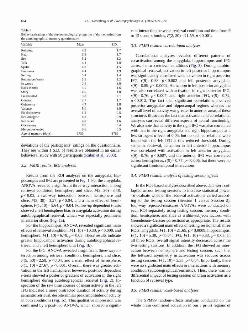
Table 1Behavioral ratings of the phenomenological properties of the memories fromthe autobiographical memory questionnaire
Variable Mean S.D.
Reliving 4.2 1.7Hear 3.9 1.7See 5.2 1.2Talk 4.1 1.8Spatial 4.8 1.5Emotions 4.4 1.9Setting 5.4 1.4Remember/know 5.9 1.2In words 2.6 1.8Back in time 4.5 1.6Story 4.6 1.8Fragmented 3.8 2.1General 2.7 1.7Coherence 4.7 1.8Valence 5.0 1.9Field/observer 1.2 0.5Real/imagine 6.3 0.9Rehearsal 4.0 1.6Once/many 0.8 0.4Merged/extended 0.6 0.5Age of memory (days) 1750 1785
deviations of the participants’ ratings on the questionnaire.They are within 1 S.D. of results we obtained in an earlierbehavioral study with 50 participants (Rubin et al., 2003).
3.2. FMRI results: ROI analyses
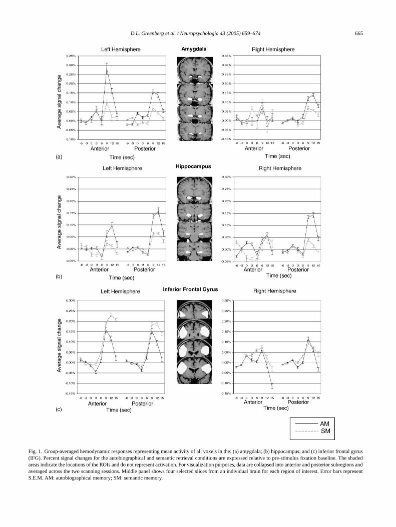
Results from the ROI analyses on the amygdala, hip-pocampus and IFG are presented inFig. 1. For the amygdala,ANOVA revealed a significant three-way interaction amongretrieval condition, hemisphere and slice,F(3, 30) = 3.48,p< 0.03, a two-way interaction between hemisphere andslice,F(3, 30) = 3.27,p< 0.04, and a main effect of hemi-sphere,F(1, 10) = 5.64,p< 0.04. Follow-up dependentt-testsshowed a left hemisphere bias in amygdala activation duringautobiographical retrieval, which was especially prominentin anterior slices (Fig. 1a).
For the hippocampus, ANOVA revealed significant maineffects of retrieval condition,F(1, 10) = 10.30,p< 0.009, andhemisphere,F(1, 10) = 6.78,p< 0.03. These results indicategreater hippocampal activation during autobiographical re-trieval and a left hemisphere bias (Fig. 1b).
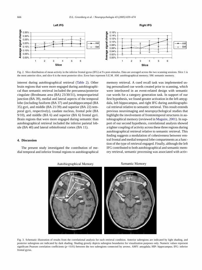
For the IFG, ANOVA revealed a significant three-way in-teraction among retrieval condition, hemisphere, and slice,F(5, 50) = 2.58,p< 0.04, and a main effect of hemisphere,F(1, 10) = 27.67,p< 0.001. Overall, there was greater acti-v dentt righths leftI rings ivityi asc ifi-
cant interaction between retrieval condition and time from 9to 15 s post-stimulus,F(2, 20) = 21.59,p< 0.001.
3.3. FMRI results: correlational analyses
Correlational analyses revealed different patterns ofco-activation among the amygdala, hippocampus and IFGacross the two retrieval conditions (Fig. 3). During autobio-graphical retrieval, activation in left posterior hippocampuswas significantly correlated with activation in right posteriorIFG, r(9) = 0.83, p< 0.002 and left posterior amygdala,r(9) = 0.89,p< 0.0002. Activation in left posterior amygdalawas also correlated with activation in right posterior IFG,r(9) = 0.76, p< 0.007, and right anterior IFG,r(9) = 0.72,p< 0.012. The fact that significant correlations involvedposterior amygdalar and hippocampal regions whereas theoverall level of activity was greater in anterior areas of thesestructures illustrates the fact that activation and correlationalanalyses can reveal different aspects of neural functioning.We also note that activity in the right IFG was also correlatedwith that in the right amygdala and right hippocampus at aless stringentα level of 0.05, but no such correlations werefound with the left IFG at this reduced threshold. Duringsemantic retrieval, activation in left anterior hippocampuswas correlated with activation in left anterior amygdala,r teda os
3
e col-l ower.T ord-i n 2),f d one ondi-t withG esultss hreeR s,Fa s thet nter-a h thatt rosst ew oryc s nod as af
3
thew of
ation in the left hemisphere; however, post-hoc depen-tests showed a posterior gradient of activation in theemisphere during autobiographical retrieval (Fig. 2). In-pection of the raw time courses of mean activity in theFG indicated a more protracted duration of activity duemantic retrieval, despite similar peak amplitudes of actn both conditions (Fig. 1c). This qualitative impression wonfirmed by a post-hoc ANOVA, which showed a sign
(9) = 0.76,p< 0.007, and the anterior IFG was correlacross hemispheres,r(9) = 0.77,p< 0.006, but there were nignificant frontotemporal interactions.
.4. FMRI results: analysis of testing session effects
In the ROI-based analyses described above, data werapsed across testing sessions to increase statistical po evaluate whether the retrieval activations varied acc
ng to the testing session (Session 1 versus Sessioour-way repeated-measures ANOVAs were conducteach ROI separately using testing session, memory c
ion, hemisphere, and slice as within-subjects factors,reenhouse–Geisser corrections as appropriate. The r
howed a significant main effect of testing session in all tOIs: amygdala,F(1, 10) = 21.65,p< 0.0009; hippocampu(1, 10) = 5.38,p< 0.04; IFG,F(1, 10) = 6.33,p< 0.03. Inll three ROIs, overall signal intensity decreased acros
wo testing sessions. In addition, the IFG showed an iction between hemisphere and testing session, suc
he leftward asymmetry in activation was reduced acesting sessions,F(1, 10) = 5.53,p< 0.04. Importantly, therere no significant main effects or interactions with memondition (autobiographical/semantic). Thus, there waifferential impact of testing session on brain activation
unction of retrieval type.
.5. FMRI results: voxel-based analyses
The SPM99 random-effects analysis conducted onhole brain confirmed activation in our a priori regions
D.L. Greenberg et al. / Neuropsychologia 43 (2005) 659–674 665
Fig. 1. Group-averaged hemodynamic responses representing mean activity of all voxels in the: (a) amygdala; (b) hippocampus; and (c) inferior frontal gyrus(IFG). Percent signal changes for the autobiographical and semantic retrieval conditions are expressed relative to pre-stimulus fixation baseline. The shadedareas indicate the locations of the ROIs and do not represent activation. For visualization purposes, data are collapsed into anterior and posteriorsubregions andaveraged across the two scanning sessions. Middle panel shows four selected slices from an individual brain for each region of interest. Error bars representS.E.M. AM: autobiographical memory; SM: semantic memory.
666 D.L. Greenberg et al. / Neuropsychologia 43 (2005) 659–674
Fig. 2. Slice distribution of mean activity in the inferior frontal gyrus (IFG) at 9 s post-stimulus. Data are averaged across the two scanning sessions. Slice 1 isthe most anterior slice, and slice 6 is the most posterior slice. Error bars represent S.E.M. AM: autobiographical memory; SM: semantic memory.
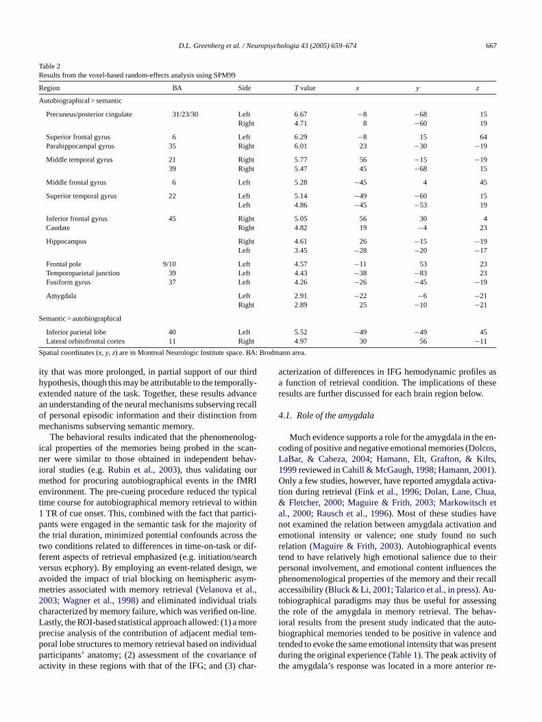
interest during autobiographical retrieval (Table 2). Otherbrain regions that were more engaged during autobiographi-cal than semantic retrieval included the precuneus/posteriorcingulate (Brodmann area (BA) 23/30/31), temporoparietaljunction (BA 39), medial and lateral aspects of the temporallobe (including fusiform (BA 37) and parahippocampal (BA35) gyri, and middle (BA 21/39) and superior (BA 22) tem-poral gyri, respectively), caudate nucleus, frontal pole (BA9/10), and middle (BA 6) and superior (BA 6) frontal gyri.Brain regions that were more engaged during semantic thanautobiographical retrieval included the inferior parietal lob-ule (BA 40) and lateral orbitofrontal cortex (BA 11).
4. Discussion
The present study investigated the contribution of me-dial temporal and inferior frontal regions to autobiographical
memory retrieval. A cued recall task was implemented us-ing personalized cue words created prior to scanning, whichwere interleaved in an event-related design with semanticcue words for a category generation task. In support of ourfirst hypothesis, we found greater activation in the left amyg-dala, left hippocampus, and right IFG during autobiographi-cal retrieval relative to semantic retrieval. This result extendsprevious neuroimaging and neuropsychological studies thathighlight the involvement of frontotemporal structures in au-tobiographical memory (reviewed inMaguire, 2001). In sup-port of our second hypothesis, correlational analysis showeda tighter coupling of activity across these three regions duringautobiographical retrieval relative to semantic retrieval. Thisfinding suggests a modulation of cohesiveness between ven-tral frontal and medial temporal lobe compartments as a func-tion of the type of retrieval engaged. Finally, although the leftIFG contributed to both autobiographical and semantic mem-ory retrieval, semantic processing was associated with activ-
F is for e dp depict uests ubregio inferiorf
ig. 3. Schematic illustration of results from the correlational analysosterior subregions are indicated by dark shading. Shading grosslyignificant Pearson correlation coefficients (p< 0.01) between the two srontal gyrus.
ach retrieval condition. Anterior subregions are indicated by lightshading, ans subregion boundaries for visualization purposes only. Numeric valrepresenns connected by arrows. AMY: amygdala; HIP: hippocampus; IFG:
D.L. Greenberg et al. / Neuropsychologia 43 (2005) 659–674 667
Table 2Results from the voxel-based random-effects analysis using SPM99
Region BA Side T value x y z
Autobiographical > semantic
Precuneus/posterior cingulate 31/23/30 Left 6.67 −8 −68 15Right 4.71 8 −60 19
Superior frontal gyrus 6 Left 6.29 −8 15 64Parahippocampal gyrus 35 Right 6.01 23 −30 −19
Middle temporal gyrus 21 Right 5.77 56 −15 −1939 Right 5.47 45 −68 15
Middle frontal gyrus 6 Left 5.28 −45 4 45
Superior temporal gyrus 22 Left 5.14 −49 −60 15Left 4.86 −45 −53 19
Inferior frontal gyrus 45 Right 5.05 56 30 4Caudate Right 4.82 19 −4 23
Hippocampus Right 4.61 26 −15 −19Left 3.45 −28 −20 −17
Frontal pole 9/10 Left 4.57 −11 53 23Temporoparietal junction 39 Left 4.43 −38 −83 23Fusiform gyrus 37 Left 4.26 −26 −45 −19
Amygdala Left 2.91 −22 −6 −21Right 2.89 25 −10 −21
Semantic > autobiographical
Inferior parietal lobe 40 Left 5.52 −49 −49 45Lateral orbitofrontal cortex 11 Right 4.97 30 56 −11
Spatial coordinates (x, y, z) are in Montreal Neurologic Institute space. BA: Brodmann area.
ity that was more prolonged, in partial support of our thirdhypothesis, though this may be attributable to the temporally-extended nature of the task. Together, these results advancean understanding of the neural mechanisms subserving recallof personal episodic information and their distinction frommechanisms subserving semantic memory.
The behavioral results indicated that the phenomenolog-ical properties of the memories being probed in the scan-ner were similar to those obtained in independent behav-ioral studies (e.g.Rubin et al., 2003), thus validating ourmethod for procuring autobiographical events in the fMRIenvironment. The pre-cueing procedure reduced the typicaltime course for autobiographical memory retrieval to within1 TR of cue onset. This, combined with the fact that partici-pants were engaged in the semantic task for the majority ofthe trial duration, minimized potential confounds across thetwo conditions related to differences in time-on-task or dif-ferent aspects of retrieval emphasized (e.g. initiation/searchversus ecphory). By employing an event-related design, weavoided the impact of trial blocking on hemispheric asym-metries associated with memory retrieval (Velanova et al.,2003; Wagner et al., 1998) and eliminated individual trialscharacterized by memory failure, which was verified on-line.Lastly, the ROI-based statistical approach allowed: (1) a moreprecise analysis of the contribution of adjacent medial tem-p dualp ce ofa ar-
acterization of differences in IFG hemodynamic profiles asa function of retrieval condition. The implications of theseresults are further discussed for each brain region below.
4.1. Role of the amygdala
Much evidence supports a role for the amygdala in the en-coding of positive and negative emotional memories (Dolcos,LaBar, & Cabeza, 2004; Hamann, Elt, Grafton, & Kilts,1999reviewed inCahill & McGaugh, 1998; Hamann, 2001).Only a few studies, however, have reported amygdala activa-tion during retrieval (Fink et al., 1996; Dolan, Lane, Chua,& Fletcher, 2000; Maguire & Frith, 2003; Markowitsch etal., 2000; Rausch et al., 1996). Most of these studies havenot examined the relation between amygdala activation andemotional intensity or valence; one study found no suchrelation (Maguire & Frith, 2003). Autobiographical eventstend to have relatively high emotional salience due to theirpersonal involvement, and emotional content influences thephenomenological properties of the memory and their recallaccessibility (Bluck & Li, 2001; Talarico et al., in press). Au-tobiographical paradigms may thus be useful for assessingthe role of the amygdala in memory retrieval. The behav-ioral results from the present study indicated that the auto-biographical memories tended to be positive in valence andt esentd ft or re-
oral lobe structures to memory retrieval based on indiviarticipants’ anatomy; (2) assessment of the covarianctivity in these regions with that of the IFG; and (3) ch
ended to evoke the same emotional intensity that was pruring the original experience (Table 1). The peak activity o
he amygdala’s response was located in a more anteri

668 D.L. Greenberg et al. / Neuropsychologia 43 (2005) 659–674
gion of the left hemisphere, although the correlation withother brain regions was stronger in its more posterior aspect.This difference may reflect differential involvement of amyg-daloid subnuclei, which are difficult to resolve at 1.5T. Al-though we observed amygdala activation in the present study,many previous studies have not reported amygdala responses(e.g.Conway et al., 1999, 2001, 2003; Maguire & Mummery,1999; Maguire et al., 2000; Piefke et al., 2003; Ryan et al.,2001), even when specific emotions were probed (Damasioet al., 2000; George, Ketter, Parekh, Herscovitch, & Post,1996; Lane et al., 1997; Liotti et al., 2000; Mayberg et al.,1999; Pardo, Pardo, & Raichle, 1993). Moreover, there arefew autobiographical memory studies in neurologic patientswith amygdala damage (e.g.Mori et al., 1999).
Several factors contribute to the difficulty in elicitingamygdala activation during autobiographical retrieval. First,amygdala responses to emotional stimuli tend to habituateover time, especially in blocked-design experiments (e.g.Breiter et al., 1996; Taylor et al., 1998). Second, MRI-relatedsusceptibility artifact in the vicinity of the amygdala variesacross participants (LaBar et al., 2001). This issue may beparticularly formidable to voxel-wise statistical proceduresthat require precise spatial overlap of activity across individ-uals to characterize group-averaged results. Such variabilitycan be minimized by tailored pulse sequences that reduces tae timec ex-a tin-g eac-t nall al at-t it-s imec e dif-f , ther mo-t oni cuei alaa ofiler -t ultantf re-v iouse -sh emi-s pporta thana s in-c eses mpo-r ievalp func-
tions the amygdala contributes to autobiographical retrieval.ROI-based statistical analyses that do not assume a canonicalhemodynamic response profile time-locked to cue onset maybe important for disentangling these alternatives.
4.2. Role of the hippocampus
Lesion evidence indicates that the hippocampus plays avital role in episodic memory (Scoville & Milner, 1957; forreviews, seeMcClelland et al., 1995; Squire, 1992), and func-tional neuroimaging evidence shows that this region is ac-tivated not only during episodic encoding but also duringepisodic retrieval (for a review, seeCabeza & Nyberg, 2000).The role of the hippocampus in the retrieval of remote mem-ories is a matter of dispute (Knowlton & Fanselow, 1998;Nadel & Moscovitch, 1997, 1998). Under one view, gener-ally called “consolidation theory,” the hippocampus and sur-rounding areas are required for the encoding of new memoriesand the retrieval of recent memories, but are not required forthe retrieval of remote memories (Alvarez & Squire, 1994;McClelland et al., 1995; Murre, 1999;Murre, Graham, &Hodges, 2001; Squire, 1992). The precise definition of “re-mote” is disputed as well, but patients with bilateral hip-pocampal damage have retrograde amnesias that last any-where from months or years to decades. The second view,m aysr thisv sultsi palr oriesw palr po-rS yd tivelyr sw l ofm
xpla-n t hip-p riestt naln to bel int ity oft leftl re-v ly,t
inga ationo ask.I anticm arlyd
usceptibility artifact in the medial temporal lobes (Posse el., 2003; Wang, Song, McCarthy, & LaBar, in press) or bymploying ROI-based analytic approaches. Third, theourse of amygdala activity may vary depending on thect function it serves. In this regard, it is important to disuish whether amygdala activation reflects emotional r
ions or feeling states evoked following recall of an emotioife event, mnemonic processes associated with retrievempt/initiation, or emotional evaluation of the recall cueelf (LaBar, 2003). These processes would have different tourses of activation relative to cue onset and may haverent sensitivities to control tasks. In the present studyetrieval cue words themselves were relatively low in eional impact (see Section2); therefore, amygdala activatis unlikely due to decoding of the emotional salience oftself. Furthermore, the hemodynamic profile of amygdctivation did not have a temporal lag or sustained prelative to the other regions involved (Fig. 1a). These feaures suggest that the amygdala is not signaling a reseeling state induced following retrieval of the memory. Pious studies that compared fictitious versus non-fictitmotional events (Markowitsch et al., 2000) or personal verus non-personal traumatic memories (Rausch et al., 1996)ave also reported amygdala activation, although its hpheric distribution has varied. These studies further sun emotional memory function of the amygdala rathern emotional perception function since the control taskluded presentation of emotionally salient information. Thtudies, however, used blocked designs that could not teally distinguish responses at different stages of the retrrocess. Further studies are needed to elucidate which
ultiple-trace theory, holds that the hippocampus is alwequired for the retrieval of episodic memories. Underiew, repeated retrieval of autobiographical memories ren multiple “traces” of the memory within the hippocamegion. For this reason, it is hypothesized that older memill have more traces distributed throughout the hippocam
egion, and will therefore be less susceptible to medial temal damage (Nadel & Moscovitch, 1997, 1998, 2001; Nadel,amsonovich, Ryan, & Moscovitch, 2000). The present studoes not address this issue, as the memories were relaecent (4.8 years old on average;Table 1), and both theorieould predict hippocampal activation during the retrievaemories this old.Recent fMRI evidence suggests some more detailed e
ations of our results. One study has shown that the lefocampus may be more involved in retrieving older memo
han the right hemisphere (Maguire & Frith, 2003). This pat-ern could explain why hippocampal activations in functioeuroimaging studies of autobiographical memory tend
eft lateralized (Maguire, 2001). Consistent with this trend,he present study the activation and functional connectivhe hippocampus during autobiographical memory wereateralized (Fig. 1); however, the voxel-based analysis dideal right-sided activation (Table 2) and, as noted previoushe memories were on average less than 5 years old.
Importantly, the activation of the hippocampus durutobiographical memory was greater than the activf this region during a demanding semantic retrieval t
t is a controversial issue whether episodic and sememory, as two forms of declarative memory, are similependent on the hippocampus (e.g.Squire & Zola, 1998),

D.L. Greenberg et al. / Neuropsychologia 43 (2005) 659–674 669
or whether episodic memory is more dependent on the hip-pocampus than semantic memory (e.g.Aggleton & Brown,1999; Mishkin, Vargha-Khadem, & Gadian, 1998; Tulving& Markowitsch, 1998; Vargha-Khadem, Gadian, & Mishkin,2001). The present results suggest that the hippocampus isinvolved in both forms of declarative memory, but it plays agreater role in episodic than in semantic memory.
It is important to emphasize that the hippocampus wasmore activated during autobiographical retrieval than dur-ing semantic retrieval, even though the latter was tempo-rally extended. This fact suggests that the difference isnot merely related to the amount of information retrievalbut the nature of the information retrieved. Recent func-tional neuroimaging studies of episodic retrieval have asso-ciated the hippocampus with the retrieval of specific rela-tional/contextual information, or recollection (e.g.Eldridgeet al., 2000; Yonelinas, Hopfinger, Buonocore, Kroll, &Baynes, 2001). Lesion studies have yielded complementaryfindings. In episodic memory tasks, people tend to producemore “remember” responses after conceptually-based en-coding (Gardiner & Parkin, 1990). Unilateral temporal lobeepilepsy or excision, however, reduces this effect; for left-hemisphere patients, the effect was eliminated for words,while for right-hemisphere patients, it was eliminated forfaces (Moscovitch & McAndrews, 2002). Thus, both thei pusp pe-r f andv veng odicm thisi
4
forr enfi re-g giont thisi thec nec-t dur-i t ac-t seti oundo n3 witht re-d d notg ovides( em-p oreg
In contrast with right IFG, left IFG was more involved insemantic retrieval than in autobiographical retrieval. In par-ticular, the duration of the left IFG activation was longer dur-ing semantic than during autobiographical retrieval (Fig. 1c).Even if this difference was partly due to prolonged exemplargeneration processes, it challenges the notion that left pre-frontal regions play a special role in autobiographical retrieval(Conway et al., 1999; Maguire et al., 2001a,b). For example,Conway et al. (1999)found left prefrontal regions, includ-ing IFG, were more activated during the retrieval of autobio-graphical memories in response to common words (Crovitztechnique) than during cued-recall of overlearnt word-pairs.Given that autobiographical trials were short (4.8 s), the leftprefrontal activation could have reflected primarily the gen-eration rather than the monitoring of autobiographical mem-ories. Recent functional neuroimaging evidence suggeststhat generation processes depend on left prefrontal regions,whereas monitoring processes depend on right prefrontal re-gions (Cabeza et al., 2003). Thus, the fact that in the presentstudy right IFG played a more important role in autobiograph-ical retrieval than left IFG could be partly a consequence ofusing participant-specific retrieval cues, which reduced thetime required for memory generation and allowed more timefor monitoring the retrieval output.
4
phi-c -s andiMF re-e ativei entd rea-s re-p nott h hasaS d of-t rea-s allyc -t wedn ieval( hip-p anticc tionr r if itr botht e re-e eaterc then sionb dged
maging and the lesion literature show that the hippocamlays a critical role in the recollection of previously exienced events. Because of the involvement of the selisuospatial imagery, recollection is likely to play an ereater role in autobiographical than in laboratory episemory, and we recently found evidence consistent with
dea using fMRI (Cabeza et al., in press).
.3. Role of the IFG
Markowitsch (1995)has specifically proposed a roleight IFG during retrieval of old episodic memories, givndings of retrograde amnesia following damage to thision and the uncinate fasciculus that connects this re
o the anteromedial temporal lobe. Our findings supportdea, especially when considered in combination withorrelational analysis that showed greater functional conivity among the right IFG, amygdala and hippocampusng autobiographical retrieval. One caveat to this is thaivity in the right IFG correlated more strongly with theemporal lobe regions in the left hemisphere (Fig. 3) thann the right hemisphere, where such correlations were fnly at a reduced statistical threshold ofp< 0.05 (see Sectio). Nonetheless, no temporal lobe regions correlatedhe left IFG during autobiographical retrieval (even at auced threshold), and the frontotemporal interactions dieneralize to semantic retrieval. These results thus prome evidence for the strong version of theMarkowitsch1995) hypothesis, although they may indicate frontotoral interactions during autobiographical retrieval menerally.
.4. Limitations and future directions
The tasks used in neuroimaging studies of autobiograal memory have varied widely (Maguire, 2001). The precan cue generation paradigm, which was used heren several previous studies (Fink et al., 1996; Maguire &
ummery, 1999; Maguire et al., 2000, 2001; Maguire &rith, 2003), has been criticized because it may permitncoding of the memories. We do not feel that this altern
nterpretation explains the greater hippocampal involvemuring autobiographical retrieval in our study for threeons. First, during debriefing, participants in this studyorted recalling the memory of the original experience,
he rating session or the previous scanning session, whiclso been the case in other studies (Maguire & Frith, 2003).econd, data from the AMQ showed that participants ha
en thought or talked about the events, and there is littleon to expect that one additional retrieval would drastichange the memory (see alsoPiefke et al., 2003). Third, a staistical comparison of the two fMRI testing sessions shoo interaction between session and type of memory retrautobiographical/semantic). One might expect greaterocampal activation in the autobiographical versus semontrast during the first scanning session if such activaeflects the novel association of the event with the cue oeflects re-encoding of the event (since by session two,he autobiographical and semantic memories would bncoded). On the positive side, this design permits grontrol over the time course of retrieval and reducesumber of retrieval failures during the scanning sesy using cues that the participants themselves have ju

670 D.L. Greenberg et al. / Neuropsychologia 43 (2005) 659–674
to be effective. This design also allowed us to comparethe characteristics of participants’ memories to those ob-tained in previous behavioral studies, thereby helping toensure that participants retrieve full-blown autobiograph-ical memories in the magnet instead of autobiographicalfacts.
On the other hand, the memory task used in this study didpreclude an examination of some relevant questions. Specifi-cally, several earlier studies have compared brain activation inrecent and remote memories (Conway et al., 1999; Maguire& Frith, 2003; Maguire, Henson, Mummery, & Frith, 2001b;Piefke et al., 2003). In the present study, we used a task thatpermitted relatively unrestricted generation of autobiograph-ical memories in order to be comparable to the semantic task.An examination of the effects of remoteness would requireseveral additional constraints on memory generation. First,given the distribution of memories across the lifespan (Rubin& Schulkind, 1997), participants would have to retrieve acertain number of memories from the recent and remote pe-riods (e.g.Piefke et al., 2003). Second, the remote memorieswould need to be 20–30 years old or older, because neu-ropsychological (Squire, 1992) and neuroimaging (Maguire& Frith, 2003) evidence shows that consolidation effects arenot necessarily apparent before this time. In this study, how-ever, memories were 4.8 years old on average, and even theo ore-o thatd utedt emo-r otp ed ust hicalm
ticu-l heyc2 gen-e acti-v o thed asksd ren-t thea orya ition,t h cue.T n mah s en-g ecifi-c hicalr tiven asksi atelyu ts). Ina nt oft phi-c lative
to the pre-stimulus baseline without reference to the controltask.
Our experimental design consisted of two retrieval ses-sions. The direct comparison across retrieval sessions did notyield differences in the pattern of brain activation. For thisreason, data were combined across testing sessions. Nonethe-less, the experience of retrieving in session 1 did have a long-lasting impact, since the degree of activation was reducedoverall in session 2. Because the activity we report is primar-ily carried by data obtained from session 1, a second retrievalsession may not be necessary to incorporate in future studiesemploying similar designs, provided that sufficient power isobtained from one retrieval session.
Due to the targeted nature of our hypotheses, we did notinvestigate how the frontotemporal regions interacted withother brain regions involved in the network that supportsautobiographical memory retrieval (Maguire et al., 2001a,b;Piefke et al., 2003; Ryan et al., 2001), so the pattern of cor-relations we report here may be extensible to other parts ofthe brain. The whole-brain SPM analysis did reveal activa-tions in other brain regions that have been reported in previ-ous autobiographical studies, including the precuneus, me-dial and lateral aspects of the temporal lobe, and the frontalpole. Given the complex, multifactorial nature of autobio-graphical memory, it will be important to understand ther sys-t romt ,2 bio-g etrics aphi-cet eno-l eriedb cil-i tem-p cessm apt ionsa re-t pli-c ory.
A
un-d a-t res-s 01D L.K andt low-s
ldest memories were no more than 20 years old. Mver, the levels of detail must be equated to ensureifferences in hippocampal activation cannot be attrib
o the greater number of details present in recent mies (Maguire & Frith, 2003). While our approach did nermit us to address the issue of remoteness, it allow
o examine a more naturalistic sample of autobiograpemories.Furthermore, autobiographical retrieval tasks are par
arly difficult to control in neuroimaging studies because tan differ in many respects to other retrieval tasks (Maguire,001). We chose a prototypical semantic task (categoryration) as a control condition to determine whether theations in the autobiographical retrieval task extended tomain of semantic retrieval. It is possible that these tiffered in other cognitive features that account for diffe
ial activation in frontotemporal regions. For example, inutobiographical condition, there is only one correct memssociated with each cue, whereas in the semantic cond
here are multiple correct exemplars associated with eachus, executive processes related to response selectioave differed across the conditions (although both taskaged left IFG). The category generation task was spally chosen to control for other aspects of autobiograpetrieval, particularly its temporally-extended and iteraature, which would not have been controlled by other t
n which semantic processing terminates almost immedipon cue presentation (e.g. abstract/concrete judgmenny event, the ROI analyses clearly implicate involveme
he amygdala, hippocampus and IFG during autobiograal retrieval because their time courses were extracted re
y
elationship between these regions and other distributedems important for recollection of people and events fhe personal past (Greenberg & Rubin, 2003; Paller et al.003). A remaining challenge is to decompose autoraphical networks into their constituent parts. Paramtudies that vary one or more aspects of autobiogral recollection (e.g. emotional tone or remoteness;Piefket al., 2003; Maguire & Frith, 2003) will be critical in
his regard. Taking on-line measures of the phenomogical properties of the memories, such as those quy our autobiographical memory questionnaire, will fa
tate these efforts. Finally, as mentioned above, theoral lag inherent in the autobiographical retrieval proay be exploited in future studies to more carefully m
he relationship between activation of different brain regnd their association with different stages of memory
rieval. The results of such studies will have broader imations for biologically-based theories of episodic mem
cknowledgements
This work was supported by the McDonnell Pew Foation (D.C.R.), a Young Investigator Award from the N
ional Alliance for Research on Schizophrenia and Depion (K.S.L.), National Institutes of Health PHS grants RA14094 (K.S.L.) and R01 AG023123 (D.C.R.), a Ruthirschstein National Research Service Award (D.L.G.),
he Society for Neuroscience Minority Neuroscience Felhip Program (H.J.R.).

D.L. Greenberg et al. / Neuropsychologia 43 (2005) 659–674 671
Appendix A. Autobiographical memory questionnaire
Listed below are the questions from our autobiographical memory questionnaire, along with the brief variable name we use foreach.For questions a–g, the scales ranged from 1 (not at all), to 3 (vaguely), to 5 (distinctly), to 7 (as clearly as if it were happeningright now).
a. Reliving As I remember the event, I feel as though I am reliving the original event.
b. Hear As I remember the event, I can hear it in my mind.
c. See As I remember the event, I can see it in my mind.
d. Talk As I remember the event, I or other people are talking.
e. Spatial As I remember the event, I know its spatial layout.
f. Emotions As I remember the event, I can feel now the emotional intensity that I felt then.
g. Setting As I remember the event, I can recall the setting where it occurred.
For questions h–n, the scales ranged from 1 (not at all), to 3 (vaguely), to 5 (distinctly), to 7 (as much as any memory).
h. Remember/know Sometimes people know something happened to them without being able to actually remember it. As Ithink about the event, I can actually remember it rather than just knowing that it happened.
i. In words As I remember the event, it comes to me in words.
j. Back in time As I remember the event, I feel that I travel back to the time when it happened, that I am a subject in itagain, rather than an outside observer tied to the present.
k. Story As I remember the event, it comes to me in words or in pictures as a coherent story or episode and notas an isolated fact, observation, or scene.
l. Fragmented My memory of the event is fragmented into specific details with missing bits.
m. General My memory for the event is only as detailed as the general knowledge of this type of event that I wouldexpect most people to have.
n. Coherence My memory of the event has a personal coherence: it fits easily into a story I would tell about that partof my life.
The remaining questions had unique scales.
o. Valence Please rate the emotional valence or the kinds of emotions it involves (1 = 100% negative; 7 = 100%positive).
p. Field/observer As I remember the event, I imagine it again through my own eyes seeing what I would have seen then,or as an observer from a different perspective than the one I had (1 = own eyes; 2 = observer; 3 = can’ttell).
q. Real/imagine I believe the event in my memory really occurred in the way I remember it and that I have not imaginedor fabricated anything that did not occur (scale: 1 = 100% imaginary; 7 = 100% real).
r. Rehearsal Since it happened, I have thought or talked about this event (scale: 1 = not at all; 7 = as often as anyevent in my life).
s. Once/many To the best of your knowledge, is the memory of an event that occurred once at one particular time andplace, a summary or merging of many similar or related events, or for events that occurred over a fairlycontinuous extended period of time lasting more than a day (scale: 1 = once; 2 = merging; 3 = extended).

672 D.L. Greenberg et al. / Neuropsychologia 43 (2005) 659–674
Responses to this question were recoded to produce two scales. Once/many had a value of 1 if the subject judged the memoryto take place within a single day and 0 if it took longer. Merged/extended had a value of 0 if the event lasted longer than a dayand was extended in a fairly continuous manner over a period of time and 1 if it was the merging of many discrete events.t. Age of memory Please date the memory (month/day/year) as accurately as you can. Please fill in a month, day, and year
even if you must estimate. If the memory extended over a period of time, report the approximate middleof the period (scored as retention interval in days).
References
Aggleton, J. P., & Brown, M. W. (1999). Episodic memory, amnesia,and the hippocampal-anterior thalamic axis.Behavioral and BrainSciences, 22, 425–489.
Amaral, D. G., Price, J. L., Pitk̈anen, A., & Carmichael, S. T. (1992).Anatomical organization of the primate amygdaloid complex. In J. P.Aggleton (Ed.),The amygdala: Neurobiological aspects of emotion,memory, and mental dysfunction(pp. 1–66). New York: Wiley-Liss.
Andreasen, N. C., O’Leary, D. S., Cizadlo, T., Arndt, S., Rezai, K.,Watkins, G. L., et al. (1995). Remembering the past: two facets ofepisodic memory explored with positron emission tomography.Amer-ican Journal of Psychiatry, 11, 1576–1585.
Baddeley, A. D. (1992). What is autobiographical memory? In M. A. Con-way, D. C. Rubin, H. Spinnler, & W. A. Wagenaar (Eds.),Theoreticalperspectives on autobiographical memory(pp. 13–29). Dordrecht, TheNetherlands: Kluwer Academic Publishers.
Baddeley, A., & Wilson, B. (1986). Amnesia, autobiographical memory,and confabulation. In D. C. Rubin (Ed.),Autobiographical memory(pp. 225–252). New York: Cambridge University Press.
Bahrick, H. P. (1979). Maintenance of knowledge: Questions about mem-ory we forgot to ask.Journal of Experimental Psychology: General,108, 296–308.
Battig, W. F., & Montague, W. E. (1969). Category norms for verbalitems in 56 categories: A replication and extension of the Con-necticut category norms.Journal of Experimental Psychology, 80, 1–45.
Bluck, S., & Li, K. Z. H. (2001). Predicting memory completeness andaccuracy: Emotion and exposure in repeated autobiographical recall.Applied Cognitive Psychology, 15, 145–158.
B . L.,uman
B ubine
B binory
C aliza-val:-
C ical-
C e, M.,bio-hoto
C nal
C upts,ical
locus for the ecphory of old episodic memories.Journal of Neurology,Neurosurgery& Psychiatry, 61, 304–310.
Christianson, S. -̊A. (1992).The handbook of emotion and memory: Re-search and theory. Hillsdale, NJ: Lawrence Erlbaum Associates.
Christianson, S. -̊A., & Safer, M. (1995). Emotional events and emotionsin autobiographical memories. In D. C. Rubin (Ed.),Rememberingour past: Studies in autobiographical memory(pp. 218–243). NewYork: Cambridge University Press.
Conway, M. A., Pleydell-Pearce, C. W., Whitecross, S. E., & Sharpe, H.(2003). Neurophysiological correlates of memory for experienced andimagined events.Neuropsychologia, 41, 334–340.
Conway, M. A., Pleydell-Pearce, C. W., & Whitecross, S. E. (2001). Theneuroanatomy of autobiographical memory: A slow cortical potentialstudy of autobiographical memory retrieval.Journal of Memory andLanguage, 45, 493–524.
Conway, M. A., Turk, D. J., Miller, S. L., Logan, J., Nebes, R. D.,Meltzer, C. C., et al. (1999). A positron emission tomography (PET)study of autobiographical memory retrieval.Memory, 7, 679–702.
Convit, A., McHugh, P., Wolf, O. T., de Leon, M. J., Bobinski, M., &De Santi, S. (1999). MRI volume of the amygdala: A reliable methodallowing separation from the hippocampal formation.Psychiatry Re-search: Neuroimaging Section, 90, 113–123.
Crovitz, H. F. (1986). Loss and recovery of autobiographical memoryafter head injury. In D. C. Rubin (Ed.),Autobiographical memory(pp. 273–290). New York: Cambridge University Press.
Crovitz, H. F., & Schiffman, H. (1974). Frequency of episodic memoriesas a function of their age.Bulletin of the Psychonomic Society, 4,517–518.
Damasio, A. R., Grabowski, T. J., Bechara, A., Damasio, H., Ponto, L.L. B., Parvisi, J., et al. (2000). Subcortical and cortical brain activity
e
D tem-eval.
D n thebetter
D nd
E S.role
F sler,own
G xpe-
G t, R.ring
G , P.,and
reiter, H. C., Etcoff, N. L., Whalen, P. J., Kennedy, W. A., Rauch, SBuckner, R. L., et al. (1996). Response and habituation of the hamygdala during visual processing of facial expression.Neuron, 17,875–887.
rewer, W. F. (1986). What is autobiographical memory? In D. C. R(Ed.), Autobiographical memory(pp. 25–49). New York: CambridgUniversity Press.
rewer, W. F. (1995). What is recollective memory? In D. C. Ru(Ed.), Remembering our past: Studies in autobiographical mem(pp. 19–66).
abeza, R., Kester Locantore, J., & Anderson, N. D. (2003). Latertion of prefrontal cortex activity during episodic memory retrieEvidence for the production-monitoring hypothesis.Journal of Cognitive Neuroscience, 15, 249–259.
abeza, R., & Nyberg, L. (2000). Imaging cognition II: An empirreview of 275 PET and fMRI studies.Journal of Cognitive Neuroscience, 12, 1–47.
abeza, R., Prince, S. E., Daselaar, S. M., Greenberg, D., BuddDolcos, F., et al. Brain activity during episodic retrieval of autographical and laboratory events: An fMRI study using a novel pparadigm.Journal of Cognitive Neuroscience(in press).
ahill, L., & McGaugh, J. L. (1998). Mechanisms of emotioarousal and lasting declarative memory.Trends in Neurosciences, 21,294–299.
alabrese, P., Markowitsch, H. J., Durwen, H. F., Widlitzek, H., HaM., Holinka, B., et al. (1996). Right temporofrontal cortex as crit
during the feeling of self-generated emotions.Nature Neuroscienc,3, 1049–1056.
olan, R. J., Lane, R., Chua, P., & Fletcher, P. (2000). Dissociableporal lobe activations during emotional episodic memory retriNeuroImage, 11, 203–209.
olcos, F., LaBar, K. S., & Cabeza, R. (2004). Interactions betweeamygdala and the medial temporal lobe memory system predictmemory for emotional events.Neuron, 42, 855–863.
uvernoy, H. M. (1999).The human brain: Surface, blood supply, athree-dimensional sectional anatomy. New York: Springer-Verlag.
ldridge, L. L., Knowlton, B. J., Furmanski, C. S., Bookheimer,Y., & Engle, S. A. (2000). Remembering episodes: A selectivefor the hippocampus during retrieval.Nature Neuroscience, 3, 1149–1152.
ink, G., Markowitsch, H. J., Reinkemeier, M., Bruckbauer, T., KesJ., & Heiss, W. -D. (1996). Cerebral representation of one’spast: Neural networks involved in autobiographical memory.Journalof Neuroscience, 16, 4275–4282.
ardiner, J. M., & Parkin, A. J. (1990). Attention and recollective erience in recognition memory.Memory& Cognition, 18, 579–583.
eorge, M. S., Ketter, T. A., Parekh, P. I., Herscovitch, P., & PosM. (1996). Gender differences in regional cerebral blood flow dutransient self-induced sadness or happiness.Biological Psychiatry, 40,859–871.
eorge, M. S., Ketter, T. A., Parekh, P. I., Horwitz, B., Herscovitch& Post, R. M. (1995). Brain activity during transient sadness
D.L. Greenberg et al. / Neuropsychologia 43 (2005) 659–674 673
happiness in healthy women.American Journal of Psychiatry, 152,341–351.
Greenberg, D. L., & Rubin, D. C. (2003). The neuropsychology of auto-biographical memory.Cortex, 39, 687–728.
Hamann, S. B. (2001). Cognitive and neural mechanisms of emotionalmemory.Trends in Cognitive Sciences, 9, 394–400.
Hamann, S. B., Elt, T., Grafton, S., & Kilts, C. (1999). Amygdala activityrelated to enhanced memory for pleasant and aversive stimuli.NatureNeuroscience, 2, 289–293.
Knowlton, B. J., & Fanselow, M. S. (1998). The hippocampus, consol-idation and on-line memory.Current Opinion in Neurobiology, 8,293–296.
Kroll, N. E. A., Markowitsch, H. J., Knight, R. T., & von Cramon, D.Y. (1997). Retrieval of old memories: The temporofrontal hypothesis.Brain, 120, 1377–1399.
LaBar, K. S. (2003). Emotional memory functions of the human amyg-dala.Current Neurology and Neuroscience Reports, 3, 363–364.
LaBar, K. S., Gitelman, D. R., Mesulam, M. -M., & Parrish, T. B. (2001).Impact of signal-to-noise on functional MRI of the human amygdala.Neuroreport, 12, 3461–3464.
Lane, R. D., Reiman, E. M., Bradley, M. M., Lang, P. J., Ahern, G. L.,Davidson, R. J., et al. (1997). Neuroanatomical correlates of pleasantand unpleasant emotion.Neuropsychologia, 35, 1437–1444.
Larsen, S. (1998). What is it like to remember? On phenomenal qualitiesof memory. In C. P. Thompson, D. J. Herrmann, D. Bruce, J. D.Read, D. G. Payne, & M. P. Toglia (Eds.),Autobiographical memory:Theoretical and applied perspectives(pp. 163–190). Mahwah, NJ:Erlbaum.
Lepage, M., Habib, R., & Tulving, E. (1998). Hippocampal PET acti-vations of memory encoding and retrieval: The HIPER model.Hip-
L oth,lated
L , P.,essers.
M ventdon
M ip-ories.
M D.ara-
M ofsion
M ofsys-
M ctsand
M d in
M cu,ntalory
M R.nc-sion
and normal sadness.American Journal of Psychiatry, 156, 675–682.
McClelland, J., McNaughton, B., & O’Reilly, R. (1995). Why there arecomplementary learning systems in the hippocampus and neocortex:Insights from the successes and failures of connectionist models oflearning and memory.Psychological Review, 102, 419–457.
McNally, R. J., Litz, B. T., Prassas, A., Shin, L. M., & Weathers, F.W. (1994). Emotional priming of autobiographical memory in post-traumatic stress disorder.Cognition& Emotion, 8, 351–367.
Mishkin, M., Vargha-Khadem, F., & Gadian, D. G. (1998). Amnesiaand the organization of the hippocampal system.Hippocampus, 8,212–216.
Mori, E., Ikeda, M., Hirono, N., Kitagaki, H., Imamura, T., & Shimomura,T. (1999). Amygdalar volume and emotional memory in Alzheimer’sdisease.American Journal of Psychiatry, 156, 216–222.
Moscovitch, D. A., & McAndrews, M. P. (2002). Material-specific deficitsin “remembering” in patients with unilateral temporal lobe epilepsyand excisions.Neuropsychologia, 40, 1335–1342.
Moscovitch, M. (1989). Confabulation and the frontal systems. In H. L.Roediger & F. I. M. Craik (Eds.),Varieties of memory and conscious-ness: Essays in honour of Endel Tulving(pp. 133–160). Hillsdale, NJ:Lawrence Erlbaum Associates.
Moscovitch, M., & Melo, B. (1997). Strategic retrieval and the frontallobes: Evidence from confabulation and amnesia.Neuropsychologia,35, 1017–1034.
Murre, J. M. J., Graham, K. S., & Hodges, J. R. (2001). Semantic demen-tia: Relevance to connectionist models of long-term memory.Brain,124, 647–675.
Nadel, L., & Moscovitch, M. (1997). Memory consolidation, retrogradeamnesia, and the hippocampal complex.Current Opinion in Neurobi-
N or-
N ng-
N pleand
N ries.
O S.sia in
P . B.,ition.
P rre-
P G.the
P G. J.,yg-
R rr,dy ofy and
R m
R . The
pocampus, 8, 313–322.evine, B., Black, S. E., Cabeza, R., Sinden, M., McIntosh, A. R., T
J. P., et al. (1998). Episodic memory and the self in a case of isoretrograde amnesia.Brain, 121, 1951–1973.
iotti, M., Mayberg, H. S., Brannan, S. K., McGinnis, S., Jerabek& Fox, P. T. (2000). Differential limbic-cortical correlates of sadnand anxiety in healthy subjects: Implications for affective disordBiological Psychiatry, 48, 30–42.
aguire, E. A. (2001). Neuroimaging studies of autobiographical ememory.Philosophical Transactions of the Royal Society of LonB: Biological Sciences, 356, 1441–1451.
aguire, E. A., & Frith, C. D. (2003). Lateral asymmetry in the hpocampal response to the remoteness of autobiographical memJournal of Neuroscience, 23, 5302–5307.
aguire, E. A., Henson, R. N. A., Mummery, C. J., & Frith, C.(2001). Activity in prefrontal cortex, not hippocampus, varies pmetrically with the increasing remoteness of memory.Neuroreport,124, 1156–1170.
aguire, E. A., & Mummery, C. J. (1999). Differential modulationa common memory retrieval network revealed by positron emistomography.Hippocampus, 9, 54–61.
aguire, E. A., Mummery, C. J., & B̈uchel, C. (2000). Patternshippocampal-cortical interaction dissociate temporal lobe memorytems.Hippocampus, 10, 475–482.
aguire, E. A., Vargha-Khadem, F., & Mishkin, M. (2001). The effeof bilateral hippocampal damage on fMRI regional activationsinteractions during memory retrieval.Brain, 124, 1156–1170.
arkowitsch, H. J. (1995). Which brain regions are critically involvethe retrieval of old episodic memory?Brain Research Reviews, 21,117–127.
arkowitsch, H. J., Thiel, A., Reinkemeier, M., Kessler, J., KoyunA., & Heiss, W. -D. (2000). Right amygdalar and temporofroactivation during autobiographic, but not during fictitious memretrieval.Behavioral Neurology, 12, 181–190.
ayberg, H. S., Liotti, M., Brannan, S. K., McGinnis, S., Mahurin,K., Jerabek, P. A., et al. (1999). Reciprocal limbic-cortical fution and negative mood: Converging PET findings in depres
ology, 7, 217–227.adel, L., & Moscovitch, M. (1998). Hippocampal contributions to c
tical plasticity.Neuropharmacology, 37, 431–439.adel, L., & Moscovitch, M. (2001). The hippocampal complex and lo
term memory revisited.Trends in Cognitive Sciences, 5, 228–230.adel, L., Samsonovich, A., Ryan, L., & Moscovitch, M. (2000). Multi
trace theory of human memory: Computational, neuroimaging,neuropsychological results.Hippocampus, 10, 352–368.
igro, G., & Neisser, U. (1983). Point of view in personal memoCognitive Psychology, 15, 467–482.
’Connor, M., Butters, N., Miliotis, P., Eslinger, P., & Cermak, L.(1992). The dissociation of anterograde and retrograde amnea patient with herpes simplex encephalitis.Journal of Clinical andExperimental Neuropsychology, 14, 159–178.
aller, K. A., Ranganath, C., Gonsalves, B., LaBar, K. S., Parrish, TGitelman, D. R., et al. (2003). Neural correlates of person recognLearning& Memory, 10, 253–260.
ardo, J. V., Pardo, P. J., & Raichle, M. E. (1993). Neural colates of self-induced dysphoria.American Journal of Psychiatry, 150,713–719.
iefke, M., Weiss, P. H., Zilles, K., Markowitsch, H. J., & Fink,(2003). Differential remoteness and emotional tone modulateneural correlates of autobiographical memory.Brain, 126, 650–668.
osse, S., Fitzgerald, D., Gao, K., Habel, U., Rosenberg, D., Moore,et al. (2003). Real-time fMRI of temporolimbic regions detects amdala activation during single-trial self-induced sadness.NeuroImage,18, 760–768.
ausch, S. L., van der Kolk, B. A., Fisler, R. E., Alpert, N. M., OS. P., Savage, C. R., et al. (1996). A symptom provocation stuposttraumatic stress disorder using positron emission tomographscript-driven imagery.Archives of General Psychiatry, 53, 380–387.
eisberg, D. (1992).Auditory imagery. Hillsdale, NJ: Lawrence ErlbauAssociates.
eisberg, D., Heuer, F., MacLean, J., & O’Shaughnessy, M. (1988)quantity, not the quality, of affect predicts memory vividness.Bulletinof the Psychonomic Society, 26, 100–103.

674 D.L. Greenberg et al. / Neuropsychologia 43 (2005) 659–674
Rubin, D. C. (1995).Memory in oral traditions: The cognitive psychol-ogy of epic, ballads, and counting-out rhymes. New York: OxfordUniversity Press.
Rubin, D. C. (1998). Beginnings of a theory of autobiographical remem-bering. In C. P. Thompson, D. J. Hermann, D. Bruce, J. D. Reed, D.G. Payne, & M. P. Toglia (Eds.),Autobiographical memory: Theoret-ical and applied perspectives(pp. 47–67). Mahwah, NJ: Erlbaum.
Rubin, D. C., & Greenberg, D. L. (1998). Visual memory-deficit amne-sia: A distinct amnesic presentation and etiology.Proceedings of theNational Academy of Sciences of the United States of America, 95,5413–5416.
Rubin, D. C., & Greenberg, D. L. (2003). The role of narrative in recol-lection: a view from cognitive and neuropsychology. In G. Fireman,T. McVay, & O. Flanagan (Eds.),Narrative and consciousness: Liter-ature, psychology, and the brain. New York: Oxford University Press.
Rubin, D. C., Groth, E., & Goldsmith, D. J. (1984). Olfactory cuingof autobiographical memory.American Journal of Psychology, 97,493–507.
Rubin, D. C., Schrauf, R. W., & Greenberg, D. L. (2003). Belief andrecollection of autobiographical memories.Memory& Cognition, 31,887–901.
Rubin, D. C., & Schulkind, M. D. (1997). Distribution of important andword-cued autobiographical memories in 20-, 35-, and 70-year-oldadults.Psychology and Aging, 12, 524–535.
Ryan, L., Nadel, L., Keil, K., Putnam, K., Schnyer, D., Trouard, T., etal. (2001). Hippocampal complex and retrieval of recent and very re-mote autobiographical memories: Evidence from functional magneticresonance imaging in neurologically intact people.Hippocampus, 11,707–714.
Schacter, D. L., Norman, K. A., & Koutstaal, W. (1998). The cognitivey
S tiva-val.
S em-the
S bi-
S from
S ory,
S ., &pus.
States
T e
T icts
Talland, G. A. (1965).Deranged memory. New York: Academic Press.Taylor, S. F., Liberzon, I., Fig, L. M., Decker, L. R., Minoshima, S.,
& Koeppe, R. A. (1998). The effect of emotional content on vi-sual recognition memory: A PET activation study.NeuroImage, 8,188–197.
Thompson-Schill, S. L. (2003). Neuroimaging studies of semantic mem-ory: Inferring “how” from “where”. Neuropsychologia, 41, 280–292.
Tulving, E. (1972). Episodic and semantic memory. In E. Tulving & W.Donaldson (Eds.),Organisation of memory(pp. 381–403). New York:Academic Press.
Tulving, E. (1983).Elements of episodic memory. New York: OxfordUniversity Press.
Tulving, E. (1985). Memory and consciousness.Canadian Psychology,26, 1–12.
Tulving, E., & Markowitsch, H. J. (1998). Episodic and declarative mem-ory: Role of the hippocampus.Hippocampus, 8, 198–204.
Ungerleider, L. M., & Mishkin, M. (1982). Two cortical visual systems.In D. J. Ingle, M. A. Goodale, & R. J. W. Mansfield (Eds.),Analysisof visual behavior(pp. 549–586). Cambridge, MA: MIT Press.
Vargha-Khadem, F., Gadian, D. G., & Mishkin, M. (2001). Dissocia-tions in cognitive memory: The syndrome of developmental amnesia.Philosophical Transactions of the Royal Society of London SeriesB: Biological Sciences of the United States of America, 356, 1435–1440.
Velanova, K., Jacoby, L. L., Wheeler, M. E., McAvoy, M. P., Petersen, S.E., & Buckner, R. L. (2003). Functional–anatomic correlates of sus-tained and transient processing components engaged during controlledretrieval.Journal of Neuroscience, 23, 8460–8470.
Voyvodic, J. T. (1999). Real-time fMRI paradigm control, physiology,
W 98).nce
W re-nc-
W cti-netic
W oryness.
W aph-iese
Y -
Y A.,pital-
ory:
neuroscience of constructive memory.Annual Review of Psycholog,49, 289–318.
chacter, D. L., & Wagner, A. D. (1999). Medial temporal lobe actions in fMRI and PET studies of episodic encoding and retrieHippocampus, 8, 7–24.
chrauf, R. W., & Rubin, D. C. (1998). Bilingual autobiographical mory in older adult immigrants: A test of cognitive explanations ofreminiscence bump and the linguistic encoding of memories.Journalof Memory& Language, 39, 437–457.
coville, W. B., & Milner, B. (1957). Loss of recent memory afterlateral hippocampal lesions.Journal of Neurology, Neurosurgery&Psychiatry, 20, 11–21.
quire, L. R. (1992). Memory and the hippocampus: A synthesisfindings with rats, monkeys, and humans.Psychological Review, 99,195–231.
quire, L. R., & Zola, S. (1998). Episodic memory, semantic memand amnesia.Hippocampus, 8, 205–211.
trange, B. A., Fletcher, P. C., Henson, R. N. A., Friston, K. JDolan, R. J. (1999). Segregating the functions of human hippocamProceedings of the National Academy of Sciences of the Unitedof America, 96, 4034–4039.
alairach, J., & Tournoux, P. (1988).Co-planar stereotaxic atlas of thhuman brain. New York: Thieme.
alarico, J., LaBar, K. S., & Rubin, D. C. Emotional intensity predautobiographical memory experience (in press).
and behavior combined with near real-time statistical analysis.Neu-roImage, 10, 91–106.
agner, A. D., Desmond, J. E., Glover, G., & Gabrieli, J. D. E. (19Prefrontal cortex and recognition memory: Functional-MRI evidefor context-dependent retrieval processes.Brain, 121, 1985–2002.
agner, A. D., Maril, A., Bjork, R. A., & Schacter, D. L. (2001). Pfrontal contributions to executive control: fMRI evidence for futional distinctions within lateral prefrontal cortex.NeuroImage, 14,1337–1347.
ang, L., Song, A. W., McCarthy, G., & LaBar, K. S. Amygdala avation to sad pictures during high-field (4 Tesla) functional magresonance imaging.Emotion(in press).
heeler, M. A., Stuss, D. T., & Tulving, E. (1997). Toward a theof episodic memory: The frontal lobes and autonoetic consciousPsychological Bulletin, 121, 331–354.
illiams, J. M. G. (1995). Depression and the specificity of autobiogrical memory. In D. C. Rubin (Ed.),Remembering our past: Studin autobiographical memory(pp. 244–270). New York: CambridgUniversity Press.
amasaki, H., LaBar, K. S., & McCarthy, G. (2002).Dissociable prefrontal brain systems for attention and emotion..
onelinas, A. P., Hopfinger, J. B., Buonocore, M. H., Kroll, N. E.& Baynes, K. (2001). Hippocampal, parahippocampal and occitemporal contributions to associative and item recognition memAn fMRI study. Neuroreport, 12, 359–363.