
CNS: The BRAIN HA&P Male – 3.5 lbs; Female – 3.2 lbs Brain mass: body mass is equal!
34
CNS: The BRAIN HA&P Male – 3.5 lbs; Female – 3.2 lbs Brain mass: body mass is equal!
-
Upload
shonda-chandler -
Category
Documents
-
view
217 -
download
1
Transcript of CNS: The BRAIN HA&P Male – 3.5 lbs; Female – 3.2 lbs Brain mass: body mass is equal!

CNS: The BRAIN HA&P
Male – 3.5 lbs; Female – 3.2 lbs
Brain mass: body mass is equal!

Embryonic Development 3 weeks old – neural plate
forms from ectoderm , and invaginates to form neural grove which deepens to form the neural tube by week four
Neural tube sinks deeper away from ectoderm and differentiates into brain anteriorly and spinal cord develops caudally
Central cavity of neural tube enlarges to form ventricles


Embryonic Brain Development

Space Restriction on Brain Development
Flextures develop bending forbrain towards brain stem
Cerebral hemispheres grow posteriorly and laterally enveloping diencephalon & midbrain
By 26th week cerebral hemispheres form convolutions to increase SA (more neurons to occupy limited space)

Regions of the Brain Cerebral Hemispheres
Longitudinal fissure seperates R/L Hemisperes
4 Lobes: Frontal, Parietal, Occipital, and Temporal
Motor, Sensory, and Cognitive Functions
Diencephelon Thalmus, Hypothalmus, and
Epithalmus Brain Stem
Medulla Oblongata, Pons, Midbrain Cerebellum
Subconscious coordinated movement and agility

Protection of the Brain
Bone Meninges
3 connective tissue membranes
Protect bv Contain CSF
Cerebral spinal Fluid (CSF) Blood Brain Barrier

Meninges Dura Mater (tough mother)
2 layers of strong fibrous connective tissue Periosteal layer connects to periosteum of
skull Subdural space
Arachnoid (spider) Middle loose brain covering Subarchnoid space
w/weblike extensions that attach to the pia mater Filled w/CSF Large bv to brain
Pia Mater (gentle mother) Delicate connecctive tissue Rich w/tiny bv Clings to brain following convolutions

What is meningitis?
A. Inflammation of meninges
B. Bacterial or viral infection
C. Diagnosed by a lumbar spinal tap
D. All of the aboveInflammation of m
eninges
Bacteria
l or v
iral in
fection
Diagnose
d by a lu
mbar ...
All of t
he above
25% 25%25%25%

Cerebral Spinal Fluid Liquid Cushion from blows and
trauma Buoyancy reduces brain weight by
97%! Provides nourishment (similar to
blood plasma) Abundant Na+, Cl-, H+, Less Ca2+
and K+ Formed by choroid plexus
(clusters of cappillaries) @ roof of ea/ventricle (ependymal cells)
Adults – 500mL produced daily, 150mL replaced ea/8 hrs
Removes waste products Circulates through ventricles to
central canal of spinal cord and subarachnoid space

Ventricles Continues w/ea other & central canal of spinal
cord Lateral ventricles (pair) 3rd ventricle 4th ventricle Cerebral aquaduct

Blood-Brain Barrier Maintains stable environment for the
brain Capillaries are the least permeable in
the body (tight junctions) Selective
Glucose, a.a., some electrolytes move by facilitated diffusion
Bloodbourne metabolic wastes, proteins, some toxins, and most drugs are denied entry
Ineffective against fats, fatty acids, O2, CO2, alcohol, nicotine, anesthetics
Incomplete in newborn and premature Lack of brain barrier in hypothalmus
and brain stem (moniter blood, toxins, and vomiting reflex!)

The ___________ is the outermost meninx, and forms supportive and protective partitions between some portions of the brain.
A. Dura materB. ArachnoidC. Pia materD. VentriclesE. Blood brain
barrier
Dura mater
Arachnoid
Pia mater
Ventricle
s
Blood brain barri
er
20% 20% 20%20%20%

Cerebral Hemispheres
83% of total brain mass! Gyri – elevated ridges Sulci – shallow grooves Fissures – deep grooves Anatomical landmarks divide cerebrum into 5 lobes
Longitudinal fissure Transverse cerebral fissure Central sulcus Lateral sulcus
Each hemisphere has 3 regions Cortex – superficial gray matter Internal white matter Basal nuclei – islands of gray matter deep within white
matter

Cerebral Cortex Conscious mind Gray matter: neuron somas,
dendrites, glia, and bv 2-4 mm but 40% of brain mass due
to convolutions Billions of neurons in 6 layers Ea/hemisphere is concerned
w/sensory & motor functions of opposite side of body
Divided into localized domains w/specific functions Motor – control voluntary movements Sensory – conscious awareness of
sensation Association – receiving input from
multiple senses and sending output to multiple areas (complex connections)


Motor Areas Primary motor cortex
Precentral gyrus of frontal lobe of ea/hemisphere Skilled voluntary movements of skeletal muscles
Premotor cortex Anterior to precentral gyrus Learned motor skills of patterned nature (playing
a musical instrument) Coordinates several muscle groups
Broca’s area Anterior to inferior region of premotor area Usually only in left hemisphere Motor speech area (directs muscles used in
speech) Frontal eye field
Anterior to premotor cortex and superior to Broca’s area
Controls voluntary movements of eyes

Sensory Areas Primary Somatosensory cortex
Postcentral gyrus of parietal lobe Receive information from sensory receptors in skin Neurons identify body region being stimulated
(opposite sides of body correlate w/hemispheres) Somatosensory Association cortex
Posterior to primary somatosensory cortex Integrates sensory inputs to understand object
being felt Visual Areas
Extreme posterior tip of occipital lobe & buried deep in medial occipital lobe
Receives visual info from retina of eye Auditory Areas
Superior margin of temporal lobe

Sensory Areas cont’d Olfactory Cortex
Medial aspect of temporal lobe Olfactory tracts from superior nasal cavities
Gustatory Cortex Insula (deep to the temporal lobe) Perception of taste
Visceral Sensory Area Cortex of insula posterior to gustatory cortex Stomach pains, full bladder, lungs bursting, etc…
Vestibular (equilibrium) cortex Posterior region of insula, deep to temporal lobe Awareness of balance and your place in space

Multimodal Association Areas Anterior association area
AKA pre-frontal cortex (frontal lobe) Intellect, cognition, recall, personality, abstract ideas Judgement, reasoning, persistence & planning Matures slowly
Posterior association area Large region of temporal, parietal, and occipital lobes Recognizing patterns, faces Wernicke’s Area – understanding written and spoken
language Limbic association area
Cingulate gyrus, parahippocampal gyrus, and hippocampus
Emotional impact & memories

Are you Left or Right Brained?

Lateralization of Cortical Functioning Both cerebral hemispheres are used for almost
every activity, however there is some division of labor
90% of people have left hemisphere dominance Meaning left hemisphere has greater control over
language abilities, math and logic And the right hemisphere is associated with intuition,
emotion, artistic and musical skills, poetic, creative, and far better at recognizing faces
Typically right handed 10% of people the roles of hemispheres is
reversed or shared equally Typically left handed and male

Cerebral White Matter Responsible for communication
between cerebral areas and between cerebral cortex and lower CNS centers
Myelinated fibers bundled into large tracts Commissures
connects gray areas of two hemispheres (horizontal)
Corpus Callosum –largest (superior to lateral vesicles)
Association Fibers Connect different parts of same
hemisphere (horizontal) Projection Fibers
Connect cortex to bodys receptors and effectors
vertical
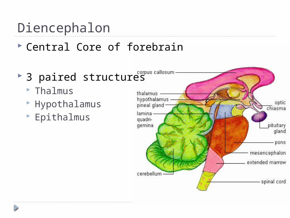
Diencephalon Central Core of forebrain
3 paired structures Thalmus Hypothalamus Epithalmus

Thalamus 80% of diencephalon Large number of
nuclei Gateway to cerebral
cortex Afferent impulses
from all senses converge
Information is sorted and relayed to appropriate area of cortex
Crude recognition as pleasant or unpleasant

Hypothalamus
Below the thalamus, above brain stem
Extends from optic chiasma to mammilary bodies (nuclei that relay olfactory pathways)
Infundibulum connects to pituitary gland
Visceral control center of body, vital for homeostatic control

Homeostatic Roles of Hypothalamus ANS
Influences hr, bp, pupils, etc Emotional response (limbic system)
Perception of pleasure, fear, rage, and sex drive Body Temperature regulation
Monitors & initiates sweating or shivering Regulation of food intake
response to changes in blood glucose/a.a., hormones Regulation of water balance and thirst
Response to concentrations of bodily fluids Regulation of sleep wake cycles
In response to light (visual) cues Endocrine system functioning
Controls secretions of pituitary gland Produces hormones ADH and oxytocin

Epithalamus Dorsal portion of
diencephelon, roof of 3rd ventricle
Pineal gland extends from posterior border
Helps regulate sleep wake cycle

Brain Imaging Compare PET, fMRI, CAT, EEG, cerebral
angiography

Brain Stem Survival Behaviors Pathway for fiber tracts Innervation of the head 3 parts:
Midbrain Descending motor tracts Cerebral aqueduct (connects 3rd & 4th ventricles Visual reflex centers Sound (startle)reflex
Pons Bulging region Deep tracts connect brain and spinal cord Superficial fibers connect motor cortex and cerebellum Help medulla maintain breathing rhythm
Medulla Oblongata

Medulla Oblongata Motor cortex fibers cross over to opposite side
of body before spinal cord Relay sensory information Auditory relays Maintains equilibrium Autonomic reflex center
Force and rate of heart contraction Vasomotor center Rate and depth of breathing Vomiting, hiccupping, swallowing, coughing,
sneezing

Cerebellum
11%of total brain mass Dorsal to pons & medulla Using input from cerebral motor
cortex and sensory receptors provides precise timing of skeletal muscle contraction subconsciously Smooth coordinated movements Agility

Brain Activities:1. Limbic System2. Reticular Formation3. Sleep4. Language5. Memory6. Brain wave patterns and EEG
Each group will create a brief (8 ppt slides) presentation w/pictures on their assigned brain topic of interest! You may use textbooks or internet, be sure to cite sources!

Cranial Nerves


![[Howard Bloom] Global Brain the Evolution of Mass(Bookos.org)](https://static.fdocuments.in/doc/165x107/55cfe3d25503467d968b647e/howard-bloom-global-brain-the-evolution-of-massbookosorg.jpg)


![Presentation ice child brain foundation - lbs adf 10.06.2011.ppt [enregistrement automatique]](https://static.fdocuments.in/doc/165x107/554e93afb4c90573338b4fa8/presentation-ice-child-brain-foundation-lbs-adf-10062011ppt-enregistrement-automatique.jpg)


