cmvIL-10 Stimulates the Invasive Potential of MDA-MB-231 ...
9
e University of San Francisco USF Scholarship: a digital repository @ Gleeson Library | Geschke Center Biology Faculty Publications Biology 2014 cmvIL-10 Stimulates the Invasive Potential of MDA-MB-231 Breast Cancer Cells Cendy Valle Oseguera [email protected] Juliet Spencer University of San Francisco, [email protected] Follow this and additional works at: hp://repository.usfca.edu/biol_fac Part of the Biology Commons , and the Oncology Commons is Article is brought to you for free and open access by the Biology at USF Scholarship: a digital repository @ Gleeson Library | Geschke Center. It has been accepted for inclusion in Biology Faculty Publications by an authorized administrator of USF Scholarship: a digital repository @ Gleeson Library | Geschke Center. For more information, please contact [email protected]. Recommended Citation Valle Oseguera CA, Spencer JV (2014) cmvIL-10 Stimulates the Invasive Potential of MDA-MB-231 Breast Cancer Cells. PLoS ONE 9(2): e88708. doi:10.1371/journal.pone.0088708
Transcript of cmvIL-10 Stimulates the Invasive Potential of MDA-MB-231 ...

The University of San FranciscoUSF Scholarship: a digital repository @ Gleeson Library |Geschke Center
Biology Faculty Publications Biology
2014
cmvIL-10 Stimulates the Invasive Potential ofMDA-MB-231 Breast Cancer CellsCendy Valle [email protected]
Juliet SpencerUniversity of San Francisco, [email protected]
Follow this and additional works at: http://repository.usfca.edu/biol_fac
Part of the Biology Commons, and the Oncology Commons
This Article is brought to you for free and open access by the Biology at USF Scholarship: a digital repository @ Gleeson Library | Geschke Center. Ithas been accepted for inclusion in Biology Faculty Publications by an authorized administrator of USF Scholarship: a digital repository @ GleesonLibrary | Geschke Center. For more information, please contact [email protected].
Recommended CitationValle Oseguera CA, Spencer JV (2014) cmvIL-10 Stimulates the Invasive Potential of MDA-MB-231 Breast Cancer Cells. PLoS ONE9(2): e88708. doi:10.1371/journal.pone.0088708

cmvIL-10 Stimulates the Invasive Potential of MDA-MB-231 Breast Cancer CellsCendy A. Valle Oseguera, Juliet V. Spencer*
Department of Biology, University of San Francisco, San Francisco, California, United States of America
Abstract
Cancer is the result of unregulated cell growth that leads to tumor formation, and in many cases, metastases. Althoughthere are several risk factors associated with cancer, one area that remains poorly understood is the impact of infectiousdisease. Human cytomegalovirus (HCMV) is a member of the herpesvirus family that is highly prevalent in the population.HCMV usually causes clinical disease only in immune compromised individuals, but recent evidence suggests that HCMVmay be strongly associated with some forms of cancer, particularly glioblastoma and breast cancer. We investigated thepossibility that cmvIL-10, a viral cytokine with homology to human IL-10 that is secreted from infected cells, could act in aparacrine manner to alter the tumor microenvironment, induce cell signaling, and increase the invasive potential of cancercells. We found that human MDA-MB-231 breast cancer cells express the IL-10 receptor and that exposure to cmvIL-10results in activation of Stat3, a transcription factor strongly associated with enhanced metastatic potential and chemo-resistance. In addition, cmvIL-10 stimulated an increase in DNA synthesis and cell proliferation, protected MDA-MB-231 cellsfrom etoposide-induced apoptosis, and also greatly enhanced chemotaxis toward epidermal growth factor (EGF). Theseresults suggest a significant and wide-ranging role for cmvIL-10 in the progression of breast cancer and could have broadimplications for the diagnosis and treatment of cancer in HCMV-positive patients.
Citation: Valle Oseguera CA, Spencer JV (2014) cmvIL-10 Stimulates the Invasive Potential of MDA-MB-231 Breast Cancer Cells. PLoS ONE 9(2): e88708.doi:10.1371/journal.pone.0088708
Editor: Nancy M Sawtell, Cincinnati Childrens Hospital Medical Center, United States of America
Received September 30, 2013; Accepted January 10, 2014; Published February 10, 2014
Copyright: � 2014 Valle Oseguera and Spencer. This is an open-access article distributed under the terms of the Creative Commons Attribution License, whichpermits unrestricted use, distribution, and reproduction in any medium, provided the original author and source are credited.
Funding: This work was supported by the National Institutes of Health (www.nih.gov) grant R15CA158767 and USF Faculty Development Funds (www.usfca.edu). The funders had no role in study design, data collection and analysis, decision to publish, or preparation of the manuscript.
Competing Interests: The authors have read the journal’s policy and have the following conflict: Juliet V. Spencer serves as an Academic Editor for PLOS ONE.This does not alter the authors’ adherence to all the PLOS ONE policies on sharing data and materials.
* E-mail: [email protected]
Introduction
Breast cancer is the second leading cause of cancer deaths in the
United States [1]. Many cancer patients do not die from local
complications of their primary tumor growth, but rather from the
malignant spread of the tumor. Approximately 30% of patients
diagnosed with a solid tumor already have a clinically detectable
metastasis, and for the remaining 70%, metastases are continually
being formed throughout the life of the tumor [2]. While there are
recognized genetic, environmental, and behavioral risk factors
associated with breast cancer, little is known about the connection
between infectious agents and breast cancer development or
progression.
Human cytomegalovirus (HCMV) is a widespread pathogen
that infects more than 70% of the general population [3]. In most
individuals, primary infection with HCMV is asymptomatic;
however, serious symptoms can occur in patients with compro-
mised immune systems. HCMV pneumonitis greatly impacts the
morbidity and mortality of transplant recipients, and HIV patients
are frequently diagnosed with severe HCMV retinitis [3]. HCMV
can be transmitted from mother to child during pregnancy, and
infection can result in serious congenital defects, including
deafness, mental retardation, and other neurological deficien-
cies[4].
The possible relationship between HCMV and cancer has been
investigated for some time. The development of more sensitive
detection methods has recently shown a very strong link between
HCMV infection and glioblastoma, prostate cancer, and breast
cancer [5–9]. While HCMV is not generally regarded as an
oncogenic virus, the term oncomodulation has been proposed to
describe the increased malignancy associated with HCMV-
infected tumor cells [10]. The molecular mechanisms for
oncomodulation include cell cycle dysregulation by immediate
early proteins IE1 and IE2 [11], which promote entry into S
phase, as well as the activity of the UL97 protein which
phosphorylates and inactivates tumor suppressor Rb [12]. Recent
studies of human breast biopsy samples have revealed abundant
expression of IE1 [9]. In addition, the HCMV UL36, UL37, and
UL38 gene products all interfere with caspase function and convey
resistance to apoptosis [13,14]. HCMV-infected neuroblastoma
cells have been observed to down-regulate adhesion molecules and
exhibit increased motility [15]. In prostate cancer and glioma cells,
HCMV infection resulted in increased migration and invasion that
was dependent on phosphorylation of focal adhesion kinase (FAK)
[6,16].
The ability to evade recognition from the immune system is also
essential for cancer cells, and HCMV is highly adept at
manipulating the host immune system [17]. The cmvIL-10 protein
is a homolog of human IL-10 encoded by the UL111A gene
product of HCMV [18]. Despite having only 27% sequence
identity to human IL-10, cmvIL-10 binds to the cellular IL-10
receptor (IL-10R) and displays many of the immune suppressive
functions of human IL-10 [19,20]. Interestingly, elevated levels of
IL-10 are frequently detected in the serum of cancer patients and
PLOS ONE | www.plosone.org 1 February 2014 | Volume 9 | Issue 2 | e88708

correlate with poor prognosis [21–24], suggesting that IL-10 may
contribute to immune suppression and protect tumor cells from
cytotoxic T lymphocytes by down-regulation of class I and class II
MHC. In vitro, IL-10 has been found to promote resistance to
apoptosis in human breast and lung cancer cell lines [25,26].
Furthermore, constitutive activation of Stat3, the primary
downstream activator associated with IL-10 signaling, correlates
with poor prognosis in ovarian cancer and is considered a key
factor in the development of metastasis and resistance to
chemotherapeutic agents [27]. Given that cmvIL-10 retains many
biological functions of human IL-10, including stimulation of B
cell growth and activation of Stat3 in monocytes and dendritic
cells [20,28,29], we investigated whether the viral cytokine might
also induce changes in human breast cancer cells that could
ultimately promote tumor metastasis.
Results and Discussion
In order to determine whether cmvIL-10 could have an impact
on tumor cell physiology, we first examined whether breast cancer
cells expressed the IL-10R. The MDA-MB-231 breast adenocar-
cinoma cell line was stained with antibody directed against the
alpha chain of the human IL-10R complex and examined via flow
cytometry. As shown in Figure 1A, there was low-level expression
of the IL-10R complex detected on the surface of these cells. To
study receptor distribution in greater detail, the cells were grown
on glass cover slips, permeabilized, and visualized with immuno-
fluorescence microscopy. The findings were consistent with the
flow cytometry results in that the IL-10R complex was detected on
the cell surface (Fig. 1B). However, additional receptor was also
observed throughout the inside of the cell, suggesting that the IL-
10R complex undergoes constitutive recycling in breast cancer
cells and that surface levels are likely to be variable. After
treatment with purified recombinant cmvIL-10, there was a
distinct redistribution of IL-10R (Fig. 1B), indicating receptor
internalization occurred rapidly after ligand engagement. Al-
though HCMV infection has been noted in many primary breast
tumor samples [9,30], there was no evidence that the MDA-MB-
231 breast cancer cell line was infected. Expression of the IE1 gene
product could not be detected in these cells by RT-PCR (Fig. 1C)
or immunofluorescence staining (Fig. 1D). Human foreskin
fibroblasts that were infected with the AD169 strain of HCMV
served as a positive control for IE1 expression, which was found to
be localized predominantly to the nucleus, as expected (Fig 1D).
These results demonstrated that uninfected tumor cells express the
IL-10R complex and have the ability to respond to cmvIL-10 in
the tumor microenvironment.
One of the earliest indicators of cmvIL-10 signaling is
phosphorylation of Stat3 by the receptor-associated kinase
JAK1. MDA-MB-231 cells were treated with either cmvIL-10,
human IL-10 (hIL-10) or interferon-gamma (IFNc), then cell
lystates were examined by Western blot (Fig 2A). The expected
83 kD band corresponding to phosphorylated Stat3 (pStat3) was
detected in cells treated with cmvIL-10 or hIL-10, but not in
control cells exposed to PBS or IFNc. Exposure to cmvIL-10
specifically activated Stat3, but did not globally activate other
cellular effectors, such as Stat1, which was phosphorylated in
response to IFNc treatment only. To confirm Stat3 activation,
cells were treated with varying doses of cmvIL-10 and then a cell-
based ELISA was performed to detect pStat3. The amount of
Stat3 phosphorylation increased in a dose-dependent manner with
higher concentrations of cmvIL-10, as shown in Figure 2B. To
confirm that Stat3 activation was solely due to treatment with
cmvIL-10, MDA-MB-231 cells were examined for the production
of endogenous hIL-10 by both ELISA (data not shown) and
Western blot (Fig. 3C). No hIL-10 could be detected in the cell
supernatants, confirming that Stat3 was not being activated by an
autocrine signaling mechanism. Likewise, cmvIL-10 could not be
detected in the supernatants of MDA-MB-231 cells (Fig. 1C), a
result that was expected because we found that these cells were not
infected with HCMV (Fig. 1C and D). Human serpin E1 (also
known as PAI, plasminogen activator inhibitor-1), which is
secreted from many cancer cells [31], served as a positive control
and was detected in cell supernatants in increasing concentrations
over time. Taken together, these results demonstrate that
exogenous cmvIL-10 can trigger phosphorylation and activation
of the transcription factor Stat3 in human breast cancer cells.
Over-activation of Stat3 has been documented in glioblastoma,
ovarian and breast cancers, which suggests that stimulation of this
signaling pathway by cmvIL-10 could contribute to malignancy.
To elucidate downstream effects of cmvIL-10 signaling and
Stat3 activation, cell proliferation was examined. MDA-MB-231
breast cancer cells were cultured in the presence of increasing
doses of cmvIL-10, and cell growth was evaluated. Cell viability
was measured by the addition of a luciferin substrate at the
indicated time points, and the resulting luminescence is propor-
tional to the amount of ATP present, reflecting the number of
viable cells in the well. As shown in Figure 3A, cells exposed to
cmvIL-10 exhibited greater growth than control cells. Overall cell
growth increased for 72 hrs and then fell, possibly due to crowding
in the wells. Subsequent assays utilized 100 ng/ml cmvIL-10 for
72 hrs, which resulted in significantly higher cell growth than
control cultures (Fig 3B, * = p,0.05). In addition, BrdU
incorporation was used to quantify the rate of DNA synthesis,
which was found to be significantly higher in cells exposed to
cmvIL-10 compared to the control cell lines (Fig. 3C). The level of
proliferation induced by cmvIL-10 was comparable to that of hIL-
10. Standard cell counts taken at each time point also revealed that
cultures treated with either cmvIL-10 or hIL-10 had higher cell
numbers than control cultures (Fig. 3D). The enhanced prolifer-
ative effect was specific to cmvIL-10 and hIL-10, as treatment with
other cytokines did not increase cell proliferation. As shown in
Figure 3E, treatment with IFNc or IL-6 actually inhibited cell
growth. Finally, treatment of cells with either a Stat3 or Jak1
inhibitor blocked the proliferative effects of cmvIL-10, confirming
that these results are mediated in part by the Jak1/Stat3 signaling
cascade. These results clearly demonstrate that cmvIL-10 specif-
ically stimulates cell proliferation and increases the rate of DNA
synthesis in human breast cancer cells.
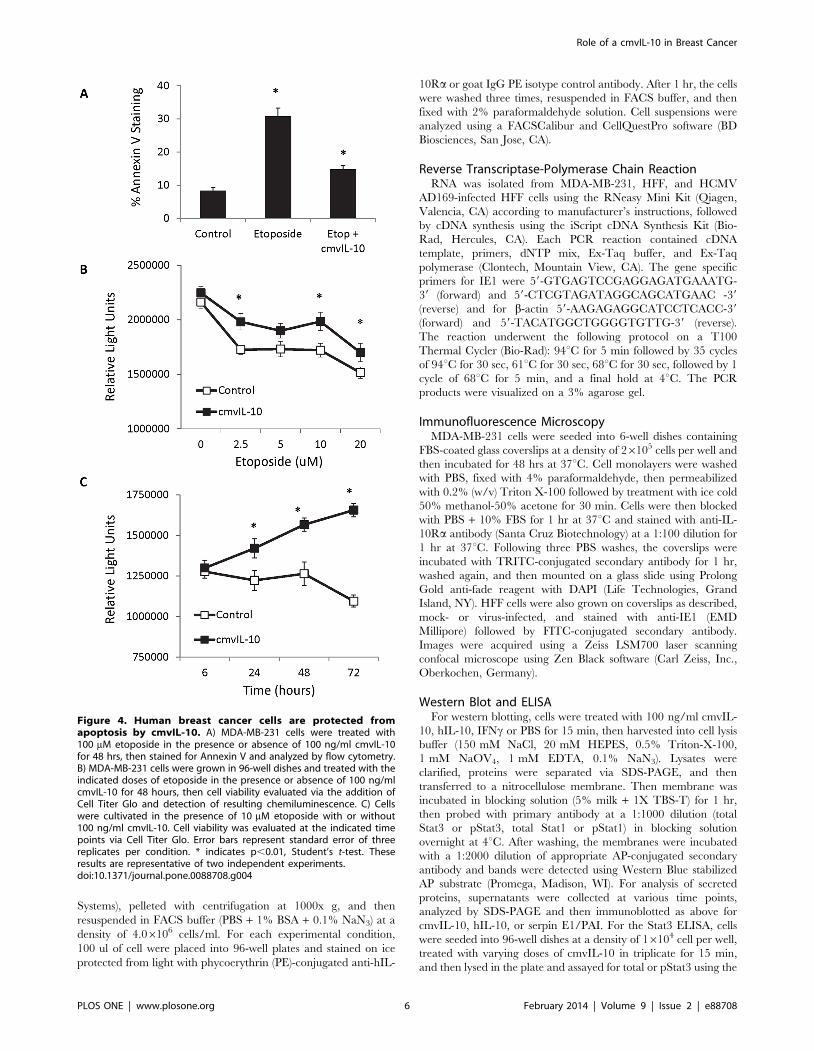
To investigate whether cmvIL-10 could protect cells from
apoptosis, MDA-MB-231 breast cancer cells were treated with
etoposide, an inhibitor of topoisomerase II that is widely used in
the treatment of cancer based on its ability to induce cell death.
After exposure to etoposide, 30.9% of cells stained positive for
Annexin V via flow cytometry, as shown in Figure 4A. In contrast,
when cultures were incubated with cmvIL-10 prior to etoposide
treatment, only 14.8% of cells stained positive for Annexin V,
indicating that cmvIL-10 was able to prevent induction of
apoptosis in human breast cancer cells. Cultivation of cells with
varying doses of etoposide revealed that cmvIL-10 increased
overall cell viability (Fig. 4B), and cells exposed to cmvIL-10 were
able to overcome etoposide-induced effects over time and
proliferate robustly (Fig. 4C).
Because the migration of cancer cells away from the primary
tumor is one of the critical early factors in the formation of
metastasis, we next examined cell motility. MDA-MB-231 breast
cancer cells express the epidermal growth factor (EGF) receptor;
therefore, EGF was utilized as a chemo-attractant in a modified
Role of a cmvIL-10 in Breast Cancer
PLOS ONE | www.plosone.org 2 February 2014 | Volume 9 | Issue 2 | e88708

Boyden chamber assay. The cells were placed in the top chamber
separated from the EGF in the lower chamber by a porous (8 mm)
filter, and after 5 hrs cells that had traversed the filter into the
lower chamber were harvested and quantified. The cells migrated
toward EGF and exhibited a standard bell-shaped curve for
chemotaxis with a maximal response at 10 ng/ml EGF (Fig. 5A,
gray bars). When both EGF and cmvIL-10 were present in the
lower chamber, the migration response was significantly increased
(Fig. 5A, black bars), and the effect of cmvIL-10 was comparable
to that of hIL-10 (Fig. 5A, white bars). cmvIL-10 alone did not
stimulate cell movement (Fig. 5B, gray bars); however, exposure to
the viral cytokine significantly enhanced cell migration toward
EGF (Fig 5B, black bars). Exposure to hIL-10 alone also failed to
stimulate cell movement (data not shown). These results demon-
strate that can cmvIL-10 work synergistically with other growth
factors and mitogens present in the tumor microenvironment, such
as EGF, to promote increased cell movement.
The hallmarks of cancer, characteristics such as sustained
proliferative activity, resistance to apoptosis, and the ability to
invade surrounding tissues which distinguish cancer cells from
normal cells, were first described by Hanahan and Weinberg in
2000 [32]. The list was subsequently updated in 2011 to include
additional characteristics, most notably, the ability to evade the
immune system [33]. cmvIL-10 is well-documented to function as
an immune suppressive cytokine that inhibits both immune cell
activation and inflammatory cytokine production [19,20]. Here,
we show for the first time that cmvIL-10 can also enhance other
properties associated with tumor cells. While this study made use
of MDA-MB-231 breast cancer cells that already exhibit robust
proliferation, treatment with cmvIL-10 was able to promote a
significantly greater rate of growth. The rapid proliferation of cells
Figure 1. Human breast cancer cells express the IL-10 receptor. A) MDA-MB-231 cells were stained with anti-IL-10R-PE antibody (black line) orisotype control (gray line) and analyzed by flow cytometry. B) Cells were untreated or treated with 100 ng/ml cmvIL-10 for 15 min and stained for IL-10R followed by TRITC-conjugated secondary antibody, then visualized by fluorescence microscopy. Red corresponds to IL-10R, blue corresponds toDAPI staining of the nucleus. C) RNA was harvested from MDA-MB-231 cells and mock- or HCMV-infected HFF cells (MOI = 1, 72 hrs post-infection),reverse-transcribed and IE1 or b-actin gene specific primers were used for PCR. D) MDA-MB-231 and mock- or HCMV-infected HFFs were cultured onglass coverslips, fixed and stained for IE1 followed by FITC-conjugated secondary (green). These results are representative of three independentexperiments.doi:10.1371/journal.pone.0088708.g001
Role of a cmvIL-10 in Breast Cancer
PLOS ONE | www.plosone.org 3 February 2014 | Volume 9 | Issue 2 | e88708

at the primary tumor site increases the chances that any individual
cell or group of cells might break away and initiate metastasis
formation. Although evidence suggests that viral DNA and
proteins can be found in tumors, no evidence of infection of the
MDA-MB-231 cell line was found here and the role that virus
infection plays in tumor development remains unclear. HCMV
infects a wide range of cell types, including monocytes and
dendritic cells which are mobile, circulating throughout the body
and entering various tissues. It seems very likely that infected cells
could enter a tissue containing a developing tumor, secrete cmvIL-
10, and ultimately stimulate progression toward malignancy.
In addition to increased growth, we detected rapid phosphor-
ylation and activation of Stat3 in breast cancer cells. This is
significant because Stat3 is a transcription factor whose activation
blocks normal programmed cell death pathways [34]. Elevated
levels of pStat3 have been detected in gastric cancer [35] and are
also associated with enhanced metastatic potential and chemo-
resistance in ovarian cancer [27,36]. In addition, constitutive
activation of pStat3 in breast cancer cells has been observed to
lead to increased expression of the anti-apoptotic protein survivin
[37], which is consistent with our observations that exposure to
cmvIL-10 protected cells from etoposide induced apoptosis.
Therefore, when HCMV-infected cells secrete cmvIL-10 into
their microenvironment, the viral cytokine could potentially
impact neighboring cells by activating the IL-10R pathway,
causing overstimulation of Stat3, enabling tumor cells to grow
uncontrollably and resist the induction of apoptosis.
The ability of cancer cells to invade other tissues and spread to
distant organs is an often fatal characteristic of malignant tumors.
One of the key steps in the process of invasion is cell motility. Our
results demonstrate that while cmvIL-10 itself does not trigger cell
movement, the viral cytokine can enhance movement toward
other growth factors such as EGF. Subsequent studies will be
necessary to investigate whether cmvIL-10 can truly promote
invasion through the extracellular matrix and into surrounding
tissues, which might be expected to involve not only motility, but
production of proteolytic enzymes.
HCMV is endemic in the human population and may be
associated with several malignancies. Although various HCMV
gene products have been shown to alter the cell cycle or inhibit
apoptosis of virus-infected tumor cells, the possibility that factors
secreted from virus-infected cells could affect tumor progression
has not yet previously been examined. We demonstrate for the first
time a novel effect of a secreted viral cytokine, cmvIL-10, in
enhancing the invasive potential of human breast cancer cells.
Although only a single cell line was tested here, MDA-MB-231
cells are widely used for breast cancer research because they are an
excellent model for tumor cells, and these results are likely to be
relevant to other cell lines as well as primary tumor cells. These
findings suggest that the presence of cmvIL-10 in the tumor
microenvironment could promote tumor progression and stimu-
late formation of metastases. These findings could ultimately lead
to new therapeutic strategies for HCMV-positive breast cancer
patients that include administration of antiviral drugs or anti-
cmvIL-10 neutralizing antibodies in concert with traditional
chemotherapy.
Materials and Methods
Cells & ReagentsMDA-MB-231 human breast cancer cells (American Type
Culture Collection, Manassas, VA) were cultured in L-15
Leibovitz’s Medium (Corning, Manassas, VA) supplemented with
10% fetal bovine serum (Atlanta Biologicals, Flowery Branch, GA)
and maintained at 37uC with atmospheric CO2 according to the
suppliers instructions. Purified recombinant cmvIL-10, hIL-10,
IFNc, IL-6 and anti-cmvIL-10, anti-hIL-10, anti-IL-10R, and
anti-serpin E1/PAI antibodies were purchased from R&D Systems
(Minneapolis, MN). Total Stat3, pStat3 (Y705), total Stat1, and
pStat1 (Y701) antibodies were from Cell Signaling Technology
(Danvers, MA). The Stat3 inhibitor was from Santa Cruz
Figure 2. cmvIL-10 induces Stat3 phosphorylation in humanbreast cancer cells. A) MDA-MB-231 cells were treated with 100 ng/ml cmvIL-10, hIL-10, or IFNc for 15 min, then lysed and Western blottedwith the indicated antibodies. B) Cells were grown in 96-well dishes andtreated with the indicated doses of cmvIL-10 for 15 min before lysis inthe well followed by quantification of total vs. pStat3 levels. Results arerepresented as the normalized ratio of pStat3 to total Stat3 in relativefluorescence units (RFUs). * = p , 0.01, Student’s t-test. Error barsrepresent standard error for three replicates of each condition. C) MDA-MB-231 cells were cultured and supernatants collected at the indicatedtime points were subjected to SDS-PAGE followed by immunoblottingwith the indicated antibodies. Control indicates purified recombinantprotein (cmvIL-10, hIL-10, or serpin E1/PAI) was loaded as a positivecontrol for each respective antibody. Results are representative of threeindependent experiments.doi:10.1371/journal.pone.0088708.g002
Role of a cmvIL-10 in Breast Cancer
PLOS ONE | www.plosone.org 4 February 2014 | Volume 9 | Issue 2 | e88708

Biotechnology (Dallas, TX), the Jak1 and p38 MAPK inhibitors
were from Calbiochem/EMD Millipore (Billerica, MA). Etoposide
was from Cayman Chemicals (Ann Arbor, MI) and purified
recombinant human EGF was from Peprotech (Rocky Hill, NJ).
The HCMV strain AD169 (ATCC) was propagated in human
foreskin fibroblasts (HFF, also from ATCC), maintained in
Dulbecco’s modification of Eagle’s medium (Corning) containing
15% fetal bovine serum.
Flow CytometryMonolayer cultures of MDA-MB-231 cells were harvested via
gentle scraping according to manufacturer’s instructions (R&D
Figure 3. cmvIL-10 stimulates proliferation and increases DNA synthesis in human breast cancer cells. A) MDA-MB-231 cells were grownin 96-well dishes and treated with the indicated doses of cmvIL-10. Cell Titer Glo was added at the indicated time points to measure cell viability,represented as relative light units (RLUs) based on the resulting chemiluminescence. B) Cell growth in the presence or absence of 100 ng/ml cmvIL-10via Cell Titer Glo. C) BrdU incorporation in the presence of 100 ng/ml cmvIL-10 or hIL-10. D) Standard cell counts of cultures from 6-well dishescontaining 100 ng/ml cmvIL-10 or hIL-10 using a hemacytometer. E) BrdU incorporation was assessed at 72 hrs in cells cultured in the presence of10 ng/ml of each indicated cytokine. F) Cells were treated with 10 mM of each indicated inhibitor or an equivalent volume of DMSO in the presence ofabsence of 10 ng/ml cmvIL-10 and BrdU incorporation measured after 72 hrs. Error bars represent standard error among three replicates for eachcondition. * indicates p,0.05, Student’s t-test. These results are representative of three independent experiments.doi:10.1371/journal.pone.0088708.g003
Role of a cmvIL-10 in Breast Cancer
PLOS ONE | www.plosone.org 5 February 2014 | Volume 9 | Issue 2 | e88708
Systems), pelleted with centrifugation at 1000x g, and then
resuspended in FACS buffer (PBS + 1% BSA + 0.1% NaN3) at a
density of 4.06106 cells/ml. For each experimental condition,
100 ul of cell were placed into 96-well plates and stained on ice
protected from light with phycoerythrin (PE)-conjugated anti-hIL-
10Ra or goat IgG PE isotype control antibody. After 1 hr, the cells
were washed three times, resuspended in FACS buffer, and then
fixed with 2% paraformaldehyde solution. Cell suspensions were
analyzed using a FACSCalibur and CellQuestPro software (BD
Biosciences, San Jose, CA).
Reverse Transcriptase-Polymerase Chain ReactionRNA was isolated from MDA-MB-231, HFF, and HCMV
AD169-infected HFF cells using the RNeasy Mini Kit (Qiagen,
Valencia, CA) according to manufacturer’s instructions, followed
by cDNA synthesis using the iScript cDNA Synthesis Kit (Bio-
Rad, Hercules, CA). Each PCR reaction contained cDNA
template, primers, dNTP mix, Ex-Taq buffer, and Ex-Taq
polymerase (Clontech, Mountain View, CA). The gene specific
primers for IE1 were 59-GTGAGTCCGAGGAGATGAAATG-
39 (forward) and 59-CTCGTAGATAGGCAGCATGAAC -39
(reverse) and for b-actin 59-AAGAGAGGCATCCTCACC-39
(forward) and 59-TACATGGCTGGGGTGTTG-39 (reverse).
The reaction underwent the following protocol on a T100
Thermal Cycler (Bio-Rad): 94uC for 5 min followed by 35 cycles
of 94uC for 30 sec, 61uC for 30 sec, 68uC for 30 sec, followed by 1
cycle of 68uC for 5 min, and a final hold at 4uC. The PCR
products were visualized on a 3% agarose gel.
Immunofluorescence MicroscopyMDA-MB-231 cells were seeded into 6-well dishes containing
FBS-coated glass coverslips at a density of 26105 cells per well and
then incubated for 48 hrs at 37uC. Cell monolayers were washed
with PBS, fixed with 4% paraformaldehyde, then permeabilized
with 0.2% (w/v) Triton X-100 followed by treatment with ice cold
50% methanol-50% acetone for 30 min. Cells were then blocked
with PBS + 10% FBS for 1 hr at 37uC and stained with anti-IL-
10Ra antibody (Santa Cruz Biotechnology) at a 1:100 dilution for
1 hr at 37uC. Following three PBS washes, the coverslips were
incubated with TRITC-conjugated secondary antibody for 1 hr,
washed again, and then mounted on a glass slide using Prolong
Gold anti-fade reagent with DAPI (Life Technologies, Grand
Island, NY). HFF cells were also grown on coverslips as described,
mock- or virus-infected, and stained with anti-IE1 (EMD
Millipore) followed by FITC-conjugated secondary antibody.
Images were acquired using a Zeiss LSM700 laser scanning
confocal microscope using Zen Black software (Carl Zeiss, Inc.,
Oberkochen, Germany).
Western Blot and ELISAFor western blotting, cells were treated with 100 ng/ml cmvIL-
10, hIL-10, IFNc or PBS for 15 min, then harvested into cell lysis
buffer (150 mM NaCl, 20 mM HEPES, 0.5% Triton-X-100,
1 mM NaOV4, 1 mM EDTA, 0.1% NaN3). Lysates were
clarified, proteins were separated via SDS-PAGE, and then
transferred to a nitrocellulose membrane. Then membrane was
incubated in blocking solution (5% milk + 1X TBS-T) for 1 hr,
then probed with primary antibody at a 1:1000 dilution (total
Stat3 or pStat3, total Stat1 or pStat1) in blocking solution
overnight at 4uC. After washing, the membranes were incubated
with a 1:2000 dilution of appropriate AP-conjugated secondary
antibody and bands were detected using Western Blue stabilized
AP substrate (Promega, Madison, WI). For analysis of secreted
proteins, supernatants were collected at various time points,
analyzed by SDS-PAGE and then immunoblotted as above for
cmvIL-10, hIL-10, or serpin E1/PAI. For the Stat3 ELISA, cells
were seeded into 96-well dishes at a density of 16104 cell per well,
treated with varying doses of cmvIL-10 in triplicate for 15 min,
and then lysed in the plate and assayed for total or pStat3 using the
Figure 4. Human breast cancer cells are protected fromapoptosis by cmvIL-10. A) MDA-MB-231 cells were treated with100 mM etoposide in the presence or absence of 100 ng/ml cmvIL-10for 48 hrs, then stained for Annexin V and analyzed by flow cytometry.B) MDA-MB-231 cells were grown in 96-well dishes and treated with theindicated doses of etoposide in the presence or absence of 100 ng/mlcmvIL-10 for 48 hours, then cell viability evaluated via the addition ofCell Titer Glo and detection of resulting chemiluminescence. C) Cellswere cultivated in the presence of 10 mM etoposide with or without100 ng/ml cmvIL-10. Cell viability was evaluated at the indicated timepoints via Cell Titer Glo. Error bars represent standard error of threereplicates per condition. * indicates p,0.01, Student’s t-test. Theseresults are representative of two independent experiments.doi:10.1371/journal.pone.0088708.g004
Role of a cmvIL-10 in Breast Cancer
PLOS ONE | www.plosone.org 6 February 2014 | Volume 9 | Issue 2 | e88708

Cell-based Stat3 ELISA kit according to manufacturer’s instruc-
tions (R&D Systems). The detection of hIL-10 in supernatants
from MDA-MB-231 cell cultures was performed using the IL-10
ELISA DuoSet kit as directed (R&D Systems).
Cell Proliferation and Apoptosis AssaysCells were seeded into 96-well dishes at a density of 16104 cell
per well in complete medium with varying doses of cmvIL-10, and
then cell viability measured at the indicated time points using the
Cell Titer Glo Assay kit according to manufacturer’s instructions
(Promega). DNA synthesis was measured in cells prepared in the
same way using the BrdU Cell Proliferation ELISA Kit (Roche,
Figure 5. Human breast cancer cells exhibit enhanced chemotaxis when exposed to cmvIL-10. MDA-MB-231 cells were seeded at adensity of 26105 cells in a total volume of 0.1 ml in the upper chamber of an 8 mm trans-well filter. A) Complete media containing the indicatedconcentrations of EGF in the presence or absence of 100 ng/ml cmvIL-10 or hIL-10 was placed in the lower chamber. After 5 hrs, cells in the lowerchamber were harvested and quantified by the addition of Cell Titer Glo to measureme luminescence. B) Complete medium containing the indicatedconcentrations of cmvIL-10 in the presence or absence of 1 ng/ml EGF. Cells traversing the filter after 5 hrs were quantified as described. Error barsrepresent standard error. * indicates p , 0.05, Student’s t-test. Results are representative of three independent experiments.doi:10.1371/journal.pone.0088708.g005
Role of a cmvIL-10 in Breast Cancer
PLOS ONE | www.plosone.org 7 February 2014 | Volume 9 | Issue 2 | e88708

Basel, Switzerland). For experiments using inhibitors, cells were
cultivated in 96-well dishes as above with a final concentration of
10 mM inhibitor and BrdU incorporation evaluated after 72 hrs.
For cell counts, cells were seeded into 6-well plates at a density of
26105 cells per well, harvested via trypsinization, and counted
using a hemacytometer at the indicated time points. The TACS
Annexin V-FITC Detection Kit (Trevigen, Gaithersburg, MD)
was used to stain cells harvested from 70% confluent T75 flasks
that had been treated with 100 mM etoposide for 48 hrs in the
presence or absence of 100 ng/ml cmvIL-10. Cells were then
analyzed via flow cytometry to detect fluorescence. For viability
assays, Cell Titer Glo reagent was utilized to quantify cells that
had been seeded into 96-well dishes at a density of 16104 cell per
well in complete medium with varying doses of etoposide in the
presence or absence of 100 ng/ml cmvIL-10 as indicated.
Migration AssaysCells were harvested and resuspended at a density of 26106 cells
per ml in complete medium. A total volume of 0.1 ml cell
suspension (26105 cells) was placed in the upper chamber of a
ThinCert filter with 8 mm pores in a 24-well plate (Greiner Bio-
One North America, Monroe, CA). A total volume of 0.6 ml of
media plus the indicated concentrations of human EGF and/or
cmvIL-10 or hIL-10 was added to the lower chamber of each well,
and plates were incubated for 5 hrs at 37uC. Medium from the
lower chamber was collected, used to rinse the bottom of the filter
twice, and then centrifuged at 1000 rpm for 10 min. The cell pellet
was resuspended in 0.1 ml media and transferred to a white 96-
well plate. Viable cell number was quantified using the Cell Titer
Glo Assay kit according to the manufacturer’s protocol.
Statistical AnalysisStatistical analysis was performed using the two-tailed Student’s
t-test.
Acknowledgments
The authors thank Carolyn Tu, Robin Bishop, Kathleen Arnolds, and Hai
Nguyen for technical assistance and helpful discussions.
Author Contributions
Conceived and designed the experiments: CVO JVS. Performed the
experiments: CVO. Analyzed the data: CVO JVS. Contributed reagents/
materials/analysis tools: CVO JVS. Wrote the paper: CVO JVS.
References
1. Key TJ, Verkasalo PK, Banks E (2001) Epidemiology of breast cancer. LancetOncol 2: 133–140.
2. John A, Tuszynski G (2001) The role of matrix metalloproteinases in tumor
angiogenesis and tumor metastasis. Pathol Oncol Res 7: 14–23.3. de la Hoz RE, Stephens G, Sherlock C (2002) Diagnosis and treatment
approaches of CMV infections in adult patients. J Clin Virol 25 Suppl 2: S1–12.4. Damato EG, Winnen CW (2002) Cytomegalovirus infection: perinatal
implications. J Obstet Gynecol Neonatal Nurs 31: 86–92.5. Cobbs CS, Harkins L, Samanta M, Gillespie GY, Bharara S, et al. (2002)
Human cytomegalovirus infection and expression in human malignant glioma.
Cancer Res 62: 3347–3350.6. Cobbs CS, Soroceanu L, Denham S, Zhang W, Britt WJ, et al. (2007) Human
cytomegalovirus induces cellular tyrosine kinase signaling and promotes gliomacell invasiveness. J Neurooncol 85: 271–280.
7. Mitchell DA, Xie W, Schmittling R, Learn C, Friedman A, et al. (2008) Sensitive
detection of human cytomegalovirus in tumors and peripheral blood of patientsdiagnosed with glioblastoma. Neuro Oncol 10: 10–18.
8. Samanta M, Harkins L, Klemm K, Britt WJ, Cobbs CS (2003) High prevalenceof human cytomegalovirus in prostatic intraepithelial neoplasia and prostatic
carcinoma. J Urol 170: 998–1002.9. Taher C, de Boniface J, Mohammad AA, Religa P, Hartman J, et al. (2013)
High prevalence of human cytomegalovirus proteins and nucleic acids in
primary breast cancer and metastatic sentinel lymph nodes. PLoS One 8:e56795.
10. Michaelis M, Doerr HW, Cinatl J (2009) The story of human cytomegalovirusand cancer: increasing evidence and open questions. Neoplasia 11: 1–9.
11. Sanchez V, Spector DH (2008) Subversion of cell cycle regulatory pathways.
Curr Top Microbiol Immunol 325: 243–262.12. Hume AJ, Finkel JS, Kamil JP, Coen DM, Culbertson MR, et al. (2008)
Phosphorylation of retinoblastoma protein by viral protein with cyclin-dependent kinase function. Science 320: 797–799.
13. McCormick AL (2008) Control of apoptosis by human cytomegalovirus. CurrTop Microbiol Immunol 325: 281–295.
14. Skaletskaya A, Bartle LM, Chittenden T, McCormick AL, Mocarski ES, et al.
(2001) A cytomegalovirus-encoded inhibitor of apoptosis that suppresses caspase-8 activation. Proc Natl Acad Sci U S A 98: 7829–7834.
15. Blaheta RA, Beecken WD, Engl T, Jonas D, Oppermann E, et al. (2004) Humancytomegalovirus infection of tumor cells downregulates NCAM (CD56): a novel
mechanism for virus-induced tumor invasiveness. Neoplasia 6: 323–331.
16. Blaheta RA, Weich E, Marian D, Bereiter-Hahn J, Jones J, et al. (2006) Humancytomegalovirus infection alters PC3 prostate carcinoma cell adhesion to
endothelial cells and extracellular matrix. Neoplasia 8: 807–816.17. Scalzo AA, Corbett AJ, Rawlinson WD, Scott GM, Degli-Esposti MA (2007)
The interplay between host and viral factors in shaping the outcome of
cytomegalovirus infection. Immunol Cell Biol 85: 46–54.18. Kotenko SV, Saccani S, Izotova LS, Mirochnitchenko OV, Pestka S (2000)
Human cytomegalovirus harbors its own unique IL-10 homolog (cmvIL-10).Proc Natl Acad Sci U S A 97: 1695–1700.
19. Slobedman B, Barry PA, Spencer JV, Avdic S, Abendroth A (2009) Virus-encoded homologs of cellular interleukin-10 and their control of host immune
function. J Virol 83: 9618–9629.
20. Spencer JV, Lockridge KM, Barry PA, Lin G, Tsang M, et al. (2002) Potent
immunosuppressive activities of cytomegalovirus-encoded interleukin-10. J Virol
76: 1285–1292.
21. Llanes-Fernandez L, Alvarez-Goyanes RI, Arango-Prado Mdel C, Alcocer-
Gonzalez JM, Mojarrieta JC, et al. (2006) Relationship between IL-10 and
tumor markers in breast cancer patients. Breast 15: 482–489.
22. Nicolini A, Carpi A, Rossi G (2006) Cytokines in breast cancer. Cytokine
Growth Factor Rev 17: 325–337.
23. Asadullah K, Sterry W, Volk HD (2003) Interleukin-10 therapy–review of a new
approach. Pharmacol Rev 55: 241–269.
24. Althwani AN, Najm MA (2011) The Role of Interleukin-10 in Women with
metastatic Invasive Ductal Carcinoma. J Fac Med - Baghdad 53: 289–292.
25. Zeng L, O’Connor C, Zhang J, Kaplan AM, Cohen DA (2009) IL-10 promotes
resistance to apoptosis and metastatic potential in lung tumor cell lines.
Cytokine.
26. Zheng M, Bocangel D, Doneske B, Mhashilkar A, Ramesh R, et al. (2007)
Human interleukin 24 (MDA-7/IL-24) protein kills breast cancer cells via the
IL-20 receptor and is antagonized by IL-10. Cancer Immunol Immunother 56:
205–215.
27. Zhang X, Liu P, Zhang B, Wang A, Yang M (2010) Role of STAT3 decoy
oligodeoxynucleotides on cell invasion and chemosensitivity in human epithelial
ovarian cancer cells. Cancer Genet Cytogenet 197: 46–53.
28. Spencer JV, Cadaoas J, Castillo PR, Saini V, Slobedman B (2008) Stimulation of
B lymphocytes by cmvIL-10 but not LAcmvIL-10. Virology 374: 164–169.
29. Spencer JV (2007) The cytomegalovirus homolog of interleukin-10 requires
phosphatidylinositol 3-kinase activity for inhibition of cytokine synthesis in
monocytes. J Virol 81: 2083–2086.
30. Harkins LE, Matlaf LA, Soroceanu L, Klemm K, Britt WJ, et al. (2010)
Detection of human cytomegalovirus in normal and neoplastic breast
epithelium. Herpesviridae 1: 8.
31. Wilkins-Port CE, Higgins CE, Freytag J, Higgins SP, Carlson JA, et al. (2007)
PAI-1 is a Critical Upstream Regulator of the TGF-beta1/EGF-Induced
Invasive Phenotype in Mutant p53 Human Cutaneous Squamous Cell
Carcinoma. J Biomed Biotechnol 2007: 85208.
32. Hanahan D, Weinberg RA (2000) The hallmarks of cancer. Cell 100: 57–70.
33. Hanahan D, Weinberg RA (2011) Hallmarks of cancer: the next generation. Cell
144: 646–674.
34. Levy DE, Lee C-K (2002) What does Stat3 do? Journal of Clinical Investigation
109: 1143–1148.
35. Deng J-Y, Sun D, Liu X-Y, Pan Y, Liang H (2010) STAT-3 correlates with
lymph node metastasis and cell survival in gastric cancer. World Journal of
Gastroenterology 16: 5380-5387.
36. Yu H, Pardoll D, Jove R (2009) STATs in cancer inflammation and immunity: a
leading role for STAT3. Nat Rev Cancer 9: 798–809.
37. Gritsko T, Williams A, Turkson J, Kaneko S, Bowman T, et al. (2006) Persistent
activation of stat3 signaling induces survivin gene expression and confers
resistance to apoptosis in human breast cancer cells. Clinical Cancer Research
12: 11–19.
Role of a cmvIL-10 in Breast Cancer
PLOS ONE | www.plosone.org 8 February 2014 | Volume 9 | Issue 2 | e88708