CME Upper Eyelid Reconstruction - lipteh.com from PRS/0412-Upper eyelid... · tightly adherent to...
10
CME Upper Eyelid Reconstruction Lisa M. DiFrancesco, M.D., Mark A. Codner, M.D., and Clinton D. McCord, M.D. Atlanta, Ga. Learning Objectives: After studying this article, the participant should be able to: 1. Understand upper eyelid anatomy and function. 2. Analyze upper eyelid defects. 3. Understand an algorithm of upper eyelid reconstruction. 4. Have a basic understanding of techniques for upper eyelid reconstruction. There are several options available for upper eyelid re- construction that depend on the extent of involvement of the anterior and posterior lamella. Knowledge of the anat- omy will ensure that in addition to the creation of an aes- thetically acceptable eyelid reconstruction, a functional up- per lid will be restored. The purpose of this article is to outline the anatomy of the eyelid, to analyze the components of eyelid defects, and to provide options for lid reconstruc- tion. (Plast. Reconstr. Surg. 114: 98e, 2004.) Any reconstructive procedure attempts to optimally restore form and function. Specifi- cally, the eyelid is critical in protection of the eye and preservation of vision. To ensure an optimal outcome, it is necessary to understand the specialized anatomic structures and func- tion of the orbit, eyelid, and globe. Once basic anatomy and function are understood, analysis of eyelid defects can be simplified when bro- ken down into separate components. The workhorses of reconstruction, such as skin and muscle flaps, are described, as well as their techniques. Even the most complex eyelid problems can be addressed. This article will outline the various techniques available for eyelid reconstruction. ANATOMY Superficial skin anatomy includes the brow, upper eyelid, lower eyelid, and midface. The upper eyelid rests 2 mm below the superior limbus on forward gaze, whereas the lower eye- lid is positioned at the inferior limbus. The supratarsal crease in the Occidental eyelid is generally 8 mm from the lid margin in men and 10 mm in women. The upper eyelid crease is formed by the dermal insertion of the levator aponeurosis to the orbital septum. In the Asian eyelid, this distance is only 2 to 3 mm from the lid margin as a result of fusion of the levator aponeurosis extensions with the orbital sep- tum. The pretarsal space is the area superior to the eyelid margin where the skin is adherent to the tarsal plate. The orbital sulcus is defined by the space between the superior orbital rim and the upper eyelid crease. Loss of the orbital sulcus is seen with relaxation of the upper septum or herniation of the preaponeurotic orbital fat pads (Fig. 1). The eyelid is divided into two main layers. The anterior lamella consists of skin and mus- cle, and the posterior lamella is formed by the tarsal plate and conjunctiva. The medial and lateral canthi are important support structures in normal eyelid function and bony fixation. They represent the confluence of the orbicu- laris muscle, tarsal plate, eyelid retractors, or- bital septum, and cheek ligaments. At the me- dial canthus this complex anatomic interplay provides the mechanism for the lacrimal pump. The lateral canthus inserts posterior on the lateral orbital rim, pulling the eyelid against the globe. The lateral canthus allows for bony stabilization of the eyelid during eye- lid movement. Analysis of an acquired defect should include evaluation of the anterior la- mella, posterior lamella, and medial and lat- eral canthi to facilitate reconstruction. The eyelid skin is the thinnest in the body, particularly on the medial aspect of the upper eyelid. At the eyelid margin the tarsal plates are From Paces Plastic Surgery. Received for publication January 15, 2003; revised April 24, 2003. DOI: 10.1097/01.PRS.0000142743.57711.48 98e
-
Upload
truongphuc -
Category
Documents
-
view
216 -
download
0
Transcript of CME Upper Eyelid Reconstruction - lipteh.com from PRS/0412-Upper eyelid... · tightly adherent to...

CME
Upper Eyelid ReconstructionLisa M. DiFrancesco, M.D., Mark A. Codner, M.D., and Clinton D. McCord, M.D.Atlanta, Ga.
Learning Objectives: After studying this article, the participant should be able to: 1. Understand upper eyelid anatomyand function. 2. Analyze upper eyelid defects. 3. Understand an algorithm of upper eyelid reconstruction. 4. Have a basicunderstanding of techniques for upper eyelid reconstruction.
There are several options available for upper eyelid re-construction that depend on the extent of involvement ofthe anterior and posterior lamella. Knowledge of the anat-omy will ensure that in addition to the creation of an aes-thetically acceptable eyelid reconstruction, a functional up-per lid will be restored. The purpose of this article is tooutline the anatomy of the eyelid, to analyze the componentsof eyelid defects, and to provide options for lid reconstruc-tion. (Plast. Reconstr. Surg. 114: 98e, 2004.)
Any reconstructive procedure attempts tooptimally restore form and function. Specifi-cally, the eyelid is critical in protection of theeye and preservation of vision. To ensure anoptimal outcome, it is necessary to understandthe specialized anatomic structures and func-tion of the orbit, eyelid, and globe. Once basicanatomy and function are understood, analysisof eyelid defects can be simplified when bro-ken down into separate components. Theworkhorses of reconstruction, such as skin andmuscle flaps, are described, as well as theirtechniques. Even the most complex eyelidproblems can be addressed. This article willoutline the various techniques available foreyelid reconstruction.
ANATOMY
Superficial skin anatomy includes the brow,upper eyelid, lower eyelid, and midface. Theupper eyelid rests 2 mm below the superiorlimbus on forward gaze, whereas the lower eye-lid is positioned at the inferior limbus. Thesupratarsal crease in the Occidental eyelid isgenerally 8 mm from the lid margin in menand 10 mm in women. The upper eyelid crease
is formed by the dermal insertion of the levatoraponeurosis to the orbital septum. In the Asianeyelid, this distance is only 2 to 3 mm from thelid margin as a result of fusion of the levatoraponeurosis extensions with the orbital sep-tum. The pretarsal space is the area superior tothe eyelid margin where the skin is adherentto the tarsal plate. The orbital sulcus is defined bythe space between the superior orbital rim andthe upper eyelid crease. Loss of the orbital sulcusis seen with relaxation of the upper septum orherniation of the preaponeurotic orbital fat pads(Fig. 1).
The eyelid is divided into two main layers.The anterior lamella consists of skin and mus-cle, and the posterior lamella is formed by thetarsal plate and conjunctiva. The medial andlateral canthi are important support structuresin normal eyelid function and bony fixation.They represent the confluence of the orbicu-laris muscle, tarsal plate, eyelid retractors, or-bital septum, and cheek ligaments. At the me-dial canthus this complex anatomic interplayprovides the mechanism for the lacrimalpump. The lateral canthus inserts posterior onthe lateral orbital rim, pulling the eyelidagainst the globe. The lateral canthus allowsfor bony stabilization of the eyelid during eye-lid movement. Analysis of an acquired defectshould include evaluation of the anterior la-mella, posterior lamella, and medial and lat-eral canthi to facilitate reconstruction.
The eyelid skin is the thinnest in the body,particularly on the medial aspect of the uppereyelid. At the eyelid margin the tarsal plates are
From Paces Plastic Surgery. Received for publication January 15, 2003; revised April 24, 2003.
DOI: 10.1097/01.PRS.0000142743.57711.48
98e

tightly adherent to the skin. The tarsal plate isa fibrous structure that provides the skeletalsupport for the eyelid. In the upper eyelid, itmeasures 25 mm in length, is 1 mm thick, andhas a maximal central height of 10 mm. Thetarsal plate is centralized over the midpupillaryline in childhood and migrates laterally withaging. Along with the conjunctiva, the tarsalplates form the posterior lamella.
The orbicularis muscle is divided into pre-tarsal, preseptal, and orbital components. Thepretarsal and preseptal orbicularis are each di-vided into a deep layer and a superficial layer,both medially and laterally. The medial canthaltendon is composed of superficial pretarsaland preseptal fibers. The deep pretarsal andpreseptal fibers intersect posterior to the lacri-mal sac and function as the lacrimal pump.The deep pretarsal and deep preseptal orbicu-laris insert on Whitnall’s tubercle laterallywithin the orbital rim. Riolan’s muscle repre-sents a strip of pretarsal muscle and forms thegray line of the eyelid, which is important foralignment of full-thickness defects.1
The orbital septum is posterior to the orbic-ularis muscle and forms the anterior barrier tothe orbital contents. In the upper lid, the sep-tum inserts on the levator aponeurosis approx-imately 10 mm above the eyelid margin justsuperior to the tarsal plate. The orbital septumattaches to the orbital rim at the arcus margi-nalis. The preaponeurotic fat pads (postseptal)are anterior extensions of orbital fat and aredivided into nasal and medial compartments.The trochlea divides the nasal and medial fat
pads. The lacrimal gland is located lateral tothe fat pads.
The eyelid retractor muscles include the le-vator palpebrae superioris and Müller’s mus-cle, which are responsible for eyelid elevation.The levator palpebrae superioris originatesalong the lesser wing of the sphenoid and isinnervated by cranial nerve III. The levatormuscle extends anteriorly approximately 40mm to Whitnall’s ligament. Whitnall’s liga-ment attaches superiorly to the levator muscleand changes the direction of levator musclepull to a superior-inferior direction. The leva-tor muscle extends inferiorly and transitionsinto a dense aponeurosis approximately 10mm inferior to Whitnall’s ligament. The leva-tor aponeurosis inserts on the anterior surfaceof the tarsal plate. Müller’s muscle is a smoothmuscle that is innervated by the sympatheticnervous system. It originates at the level ofWhitnall’s ligament posterior to the levatoraponeurosis and inserts on the superior tarsalplate.2
The medial canthus represents a fixed-pointfulcrum that is necessary for eyelid function.The medial canthus is a complex structure thatconsists of an anterior and posterior reflectionof the medial canthal tendon that envelops thelacrimal sac. The tendons insert on the ante-rior and posterior lacrimal crest, respectively.
The lateral canthal tendon is approximately5 mm to 7 mm in length and provides dynamicsupport by connecting the tarsal plates to Whit-nall’s tubercle along the lateral orbital rim.The lateral canthus also consists of anterior
FIG. 1. Anatomy of the preaponeurotic space demonstrating the struc-tures encountered during eyelid reconstruction.
Vol. 114, No. 7 / UPPER EYELID RECONSTRUCTION 99e

and posterior fibrous attachments of the tarsalplate and fibers of the pretarsal orbicularis.1Eisler’s fat pad can be found anterior to thelateral canthal tendon and is a useful anatomiclandmark (Fig. 2).
The arterial supply to the upper eyelid isprimarily from branches of the ophthalmic ar-tery that form two main arcades. The periph-eral arcade is located between Müller’s muscleand the levator aponeurosis on the upper bor-der of the tarsus; the marginal arcade is in thepretarsal space 2 to 3 mm above the eyelidmargin (Fig. 3). The superior palpebral veincourses parallel to the eyelid margin with ananastomosis between the angular vein and thesupraorbital vein. The lymphatic drainage ofthe upper eyelid is to the preauricular nodes,whereas the lymphatic drainage of the lateralcanthus is to the submandibular nodes. Evalu-ation of the appropriate lymphatic basin is im-portant for treatment of cancers such as squa-mous cell carcinoma, melanoma, andsebaceous carcinoma. Sensory innervation ofthe upper eyelid is divided between the su-pratrochlear nerve medially and a combinationof the supraorbital nerve and the lacrimalnerve laterally. The facial nerve cranial nerveVII supplies the motor innervation to the eyelidthrough the zygomatic and buccal branches.2 Re-cent data collected by the authors, however, sug-gest that the buccal branches have a more signif-icant contribution to motor function.
RECONSTRUCTION
Understanding the complex anatomy of theupper eyelid enables the surgeon to accurately
analyze upper eyelid defects. The division ofthe eyelid into its component structures facili-tates the approach to reconstruction.
DIRECT REPAIR
Benign lesions or biopsy defects often createsmall defects amenable to direct repair. This ispossible with defects of up to 30 percent of theeyelid in younger patients and up to 40 percentof the eyelid in patients with eyelid skin laxity.Full-thickness incisions should be made per-pendicular to the eyelid margin and extend tothe superior tarsal margin to prevent notching(Figs. 4 and 5). Superior to the tarsal plate theincision is connected to a horizontal sutureline hidden in the eyelid crease. Tension onthe upper eyelid closure can result in mechan-ical ptosis; a lateral cantholysis may be per-formed to reduce closure under tension. It iscritical that the eyelid margin, gray line, andlash line are approximated exactly to preventeyelid inversion, which can cause corneal irri-tation. Next, the repair is completed in layers.The conjunctival sutures are placed with theknot buried away from the conjunctival sur-face. Finally, the anterior lamella is repaired.Any remaining excess tissue may be removedby two Burrow’s triangles. Postoperative careconsists of ophthalmic antibiotic ointment andsuture removal at 7 days.3
SKIN GRAFT
A full-thickness graft can adequately recon-struct small superficial anterior lamellar de-fects. The skin graft may also be used as avertically shortened eyelid with a split level with
FIG. 2. Cross-section anatomy of the eyelid and orbit demonstrating theposterior insertion of the medial and lateral canthus.
100e PLASTIC AND RECONSTRUCTIVE SURGERY, December 2004

a posterior lamellar graft.2 Upper eyelid skinis the best donor site because of the match incolor and thickness. Excess skin from thecontralateral upper lid can be used but mayonly be available in limited quantities. Alter-native harvest sites for skin include the su-praclavicular skin, preauricular skin, and thelateral cervical skin. An elliptical incision ismade in the eyelid, and using iris scissors, theskin is separated from the muscle. The donorsite is closed primarily. The skin graft is insetand a bolster dressing is placed. The medialeyelid skin is avoided as a donor site to pre-vent lagophthalmos.
There are limited indications for a split-thickness skin graft; an example would beburns or other eyelid trauma involving exten-sive loss of upper and lower eyelid skin. Whendesigning a split-thickness skin graft, 50 to 70percent overcorrection should be incorpo-rated to allow for secondary contraction. In theupper lid, separate grafts are placed in the pre-tarsal area and the orbital sulcus, with the sutureline hidden in the eyelid crease.1
POSTERIOR LAMELLA
Sliding Tarsoconjunctival Flap
The sliding tarsoconjunctival flap (tarsal ex-tension flap) is a transposition flap based onsmall adjacent conjunctival defects involvingthe medial or lateral upper eyelid.3 The upperlid is everted over a Desmarres retractor. An
incision is made in the adjacent conjunctiva 4mm superior to the eyelid margin to avoidpotential margin eversion. The incision is ex-tended parallel to the lid margin with the flaplength in a 1:1 ratio with the defect. Finally, theincision is carried superiorly to the edge of thetarsal plate. The blood supply is from the su-perior peripheral arcade, which is included inthe base of the flap. Westcott scissors are usedto dissect the tarsal flap from the levator apo-neurosis and Müller’s muscle of the remainingeyelid, and the flap is transposed into position(Fig. 6). Suture fixation secures the flap to theperiosteum of the orbital rim. The tarsal plate isaligned with 6-0 silk. The conjunctiva is repairedwith 6-0 plain catgut inverted sutures. The re-maining anterior lamellar defect is repaired witha skin/muscle flap or a full-thickness skin graft.The transposition graft provides a highly vascu-larized bed for the skin graft. The eye is suturedclosed and a bolster dressing is left in place for 1week postoperatively.
Free Tarsoconjunctival Flap
If the ipsilateral eyelid skin is not availablefor reconstruction, then the contralateral up-per eyelid can be used as a free tarsoconjunc-tival flap. An incision is made 4 mm from thelid margin to preserve the integrity of the eye-lid with length equal to the defect length. Par-allel vertical incisions are made and extendedto the superior margin of the tarsal plate. The
FIG. 3. Arterial supply to the upper lid.
Vol. 114, No. 7 / UPPER EYELID RECONSTRUCTION 101e

flap is dissected free from the levator apo-neurosis and Müller’s muscle, and the donorsite is left to reepithelialize. The free graft isinset with 6-0 plain catgut sutures.2
Other Autologous OptionsOral mucosal grafts from the upper lip,
lower lip, or buccal mucosa may be used as aconjunctival lining. Although oral mucosa does
FIG. 4. Upper lid marginal tumors require full-thickness skin parallel excision of the lid andtarsal plate to avoid postoperative notching.
FIG. 5. (Left) Outline of upper lid resection. (Right) Parallel full-thickness excisionthrough tarsal plate.
102e PLASTIC AND RECONSTRUCTIVE SURGERY, December 2004

not have sufficient integrity for tarsal replace-ment, the smooth, nonkeratinizied epitheliumis important to avoid irritation to the corneaduring blinking after reconstruction of centralupper eyelid defects. Ear cartilage should notbe exposed directly to the cornea because ofthe risk of abrasion and irritation.
ANTERIOR LAMELLA
Semicircular Rotation Flap
Tenzel and Stewart described the semicircu-lar rotation flap for defects of 40 to 60 percent
of the eyelid.4,5 The tarsal edges of the defectmust be parallel for reconstruction. The flap isdesigned as a superiorly based musculocutane-ous flap starting at the lateral canthus andextending in a semicircle with a diameter of atleast 3 cm (Fig. 7). The plane of dissection isdeep to the orbicularis muscle. Once the flap isdissected, a selective lateral canthotomy is per-formed on the upper lateral canthal tendon,preserving the anterior tendon. The flap isadvanced into the defect and the tarsal plate isreapproximated first with 7-0 silk. The lateralcanthus is reconstructed with a lateral cantho-plasty, with the skin/muscle flap sutured to theperiosteum at the point the flap overlaps theorbital rim with 4-0 Prolene. The anterior andposterior components of the lateral canthusrequire reconstruction by suturing the tarsalplate to the inner aspect of the orbital rim andthe skin/muscle flap to the anterior aspect ofthe orbital rim. The conjunctiva is closed withinverted 6-0 plain catgut sutures, and the skin/muscle flap is closed with 6-0 nylon. The donorsite is closed, and removal of a Burrow’s trian-gle may be necessary for a residual dog-ear.The Tenzel flap lateralizes the defect to createa continuous eyelash line for eye protection.
Bridge Flap
The Cutler-Beard6 or bridge flap7 is a full-thickness lower lid bridge flap designed forbroad, shallow upper eyelid defects, including
FIG. 6. Sliding tarsoconjunctival flap. The upper borderof the tarsal plate is used to reconstruct posterior lamellardefects based on the lateral superior arcade.
FIG. 7. The semicircular rotation or Tenzel flap is used for defects of 40 to 60 percent of the uppereyelid. Lateral canthotomy is required to rotate the flap and lateral remaining lid margin.
Vol. 114, No. 7 / UPPER EYELID RECONSTRUCTION 103e

total eyelid reconstruction (Fig. 8). An incisionis made 5 mm inferior to the lower eyelidmargin to preserve lid margin vascularity. Thewidth of the flap corresponds to the eyeliddefect. Vertical incisions are made inferiorly tothe fornix and the inferior orbital rim for theskin/muscle flap. The flap is maximally mobi-lized for superior placement. Relaxing inci-sions in the capsulopalpebral fascia are madeto allow passage of the flap under the eyelidmargin bridge to the upper eyelid. The con-junctival layer is sutured with inverted 6-0 plaincatgut sutures. Because the flap lacks tarsalsupport, the ear cartilage graft is harvestedfrom the flat scaphoid donor site rather thenthe conchal cartilage. The cartilage is placedanterior to the conjunctiva to avoid cornealirritation and sutured with 7-0 silk along thelateral and medial borders (Figs. 9 and 10).The superior edge of the cartilage graft is su-tured to the levator aponeurosis to maintain lidfunction. Finally, the skin/muscle flap is su-tured with 7-0 silk to the cutaneous border ofthe defect.
Division of the flap traditionally occurs at 6weeks. The flap is transected 2 mm below theoptimal position for the upper eyelid. A 1- to2-mm section of skin and muscle is resected,
leaving a conjunctival border that is rotatedanteriorly and secured to the skin. This pre-vents corneal irritation from exposure to thekeratinized skin.
The donor site is repaired by insetting theresidual skin bridge with deepithelialization.The edges are reapproximated and the con-junctival surface is allowed to reepithelialize.Lateral canthoplasty of the lower lid is requiredto avoid lower lid malposition or ectropion atthe donor site.
PEDICLED LOWER LID SHARING
Originally described by Mustarde,8 broad,shallow, full-thickness defects of the upper lidmargin may be addressed with a pedicled flapfrom the lower eyelid (Fig. 11). The flap isbased in the central portion of the lower lidand is the only flap that allows for primarylower lid closure. A full-thickness, 5-mm verti-cal incision is then made at the lateral limbus.Because a selective lateral canthotomy of theinferior tendon is made, the blood supply ispreserved from the medial inferior marginalarcade. The inferior incision is then made par-allel to the lid margin below the marginal ar-tery until adequate mobilization is achieved toreach the upper lid defect. The flap is rotated
FIG. 8. The bridge flap or Cutler-Beard flap is used for total upper lid reconstruction. Theflap is a biplanar flap passed under the lower lid margin.
104e PLASTIC AND RECONSTRUCTIVE SURGERY, December 2004

superiorly and the lateral lower lid tarsal plateis sutured to the medial upper eyelid tarsalplate. Care must be taken to align the grey lineand the lash line.
Traditional flap division occurs in 6 weeks,although earlier flap division can be per-formed successfully. An angled incision ismade and the remainder of the flap is rotatedinto the upper eyelid defect. The lateral mar-gin of the defect gray line and lash line isaligned with the flap. The donor site is re-paired by allowing lateral advancement of thelower lid margin and reapproximation of thelid margin. If there is concern for excess ten-sion or lower lid malposition, a lateral cantho-tomy and canthoplasty should be performed.
FIG. 9. Left, total upper eyelid defect following resection of squamous cell cancer. Right, outline of Cutler-Beard flap inferiorto lower lid marginal blood supply.
FIG. 10. Left, ear cartilage is used in the biplanar flap for tarsal plate replacement. Right, final result afterdivision of flap.
FIG. 11. Lid sharing or Mustarde lid flap utilizes a two-stagetransposition of the lower lid margin, including lashes.
Vol. 114, No. 7 / UPPER EYELID RECONSTRUCTION 105e
Periorbital Donor Sites
The periorbital donor sites are an option forlarge-volume tissue loss or when adequate eye-lid tissue is not available. The surroundingperiorbital skin, however, is of a different qual-ity and thickness.
Temporal Forehead Flap
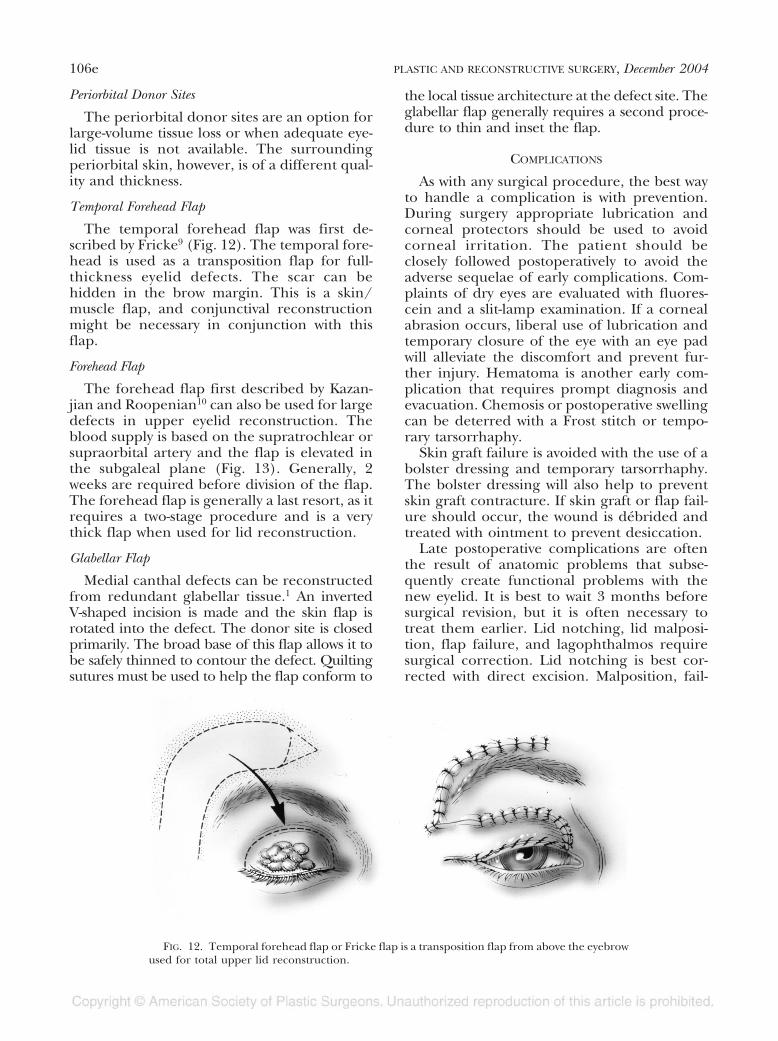
The temporal forehead flap was first de-scribed by Fricke9 (Fig. 12). The temporal fore-head is used as a transposition flap for full-thickness eyelid defects. The scar can behidden in the brow margin. This is a skin/muscle flap, and conjunctival reconstructionmight be necessary in conjunction with thisflap.
Forehead Flap
The forehead flap first described by Kazan-jian and Roopenian10 can also be used for largedefects in upper eyelid reconstruction. Theblood supply is based on the supratrochlear orsupraorbital artery and the flap is elevated inthe subgaleal plane (Fig. 13). Generally, 2weeks are required before division of the flap.The forehead flap is generally a last resort, as itrequires a two-stage procedure and is a verythick flap when used for lid reconstruction.
Glabellar Flap
Medial canthal defects can be reconstructedfrom redundant glabellar tissue.1 An invertedV-shaped incision is made and the skin flap isrotated into the defect. The donor site is closedprimarily. The broad base of this flap allows it tobe safely thinned to contour the defect. Quiltingsutures must be used to help the flap conform to
the local tissue architecture at the defect site. Theglabellar flap generally requires a second proce-dure to thin and inset the flap.
COMPLICATIONS
As with any surgical procedure, the best wayto handle a complication is with prevention.During surgery appropriate lubrication andcorneal protectors should be used to avoidcorneal irritation. The patient should beclosely followed postoperatively to avoid theadverse sequelae of early complications. Com-plaints of dry eyes are evaluated with fluores-cein and a slit-lamp examination. If a cornealabrasion occurs, liberal use of lubrication andtemporary closure of the eye with an eye padwill alleviate the discomfort and prevent fur-ther injury. Hematoma is another early com-plication that requires prompt diagnosis andevacuation. Chemosis or postoperative swellingcan be deterred with a Frost stitch or tempo-rary tarsorrhaphy.
Skin graft failure is avoided with the use of abolster dressing and temporary tarsorrhaphy.The bolster dressing will also help to preventskin graft contracture. If skin graft or flap fail-ure should occur, the wound is débrided andtreated with ointment to prevent desiccation.
Late postoperative complications are oftenthe result of anatomic problems that subse-quently create functional problems with thenew eyelid. It is best to wait 3 months beforesurgical revision, but it is often necessary totreat them earlier. Lid notching, lid malposi-tion, flap failure, and lagophthalmos requiresurgical correction. Lid notching is best cor-rected with direct excision. Malposition, fail-
FIG. 12. Temporal forehead flap or Fricke flap is a transposition flap from above the eyebrowused for total upper lid reconstruction.
106e PLASTIC AND RECONSTRUCTIVE SURGERY, December 2004

ure, or lagophthalmos requires additional tis-sue recruitment for correction. In addition,corneal irritation from trichiasis seen with op-posite lid flaps can be eliminated with excisionor cryotherapy.
CONCLUSIONS
Upper eyelid reconstruction can be chal-lenging to a surgeon, given the specializedanatomy and function of the eyelid. Successfulaesthetic and functional reconstruction beginswith analysis of a complex defect and divisioninto its separate components. Next, an algo-rithmic approach is used to determine the op-timal reconstruction. Finally, meticulous surgi-cal technique and avoidance of complicationswill result in an optimal reconstruction.
Mark A. Codner, M.D.3200 Downwood Circle, Suite 640Atlanta, Ga. [email protected]
REFERENCES
1. Codner, M. A. Reconstruction of the eyelids and orbit.In B. Achauer (Ed.), Plastic Surgery: Indications, Oper-
ations, and Outcomes, Vol. 3. St. Louis: Mosby, 2000. Pp.1425-1454.
2. McCord, C. D. Eyelid Surgery. Philadelphia: Lippincott-Raven, 1995. Pp. 252-269.
3. McCord, C. D., and Wesley, R. Reconstruction of uppereyelid and medial canthus. In C. D. McCord and M.Tannenbaum (Eds.), Oculoplastic Surgery. New York:Raven, 1995. Pp. 99-117.
4. Tenzel, R. R. Reconstruction of the central one half ofan eyelid. Arch. Ophthalmol. 93: 125, 1975.
5. Tenzel, R. R., and Stewart, W. B. Eyelid reconstructionby semi-circular flap technique. Trans. Am. Soc. Oph-thalmol. Otolaryngol. 85: 1164, 1978.
6. Culter, N., and Beard, C. A method for partial and totalupper lid reconstruction. Am. J. Ophthalmol. 39: 1,1955.
7. Smith, B., and Obear, M. F. Bridge flap technique forreconstruction of large upper lid defects. Plast. Recon-str. Surg. 38: 45, 1966.
8. Mustarde, J. C. Eyelid reconstruction. Orbit 1: 33, 1982.9. Fricke, J. C. G. Die Bildung neuer Augenlider (Blepharo-
plastik) nach Zerstorungen und dadurch hervorgebrachtenAuswartswendungen derselben. Hamburg: Perthes andBessler, 1929.
10. Kazanjian, V. H., and Roopenian, A. Median foreheadflaps in the repair of defects of the nose and sur-rounding areas. Trans. Am. Acad. Ophthalmol. Otolar-yngol. 60: 557, 1956.
FIG. 13. A forehead flap can be used for total upper lid reconstruction combined with amucosal graft for lining against the cornea.
Vol. 114, No. 7 / UPPER EYELID RECONSTRUCTION 107e