CloverleafSkull and Thanatophoric Dwarfism · cage, short extremities, and relatively normal length...
9
Archives of Disease in Childhood, 1971, 46, 656. Cloverleaf Skull and Thanatophoric Dwarfism Report of Four Cases, Two in the Same Sibship M. W. PARTINGTON, F. GONZALES-CRUSSI, S. G. KHAKEE, and D. G. WOLLIN From Derbyshire Children's Hospital, Derby, England; and the Departments of Paediatrics, Pathology and Radiology Queen's University, Kingston) Ontario, Canada Partington, M. W., Gonzales-Crussi, F., Khakee, S. G., and Wollin, D. G. (1971). Archives of Disease in Childhood, 46, 656. Cloverleaf skull and thanatophoric dwarfism: report of four cases, two in the same sibship. Four cases of the cloverleaf skull syndrome are reported, 3 from Britain and 1 from Canada in a family of German/Irish descent. All cases had generalized chondrodys- plastic changes and died at or just after birth. It is suggested that a cloverleaf skull is a previously unrecognized feature of thanatophoric dwarfism. Two affected girls from the same sibship are reported for the first time, suggesting an autosomal recessive type of inheritance. A review of the published material indicates that there may be three distinct syndromes in patients with the cloverleaf skull deformity. (1) The cloverleaf skull is associated with thanatophoric dwarfism and death in the perinatal period. (2) There are localized bony lesions of the skeleton outside the skull. (3) The skeleton outside the skull is normal. In the last two syndromes death may occur at birth, but survival into later childhood is the rule. The cloverleaf skull or Kleeblattschadel is a con- genitally enlarged and trilobed skull. There are now over 20 published cases, most of which have been reported from Germany (Holtermiiller and Wiede- mann, 1960; Liebaldt, 1964; Lenz, 1964) or in families of German descent living in North America (Angle, McIntire, and Moore, 1967; Wollin, Binnington, and Partington, 1968; Fein- gold, O'Connor, and Berkman, 1969). Outside the skull a few cases have had normal skeletons, but most have had either localized bony abnormali- ties or generalized chondrodysplastic changes. In 1968 we reported an 8-year-old boy from Canada with a cloverleaf skull and no other bony abnormalities. We also presented limited data on a 3-month-old child from Louisville, Kentucky, whose deformities were probably confined to the skull. In this paper we present 4 further examples of the cloverleaf skull syndrome, 3 from Britain and 1 from Canada, all of which had widespread chondrodysplastic changes as well. Case Histories The Bart's specimen Lenz (1964) recognized that one of the photographs shown to illustrate varieties Received 22 March 1971. of achondroplasia in a long article on pygmies by Gates (1958) was that of a case of the cloverleaf skull syndrome. This photograph was of a specimen in the Pathology Museum of St. Bartholomew's Hospital, London. The origin of the specimen is not known but there is no reason to believe that it was other than Britain. It was taken into the museum some time between 1831 and 1851. Paget described the specimen in the museum's catalogue of 1851 as the 'skeleton of a foetus at about the seventh month showing the effects of hydrocephalus and rachitis'. Bowlby, in 1884, revised the catalogue and described the specimen as the 'skeleton of a foetus, twelve inches in length, showing the condition of the extremities which is typical of cretinism'. In 1929, Shore offered a further interpretation with a description which we quote in full. Achondroplasia with Hydrocephalus 'TE 256. The skeleton of a foetus at about the seventh month, showing shortening and thickening of the long bones of the extremities, together with hydrocephalus. The hydrocephalus is of unusual form owing to premature synostosis of a part of the cranial vault. The frontals are widely separated from one another, but are partially fused with the parietals, while the sagittal and lambdoidal sutures are completely synostosed. In the latter situations the bones are thickened, though light and porous, and this has prevented the skull from expanding in the parietal and occipital regions. Expansion has taken place chiefly in the region of the anterior 656 on June 28, 2020 by guest. Protected by copyright. http://adc.bmj.com/ Arch Dis Child: first published as 10.1136/adc.46.249.656 on 1 October 1971. Downloaded from
Transcript of CloverleafSkull and Thanatophoric Dwarfism · cage, short extremities, and relatively normal length...

Archives of Disease in Childhood, 1971, 46, 656.
Cloverleaf Skull and Thanatophoric DwarfismReport of Four Cases, Two in the Same Sibship
M. W. PARTINGTON, F. GONZALES-CRUSSI, S. G. KHAKEE, and D. G. WOLLINFrom Derbyshire Children's Hospital, Derby, England; and the Departments of Paediatrics, Pathology and Radiology
Queen's University, Kingston) Ontario, Canada
Partington, M. W., Gonzales-Crussi, F., Khakee, S. G., and Wollin, D. G.(1971). Archives of Disease in Childhood, 46, 656. Cloverleaf skull andthanatophoric dwarfism: report of four cases, two in the same sibship.Four cases of the cloverleaf skull syndrome are reported, 3 from Britain and 1 fromCanada in a family of German/Irish descent. All cases had generalized chondrodys-plastic changes and died at or just after birth. It is suggested that a cloverleaf skullis a previously unrecognized feature of thanatophoric dwarfism. Two affected girlsfrom the same sibship are reported for the first time, suggesting an autosomalrecessive type of inheritance.A review of the published material indicates that there may be three distinct
syndromes in patients with the cloverleaf skull deformity. (1) The cloverleaf skull isassociated with thanatophoric dwarfism and death in the perinatal period. (2) Thereare localized bony lesions of the skeleton outside the skull. (3) The skeleton outsidethe skull is normal. In the last two syndromes death may occur at birth, but survivalinto later childhood is the rule.
The cloverleaf skull or Kleeblattschadel is a con-genitally enlarged and trilobed skull. There arenow over 20 published cases, most ofwhich have beenreported from Germany (Holtermiiller and Wiede-mann, 1960; Liebaldt, 1964; Lenz, 1964) or infamilies of German descent living in NorthAmerica (Angle, McIntire, and Moore, 1967;Wollin, Binnington, and Partington, 1968; Fein-gold, O'Connor, and Berkman, 1969). Outsidethe skull a few cases have had normal skeletons,but most have had either localized bony abnormali-ties or generalized chondrodysplastic changes.
In 1968 we reported an 8-year-old boy fromCanada with a cloverleaf skull and no other bonyabnormalities. We also presented limited data ona 3-month-old child from Louisville, Kentucky,whose deformities were probably confined to theskull. In this paper we present 4 further examplesof the cloverleaf skull syndrome, 3 from Britain and1 from Canada, all of which had widespreadchondrodysplastic changes as well.
Case HistoriesThe Bart's specimen Lenz (1964) recognized
that one of the photographs shown to illustrate varieties
Received 22 March 1971.
of achondroplasia in a long article on pygmies by Gates(1958) was that of a case of the cloverleaf skull syndrome.This photograph was of a specimen in the PathologyMuseum of St. Bartholomew's Hospital, London. Theorigin of the specimen is not known but there is noreason to believe that it was other than Britain. It wastaken into the museum some time between 1831 and1851. Paget described the specimen in the museum'scatalogue of 1851 as the 'skeleton of a foetus at about theseventh month showing the effects of hydrocephalusand rachitis'. Bowlby, in 1884, revised the catalogueand described the specimen as the 'skeleton of a foetus,twelve inches in length, showing the condition of theextremities which is typical of cretinism'. In 1929,Shore offered a further interpretation with a descriptionwhich we quote in full.
Achondroplasia with Hydrocephalus'TE 256. The skeleton of a foetus at about the
seventh month, showing shortening and thickeningof the long bones of the extremities, together withhydrocephalus. The hydrocephalus is of unusualform owing to premature synostosis of a part ofthe cranial vault. The frontals are widely separatedfrom one another, but are partially fused with theparietals, while the sagittal and lambdoidal suturesare completely synostosed. In the latter situationsthe bones are thickened, though light and porous,and this has prevented the skull from expandingin the parietal and occipital regions. Expansionhas taken place chiefly in the region of the anterior
656
on June 28, 2020 by guest. Protected by copyright.
http://adc.bmj.com
/A
rch Dis C
hild: first published as 10.1136/adc.46.249.656 on 1 October 1971. D
ownloaded from

Cloverleaf Skull and Thanatophoric Dwarfismfontanelle and between the frontal bones, and alsolaterally in the parieto-squamosal region, where nosynostosis has occurred. The skull has thusassumed a broadly pyriform shape and measures4 inches from base to vault. The parietal bone isthrown backwards into a vertical position. Theposterior fontanelle is unclosed and its edges areeverted, roughened and prominent. In thelateral regions of the skull the bones are set apartand bowed by the pressure of the hydrocephalicfluid; thus the squamous portion of the temporallooks vertically downward, as does the externalauditory meatus. The orbits are much encroachedupon by the bulging of the skull, which affects theirupper and outer walls. There is no clear evidenceof shortening or premature synostosis of the basiscranii.
'The total length of the skeleton is 12 inches, butsome of this is due to the upward expansion of theskull. The vertebral column is natural; the ribsare thickened and unnaturally short. Both theupper and lower extremities are somewhat stunted.The arms measure but 31 inches from the shoulderto the fingertips and do not reach beyond the
FIG. 1.-Photograph of the Bart's specimen. The skullis large in comparison to the remainder of the skeleton andis typically 'cloverleaf'. Note the short thick bones withcurved radii andfibulae, normal clavicles, narrow thoraciccage, short extremities, and relatively normal length of
spine.
anterior superior spines of the ilia. The lowerlimbs, from the head of the femur to the heel, showa similar measurement. The total length of thespine is 4j inches. The diaphyses of all the longbones of the limbs are well ossified, thickened andheavy, but only of some two-thirds the lengthnatural to a seven month foetus. Some, such asthe femora and fibulae, are unnaturally curved, aswell as thickened. The epiphyses, owing to themode of preparation, are so shrunken that theircondition cannot be determined. There is nobeading of the ribs. The clavicle alone of thelimb-bones is natural in length and thickness.
'It is probable that the condition of the longbones of the limbs is due to a slight degree ofachondroplasia.'
Photographs and x-rays of the specimen are shown inFig. 1 to 4.
FIG. 2.-X-rays of the Bart's specimen. Visible ossifica-tion of epiphyses at the ends of all long bones is absent.Metacarpals, metatarsals, and associated phalanges arewell ossified. Small spurs ofperiosteal bone project at thelateral aspects of the proximal end of the humeri and distalends of thefemora. The proximal segments of the extremi-ties are proportionately shorter than the distal segments.The iliac wings are small, the roof of each acetabulum isflat. The length of the spine is relatively normal comparedto the short upper and lower exremities. The intervertebraldisc spaces are correspondingly increased. The interpedi-cular distance in the lumbar vertebrae gradually narrows
from Li to L5.
657
on June 28, 2020 by guest. Protected by copyright.
http://adc.bmj.com
/A
rch Dis C
hild: first published as 10.1136/adc.46.249.656 on 1 October 1971. D
ownloaded from

Partington, Gonzales-Crussi, Khakee, and Wollin
FIG. 3.-Photograph of the skull and facial bones of theBart's specimen. The trilobed contour of the cranialvault is the outstanding feature; the superior lobe is thelargest. Ossification of the vault is incomplete. Theorbits are shallow, particularly at the superior orbitalmargins. The maxillae and mandible are small comparedto the size of the nasal bones. The anterolateralfontanelle(pterion) extends forward to the lateral margin of each
orbit.
The Derby cases. These two cases came to lightthrough Dr. Whitelaw and Dr. Whitaker who diagnosedthem in retrospect when shown x-rays of our cases of1968.Both patients were girls from the same sibship. The
parents were unrelated. The first 3 children in thefamily (a boy born in 1957 and two girls born in 1960and 1961 respectively) were normal and there had beenno miscarriages. The mother was aged 33 and 35years, respectively, when her abnormal children wereborn; the father was a year younger. There were noabnormalities discovered in the rest of the family exceptfor a cousin of the mother who had died at the age of 15months with mental retardation and an enlarged head;no further information could be obtained about thischild. Both sides of the family were English and hadlived in England for at least 4 generations; the mother'smaiden name was Wolfe which is often regarded as ofGerman origin.
Case 1 (date of birth, 11 August 1965). During thepregnancy the mother was well until the 31st weekwhen she went into labour. She was thought to havehydramnios; no fetal parts were felt and no fetal heart
FIG. 4.-X-ray of the skull of the Bart's specimen:anteroposterior view. Two struts of thickened bone, oneon each side, appear to compress the vault and producethe trilobed deformity. A large irregular defect is presentin the posterior aspect of the vault at the junction of thethree lobes posteriorly; it is believed to represent the
posterior fontanelle (lambda).
was heard. About 3700 ml amniotic fluid were releasedafter artificial rupture of the membranes. An x-rayshowed the abnormal fetus. The first stage of labourlasted 27 hours, the second lasted 10 minutes, and thebaby was born spontaneously as a vertex delivery.The placenta was normal and weighed 568 g. Thebaby was a female child weighing 2- 02 kg who died atthe age of 5 minutes. No clinical description wasrecorded beyond the statement that the child had'hydrocephalus and achondroplasia'. X-rays weretaken after death (Fig. 5, Case 1).
Case 2 (date of birth, 26 February 1969). In thispregnancy the mother was well until the 38th weekwhen she developed swelling of the ankles and slighthypertension. She was admitted to hospital andx-ray showed the abnormal fetus presenting as a breech.Labour started spontaneously 5 days later. The firststage lasted 25 hours and the fetal heart was heard.During the second stage an episiotomy was carried outand the legs and trunk were delivered. The after-
658
on June 28, 2020 by guest. Protected by copyright.
http://adc.bmj.com
/A
rch Dis C
hild: first published as 10.1136/adc.46.249.656 on 1 October 1971. D
ownloaded from

Cloverleaf Skull and Thanatophoric Dwarfism 659_.4
FIG. 5.-Cases 1, 2, and 3. Anteroposterior and lateral x-rays showing the general topography and skeletal changes.Note the narrow airless thoraces, short curved limbs, marked platyspondyly with greatly increased intervertebral discspaces, micromelia affecting the proximal limb segments more than the distal, broad iliac wings, flat acetabula,
and broad pelves. The trilobed contour of the cranial vault in each patient is evident.
coming head was delayed at the pelvic brim. It wasnot possible to drain the head by needle because of thehardness of the bones so a craniotomy and cranioclastywere performed. The baby, a girl weighing 3a1 kg,was dead at birth. The placenta was normal macro-scopically and microscopically and it weighed 657 g.The only clinical description was 'hydrocephalus withachondroplasia'. Further x-rays were taken afterdeath (Fig. 5, Case 2).
The Kingston caseCase 3 (date of birth, 1 August 1969). The mother
was aged 37 years and the father 46 years at the time thechild was born and they were unrelated. The mother'sfamily had all originated from Germany, emigrating tothe U.S.A. three generations previously; the mother'sparents still spoke German as their native tongue.The father's family had emigrated to Canada fromNorthern Ireland three generations previously.
on June 28, 2020 by guest. Protected by copyright.
http://adc.bmj.com
/A
rch Dis C
hild: first published as 10.1136/adc.46.249.656 on 1 October 1971. D
ownloaded from
Partington, Gonzales-Crussi, Khakee, and WollinIn 1958 the mother had had an abortion at 12 weeks.
In 1959, in the 42nd week of her second pregnancy,the baby died in utero; labour was induced and a still-born female child weighing 2320 g was delivered as abreech; necropsy showed a high ventricular septaldefect but no other abnormalities. In 1960 she had anabortion at the tenth week of pregnancy. In 1961, at42 weeks of pregnancy, she developed severe pre-eclamptic toxaemia; labour was prolonged but a normalmale child weighing 3160 g was delivered who is stillalive and well.
In 1965 her fifth pregnancy was terminated bycaesarean section following a failed induction of labourat 42 weeks. Antenatal x-rays had shown an abnormalfetus with hydrocephalus. A male child weighing4360 g was delivered. The child had hydrocephalus(head circumference 46 cm), a cleft of the posterior halfof the palate and bilateral club feet. The Apgar scorewas 6 one minute after birth: the child's conditiondeteriorated and he died at 16 hours of age. Pre- andpostmortem x-rays of the skull showed an enlargedvault with fairly good ossification of the bones. Atnecropsy the brain was enlarged with an excess of CSF(700 ml). There was polymicrogyria over the frontalcortex on each side; the medial surface of the left hemi-sphere was absent and there was a small porencephaliccyst in the left occipital region. In the lower part of theaqueduct there was an area of stenosis 3 mm in length.The aqueduct was broken up by gliosis into two micro-scopic ductules: in addition there were free ependymalcells and some swelling of the axons. The cerebellumwas normal though small. The lungs showed obviouscongestion and oedema. Sections of the long bonesshowed normal enchondral ossification.
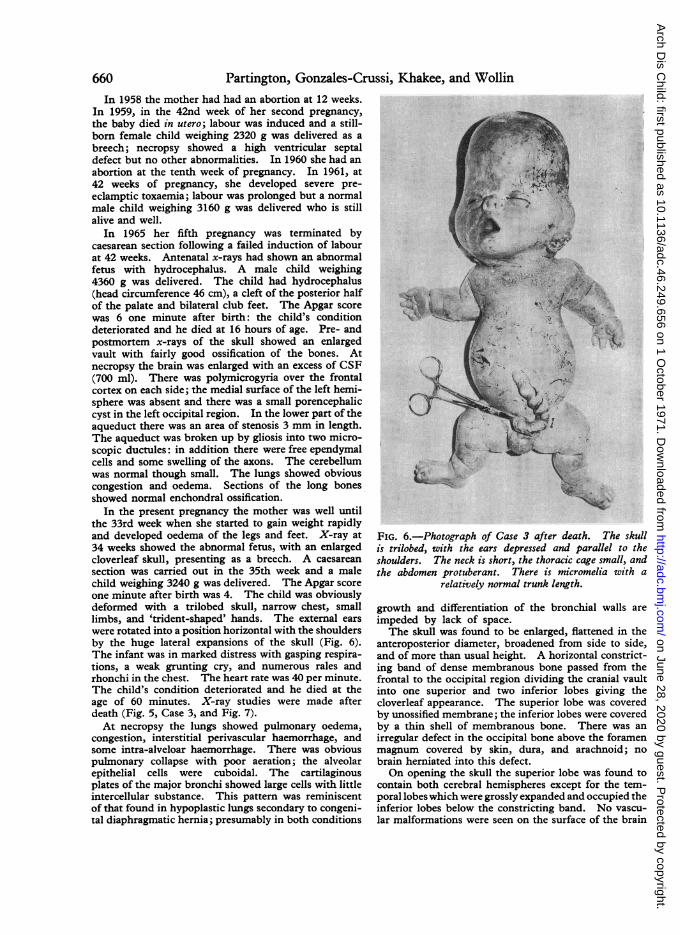
In the present pregnancy the mother was well untilthe 33rd week when she started to gain weight rapidlyand developed oedema of the legs and feet. X-ray at34 weeks showed the abnormal fetus, with an enlargedcloverleaf skull, presenting as a breech. A caesareansection was carried out in the 35th week and a malechild weighing 3240 g was delivered. The Apgar scoreone minute after birth was 4. The child was obviouslydeformed with a trilobed skull, narrow chest, smalllimbs, and 'trident-shaped' hands. The external earswere rotated into a position horizontal with the shouldersby the huge lateral expansions of the skull (Fig. 6).The infant was in marked distress with gasping respira-tions, a weak grunting cry, and numerous rales andrhonchi in the chest. The heart rate was 40 per minute.The child's condition deteriorated and he died at theage of 60 minutes. X-ray studies were made afterdeath (Fig. 5, Case 3, and Fig. 7).At necropsy the lungs showed pulmonary oedema,
congestion, interstitial perivascular haemorrhage, andsome intra-alveloar haemorrhage. There was obviouspulmonary collapse with poor aeration; the alveolarepithelial cells were cuboidal. The cartilaginousplates of the major bronchi showed large cells with littleintercellular substance. This pattern was reminiscentof that found in hypoplastic lungs secondary to congeni-tal diaphragmatic hernia; presumably in both conditions
FIG. 6.-Photograph of Case 3 after death. The skullis trilobed, with the ears depressed and parallel to theshoulders. The neck is short, the thoracic cage small, andthe abdomen protuberant. There is micromelia with a
relatively normal trunk length.
growth and differentiation of the bronchial walls areimpeded by lack of space.The skull was found to be enlarged, flattened in the
anteroposterior diameter, broadened from side to side,and of more than usual height. A horizontal constrict-ing band of dense membranous bone passed from thefrontal to the occipital region dividing the cranial vaultinto one superior and two inferior lobes giving thecloverleaf appearance. The superior lobe was coveredby unossified membrane; the inferior lobes were coveredby a thin shell of membranous bone. There was anirregular defect in the occipital bone above the foramenmagnum covered by skin, dura, and arachnoid; nobrain herniated into this defect.On opening the skull the superior lobe was found to
contain both cerebral hemispheres except for the tem-poral lobes which were grossly expanded and occupied theinferior lobes below the constricting band. No vascu-lar malformations were seen on the surface of the brain
660
on June 28, 2020 by guest. Protected by copyright.
http://adc.bmj.com
/A
rch Dis C
hild: first published as 10.1136/adc.46.249.656 on 1 October 1971. D
ownloaded from

Cloverleaf Skull and Thanatophoric Dwarfismor in its membranes. In attempting to remove thebrain the thin cerebral mantle ruptured and a largeamount of cerebrospinal fluid escaped. The brain wassoft and friable and did not retain its original contouron fixation.The base of the skull showed the gross expansion
of the middle cranial fossae which displaced the petrousportions of the temporal bones posteriorly and parallelto the margins of the foramen magnum. The posteriorfossae were small, only 5 to 6 mm in depth, due both tothe displaced petrous temporal bones and markedbasilar invagination of the cervical spine into the marginof the foramen magnum. The foramen magnum wassmall but not deformed.
After fixation the brain weighed 562 g. There washypoplasia of the cerebellum and the brainstem andfailure of differentiations of the tectal plate. Thecerebellar hemispheres were separate and there was noevidence of development of the vermis. The lateraland third ventricles were grossly dilated. The aqueductwas enlarged close to its opening into the third ventricle,but more posteriorly it was of normal calibre. Micro-scopically there was some focal periependymal gliosiswith granular calcific deposits, but it was lined bynormal ependymal cells. The fourth ventricle was nor-mal in size. The medulla oblongata was displacedcaudally into the upper cervical spine by the basilarinvagination of the skull.
FIG. 7.-Case 3. Ventriculogram performed immediately Sections taken from various regions of the skeletonafter death: right lateral view. The superior cranial showed abnormal enchondral ossification particularlyexpansion contains much of the cerebral hemispheres in the long bones of the extremities. The zone ofoccupied by grossly dilated lateral ventricles. The provisional cartilage showed inadequate orientation ofinferior expansion contains the left temporal lobe with a the cartilage cell columns which were either distortedgrossly dilated left temporal horn. The right lateral or poorly formed. The vertebral pedicles, though less
ventricle was even more dilated. severely affected, were the seat of identical changes.
FIG. 8.-Case 3. Radiographic details of the extremities show characteristic concave irregular metaphyses withperiosteal overgrowth of bone at the periphery. The long bones of the hand and foot are very short and broad.
661
on June 28, 2020 by guest. Protected by copyright.
http://adc.bmj.com
/A
rch Dis C
hild: first published as 10.1136/adc.46.249.656 on 1 October 1971. D
ownloaded from

Partington, Gonzales-Crussi, Khakee, and WollinThe spinal canal was distorted into a typical triangularshape.
DiscussionThe radiological appearances of the skulls of all
our cases correspond with those reported by othersin patients with the cloverleaf skull syndrome.Outside the skull, all four cases had micromelicdwarfism with short bowed tubular bones, andflared metaphyses, obvious flattening of the verte-bral bodies, and severe narrowing of the chest;death occurred at or soon after birth. The histo-logical findings in one case showed improperendochondral ossification. Formerly these caseswould have been classified as fetal achondroplasiaor chondrodystrophy, and this, with hydrocephalus,was the diagnosis in the Bart's specimen (Shore,1929; Gates, 1958), the two Derby cases, and someothers reported in the literature (see Table).However, it is becoming increasingly clear that thename achondroplasia has been loosely applied to a
variety of unrelated disorders for which strictdiagnostic criteria are only now emerging (Carter,1969; Rimoin et al., 1970). Recent evidence
suggests that even the generally accepted histo-logical appearances of 'classical' achondroplasiamay be incorrect because they were derived frompatients with other diseases (Rimoin'et al., 1970;Ponsetti, 1970).
In 1967, Maroteaux, Lamy, and Robert arguedon clinical, radiological, and genetic grounds thatthe conditicn known as fetal chondrodystrophy or
congenital achondroplasia was distinct from clas-
sical achondroplasia. In the former conditionthey emphasized the severity of the bony changes,similar to but more obvious than those of classicalachondroplasia, the flattening of the vertebrae, theextreme narrowing of the thorax, and death at or
soon after birth. Maroteaux et al. suggested thename thanatophoric dwarfism. Subsequent casereports from other authors (Giedion, 1968; Langeret al., 1969; Huguenin et al., 1969; Keats, Ridder-vold, and Michaelis, 1970; Kaufman et al., 1970)strongly support this view.Except for the cloverleaf skull, the findings in
our own cases meet all the criteria of thanatophoricdwarfism. Maroteaux has seen the radiographs of
BLESome Characteristics of Patients with 3 Types of Cloverleaf Skull
Type I Type II Type IIIwith Chondro- with Localized with Normal Skeletons
dysplasia Skeletal Lesions Outside Skull
Authors and cases Holtermuller and Wiedemann Holtermuller and Wiedemann Holtermuller and Wiedemann(1960; Cases 1-6, 10, 11); (1960: Case 13); Liebaldt (1960: Cases 7 and 9);Feingold et al. (1969: Case (1964: Case 2); Comings Angle et al. (1967: Case 2);2) present series (1965); Angle et al. (1967: Wollin et al. (1968: Case 1)
Case 1); Feingold et al.(1969: Case 1)
Total number 13 5 4Male 3 1 2Female 7 2 2Sex not known 3 2Birthweight (g) 2020, 2400, 3100, 3246, 3450 - 1810, 2580Pregnancy Hydramnios reported twice -
Maternal age (yr) 24, 26, 28, 34 (mean of two 'Early 20s', 27, 29, 33 24, 28pregnancies), 37, 50
Paternal age (yr) 33 (mean of 2 offspring), 46 'Early 20s', 32, 34 ,38 24, 26Sibs Affected pair in 1 sibship; 1 sib Case was 1 of dizygotic twins
with hydrocephalus andaqueduct stenosis
Racial background 7 reported from Germany; 3 1 from Germany; 2 from 2 from Germany; 2 fromfrom Britain; 1 from Hol- North America of German North America of Germanland; 1 from Canada of descent; 1 from Switzer- descentGerman/Irish descent; 1 land; 1 'Caucasian'from U.S.A.
Genetics ? Autosomal recessive Not known Not knownAge at death or at time of reporting 7 stillborn; 1 died in first hr, 1 stillborn; 1 died at 54 dy; 1 stillborn; single cases alive
1 on first dy, and 1 at 16 wk single cases still alive at 4k at 6 mth, 16 mth, and 8 yrmth, 2 yr and 14 yr
Bony anomalies outside skull Severe generalized chondro- Bony ankylosis of elbows; Nildysplasia or thanatophoric subluxation of radial heads;dwarfism subluxation ofhips; thoracic
spina bifida; cervical spinabifida; split xiphoid; fixedflexed fingers; webbing ofthird and fourth toes
662
on June 28, 2020 by guest. Protected by copyright.
http://adc.bmj.com
/A
rch Dis C
hild: first published as 10.1136/adc.46.249.656 on 1 October 1971. D
ownloaded from

Cloverleaf Skull and Thanatophoric Dwarfism 663the Kingston case and discussed them personallywith one of us (F.G-C): he agrees that, outsidethe skull, the appearances are those of thanatophoricdwarfism.
It is of great interest that a male sib of theKingston case had died soon after birth fromhydrocephalus apparently due to aqueduct stenosis.At least one form of this disorder is inherited asan X-linked recessive (Edwards, Norman, andRoberts, 1961). One might have postulated that,in the Kingston case, the cloverleaf skull was dueto aqueduct stenosis occurring in a child withthanatophoric dwarfism. However, no trace ofaqueduct stenosis was found at necropsy. Further-more, it seems unlikely that all our 4 cases and the 9others published with the same combination oflesions represent the chance association of twodifferent rare disorders but rather that they are,in some way, related. We think that a cloverleafskull may well be a previously unrecognized featureof thanatophoric dwarfism.
Burkhardt (1970) has recently reviewed thecloverleaf skull syndrome and he concludes thatthe cloverleaf deformity is due to morphogeneticforces superimposed on a basic chondrodysplasticprocess. He believes that the main factor isimproper endochondral ossification leading toforeshortening of the base of the skull. The fronto-parietal growth of the fetal brain is hampered and itenlarges upward. The lateral expansions of thebrain and skull are explained by gross increasesin the volume of the temporal lobes due to obstruc-tion of CSF circulation in the lateral ventricles.Early intracranial hypertension may also lead toincreased vascularization of the diploe accountingfor the haemangiomatous appearance of variousskull bones found in some cases. This speculationis quite plausible, but it fails to account for thosecases with cloverleaf skulls but without generalizedchondrodysplastic skeletal changes. We are equallyunable to explain this discrepancy short of postulat-ing a localized chondrodysplastic lesion in the skullin this latter group of patients.On reviewing our cases and 21 others, we
are led to believe that there are at least 2, andpossibly 3, distinct syndromes in patients withthe cloverleaf skull deformity (see Table). Inthe first syndrome, Type I, the cloverleaf skullis associated with thanatophoric dwarfism. Inthe second syndrome, Type II, there are bonylesions outside the skull, but these are localized,not generalized; death may occur at or beforebirth but usually the child survives the newbornperiod and may live for several years. In the thirdsyndrome, Type III, the prognosis is similar to
Type II but the skeleton outside the skull is normal.The distinction between Type II and Type IIIseems less justifiable on present evidence thanbetween Type I and Type II.Enough information was available to assign 22
of the 27 cases reviewed to one or another Typewith a fair degree of certainty: the 5 unassignedcases probably belong to Type II or III. No casewas found with both generalized chondrodysplasticchanges and a localized skeletal lesion. Type Iseemed the most homogenous group with onlyone anomalous case. This was a boy reported byVrolik in 1849 (Case 1 in Table I of Holtermiullerand Wiedemann, 1960), who, to judge from theauthor's description and the artist's drawing,undoubtedly had both a cloverleaf skull and thana-tophoric dwarfism but who lived to the age of16 weeks.Some characteristics of these three Types of
cloverleaf skull syndromes are shown in the Table.The amount and type of data on each case vary and,in sum, are not yet sufficient to make meaningfulinferences about aetiology.The occurrence of affected sibs in the Type I
syndrome is reported here for the first time andraises the possibility of an autosomal recessivetype of inheritance. If our cases are accepted asexamples of thanatophoric dwarfism, then thissuggests that thanatophoric dwarfism may besimilarly inherited. The preponderance of casesof all Types reported from Germany or fromfamilies of German descent supports a geneticrather than a teratogenic aetiology; of course theracial distribution may also be biased due to theway cases have come to light.
We wish to thank Dr. A. G. Stansfeld of the Depart-ment of Pathology, Mr. J. L. Thornton of the MedicalLibrary and the Department of Medical Illustration,all of St. Bartholomew's Hospital, London, for theirhelp in the study and presentation of the 'Bart's speci-men'; Dr. R. A. J. Whitelaw and Dr. R. Whitaker ofDerby for permission to publish their cases, Dr. S. W.George of Kingston for permission to publish hiscase, and Dr. C. S. Houston of the University ofSaskatchewan for comments.
REFERENCES
Angle, C. R., McIntire, M. S., and Moore, R. C. (1967). Clover-leaf skull: Kleeblattschadel-deformity syndrome. AmericanJournal of Diseases of Children, 114, 198.
Bowlby, A. A. (1884). A Descriptive Catalogue of the Anatomicaland Pathological Museum of St. Bartholomew's Hospital, vol. II,p. 17. Churchill, London.
Burkhardt, L. (1970). Pathologische Anatomie des Schadels, p. 115.Springer, Berlin.
Carter, C. 0. (1969). Diastrophic dwarfism. DevelopmentalMedicine and Child Neurology, 11, 247.
Comings, D. E. (1965). The Kleeblattschadel syndrome-agrotesque form of hydrocephalus. Journal of Pediatrics, 67,126.
7
on June 28, 2020 by guest. Protected by copyright.
http://adc.bmj.com
/A
rch Dis C
hild: first published as 10.1136/adc.46.249.656 on 1 October 1971. D
ownloaded from

664 Partington, Gonzales-Crussi, Khakee, and WollinEdwards, J. H., Norman, R. M., and Roberts, J. M. (1961). Sex-
linked hydrocephalus. Archives ofDisease in Childhood, 36,481.Feingold, M., O'Connor, J. F., and Berkman, M. (1969). Kleeblatt-
schadel syndrome. American J7ournal of Diseases of Children,118, 589.
Gates, R. R. (1958). The African pygmies. Acta GeneticaeMedicae et Gemellologiae, 7, 159.
Giedion, A. (1968). Thanatophoric dwarfism. Helvetica Paedia-trica Acta, 23, 175.
Holtermfiller, K., and Wiedemann, H. R. (1960). Kleeblattschadel-Syndrom. Medizinische Monatsschrift, 14, 439.
Huguenin, M., Godard, C., Ferrier, P. E., and Bamatter, F. (1969).Two different mutations within the same sibship: thanatophoricdwarfism and Ullrich-Feichtiger syndrome. Helvetica Paedia-trica Acta, 24, 239.
Kaufman, R. L., Rimoin, D. L., McAlister, W. H., and Kissane,J. M. (1970). Thanatophoric dwarfism. American Journalof Diseases of Children, 120, 53.
Keats, T. E., Riddervold, H. O., and Michaelis, L. L. (1970).Thanatophoric dwarfism. American Journal of Roentgenology,Radium Therapy, and Nuclear Medicine, 108, 473.
Langer, L. O., Spranger, J. W., Greinacher, I., and Herdman, R. C.(1969). Thanatophoric dwarfism. Radiology, 92, 285.
Lenz, W. (1964). Anomalien des Wachstums und der Korperform.In Humangenetik, vol. 2, p. 79. Ed. by P. E. Becker. Thieme,Stuttgart.
Liebaldt, G. (1964). Das 'Kleeblatt'-Schadel-Syndrom, alsBeitrag sur formalen Genese der Entwicklungsst6rungen des
Schadeldaches. Ergebnisse der allgemeinen Pathologie undpathologischen Anatomie, 45, 23.
Maroteaux, P., Lamy, M., and Robert, J. M. (1967). Le nanismethanatophore. Presse Medicale, 75, 2519.
Paget, J. (1851). A Descriptive Catalogue of the Anatomical Museumof St. Bartholomew's Hospital, vol. II, p. 210. Churchill,London.
Ponsetti, I. V. (1970). Skeletal growth in achondroplasia. Journalof Bone and joint Surgery, 52-A, 701.
Rimoin, D. L., Hughes, G. N., Kaufman, R. L., Rosenthal, R. E.,McAlister, W. H., and Silberberg, R. (1970). Endochondralossification in achondroplastic dwarfism. New Englandjournalof Medicine, 283, 728.
Shore, T. H. G. (1929). A Descriptive Catalogue of the PathologicalMuseum of St. Bartholomew's Hospital Medical College, vol. II,p. 1632. Adlard, London.
Vrolik, W. (1849). Tabulae ad illustrandem Embryogenesin Hominiset Mammalium, tam Naturalem quam Abnormem, Fig. 35, 36.G.M.P. Londonck, Amsterdam.
Wollin, D. G., Binnington, V. I., and Partington, M. W. (1968).Cloverleaf skull. Journal of the Canadian Association ofRadiologists, 19, 148.
Correspondence to Dr. M. W. Partington, Depart-ment of Paediatrics, Queen's University, Kingston,Ontario, Canada.
on June 28, 2020 by guest. Protected by copyright.
http://adc.bmj.com
/A
rch Dis C
hild: first published as 10.1136/adc.46.249.656 on 1 October 1971. D
ownloaded from