Cloning, Purification, and Biochemical Characterization of ...

Electronic Journal of Biotechnology 41 (2019) 48–55
⁎
Pe
Contents lists available at ScienceDirect
Electronic Journal of Biotechnology
Research article
Cloning and molecular characterization of a putative habanero pepperSERK1 cDNA expressed during somatic and zygotic embryogenesis
Doribet Jiménez-Guillen a, Daniel Pérez-Pascual a, Ramón Souza-Perera b, José Juan Zúñiga Aguilar a,⁎a Instituto Tecnológico Superior de los Ríos, Km 3 carretera Balancán-Villahermosa, Balancán 86930, Tabasco, Mexicob Unidad de Bioquímica y Biología Molecular de Plantas, Centro de Investigación Científica de Yucatán, Calle 43 No. 130, Col. Chuburná de Hidalgo, Mérida 97200, Yucatán, Mexico
Corresponding author.E-mail address: [email protected] (J.J.Z. Aguilar).
er review under responsibility of Pontificia Universidad Ca
https://doi.org/10.1016/j.ejbt.2019.07.0060717-3458/© 2019 Pontificia Universidad Católica de Valp(http://creativecommons.org/licenses/by-nc-nd/4.0/).
a b s t r a c t
a r t i c l e i n f oArticle history:Received 10 March 2019Accepted 31 July 2019Available online 6 August 2019
Background: Plant gene homologs that control cell differentiation can be used as biotechnological tools to studythe in vitro cell proliferation competence of tissue culture-recalcitrant species such as peppers. It has beendemonstrated that SERK1 homologs enhance embryogenic competence when overexpressed in transformedtissues; therefore, cloning of a pepper SERK1 homolog was performed to further evaluate its biotechnologicalpotential.Results: A Capsicum chinense SERK full-length cDNA (CchSERK1) was cloned and characterized at the molecularlevel. Its deduced amino acid sequence exhibits high identity with sequences annotated as SERK1 andpredicted-SERK2 homologs in the genomes of the Capsicum annuum CM-334 and Zunla-1 varieties,respectively, and with SERK1 homologs from members of the Solanaceae family. Transcription of CchSERK1 inplant tissues, measured by quantitative RT-PCR, was higher in stems, flowers, and roots but lower in leavesand floral primordia. During seed development, CchSERK1 was transcribed in all zygotic stages, with higherexpression at 14 days post anthesis. During somatic embryogenesis, CchSERK1 was transcribed at alldifferentiation stages, with a high increment in the heart stage and lower levels at the torpedo/cotyledonalstages.Conclusion: DNA sequence alignments and gene expression patterns suggest that CchSERK1 is the C. chinenseSERK1 homolog. Significant levels of CchSERK1 transcripts were found in tissues with cell differentiationactivities such as vascular axes and during the development of zygotic and somatic embryos. These resultssuggest that CchSERK1 might have regulatory functions in cell differentiation and could be used as abiotechnological tool to study the recalcitrance of peppers to proliferate in vitro.How to cite: Jiménez-Guillen D, Pérez-Pascual D, Souza-Perera R, et al. Cloning andmolecular characterization ofa putative Habanero pepper SERK1 cDNA expressed during somatic and zygotic embryogenesis. Electron JBiotechnol 2019;41. https://doi.org/10.1016/j.ejbt.2019.07.006.
© 2019 Pontificia Universidad Católica de Valparaíso. Production and hosting by Elsevier B.V. All rights reserved.This is an open access article under the CC BY-NC-ND license (http://creativecommons.org/licenses/by-nc-nd/4.0/).
Keywords:ArabidopsisBiotechnological toolsCapsicum annuumCapsicum chinenseCell differentiationGene expressionin vitro cell proliferationPeppersRecalcitrant speciesSolanaceaeSomatic embryogenesis
1. Introduction
According to the Product Complexity Index [1], peppers are amongthe world's most traded products mainly because of their highpungency levels, with habanero pepper (Capsicum chinense Jacq.)being one of the most pungent varieties [2]. Peppers belong to a genusin which all species are recalcitrant to in vitro proliferation throughtissue culture techniques. Limitations in regenerating whole plantsfrom a cell, tissue, and organ explants have delayed thebiotechnological breeding of pepper species [3]. Most pepperregeneration and genetic transformation protocols are not
tólica de Valparaíso.
araíso. Production and hosting by Els
reproducible or cannot be applied to other species of the genus or arebased in the in vitro regeneration of shoots [4,5]. On the other hand,enhancement of the regeneration competence in peppers through theembryogenic pathway has been achieved by the overexpression ofmaster genes that regulate cell differentiation. Transient induction ofthe Brassica napus BABY BOOM gene in pepper transgenic plantsproduced a large number of somatic embryos that could be convertedreadily to seedlings [6]. In addition, the ectopic induction of anArabidopsis thaliana gene encoding a Wuschel transcription factor inthe transgenic hypocotyl explants of pepper promoted thedevelopment of embryo globular structures [7]. Other regulatorygenes such as the Somatic Embryogenesis Receptor-like Kinase 1 fromCoffea canephora (CcSERK1) lead to an increase in the somaticembryogenesis competence when overexpressed ectopically and alsoinduced the transcription of developmental regulatory genes [8].
evier B.V. All rights reserved. This is an open access article under the CC BY-NC-ND license

49D. Jiménez-Guillen et al. / Electronic Journal of Biotechnology 41 (2019) 48–55
SERK1 homologs are broadly present in monocotyledonous anddicotyledonous plants as members of a multigene family (SERK1,SERK2, etc.) with different functions [9]; however, all the SERK1homologs have in common their accurate expression patterns intissues with high cell differentiation activity. For example, AtSERK1 isexpressed during megasporogenesis and in all cells of the embryonicsac until the fertilization [10]. Further, during somatic embryogenesis,SERK1 homologs analyzed are expressed in embryogenic cells andembryo structures but not in non-embryogenic tissues [10,11,12].SERK1 homologs are consistently expressed in somatic cells displayingembryogenic competence and later during early stages of zygotic andsomatic embryogenesis [13]; these expression patterns have led to theproposal of SERK1 as the best molecular marker of embryogeniccompetence [14].
As the transient overexpression of the C. canephora SERK1 genehomolog during somatic embryogenesis promoted the differentialexpression of key cell differentiation regulatory genes, including LEC1,BBM, and WUS [8], it is possible that the SERK1 product functions as amaster coregulator of cell differentiation that could be used to studycell proliferation in vitro. Despite the large number of SERK1 homologsisolated to date, no SERK1 homologs from pepper have been cloned.Recently, the publication of the DNA genome sequences from threeCapsicum annuum varieties has been reported online. Analyses of theZunla-1 and CM-334 pepper varieties predicted the existence of SERK1homologs; however, only the predicted SERK2 homolog in the Zunla-1variety and the SERK1 homolog in the CM-334 variety have beenannotated, with no supporting experimental data. Because cloning ofthe SERK1 homolog from peppers could be potentially exploited as abiotechnological tool to promote cell proliferation competence inthese recalcitrant species, the objective of this work was to clone theSERK1 full-length cDNA homolog from habanero pepper (C. chinenseJacq.) and to characterize it at the molecular level as a way tounderstand its possible role during the zygotic and somaticembryogenesis.
2. Materials and methods
2.1. Plant material and growth conditions
Habanero pepper plants of an orange Criollo variety were grown inpots under greenhouse conditions. Plant tissues (floral bud, flower,stem, root, and leaf) were collected at different stages of development,frozen immediately in liquid nitrogen, and stored at −80°C untilneeded for RNA extraction.
Table 1Oligonucleotide primers employed in the RT-PCR experiments.
Amplification type Primer name DNA sequence (5′ → 3′)
Degenerate RT-PCR SecoDir2 GTCTTGCAGAGTTGGGATCCYACSecoRev4 CCRTTAGCCATGTADGG
3′ RACE CchSERK1-For.1 TTCACCGGTCCCATCCCGACTTCCchSERK1-Rev1 TGGCGGTGGTGGAACAAATGGAG
5′ RACE CchSERK1-For.2 CAACCGTCTCTCAGGTGCTGTTCCACchSERK1-Rev.2 TCCTGGGCAAGGACGTCCAGTTACA
Full-length CchSERK1cDNA synthesis
For1 ACATGGGGACTTACTTTTTTCTTCTRev1 TTTTTTTCGGTTGTAAATATTTGCT
Real-Time RT-PCR qChSERK1 For CTCTCAGGTGCTGTTCCAGAqChSERK1 Rev GTGGAGGAGAGAATGGAGGACchGAPDH For GGCCTGAGCAAATCATTCATCchGAPDH Rev TACCAACGCCATGTGCTCTA
2.2. Habanero pepper somatic and zygotic embryogenesis
Direct somatic embryogenesis of habanero pepper was conductedwith modifications to the method reported by Avilés-Viñas et al. [15].In brief, hypocotyls were excised from 15-day-old in vitro germinatedpepper seedlings (MS salts supplemented with 1.1 μM GA3, 3%sucrose, 0.2% Gelrite, pH 5.8) and incubated in an embryogenesisinduction medium (MS salts supplemented with 142.36 μM cysteine-HCl, 554.93 μM myo-inositol, 29.64 μM thiamine, 4.50 μM 2,4-dichlorophenoxiacetic acid, 3% sucrose, pH 5.8) at 25 ± 2°C underconstant agitation (55 rpm) and light. Embryos at the globular, heart,torpedo, and hypocotyl developmental stages were collected within aperiod of 30–55 days after the initiation of culture.
To analyze SERK1 expression during zygotic embryogenesis, tissueswere collected at different developmental stages of the fruit [flower atanthesis; the whole fruit buds at 1 day after pollination (DAP); andwhole seeds, placenta, and pericarp at 4, 10, 14, 20, 25, 30, 35, 40, and45 days after anthesis], frozen immediately in liquid nitrogen, andstored at−80°C until they were processed for RNA extraction.
2.3. Cloning of the habanero pepper SERK1 full-length cDNA
The CchSERK1 full-length cDNA was cloned according to theprotocol described by Pérez-Pascual et al. [8]. In brief, theSMARTer™ RACE cDNA protocol was performed using total RNAisolated from fullyopened flowers, following the manufacturer'sinstructions (Clontech, California, USA). A 927 bp cDNA (GenBankaccession CAP15761) previously cloned in our lab with degenerateprimers (Table 1) was used as the RACE template. After in silicoassembling and corroboration of the DNA sequence identities of the5′ and 3′ clones in databases (http://www.ncbi.nlm.nih.gov/BLASTX/), specific primers (Table 1), directed against the 5′ and 3′ends, were designed to amplify the CcSERK1 full-length cDNA byRT-PCR assays using Advantage 2 Polymerase Mix (Clontech,California, USA) and PCR conditions as follows: initial denaturationstep at 95°C for 1 min; then 94°C for 30 s, 66°C for 30 s, and 72°Cfor 3 min, 30 cycles; and a final extension step at 72°C for 10 min.The identity of the cloned RT-PCR product was corroborated byDNA sequencing (Davis Sequencing, Piscataway, NJ) and sequenceanalysis using the Basic Local Alignment Search Tool of theNational Center for Biotechnology Information (http://blast.ncbi.nlm.nih.gov/Blast.cgi).
2.4. Phylogenetic analysis
The deduced amino acid sequence of CchSERK1 was obtained usingthe Expasy Translate Tool, an online service of the Swiss Institute ofBioinformatics (http://expasy.org/tools/dna.html). The sequence of theputative open reading frame was used to run a BLAST (Basic Local 7Alignment Search Tool, NCBI) analysis to further corroborate theidentity of the CchSERK1 encoded protein, and then, its amino acidsequence was used to run a phylogenetic analysis with the mostsimilar amino acid sequences reported, the five SERK Arabidopsishomologs, and the two SERK homologs annotated in the peppergenome sequences, using the 14 MEGA 7 program (http://www.megasoftware.net), the neighbor-joining (NJ) test, and the p-distancemethod 16 with 1000 bootstrap repeats.
2.5. Real-time quantitative analysis of gene expression
Total RNA was extracted from the corresponding tissue using anRNeasy Plant Mini Kit (Qiagen, Hilden, Germany). Synthesis ofcDNA was performed with 300 ng of total RNA isolated fromspecific adult organs of the plant, whole seeds at different stages ofdevelopment, or isolated embryos following the directions of theMaxima H Minus First Strand cDNA Synthesis Kit and dsDNasecat#K1681 (Thermo Fisher Scientific, California, USA). Analyses ofrelative gene expression were performed by quantitative real-timePCR using the Luminaris HiGreen qPCR Master Mix (Thermo FisherScientific, California, USA) and the 2−ΔΔCT method [16] in a Rotor-

50 D. Jiménez-Guillen et al. / Electronic Journal of Biotechnology 41 (2019) 48–55
Gene Q 5-Plex thermocycler (Qiagen, Valencia, CA) with 160 ng ofcDNA and 0.7 μM of each of the following primers: Forward, 5′-ACGGGATCATGCTTCTTGAG-3′, and Reverse, 5′-CCCAATCAAGCAACATGACATC-3′. The PCR conditions were as follows: 95°C for 2 min,followed by 40 cycles of 95°C for 15 s, 72°C for 30 s, and 60°C for30 s. The GAPDH gene (GenBank accession number AJ246011) wasused as the internal control (Table 1).
Fig. 1.Nucleotide anddeduced amino acid sequences of theCchSERK1 cDNA. The representativeintracellular domains of the SERK1 protein homologs are shown. The 15 invariant amino acidsplant serine/threonine-type RLKs is double-boxed. The XI subdomains of protein kinases are re
3. Results and discussion
3.1. Isolation of the CchSERK full-length cDNA
The cloned CchSERK full-length cDNA is 2673 bp long. The openreading frame encodes a putative 628-amino acid peptide, with all thecharacteristic domains of the SERK1 homolog proteins, including the
proteinmotifs and amino acid residues presented in the extracellular, transmembrane, andfound in almost all eukaryotic protein kinases are boxed. The conserved threonine in mostpresented in bold roman numbers. The putative polyadenylation signal is labeled in red.
51D. Jiménez-Guillen et al. / Electronic Journal of Biotechnology 41 (2019) 48–55
SERK1-specific signature Ser-Pro-Pro (SPP) motif located in theextracellular domain, the amino acid residues required to display afunctional kinase activity in the intracellular domain, and theconserved threonine that defines the substrate specificity of mostplant serine/threonine type RLKs [17,18] (Fig. 1). The encodedputative peptide has higher identity levels with the deduced aminoacid sequences of the genomic annotated CaSERK1 and CaSERK2homologs from pepper (C. annuum Zunla-1 and CM-334 varieties,respectively), and with the SERK1 and SERK2 protein homologmembers of the Solanaceae family (Fig. 2).
3.2. Phylogenetic analysis
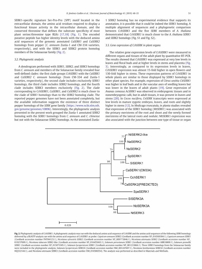
A dendrogram performed with SERK1, SERK2, and SERK3 homologsfrom C. annuum and members of the Solanaceae family revealed fourwell-defined clades: the first clade groups CchSERK1with the CaSERK1and CaSERK2 C. annuum homologs (from CM-334 and Zunla-1varieties, respectively), the second clade includes exclusively SERK1homologs, the third clade includes SERK2 homologs, and the fourthclade includes SERK3 members exclusively (Fig. 2). The cladecorresponding to CchSERK1, CaSERK1, and CaSERK2 is much closer tothe clade of SERK1 homologs than to the SERK2 homolog clade. Thereported pepper genomes have not been annotated completely, butthe available information suggests the existence of three distinctpepper homologs of the SERK gene family (https://www.ncbi.nlm.nih.gov/genome/genomes/10896). Interestingly, the phylogenetic analysispresented in the present work grouped the Zunla-1 annotated SERK2homolog with the SERK1 homologs from C. annuum and C. chinensebut not with the Solanaceae SERK2 homologs. As the annotated Zunla-
Fig. 2. Phylogenetic analysis of CchSERK1. A phylogenetic analysiswas runwith thededuced amidelivered by a BLASTP analysis ran with the amino acid sequence of CchSERK1 as probe: Capsicu(GenBank accession number PHT84123.1), Nicotiana sylvestris SERK2 (GenBank accession n019237009.1), Nicotiana tabacum SERK2-like (GenBank accession number XP_016455663.1),SERK1 (GenBank accession number XP_015073305.1), Solanum lycopersicum SERK1 (GenBankwere included in the phylogenetic analysis, Solanum lycopersicum SERK3A (GenBank accessionHQ332144.1), and Nicotiana attenuata SERK3 (GenBank accession number XM_016586554). Th
1 SERK2 homolog has no experimental evidence that supports itsannotation, it is possible that it could be indeed the SERK1 homolog. Amultiple alignment of sequences and a phylogenetic comparisonbetween CchSERK1 and the five SERK members of A. thalianademonstrated that CchSERK1 is much closer to the A. thaliana SERK1and SERK2 homologs (Fig. S1 and Fig. S2).
3.3. Gene expression of CchSERK in plant organs
The relative gene expression levels of CchSERK1 were measured indifferent organs and tissues of the adult plant by quantitative RT-PCR.The results showed that CchSERK1 was expressed at very low levels inleaves and floral buds and at higher levels in stems and placenta (Fig.3). Interestingly, as compared to its expression levels in leaves,CchSERK1 expression was almost 15-fold higher in open flowers and130-fold higher in stems. These expression patterns of CchSERK1 inwhole plants are similar to those displayed by SERK1 homologs inother plant species. For example, expression of Citrus unshiu CitSERK1was higher in leaf buds and in the vascular axes of seedling leaves butwas lower in the leaves of adult plants [19]. Gene expression ofAnanas comosus AcSERK1 was observed in embryogenic tissues and innonembryogenic calli, but in adult tissues, it was present in leaves andstems [20]. In Cocos nucifera, CnSERK transcripts were expressed atlow levels in mature zygotic embryos, leaves, and roots and slightlyhigher in stems [12]. In Medicago truncatula, in planta studies revealedthat expression of the SERK1 homolog (MtSERK1) was associated withthe primary meristems of the root and shoot and the newly formedmeristems of the lateral roots and nodule; MtSERK1 expression wasalso associated with the junction between one type of tissue or organ
no acid sequence ofCchSERK and the amino acid sequences of the following SERKhomologsmannuum SERK2 (GenBank accession number XP_016569384.1), Capsicum annuum SERK1umber XP_009773846.1), Nicotiana attenuata SERK2 (GenBank accession number XP_Solanum peruvianum SERK1 (GenBank accession number ABR18800.1), Solanum pennelliiaccession number NP_001233866.1). Three SERK3 homologs from the Solanaceae familynumber NM_001247697.1), Nicotiana benthamiana SERK3A (GenBank accession numbere analysis was performed as described in Materials and Methods.

Fig. 3. CchSERK1 transcription levels in adult organs of the plant. Transcript levels of CchSERK1were quantified by the 2−ΔΔCT qRT-PCRmethod (b) using specific oligonucleotide primersand total RNA extracted from leaves, roots, and stems of 2-week-old in vitro germinated seedlings, from 4-DAI floral buds, from open flowers of 3-month-old plants cultivated in thegreenhouse, and from placenta extracted from 4-DAI fruits (a). The bars over the columns represent the mean value ± SD of three independent experiments.
52 D. Jiménez-Guillen et al. / Electronic Journal of Biotechnology 41 (2019) 48–55
and another, and with the vascular tissue procambial cells. Based onthese data, the authors concluded that gene expression of MtSERK1 isassociated with tissue developmental changes, possibly reflectingcellular reprogramming [21]. In other reports, gene expression ofAtSERK1 was found in the procambium of the vascular bundles inroots, hypocotyls, and inflorescence stems of Arabidopsis [10,22]. Asprocambium is regarded as a primary meristematic tissue, whereprocambium cells are differentiated as xylem and phloem cells [23],and SERK1 expression becomes confined to the procambium, it istherefore proposed that SERK1 expression marks the procambiumvascular cell population to differentiate [22].
The similarity of the expression patterns developed by CchSERK1 inthe present work, with those reported for SERK1 homologs in theliterature suggests that the cloned C. chinense SERK1 homolog couldhave similar functions in the regulation of cell differentiation in adultplants.
3.4. Gene expression of CchSERK1 during zygotic embryogenesis
Analysis of the CchSERK1 gene expression during zygoticembryogenesis was performed in different tissues of the pepper fruit,as described in the Materials and methods section. When they arecompared to the relative expression found in the open flowers, theexpression patterns during the seed development showed a smallreduction at the 10, 30, and 45-DAP stages and a small increment atthe 14-DAP stage. The expression remained relatively constant in theother periods analyzed (Fig. 4). Gene expression analysis innonembryogenic tissues during fruit development revealed thatCchSERK1 was expressed at early stages in the placenta and at lowlevels in the middle developmental stages in the pericarp (Fig. S3).The peaks of CchSERK1 expression during fruit development coincide
with peaks at those stages when zygotic embryogenesis was triggeredand when the globular to embryo transition occurred. Theseexpression patterns are similar to those exhibited by other SERK1homologs during zygotic embryogenesis. In Daucus carota, SERK1transcripts were detected transiently in zygotic embryos up to theglobular stage [14]. In flowers, these transcripts were expressed from3-DAP, when fertilization occurs, to 20-DAP, which corresponds to thezygotic embryo early globular stage [24]. The authors did not findsignificant expression in leaves, stems, and roots [14]. In Zea mays,expression of ZmSERK1 was detected in all cells of the embryo sac,except in the synergid, and it was found in zygotes by 36 h afterpollination [25]. During the developmental stages of Arabidopsiszygotic embryogenesis, high levels of AtSERK1 mRNA were found inclosed floral buds and in pollinated flowers from stages 1 to 7.Quantitative analysis showed that AtSERK1 transcripts in floral budswere 10-fold higher than those in leaf tissues [10]. The expression ofMedicago truncatula MtSERK1 showed different patterns comparedwith that in other species during zygotic embryogenesis becausetranscripts were detected in globular embryos but expressionincreased at the heart stage, to diminish later at torpedo andcotyledonal stages. In the late stages of embryo development,MtSERK1 expression was relevant in cotyledons and radicle tips,especially in pro-vascular chain and epidermis. Expression was alsodetected in the cell layer that surrounds the embryo endosperm [21].
3.5. Gene expression of CchSERK1 during somatic embryogenesis
Quantitative RT-PCR analysis of CchSERK1 expression was alsoconducted during somatic embryogenesis. Results showed that thetranscription levels of CchSERK1 diminished from the globular to thecotyledonal stages but reached a remarkable fourfold peak of

Fig. 4. CchSERK1 transcription levels during different stages of seed development. Transcript levels of CchSERK1 were quantified by the 2−ΔΔCT qRT-PCR method (c) using specificoligonucleotide primers and total RNA extracted from open flowers, the whole fruit body after 1 and 4-DAI, and from the whole seeds (b) isolated at different stages from 10 to45 days of fruit development (a). The bars over the columns represent the mean value ± SD of three independent experiments.
53D. Jiménez-Guillen et al. / Electronic Journal of Biotechnology 41 (2019) 48–55
expression at the heart stage as compared to the expression levels foundin the non-induced hypocotyl explants (Fig. 5). The expression patternof CchSERK1 during the development of somatic embryos was slightlydifferent from those patterns exhibited by many other species, wherehigher expression was found at the globular stage. During the somaticembryogenesis processes occurring in A. thaliana, D. carota, and M.truncatula, the corresponding SERK1 homologs showed consistentexpression patterns, increasing at the globular stage and thendiminishing at the heart and cotyledonal stages [10,14,21].Nonetheless, gene expression patterns of SERK1 homologs, whichare distinct from this conserved behavior, are found in otherspecies. In Passiflora edulis, the expression of PeSERK, as assessed byin situ hybridization, was predominantly high in the abaxial part ofthe cotyledonal explants from 10 to 30 days after embryogenicinduction; interestingly, neither pro-embryogenic structures (20-to 22-day-old cultures) nor globular embryos (22- to 30-day-oldcultures) showed PeSERK expression. These results suggested thatin P. edulis, expression of PeSERK1 is needed when embryogeniccompetence is acquired, but not later during embryo development[26], and also demonstrate that, as in C. chinense, SERK1 homologsfrom different species may show different patterns of expressionduring somatic embryogenesis, essentially during the transition ofthe first stages.
While SERK1 is probably the bestmolecularmarker of embryogenesis,recent findings and reports in the scientific literature demonstrate that it
possesses wider biological functions. It is now accepted that the SERK1protein functions as a co-receptor that helps the main receptors to bindto their specific ligands and trigger the correspondent downstreamsignal transduction pathways [27]. Indeed, SERK receptors have beengrouped as mode-I coreceptors, which can form complexes withdifferent ligand-binding receptors, allowing SERK1 receptors toparticipate in different biological processes [28]. Based in its ability tobind different receptors, it has been demonstrated that SERK1 is alsoinvolved in the coregulation of different cell differentiation pathways,including the control of male sporogenesis [29], and the tapetumdevelopment and microspore maturation [30]. It has beendemonstrated that SERK1 complexes in vivo with the brassinosteroidreceptor (BRI1), forming the true hetero-receptor binding site for thesteroid. The heteromerization of BRI1 with SERK1 leads to the activationof a cytoplasmic signaling cascade, triggering plant growth and celldifferentiation [31]. More recently, in vivo evidence demonstrated thatSERKs (SERK1, SERK2, and SERK3) serve as co-receptors in CLE41/TDIF-PXY signaling to regulate plant vascular development. As CLE peptideshave decisive roles in the regulation of proliferation and differentiationof plant stem cells, the authors deduced that SERK1 acts as a coregulatorof these cell functions [32].
The above reports provide strong evidence that SERK1 homologsparticipate as coregulator of the cell differentiation processes of plants,including embryogenesis. Thus, it is possible that the gene expressionpatterns of the habanero pepper SERK1 homolog observed in this work,

Fig. 5. CchSERK1 transcription levels during somatic embryogenesis. Transcript levels of CchSERK1 were quantified by the 2−ΔΔCT qRT-PCR method (b) using specific oligonucleotideprimers and total RNA extracted from non-induced hypocotyl explants and globular, heart, and torpedo/cotyledonal whole embryo structures (a). The bars over the columns representthe mean value ± SD of three independent experiments.
54 D. Jiménez-Guillen et al. / Electronic Journal of Biotechnology 41 (2019) 48–55
both in whole plants and during somatic and zygotic embryogenesis,could be related to its function as a general coregulator of celldifferentiation, which is in turn needed to regulate the production ofdifferentiated cells in roots, stem, leaves, and flowers and to trigger thesomatic-embryogenic transition and the differentiation of tissues duringthe transition between every embryogenesis stage.
4. Conclusion
The alignment of the CchSERK1 cDNA sequence with the DNAsequences of different SERK1 homologs and the similarity in their geneexpression patterns constitute indirect evidence that supports theidentity of CchSERK1 as the C. chinense SERK1 homolog. The expressionpatterns in tissues with a high differentiation activity suggest thatCchSERK1 has functions in the co-regulation of both zygotic andsomatic embryogenesis, as well as in the production of vascular cells.As the deduced amino acid sequence of the encoded protein presentsall the protein residues and domains conforming a SERK1 functionalreceptor-like kinase enzyme, the cloned cDNA constitutes a powerfultool that can be used to ectopically manipulate the expression of theCchSERK1 gene; this could be useful to overcome the absence of SERK1mutants in peppers and can be used to analyze the molecularregulation of somatic embryogenesis and the recalcitrance of thegenus Capsicum to proliferate in vitro by tissue culture techniques.
Financial support
This project received financial support from Consejo Nacional deCiencia y Tecnología-FORDECyT (173407) and ComisiónIntersecretarial de Bioseguridad de los Organismos Genéticamente
Modificados (278944). DJG and DPP received CONACYT Ph.D.fellowships 255285 and 242977, respectively.
Conflict of interest
The authors declare that they have no conflict of interest.
Acknowledgments
The authors wish to thank M.C. Elidé Avilés-Berzunza for technicalassistance.
Supplementary material
https://doi.org/10.1016/j.ejbt.2019.07.006
References
[1] The Observatory of Economic Complexity. Product complexity rankings (PCI). Avail-able from Internet https://atlas.media.mit.edu/en/rankings/product/hs92/. [Cited 31October 2018].
[2] Canto-Flick A, Balam-Uc E, Bello-Bello J, et al. Capsaicinoids content in habanero pep-per (Capsicum chinense Jacq.): hottest known cultivars. HortSci Horts 2008;43(5):1344–9. https://doi.org/10.21273/HORTSCI.43.5.1344.
[3] Ochoa-Alejo N, Ramírez-Malagón R. In vitro chili pepper Biotechnology. In Vitro CellDev Biol-Plant 2001;37(6):701–29. https://doi.org/10.1007/s11627-001-0121-z.
[4] Orlinska M, Nowaczyk P. In vitro plant regeneration of 4 Capsicum spp. genotypesusing different explant types. Turkish J Biol 2015;39(1):60–8. https://doi.org/10.3906/biy-1403-89.
[5] Grozeva S, Todorova V. In vitro regeneration in pepper (Capsicum annuum L.) andcharacterization of plant–regenerants. Electron. J Biol 2015;11(1):17–22.

55D. Jiménez-Guillen et al. / Electronic Journal of Biotechnology 41 (2019) 48–55
[6] Heidmann I, De Lange B, Lambalk J, et al. Efficient sweet pepper transformation me-diated by the BABY BOOM transcription factor. Plant Cell Rep 2011;30(6):1107–15.https://doi.org/10.1007/s00299-011-1018-x PMid: 21305301.
[7] Solís-Ramos LY, González-Estrada T, Nahuath-Dzib S, et al. Overexpression ofWUSCHEL in C. chinense causes ectopic morphogenesis. Plant Cell Tiss Organ Cult2009;96(3):279–87. https://doi.org/10.1007/s11240-008-9485-7.
[8] Pérez-Pascual D, Jiménez-Guillen D, Villanueva-Alonzo H, et al. Ectopic expression ofthe Coffea canephora SERK1 homolog-induced differential transcription of genes in-volved in auxinmetabolism and in the developmental control of embryogenesis. Phys-iol Plant 2018;163(4):530–51. https://doi.org/10.1111/ppl.12709 PMid: 29607503.
[9] Li J.Multi-tasking of somatic embryogenesis receptor-like protein kinases. Curr OpPlantBiol 2010;13(5):509–14. https://doi.org/10.1016/j.pbi.2010.09.004 PMid: 20926334.
[10] Hecht V, Vielle-Calzada JP, Hartog MV, et al. The Arabidopsis Somatic EmbryogenesisReceptor Kinase 1 gene is expressed in developing ovules and embryos and enhancesembryogenic competence in culture. Plant Physiol 2001;127(3):803–16. https://doi.org/10.1104/pp.010324 PMid: 11706164.
[11] Zeng F, Zhang X, Zhu L, et al. Isolation and characterization of genes associated tocotton somatic embryogenesis by suppression subtractive hybridization and micro-array. Plant Mol Biol 2006;60(2):167–83. https://doi.org/10.1007/s11103-005-3381-x PMid:16429258.
[12] Perez-NúñezMT, Souza R, Sáenz L, et al. Detection of a SERK-like gene in coconut andanalysis of its expression during the formation of embryogenic callus and somaticembryos. Plant Cell Rep 2009;28(1):11–9. https://doi.org/10.1007/s00299-008-0616-8 PMid:18818928.
[13] Somleva MN, Schmidt EDL, De Vries SC. Embryogenic cells in Dactylis glomerata L.(Poaceae) explants identified by cell tracking and by SERK expression. Plant CellRep 2000;19(7):718–26. https://doi.org/10.1007/s002999900169 PMid:30754811.
[14] Schmidt EDL, Guzzo F, Toonen MAJ, et al. A leucine-rich repeat containing receptor-like kinase marks somatic plant cells competent to form embryos. Develop 1997;124(10):2049–62.
[15] Avilés-Viñas SA, Lecona-Guzmán CA, Canto-Flick A, et al. Morpho-histological andultrastructural study on direct somatic embryogenesis of Capsicum chinense Jacq.in liquid medium. Plant Biotechnol Rep 2013;7(3):277–86. https://doi.org/10.1007/s11816-012-0261-0.
[16] Livak KJ, Schmittgen TD. Analysis of relative gene expression data using real-timequantitative PCR and the 2−ΔΔCT method. Methods 2001;25(4):402–8. https://doi.org/10.1006/meth.2001.1262.
[17] Shah K, Vervoort J, de Vries Sacco C. Role of Threonines in the Arabidopsis thalianasomatic embryogenesis receptor kinase 1 activation loop in phosphorylation. J BiolChem 2001;276(44):41263–9. https://doi.org/10.1074/jbc.M102381200 PMid:11509554.
[18] Chen X, Chern M, Canlas PE, et al. A conserved threonine residue in thejuxtamembrane domain of the XA21 pattern recognition receptor is critical for ki-nase autophosphorylation and XA21-mediated immunity. J Biol Chem 2010;285(14):10454–63. https://doi.org/10.1074/jbc.M109.093427 PMid: 20118235.
[19] Ge X-X, Fan G-E, Chai L-J, et al. Cloning, molecular characterization and expressionanalysis of a somatic embryogenesis receptor-like kinase gene (CitSERK1-like) in
Valencia sweet orange. Acta Physiol Plant 2010;32(6):1197–207. https://doi.org/10.1007/s11738-010-0515-9.
[20] Ma J, He Y, Wu C, et al. Cloning and molecular characterization of a SERK gene tran-scriptionally induced during somatic embryogenesis in Ananas comosus cv.Shenwan. Plant Mol Biol Rep 2012;30(1):195–203. https://doi.org/10.1007/s11105-011-0330-5.
[21] Nolan KE, Kurdyukov S, Rose RJ. Expression of the SOMATIC EMBRYOGENESIS RECEP-TOR- LIKE KINASE1 (SERK1) gene is associated with developmental change in the lifecycle of the model legume Medicago truncatula. J Exp Bot 2009;60(6):1759–71.https://doi.org/10.1093/jxb/erp046 PMid: 19305022.
[22] Kwaaitaal MACJ, de Vries SC, Russinova E. Arabidopsis thaliana somatic embryogen-esis receptor kinase 1 protein is present in sporophytic and gametophytic cells andundergoes endocytosis. Protoplasma 2005;226(1):55–65. https://doi.org/10.1007/s00709-005-0111-9 PMid: 16231101.
[23] Jouannet V, Brackmann K, Greb T. (Pro)cambium formation and proliferation: twosides of the same coin? Curr Op Plant Biol 2015;23:54–60. https://doi.org/10.1016/j.pbi.2014.10.010 PMid: 25449727.
[24] Lackie S, Yeung EC. Zygotic embryo development inDaucus carota. Can J Bot 1996;74(7):990–8. https://doi.org/10.1139/b96-123.
[25] Baudino S, Hansen S, Brettschneider R. Molecular characterisation of two novelmaize LRR receptor-like kinases, which belong to the SERK gene family. Planta2001;213(1):1–10. https://doi.org/10.1007/s004250000471 PMid: 11523644.
[26] Rocha DI, Pinto DLP, Vieira LM, et al. Cellular and molecular changes associated withcompetence acquisition during passion fruit somatic embryogenesis: Ultrastructuralcharacterization and analysis of SERK gene expression. Protoplasma 2016;253(2):595–609. https://doi.org/10.1007/s00709-015-0837-y PMid: 26008651.
[27] He Y, Zhou J, Shan L, et al. Plant cell surface receptor-mediated signaling — A com-mon theme amid diversity. J Cell Sci 2018;131(2):1–11. jcs209353. https://doi.org/10.1242/jcs.209353 PMid: 29378836.
[28] Xi L, Wu XN, Gilbert M, et al. Classification and interactions of LRR receptors and co-receptors within the Arabidopsis plasma membrane–an overview. Front Plant Sci2019;10(472). https://doi.org/10.3389/fpls.2019.00472 PMid: 31057579.
[29] Albrecht C, Russinova E, Hecht V, et al. The Arabidopsis thaliana somatic embryogen-esis receptor-likE kinases1 and 2 control male sporogenesis. Plant Cell 2005;17(12):3337–49. https://doi.org/10.1105/tpc.105.036814 PMid: 16284305.
[30] Colcombet J, Boisson-Dernier A, Ros-Palau R, et al. Arabidopsis somatic embryogene-sis receptor kinases1 and 2 are essential for tapetum development and microsporematuration. Plant Cell 2005;17(12):3350–61. https://doi.org/10.1105/tpc.105.036731 PMid: 16284306.
[31] Santiago J, Henzler C, Hothorn M. Molecular mechanism for plant steroid receptoractivation by somatic embryogenesis co-receptor kinases. Science 2013;341(6148):889–92. https://doi.org/10.1126/science.1242468 PMid: 23929946.
[32] Zhang H, Lin X, Han Z, et al. SERK family receptor-like kinases function as co-recep-tors with PXY for plant vascular development. Mol Plant 2016;9(10):1406–14.https://doi.org/10.1016/j.molp.2016.07.004 PMid: 27449136.